Biological adhesion for locomotion: basic · PDF fileBiological adhesion for locomotion: ......
29
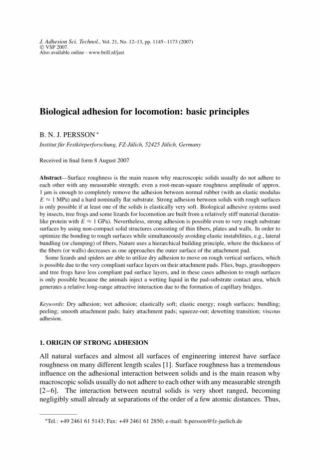
J. Adhesion Sci. Technol., Vol. 21, No. 12–13, pp. 1145–1173 (2007) VSP 2007. Also available online - www.brill.nl/jast Biological adhesion for locomotion: basic principles B. N. J. PERSSON ∗ Institut für Festkörperforschung, FZ-Jülich, 52425 Jülich, Germany Received in final form 8 August 2007 Abstract—Surface roughness is the main reason why macroscopic solids usually do not adhere to each other with any measurable strength; even a root-mean-square roughness amplitude of approx. 1 μm is enough to completely remove the adhesion between normal rubber (with an elastic modulus E ≈ 1 MPa) and a hard nominally flat substrate. Strong adhesion between solids with rough surfaces is only possible if at least one of the solids is elastically very soft. Biological adhesive systems used by insects, tree frogs and some lizards for locomotion are built from a relatively stiff material (keratin- like protein with E ≈ 1 GPa). Nevertheless, strong adhesion is possible even to very rough substrate surfaces by using non-compact solid structures consisting of thin fibers, plates and walls. In order to optimize the bonding to rough surfaces while simultaneously avoiding elastic instabilities, e.g., lateral bundling (or clumping) of fibers, Nature uses a hierarchical building principle, where the thickness of the fibers (or walls) decreases as one approaches the outer surface of the attachment pad. Some lizards and spiders are able to utilize dry adhesion to move on rough vertical surfaces, which is possible due to the very compliant surface layers on their attachment pads. Flies, bugs, grasshoppers and tree frogs have less compliant pad surface layers, and in these cases adhesion to rough surfaces is only possible because the animals inject a wetting liquid in the pad-substrate contact area, which generates a relative long-range attractive interaction due to the formation of capillary bridges. Keywords: Dry adhesion; wet adhesion; elastically soft; elastic energy; rough surfaces; bundling; peeling; smooth attachment pads; hairy attachment pads; squeeze-out; dewetting transition; viscous adhesion. 1. ORIGIN OF STRONG ADHESION All natural surfaces and almost all surfaces of engineering interest have surface roughness on many different length scales [1]. Surface roughness has a tremendous influence on the adhesional interaction between solids and is the main reason why macroscopic solids usually do not adhere to each other with any measurable strength [2–6]. The interaction between neutral solids is very short ranged, becoming negligibly small already at separations of the order of a few atomic distances. Thus, ∗ Tel.: +49 2461 61 5143; Fax: +49 2461 61 2850; e-mail: [email protected]
Transcript of Biological adhesion for locomotion: basic · PDF fileBiological adhesion for locomotion: ......
J. Adhesion Sci. Technol., Vol. 21, No. 12–13, pp. 1145–1173 (2007) VSP 2007.Also available online - www.brill.nl/jast
Biological adhesion for locomotion: basic principles
B. N. J. PERSSON ∗
Institut für Festkörperforschung, FZ-Jülich, 52425 Jülich, Germany
Received in final form 8 August 2007
Abstract—Surface roughness is the main reason why macroscopic solids usually do not adhere toeach other with any measurable strength; even a root-mean-square roughness amplitude of approx.1 µm is enough to completely remove the adhesion between normal rubber (with an elastic modulusE ≈ 1 MPa) and a hard nominally flat substrate. Strong adhesion between solids with rough surfacesis only possible if at least one of the solids is elastically very soft. Biological adhesive systems usedby insects, tree frogs and some lizards for locomotion are built from a relatively stiff material (keratin-like protein with E ≈ 1 GPa). Nevertheless, strong adhesion is possible even to very rough substratesurfaces by using non-compact solid structures consisting of thin fibers, plates and walls. In order tooptimize the bonding to rough surfaces while simultaneously avoiding elastic instabilities, e.g., lateralbundling (or clumping) of fibers, Nature uses a hierarchical building principle, where the thickness ofthe fibers (or walls) decreases as one approaches the outer surface of the attachment pad.
Some lizards and spiders are able to utilize dry adhesion to move on rough vertical surfaces, whichis possible due to the very compliant surface layers on their attachment pads. Flies, bugs, grasshoppersand tree frogs have less compliant pad surface layers, and in these cases adhesion to rough surfacesis only possible because the animals inject a wetting liquid in the pad-substrate contact area, whichgenerates a relative long-range attractive interaction due to the formation of capillary bridges.
Keywords: Dry adhesion; wet adhesion; elastically soft; elastic energy; rough surfaces; bundling;peeling; smooth attachment pads; hairy attachment pads; squeeze-out; dewetting transition; viscousadhesion.
1. ORIGIN OF STRONG ADHESION
All natural surfaces and almost all surfaces of engineering interest have surfaceroughness on many different length scales [1]. Surface roughness has a tremendousinfluence on the adhesional interaction between solids and is the main reason whymacroscopic solids usually do not adhere to each other with any measurable strength[2–6]. The interaction between neutral solids is very short ranged, becomingnegligibly small already at separations of the order of a few atomic distances. Thus,
∗Tel.: +49 2461 61 5143; Fax: +49 2461 61 2850; e-mail: [email protected]
1146 B. N. J. Persson
strong interaction is only possible if at least one of the solids is elastically very softso that the surface can bend and make atomic contact at the interface. In this casethere will be a large area of real contact, A, between the solids and a small elasticenergy, Uel, will be stored at the interface. During pull-off this stored elastic energyis (partly) given back and may help to break the interfacial bonds. However, if thebonding energy �γA, where �γ is the change in the (interfacial) free energy (perunit surface area) as two flat surfaces of the two solids are brought together, is muchlarger than Uel, the stored elastic energy can be neglected. Thus the first criterionfor strong adhesion is that at least one of the solids should be elastically soft so thatthe area of real contact is large and the stored elastic energy is small.
The second criterion for strong adhesion is that the interaction between the solidsshould involve “long dissipative bonds”. That is, during pull-off the effectiveadhesion bonds should elongate a long distance and the elastic energy stored inthe bonds at the point where they break should be dissipated in the solids ratherthan used to break the other interfacial adhesion bonds.
Synthetic adhesives often satisfy both these criteria. Thus, for example, pressuresensitive adhesives, used for Scotch� tapes, consist of thin elastically soft polymerfilms which form thin filaments (effective bonds) during pull-off, which can beextended a large distance (sometimes up to several millimeters) before they break,and the energy stored in the filament is mainly dissipated in the polymer ratherthan used to break other polymer filaments [7]. In addition, the effective elasticmodulus of pressure sensitive adhesives is very low (often only in the kPa-range)so the relative contact area, A/A0 (where A is the area of real contact and A0 thenominal or apparent contact area), can be very large even for very rough substrates,and the stored elastic energy, Uel, may be relatively small [8].
Biological adhesive systems used for locomotion cannot be built on the sameprinciples as pressure sensitive adhesives. First, pressure sensitive adhesives arerelatively weak materials (almost liquid-like) which would wear rapidly. Moreimportantly, during repeated use on (real) contaminated surfaces, the adhesivewould rapidly be covered by small particles (dust, pollen, . . . ) and since the particlescannot be (easily) removed, the adhesive would fail after just a few contact cycles.In addition, much of the adhesion strength of pressure sensitive adhesives comesfrom the formation of long polymer filaments, but for biological applications (atleast for insects), such long effective bonds would lead to disaster: the (small) insectwould need to lift its legs several millimeters in order to break the bond, which isimpossible for legs which may be shorter than a millimeter. Thus, for biologicallocomotion, the effective adhesive bonds should be long (on an atomic scale) butnot too long.
Biological adhesive systems used for locomotion are made from keratin-likematerials which are elastically relatively stiff, with an elastic modulus in the GPa-range, i.e., 103 times stiffer than normal (cross-linked) rubber and maybe 106 timesstiffer than pressure sensitive adhesives. Thus, the fundamental question is how
Biological adhesion for locomotion: basic principles 1147
Nature has been able to use this material to design an adhesive which satisfies thetwo adhesion criteria discussed above.
Using the principle of natural selection, Nature has produced adhesive pads ininsects, and some lizards and frogs which consist of non-compact material inthe form of either foam-like or fiber-like structures [9, 10]; the effective elasticproperties of these materials are much lower than of the compact material andthe first criterion above can be satisfied, in particular if the material exhibits ahierarchical (fractal-like) construction, with thinner fibers or walls close to the(outer) attachment surface. However, the fibers and walls cannot be made arbitrarilythin as this would result in a weak material and strong wear, or a collapse of thematerial as a result of the attraction between the (internal) surfaces of the solid. Ifby a mutation an insect (or lizard) would appear where the attachment system wouldfail due to this effect, the insect would get quickly eliminated by natural selection:thus one may expect the adhesive system to be highly optimized and close to thelimit of what is possible from the point of view of strength and stability.
The effective “long dissipative bonds”, which are required for strong adhesion,arise in different ways for hairy and smooth adhesive pads. For the hairy systems,long thin bent fibers adhere to the (rough) substrate. During pull-off the fibers willstraighten out before the bond between the fiber and the substrate is broken. At thepoint of “snap-off” the elastic energy stored in the straightened fiber will get lost(the fiber will vibrate for a short time and the vibration energy will, e.g., be radiatedas sound waves into the surrounding). This will result in effective bonds which arelong (compared to the atomic dimension) and dissipative (the stored elastic energywill be mainly dissipated in the solids rather than used to break other fiber adhesionbonds) [11]. For the smooth adhesive pads, the insect (or frog) injects a wettingliquid in the contact region between the toe pad and the substrate. During pull-offfrom a rough substrate, small liquid capillary bridges will form in many toe pad-asperity contact regions, and the bridges will elongate a long distance (on the atomicscale) before they break because of capillary instabilities. The (surface) energystored in the stretched capillary bridge will be dissipated in the liquid (because ofthe liquid viscosity) so again we will have effective “long dissipative bonds” actingbetween the surfaces.
2. ADHESION AND FRICTION BETWEEN ELASTIC SOLIDS WITHRANDOMLY ROUGH SURFACES
The breaking of the atomic bonds between two elastic solids during “pull-off”usually occurs by propagating an interfacial crack. For elastic solids, part of theelastic energy stored at the interface because of surface roughness will flow to thecrack tip and facilitate the interfacial bond-breaking process, resulting in the smalladhesion observed in most situations.
The adhesion between elastic solids with smooth surfaces is determined by theelastic modulus of the solids and by the change in the interfacial energy (per unit
1148 B. N. J. Persson
area) �γ when two flat surfaces of the solids are brought into contact. A similardescription is possible if the solids have small-amplitude and short-wavelengthroughness but in this case it is necessary to replace �γ with the effective interfacialenergy γeff which depends on the nature of the surface roughness and on the elasticmodulus of the solids [4, 5]. If A0 is the nominal contact area and A is the area ofreal (atomic) contact then A0γeff = A�γ−Uel. where Uel is the elastic energy storedat the interface as a result of the surface roughness, i.e., Uel is the energy necessaryto deform the rubber surface so that it makes atomic contact with the substrate overthe area A. Negligible adhesion will occur if γeff ≈ 0, i.e., if the elastic energy Uel
stored at the interface (nearly) equals the interfacial bonding energy A�γ .In the theory developed in Refs [4, 5] the interface between two contacting solids
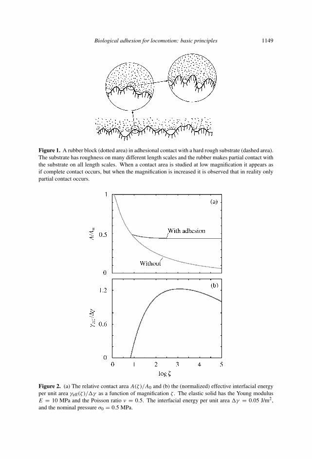
is studied as a function of the magnification ζ . The magnification refers to some(arbitrary) length scale which, e.g., could be the lateral size L of the nominalcontact area. When the system is studied at the magnification ζ , surface roughnesscomponents with wavelength λ < L/ζ cannot be observed and the (apparent)contact area (projected on the xy-plane) A(ζ ) between the solids will dependon the magnification ζ . In particular, as we increase the magnification we willobserve new surface roughness (see Fig. 1) and the area of (apparent) contact A(ζ )
will, therefore, decrease with increasing magnification. The effective interfacialfree energy (per unit surface area) will also change with the magnification withγeff(ζ ) → �γ as ζ → ζ1, where ζ1 is the highest magnification corresponding toatomic distances. The pull-off force is determined by the macroscopic interfacialfree energy γeff(1) and if γeff(1) = 0 the pull-off force will vanish, i.e., no adhesioncan be detected in a pull-off experiment. Nevertheless, the area of real (atomic)contact A(ζ1) will, in general, be enhanced by the adhesional interaction, andsince the sliding friction is determined by the area of real contact, the adhesionalinteraction may strongly increase the friction force in spite of the fact that noadhesion can be detected in a pull-off experiment. This effect has recently beenobserved for microfiber arrays of stiff polymers [13]. In this case the fiber arraysystem, owing to the high compliance due to fiber buckling and bending [12],exhibits a strongly enhanced contact area and friction as compared to a nominallysmooth surface of the same material. Nevertheless, γeff(1) = 0 and the pull-offforce vanishes.
Fig. 2 shows as an illustration the calculated [4, 5] relative contact area A(ζ )/A0
and the (normalized) effective interfacial energy per unit area γeff(ζ )/�γ as afunction of magnification when a rubber block is squeezed against a (rigid) self-affine fractal surface with the root-mean-square roughness h0 = 0.5 µm and thefractal dimension Df = 2.2. In this case the adhesional interaction gives a strongincrease in the real contact area (at the highest magnification) and hence also inthe friction force, but γeff(ζ ) = 0 for ζ < 8 so the (macroscopic) pull-off forcevanishes.
Biological adhesion for locomotion: basic principles 1149
Figure 1. A rubber block (dotted area) in adhesional contact with a hard rough substrate (dashed area).The substrate has roughness on many different length scales and the rubber makes partial contact withthe substrate on all length scales. When a contact area is studied at low magnification it appears asif complete contact occurs, but when the magnification is increased it is observed that in reality onlypartial contact occurs.
Figure 2. (a) The relative contact area A(ζ )/A0 and (b) the (normalized) effective interfacial energyper unit area γeff(ζ )/�γ as a function of magnification ζ . The elastic solid has the Young modulusE = 10 MPa and the Poisson ratio ν = 0.5. The interfacial energy per unit area �γ = 0.05 J/m2,and the nominal pressure σ0 = 0.5 MPa.
1150 B. N. J. Persson
3. ADHESION USING NON-COMPACT SOLIDS
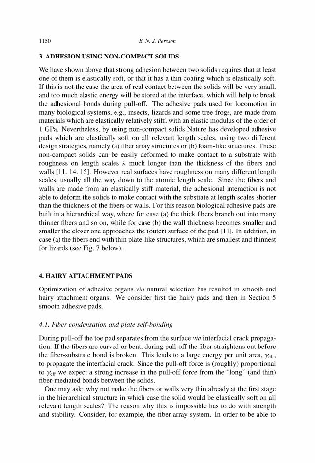
We have shown above that strong adhesion between two solids requires that at leastone of them is elastically soft, or that it has a thin coating which is elastically soft.If this is not the case the area of real contact between the solids will be very small,and too much elastic energy will be stored at the interface, which will help to breakthe adhesional bonds during pull-off. The adhesive pads used for locomotion inmany biological systems, e.g., insects, lizards and some tree frogs, are made frommaterials which are elastically relatively stiff, with an elastic modulus of the order of1 GPa. Nevertheless, by using non-compact solids Nature has developed adhesivepads which are elastically soft on all relevant length scales, using two differentdesign strategies, namely (a) fiber array structures or (b) foam-like structures. Thesenon-compact solids can be easily deformed to make contact to a substrate withroughness on length scales λ much longer than the thickness of the fibers andwalls [11, 14, 15]. However real surfaces have roughness on many different lengthscales, usually all the way down to the atomic length scale. Since the fibers andwalls are made from an elastically stiff material, the adhesional interaction is notable to deform the solids to make contact with the substrate at length scales shorterthan the thickness of the fibers or walls. For this reason biological adhesive pads arebuilt in a hierarchical way, where for case (a) the thick fibers branch out into manythinner fibers and so on, while for case (b) the wall thickness becomes smaller andsmaller the closer one approaches the (outer) surface of the pad [11]. In addition, incase (a) the fibers end with thin plate-like structures, which are smallest and thinnestfor lizards (see Fig. 7 below).
4. HAIRY ATTACHMENT PADS
Optimization of adhesive organs via natural selection has resulted in smooth andhairy attachment organs. We consider first the hairy pads and then in Section 5smooth adhesive pads.
4.1. Fiber condensation and plate self-bonding
During pull-off the toe pad separates from the surface via interfacial crack propaga-tion. If the fibers are curved or bent, during pull-off the fiber straightens out beforethe fiber-substrate bond is broken. This leads to a large energy per unit area, γeff,to propagate the interfacial crack. Since the pull-off force is (roughly) proportionalto γeff we expect a strong increase in the pull-off force from the “long” (and thin)fiber-mediated bonds between the solids.
One may ask: why not make the fibers or walls very thin already at the first stagein the hierarchical structure in which case the solid would be elastically soft on allrelevant length scales? The reason why this is impossible has to do with strengthand stability. Consider, for example, the fiber array system. In order to be able to
Biological adhesion for locomotion: basic principles 1151
Figure 3. The hierarchical nature of the lizard adhesive system is compliant on all relevant lengthscales, and deforms elastically to optimize the contact area and the bonding to the rough substrate.
Figure 4. If the fibers are too thin or long, bundling may occur (left). Similarly, if the plates are toothin or long, self-bonding may occur (right).
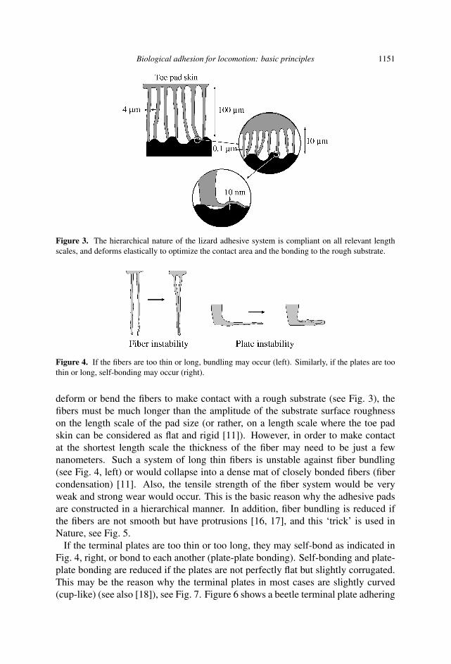
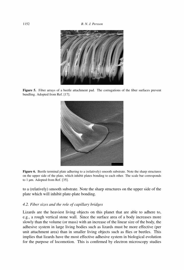
deform or bend the fibers to make contact with a rough substrate (see Fig. 3), thefibers must be much longer than the amplitude of the substrate surface roughnesson the length scale of the pad size (or rather, on a length scale where the toe padskin can be considered as flat and rigid [11]). However, in order to make contactat the shortest length scale the thickness of the fiber may need to be just a fewnanometers. Such a system of long thin fibers is unstable against fiber bundling(see Fig. 4, left) or would collapse into a dense mat of closely bonded fibers (fibercondensation) [11]. Also, the tensile strength of the fiber system would be veryweak and strong wear would occur. This is the basic reason why the adhesive padsare constructed in a hierarchical manner. In addition, fiber bundling is reduced ifthe fibers are not smooth but have protrusions [16, 17], and this ‘trick’ is used inNature, see Fig. 5.
If the terminal plates are too thin or too long, they may self-bond as indicated inFig. 4, right, or bond to each another (plate-plate bonding). Self-bonding and plate-plate bonding are reduced if the plates are not perfectly flat but slightly corrugated.This may be the reason why the terminal plates in most cases are slightly curved(cup-like) (see also [18]), see Fig. 7. Figure 6 shows a beetle terminal plate adhering
1152 B. N. J. Persson
Figure 5. Fiber arrays of a beetle attachment pad. The corrugations of the fiber surfaces preventbundling. Adopted from Ref. [17].
Figure 6. Beetle terminal plate adhering to a (relatively) smooth substrate. Note the sharp structureson the upper side of the plate, which inhibit plates bonding to each other. The scale bar correspondsto 1 µm. Adopted from Ref. [35].
to a (relatively) smooth substrate. Note the sharp structures on the upper side of theplate which will inhibit plate-plate bonding.
4.2. Fiber sizes and the role of capillary bridges
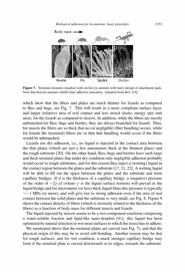
Lizards are the heaviest living objects on this planet that are able to adhere to,e.g., a rough vertical stone wall. Since the surface area of a body increases moreslowly than the volume (or mass) with an increase of the linear size of the body, theadhesive system in large living bodies such as lizards must be more effective (perunit attachment area) than in smaller living objects such as flies or beetles. Thisimplies that lizards have the most effective adhesive system in biological evolutionfor the purpose of locomotion. This is confirmed by electron microscopy studies
Biological adhesion for locomotion: basic principles 1153
Figure 7. Terminal elements (marked with circles) in animals with hairy design of attachment pads.Note that heavier animals exhibit finer adhesive structures. Adopted from Ref. [19].
which show that the fibers and plates are much thinner for lizards as comparedto flies and bugs, see Fig. 7. This will result in a more compliant surface layer,and larger (relative) area of real contact and less stored elastic energy (per unitarea), for the lizards as compared to insects. In addition, while the fibers are mostlyunbranched for flies, bugs and beetles, they are always branched for lizards. Thus,for insects the fibers are so thick that no (or negligible) fiber bundling occurs, whilefor lizards the (terminal) fibers are so thin that bundling would occur if the fiberswould be unbranched.
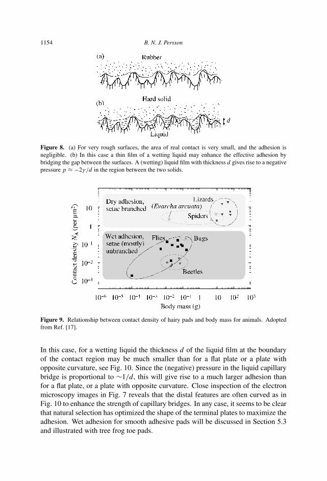
Lizards use dry adhesion, i.e., no liquid is injected in the contact area betweenthe thin plates (which are just a few nanometers thick at the thinnest place) andthe rough substrate [20]. On the other hand, flies, bugs and beetles have such largeand thick terminal plates that under dry condition only negligible adhesion probablywould occur to rough substrates, and for this reason they inject a (wetting) liquid inthe contact region between the plates and the substrate [17, 21, 22]. A wetting liquidwill be able to fill out the space between the plates and the substrate and formcapillary bridges. If d is the thickness of a capillary bridge, a (negative) pressureof the order of −2γ /d (where γ is the liquid surface tension) will prevail in theliquid bridge and for micrometer (or less) thick liquid films this pressure is typically∼−1 MPa (or more), and will give rise to strong adhesion even if the area of realcontact between the solid plates and the substrate is very small, see Fig. 8. Figure 9shows the contact density of fibers (which is inversely related to the thickness of thefibers) as a function of body mass for different insects and lizards.
The liquid injected by insects seems to be a two-component emulsion comprisinga water-soluble fraction and lipid-like nano-droplets [41]; this liquid has beenoptimized by natural selection to wet most surfaces to which the insect has to adhere.
We mentioned above that the terminal plates are curved (see Fig. 7), and that thephysical origin of this may be to avoid self-bonding. Another reason may be thatfor rough surfaces, and for wet condition, a much stronger capillary bridge mayform if the terminal plate is curved downwards at its edges, towards the substrate.
1154 B. N. J. Persson
Figure 8. (a) For very rough surfaces, the area of real contact is very small, and the adhesion isnegligible. (b) In this case a thin film of a wetting liquid may enhance the effective adhesion bybridging the gap between the surfaces. A (wetting) liquid film with thickness d gives rise to a negativepressure p ≈ −2γ /d in the region between the two solids.
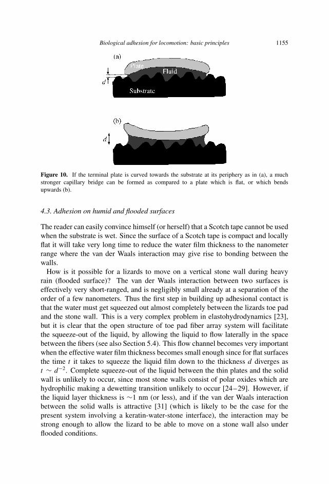
Figure 9. Relationship between contact density of hairy pads and body mass for animals. Adoptedfrom Ref. [17].
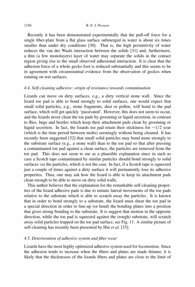
In this case, for a wetting liquid the thickness d of the liquid film at the boundaryof the contact region may be much smaller than for a flat plate or a plate withopposite curvature, see Fig. 10. Since the (negative) pressure in the liquid capillarybridge is proportional to ∼1/d, this will give rise to a much larger adhesion thanfor a flat plate, or a plate with opposite curvature. Close inspection of the electronmicroscopy images in Fig. 7 reveals that the distal features are often curved as inFig. 10 to enhance the strength of capillary bridges. In any case, it seems to be clearthat natural selection has optimized the shape of the terminal plates to maximize theadhesion. Wet adhesion for smooth adhesive pads will be discussed in Section 5.3and illustrated with tree frog toe pads.
Biological adhesion for locomotion: basic principles 1155
Figure 10. If the terminal plate is curved towards the substrate at its periphery as in (a), a muchstronger capillary bridge can be formed as compared to a plate which is flat, or which bendsupwards (b).
4.3. Adhesion on humid and flooded surfaces
The reader can easily convince himself (or herself) that a Scotch tape cannot be usedwhen the substrate is wet. Since the surface of a Scotch tape is compact and locallyflat it will take very long time to reduce the water film thickness to the nanometerrange where the van der Waals interaction may give rise to bonding between thewalls.
How is it possible for a lizards to move on a vertical stone wall during heavyrain (flooded surface)? The van der Waals interaction between two surfaces iseffectively very short-ranged, and is negligibly small already at a separation of theorder of a few nanometers. Thus the first step in building up adhesional contact isthat the water must get squeezed out almost completely between the lizards toe padand the stone wall. This is a very complex problem in elastohydrodynamics [23],but it is clear that the open structure of toe pad fiber array system will facilitatethe squeeze-out of the liquid, by allowing the liquid to flow laterally in the spacebetween the fibers (see also Section 5.4). This flow channel becomes very importantwhen the effective water film thickness becomes small enough since for flat surfacesthe time t it takes to squeeze the liquid film down to the thickness d diverges ast ∼ d−2. Complete squeeze-out of the liquid between the thin plates and the solidwall is unlikely to occur, since most stone walls consist of polar oxides which arehydrophilic making a dewetting transition unlikely to occur [24–29]. However, ifthe liquid layer thickness is ∼1 nm (or less), and if the van der Waals interactionbetween the solid walls is attractive [31] (which is likely to be the case for thepresent system involving a keratin-water-stone interface), the interaction may bestrong enough to allow the lizard to be able to move on a stone wall also underflooded conditions.
1156 B. N. J. Persson
Recently it has been demonstrated experimentally that the pull-off force for asingle fiber-plate from a flat glass surface submerged in water is about six timessmaller than under dry conditions [30]. That is, the high permittivity of waterreduces the van der Waals interaction between the solids [31] and, furthermore,a thin (a few monolayers) layer of water may separate the solids in the contactregion giving rise to the small observed adhesional interaction. It is clear that theadhesion force of a whole gecko foot is reduced substantially and this seems to bein agreement with circumstantial evidence from the observation of geckos whenrunning on wet surfaces.
4.4. Self-cleaning adhesive: origin of resistance towards contamination
Lizards can move on dirty surfaces, e.g., a dirty vertical stone wall. Since thelizard toe pad is able to bond strongly to solid surfaces, one would expect thatsmall solid particles, e.g., stone fragments, dust or pollen, will bond to the padsurface, which will get quickly ‘passivated’. However, this does not seem to occur,and the lizards never clean the toe pads by grooming or liquid secretion, in contrastto flies, bugs and beetles which keep their attachment pads clean by grooming orliquid secretion. In fact, the lizards toe pad retain their stickiness for ∼1/2 year(which is the time period between molts) seemingly without being cleaned. It hasrecently been suggested [32] that small solid particles may bond more strongly tothe substrate surface (e.g., a stone wall) than to the toe pad so that after pressinga contaminated toe pad against a clean surface, the particles are removed from thetoe pad. This does not seem to me as a plausible explanation since in such ascase a Scotch tape contaminated by similar particles should bond strongly to solidsurfaces via the particles, which is not the case. In fact, if a Scotch tape is squeezedjust a couple of times against a dirty surface it will permanently lose its adhesiveproperties. Thus, one may ask how the lizard is able to keep its attachment padsclean enough to be able to move on dirty solid walls.
This author believes that the explanation for the remarkable self-cleaning proper-ties of the lizard adhesive pads is due to minute lateral movements of the toe padsrelative to the substrate which is able to scratch away the particles. It is knownthat in order to bond strongly to a substrate, the lizard must shear the toe pad ina special direction in order to line-up (or bend) the bonding plates into a positionthat gives strong bonding to the substrate. It is suggest that motion in the oppositedirection, while the toe pad is squeezed against the (rough) substrate, will scratchaway solid particles trapped on the toe pad surface, see Fig. 11. A similar picture ofself-cleaning has recently been presented by Hui et al. [33].
4.5. Deterioration of adhesive system and fiber wear
Lizards have the most highly optimized adhesive system used for locomotion. Sincethe adhesion tends to increase when the fibers and plates are made thinner, it islikely that the thicknesses of the lizards fibers and plates are close to the limit of
Biological adhesion for locomotion: basic principles 1157
Figure 11. Removal of dirt particle by lateral shear (scratch motion).
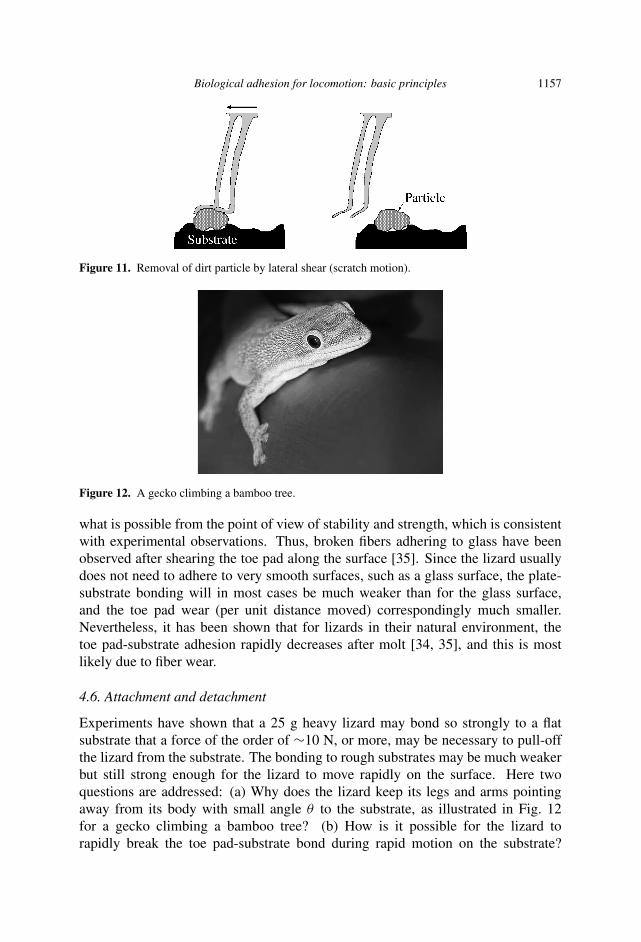
Figure 12. A gecko climbing a bamboo tree.
what is possible from the point of view of stability and strength, which is consistentwith experimental observations. Thus, broken fibers adhering to glass have beenobserved after shearing the toe pad along the surface [35]. Since the lizard usuallydoes not need to adhere to very smooth surfaces, such as a glass surface, the plate-substrate bonding will in most cases be much weaker than for the glass surface,and the toe pad wear (per unit distance moved) correspondingly much smaller.Nevertheless, it has been shown that for lizards in their natural environment, thetoe pad-substrate adhesion rapidly decreases after molt [34, 35], and this is mostlikely due to fiber wear.
4.6. Attachment and detachment
Experiments have shown that a 25 g heavy lizard may bond so strongly to a flatsubstrate that a force of the order of ∼10 N, or more, may be necessary to pull-offthe lizard from the substrate. The bonding to rough substrates may be much weakerbut still strong enough for the lizard to move rapidly on the surface. Here twoquestions are addressed: (a) Why does the lizard keep its legs and arms pointingaway from its body with small angle θ to the substrate, as illustrated in Fig. 12for a gecko climbing a bamboo tree? (b) How is it possible for the lizard torapidly break the toe pad-substrate bond during rapid motion on the substrate?
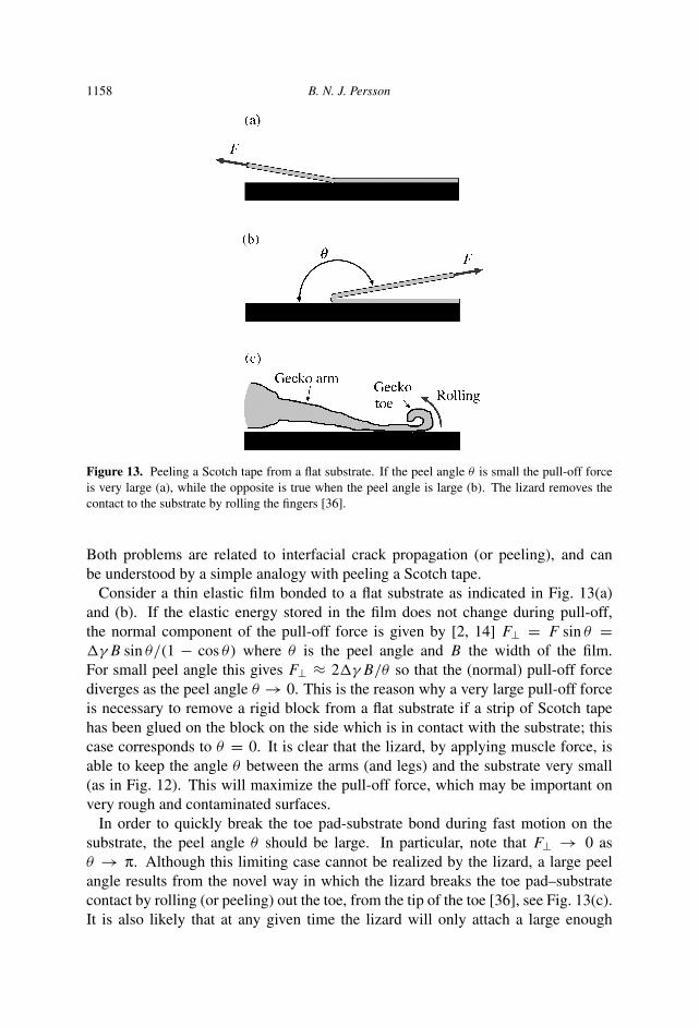
1158 B. N. J. Persson
Figure 13. Peeling a Scotch tape from a flat substrate. If the peel angle θ is small the pull-off forceis very large (a), while the opposite is true when the peel angle is large (b). The lizard removes thecontact to the substrate by rolling the fingers [36].
Both problems are related to interfacial crack propagation (or peeling), and canbe understood by a simple analogy with peeling a Scotch tape.
Consider a thin elastic film bonded to a flat substrate as indicated in Fig. 13(a)and (b). If the elastic energy stored in the film does not change during pull-off,the normal component of the pull-off force is given by [2, 14] F⊥ = F sin θ =�γB sin θ/(1 − cos θ) where θ is the peel angle and B the width of the film.For small peel angle this gives F⊥ ≈ 2�γB/θ so that the (normal) pull-off forcediverges as the peel angle θ → 0. This is the reason why a very large pull-off forceis necessary to remove a rigid block from a flat substrate if a strip of Scotch tapehas been glued on the block on the side which is in contact with the substrate; thiscase corresponds to θ = 0. It is clear that the lizard, by applying muscle force, isable to keep the angle θ between the arms (and legs) and the substrate very small(as in Fig. 12). This will maximize the pull-off force, which may be important onvery rough and contaminated surfaces.
In order to quickly break the toe pad-substrate bond during fast motion on thesubstrate, the peel angle θ should be large. In particular, note that F⊥ → 0 asθ → π. Although this limiting case cannot be realized by the lizard, a large peelangle results from the novel way in which the lizard breaks the toe pad–substratecontact by rolling (or peeling) out the toe, from the tip of the toe [36], see Fig. 13(c).It is also likely that at any given time the lizard will only attach a large enough
Biological adhesion for locomotion: basic principles 1159
fraction of the toe pad fibers to the substrate surface, as is necessary to obtainsufficient adhesion.
4.7. Uniqueness of biological adhesive systems for locomotion
In nature two different types of adhesive pads are used for locomotion, involvingeither smooth or hairy attachment pads. Hairy adhesive organs have evolvedindependently at least three times in lizards [37], at least three times in insects [38],and occur in three phylogenetically distant groups of spiders [39, 40]. This suggeststhat hairy pads represent an optimum design for attachment, and it is likely thatmany living objects on other planets in our universe will make use of similar hairyattachment organs for locomotion.
It is clear that a detailed understanding of the function of biological adhesivesystems used for locomotion may result in new improved synthetic adhesives, basedon similar principles as in biological adhesive systems. Such systems will have greatadvantages over adhesives used today, and may make new applications possiblesuch as wall-climbing robots.
5. SMOOTH ATTACHMENT PADS
All animals which use smooth attachment pads inject a wetting liquid in the contactarea in order to increase the adhesion. In this section some aspects of wet adhesionfor tree frogs are discussed, but the results may also be relevant for other animalsusing smooth adhesive pads, e.g., grasshoppers [42].
5.1. Toe pad construction
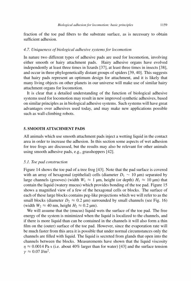
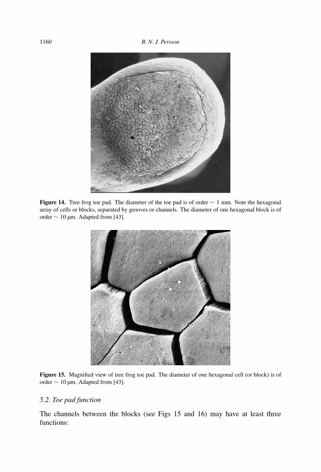
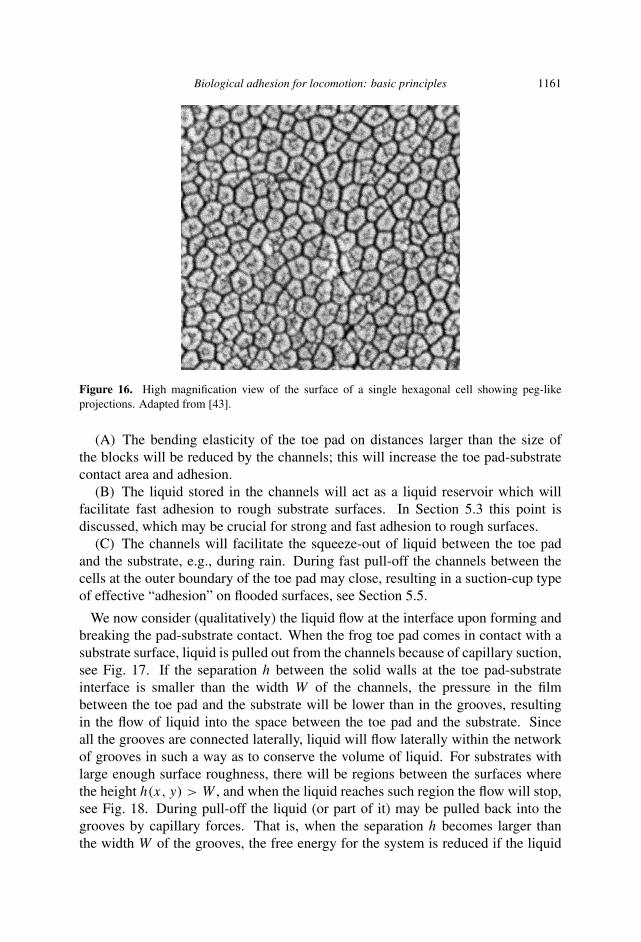
Figure 14 shows the toe pad of a tree frog [43]. Note that the pad surface is coveredwith an array of hexagonal (epithelial) cells (diameter D1 ∼ 10 µm) separated bylarge channels (grooves) (width W1 ≈ 1 µm, height (or depth) H1 ≈ 10 µm) thatcontain the liquid (watery mucus) which provides bonding of the toe pad. Figure 15shows a magnified view of a few of the hexagonal cells or blocks. The surface ofeach of these large blocks contains peg-like projections which we will refer to as thesmall blocks (diameter D2 ≈ 0.2 µm) surrounded by small channels (see Fig. 16)(width W2 ≈ 40 nm, height H2 ≈ 0.2 µm).
We will assume that the (mucus) liquid wets the surface of the toe pad. The freeenergy of the system is minimized when the liquid is localized to the channels, andif there is more liquid than can be contained in the channels it will also form a thinfilm on the (outer) surface of the toe pad. However, since the evaporation rate willbe much faster from this area it is possible that under normal circumstances only thechannels are filled with liquid. The liquid is secreted from glands that open into thechannels between the blocks. Measurements have shown that the liquid viscosityη ≈ 0.0014 Pa s (i.e. about 40% larger than for water) [43] and the surface tensionγ ≈ 0.07 J/m2.
1160 B. N. J. Persson
Figure 14. Tree frog toe pad. The diameter of the toe pad is of order ∼ 1 mm. Note the hexagonalarray of cells or blocks, separated by grooves or channels. The diameter of one hexagonal block is oforder ∼ 10 µm. Adapted from [43].
Figure 15. Magnified view of tree frog toe pad. The diameter of one hexagonal cell (or block) is oforder ∼ 10 µm. Adapted from [43].
5.2. Toe pad function
The channels between the blocks (see Figs 15 and 16) may have at least threefunctions:
Biological adhesion for locomotion: basic principles 1161
Figure 16. High magnification view of the surface of a single hexagonal cell showing peg-likeprojections. Adapted from [43].
(A) The bending elasticity of the toe pad on distances larger than the size ofthe blocks will be reduced by the channels; this will increase the toe pad-substratecontact area and adhesion.
(B) The liquid stored in the channels will act as a liquid reservoir which willfacilitate fast adhesion to rough substrate surfaces. In Section 5.3 this point isdiscussed, which may be crucial for strong and fast adhesion to rough surfaces.
(C) The channels will facilitate the squeeze-out of liquid between the toe padand the substrate, e.g., during rain. During fast pull-off the channels between thecells at the outer boundary of the toe pad may close, resulting in a suction-cup typeof effective “adhesion” on flooded surfaces, see Section 5.5.
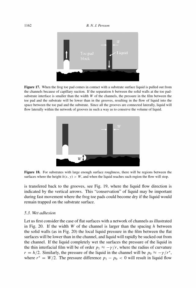
We now consider (qualitatively) the liquid flow at the interface upon forming andbreaking the pad-substrate contact. When the frog toe pad comes in contact with asubstrate surface, liquid is pulled out from the channels because of capillary suction,see Fig. 17. If the separation h between the solid walls at the toe pad-substrateinterface is smaller than the width W of the channels, the pressure in the filmbetween the toe pad and the substrate will be lower than in the grooves, resultingin the flow of liquid into the space between the toe pad and the substrate. Sinceall the grooves are connected laterally, liquid will flow laterally within the networkof grooves in such a way as to conserve the volume of liquid. For substrates withlarge enough surface roughness, there will be regions between the surfaces wherethe height h(x, y) > W , and when the liquid reaches such region the flow will stop,see Fig. 18. During pull-off the liquid (or part of it) may be pulled back into thegrooves by capillary forces. That is, when the separation h becomes larger thanthe width W of the grooves, the free energy for the system is reduced if the liquid
1162 B. N. J. Persson
Figure 17. When the frog toe pad comes in contact with a substrate surface liquid is pulled out fromthe channels because of capillary suction. If the separation h between the solid walls at the toe pad-substrate interface is smaller than the width W of the channels, the pressure in the film between thetoe pad and the substrate will be lower than in the grooves, resulting in the flow of liquid into thespace between the toe pad and the substrate. Since all the grooves are connected laterally, liquid willflow laterally within the network of grooves in such a way as to conserve the volume of liquid.
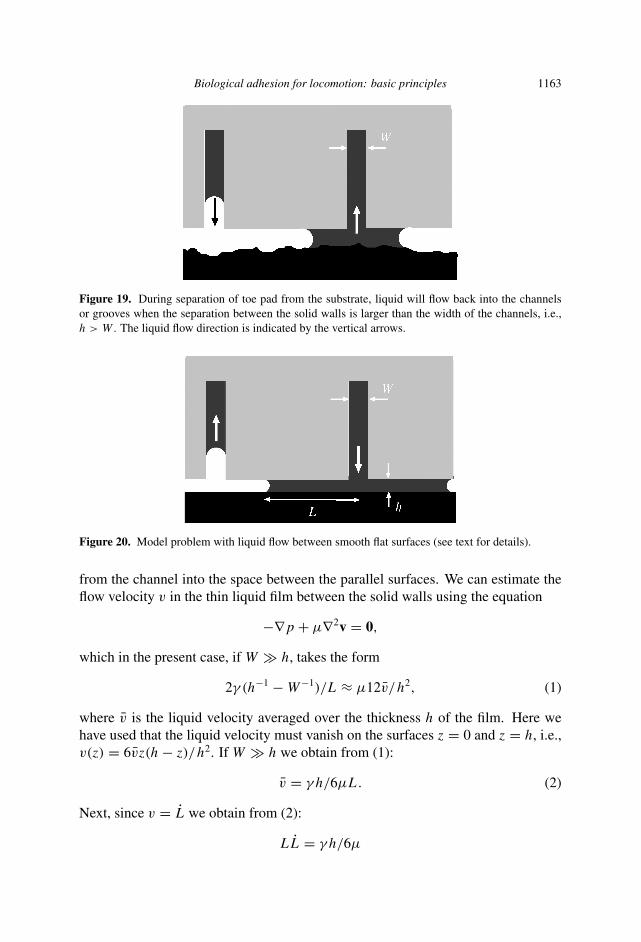
Figure 18. For substrates with large enough surface roughness, there will be regions between thesurfaces where the height h(x, y) > W , and when the liquid reaches such region the flow will stop.
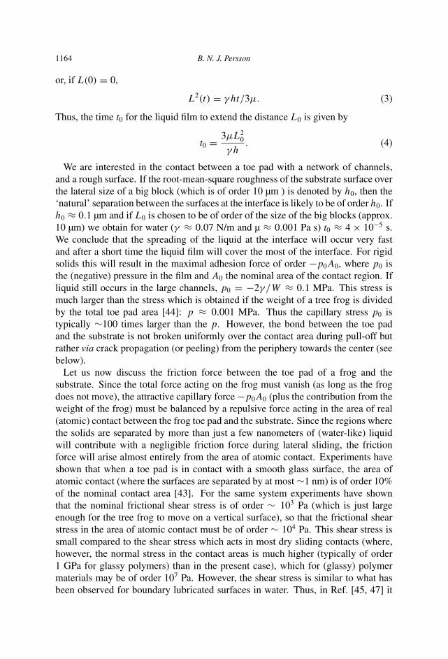
is transfered back to the grooves, see Fig. 19, where the liquid flow direction isindicated by the vertical arrows. This “conservation” of liquid may be importantduring fast movement where the frog toe pads could become dry if the liquid wouldremain trapped on the substrate surface.
5.3. Wet adhesion
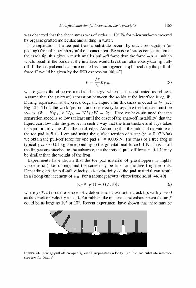
Let us first consider the case of flat surfaces with a network of channels as illustratedin Fig. 20. If the width W of the channel is larger than the spacing h betweenthe solid walls (as in Fig. 20) the local liquid pressure in the film between the flatsurfaces will be lower than in the channel, and liquid will rapidly be sucked out fromthe channel. If the liquid completely wet the surfaces the pressure of the liquid inthe thin interfacial film will be of order p1 ≈ −γ /r , where the radius of curvaturer = h/2. Similarly, the pressure of the liquid in the channel will be p0 ≈ −γ /r∗,where r∗ = W/2. The pressure difference p1 − p0 < 0 will result in liquid flow
Biological adhesion for locomotion: basic principles 1163
Figure 19. During separation of toe pad from the substrate, liquid will flow back into the channelsor grooves when the separation between the solid walls is larger than the width of the channels, i.e.,h > W . The liquid flow direction is indicated by the vertical arrows.
Figure 20. Model problem with liquid flow between smooth flat surfaces (see text for details).
from the channel into the space between the parallel surfaces. We can estimate theflow velocity v in the thin liquid film between the solid walls using the equation
−∇p + µ∇2v = 0,
which in the present case, if W � h, takes the form
2γ (h−1 − W−1)/L ≈ µ12v/h2, (1)
where v is the liquid velocity averaged over the thickness h of the film. Here wehave used that the liquid velocity must vanish on the surfaces z = 0 and z = h, i.e.,v(z) = 6vz(h − z)/h2. If W � h we obtain from (1):
v = γ h/6µL. (2)
Next, since v = L we obtain from (2):
LL = γ h/6µ
1164 B. N. J. Persson
or, if L(0) = 0,
L2(t) = γ ht/3µ. (3)
Thus, the time t0 for the liquid film to extend the distance L0 is given by
t0 = 3µL20
γ h. (4)
We are interested in the contact between a toe pad with a network of channels,and a rough surface. If the root-mean-square roughness of the substrate surface overthe lateral size of a big block (which is of order 10 µm ) is denoted by h0, then the‘natural’ separation between the surfaces at the interface is likely to be of order h0. Ifh0 ≈ 0.1 µm and if L0 is chosen to be of order of the size of the big blocks (approx.10 µm) we obtain for water (γ ≈ 0.07 N/m and µ ≈ 0.001 Pa s) t0 ≈ 4 × 10⁻⁵ s.We conclude that the spreading of the liquid at the interface will occur very fastand after a short time the liquid film will cover the most of the interface. For rigidsolids this will result in the maximal adhesion force of order −p0A0, where p0 isthe (negative) pressure in the film and A0 the nominal area of the contact region. Ifliquid still occurs in the large channels, p0 = −2γ /W ≈ 0.1 MPa. This stress ismuch larger than the stress which is obtained if the weight of a tree frog is dividedby the total toe pad area [44]: p ≈ 0.001 MPa. Thus the capillary stress p0 istypically ∼100 times larger than the p. However, the bond between the toe padand the substrate is not broken uniformly over the contact area during pull-off butrather via crack propagation (or peeling) from the periphery towards the center (seebelow).
Let us now discuss the friction force between the toe pad of a frog and thesubstrate. Since the total force acting on the frog must vanish (as long as the frogdoes not move), the attractive capillary force −p0A0 (plus the contribution from theweight of the frog) must be balanced by a repulsive force acting in the area of real(atomic) contact between the frog toe pad and the substrate. Since the regions wherethe solids are separated by more than just a few nanometers of (water-like) liquidwill contribute with a negligible friction force during lateral sliding, the frictionforce will arise almost entirely from the area of atomic contact. Experiments haveshown that when a toe pad is in contact with a smooth glass surface, the area ofatomic contact (where the surfaces are separated by at most ∼1 nm) is of order 10%of the nominal contact area [43]. For the same system experiments have shownthat the nominal frictional shear stress is of order ∼ 103 Pa (which is just largeenough for the tree frog to move on a vertical surface), so that the frictional shearstress in the area of atomic contact must be of order ∼ 104 Pa. This shear stress issmall compared to the shear stress which acts in most dry sliding contacts (where,however, the normal stress in the contact areas is much higher (typically of order1 GPa for glassy polymers) than in the present case), which for (glassy) polymermaterials may be of order 107 Pa. However, the shear stress is similar to what hasbeen observed for boundary lubricated surfaces in water. Thus, in Ref. [45, 47] it
Biological adhesion for locomotion: basic principles 1165
was observed that the shear stress was of order ∼ 104 Pa for mica surfaces coveredby organic grafted molecules and sliding in water.
The separation of a toe pad from a substrate occurs by crack propagation (orpeeling) from the periphery of the contact area. Because of stress concentration atthe crack tip, this gives a much smaller pull-off force than the force −p0A0 whichwould result if the bonds at the interface would break simultaneously during pull-off. If the toe pad can be approximated as a homogeneous spherical cup the pull-offforce F would be given by the JKR expression [46, 47]
F = 3π2
Rγeff, (5)
where γeff is the effective interfacial energy, which can be estimated as follows.Assume that the (average) separation between the solids at the interface h � W .During separation, at the crack edge the liquid film thickness is equal to W (seeFig. 21). Thus, the work (per unit area) necessary to separate the surfaces must beγeff ≈ (W − h)p0 ≈ Wp0 = W2γ /W = 2γ . Here we have assumed that theseparation speed is so low (at least until the onset of the snap-off instability) that theliquid can flow into the grooves in such a way that the film thickness always takesits equilibrium value W at the crack edge. Assuming that the radius of curvature ofthe toe pad is R ≈ 1 cm and using the surface tension of water (γ ≈ 0.07 N/m)we obtain the pull-off force for one pad F ≈ 0.006 N. The mass of a tree frog istypically m ∼ 0.01 kg corresponding to the gravitational force 0.1 N. Thus, if allthe fingers are attached to the substrate, the theoretical pull-off force ∼ 0.1 N maybe similar than the weight of the frog.
Experiments have shown that the toe pad material of grasshoppers is highlyviscoelastic (like rubber), and the same may be true for the tree frog toe pads.Depending on the pull-off velocity, viscoelasticity of the pad material can resultin a strong enhancement of γeff. For a (homogeneous) viscoelastic solid [48, 49]
γeff ≈ γ0(1 + f (T , v)
), (6)
where f (T , v) is due to viscoelastic deformation close to the crack tip, with f → 0as the crack tip velocity v → 0. For rubber-like materials the enhancement factor f
could be as large as 103 or 104. Recent experiment have shown that there may be
Figure 21. During pull-off an opening crack propagates (velocity v) at the pad-substrate interface(see text for details).
1166 B. N. J. Persson
a similar enhancement factor for the toe pad of grasshoppers and most likely forsmooth adhesive pads in general. Thus, experiments by Goodwyn et al. [42] foundγeff ≈ 10 J/m2 for the toe pads of two different types of grasshoppers, and since oneexpect γ ≈ 0.07 J/m2 due to capillary bridges, one obtains f ≈ 140 at the cracktip velocities probed in the experiments by Goodwyn et al. This would result in astrongly enhanced pull-off force which would allow the tree frog to adhere to evenvery rough surfaces inclined at any angle relative to the earth gravitational force.
The toe pad bulk viscoelasticity, which may result in a strong increase in γeff,may also be importance for sliding friction on rough substrates, and may result invery large sliding friction as observed for rubber materials. Thus, during slidingthe substrate asperities generate pulsating deformations of the pad material and ifthe pad material behaves as viscoelastic at the perturbing frequencies, a very largefriction may result as observed for rubber sliding on rough substrates [1, 50]. Wenote that this is the case even if the pad and the substrate are separated by a verythin viscous liquid film, assuming that the film thickness is smaller than the size ofthe (relevant) substrate asperities. This effect has, in fact, been observed in a recentexperiment for rubber lubricated by different organic oils and sliding on a roughsubstrate [51].
5.4. Squeeze-out
Tree frogs can adhere and move on rough (hard) vertical surfaces during heavyrain where the surfaces are flooded with water. This cannot be explained by thecapillary bridge picture since no capillary bridges can form on a flooded surface.We will discuss how the adhesion may be generated for flooded surfaces. Here wefirst consider the liquid squeeze-out, which is a prerequirement for non-negligibleadhesion and friction.
Consider the squeeze-out of liquid from the space between two solid bodies. As-sume first rigid solids with perfectly flat surfaces without draining channels. Sincein the present applications the pressure is low, we can assume an incompressibleliquid so that
∇ · v ≈ 0, (7)
−∇p + µ∇2v ≈ 0. (8)
Assume that h(t) is the separation between the surfaces at time t . We will use simple(dimensional) arguments to obtain an approximate form of h(t). Assume that thenominal contact region is circular with the diameter D0. Assume that h(t) changesby the amount �h < 0 during the time interval �t . Liquid mass conservation gives
−D20�h ≈ D0hv�t
or
h ≈ −hv/D0, (9)
Biological adhesion for locomotion: basic principles 1167
where v stands for the radial component of the liquid velocity averaged over thethickness 0 < z < h of the liquid film. Since the flow velocity vanishes on thesurfaces z = 0 and z = h the strongest spatial variation in v(x, t) will be derivedfrom the variation of v with z so that, from dimensional arguments, ∇2v ∼ v/h2.Thus, (8) gives p/D0 ≈ µv/h2, where p = FN/πD2
0 is the squeezing pressure.Combining this with (9) gives
h ≈ − αp
µD20
h3, (10)
where α is a number of order unity. An accurate calculation gives α = 4/3π. If theexternal load FN is constant it is easy to integrate (10) to obtain
1
h2(t)− 1
h2(0)≈ αpt
µD20
. (11)
Next let us assume that the substrate surface has vertical draining channels as inFig. 20. Let us first consider the situation where h is so small (but not too small— see below) that nearly all the squeeze-out of the liquid occurs via the channels.Consider first the flow in one channel. We assume that the height H1 of the channelis much larger than its width W1, see Fig. 20. In this case we expect the strongestspatial variation of v(x, t) to be derived from the variation of v with y so that, fromdimensional arguments, ∇2v ∼ v/W 2
1 , where v is the flow velocity in the channel,averaged over the channel cross-sectional area H1W1. Thus, (8) takes the form
p/D0 ≈ µv/W 21 . (12)
Let us now assume a network of channels on the surface forming a square (orhexagonal) lattice with the ‘lattice constant’ D1. Liquid mass conservation gives−hD2
0 ≈ NvH1W1, where N ≈ D0/D1 is the number of channels crossing theouter boundary of the nominal contact area. Thus we obtain
h ≈ −vH1W1
D0D1. (13)
From (12) and (13) we obtain
h ≈ −pW 31 H1
µD20D1
= − αp
µD20
h30, (14)
where
h0 =(
βW 3
1 H1
D1
)1/3
. (15)
and the dimensionless number β is of order unity. We can interpolate smoothlybetween the limits (10) and (14) using
h ≈ − αp
µD20
(h + h0)3. (16)
1168 B. N. J. Persson
Thus, the draining channels will effectively increase the separation between thesurfaces with the distance h0, and hence facilitate the squeeze-out. Equation (16) isonly valid until the film thickness h reaches some lower critical value h1 which canbe determined as follows. For h > h1 (but h < h0) the ‘bottleneck’ for squeeze-outis the viscous resistance to liquid flow in the channels. For h < h1 the ‘bottleneck’for squeeze-out is instead the viscous squeeze-out (transfer) of the liquid from theblock-substrate D1 × D1 interface area to the channels. To study this quantitatively,let us consider the squeeze-out of the liquid film from a basic unit (area ∼ D2
1) tothe surrounding draining channels. If the film is very thin the squeeze-out is veryslow and the liquid pressure in the draining channels will be similar to the external(atmospheric) pressure. In this case the squeeze-out of the thin liquid film into thedraining channels will be mathematically identical to the squeeze-out of the liquidbetween smooth surfaces studied above [equation (10)], but with D0 replaced by D1.Thus, for a very thin liquid film we have
h ≈ − αp
µD21
h3. (17)
We can determine h1 by the condition that the squeeze rates (14) and (17) are equal:
h31/D
21 ≈ h3
0/D20
or
h1 = h0
(D1
D0
)2/3
=(
βW 31 H1D1
D20
)1/3
. (18)
From the analysis above it is clear that if the squeeze-pressure p (or the force FN) isconstant the liquid film thickness will first decrease with time as ∼ t−1/2 until h(t)
reaches ∼h0 which takes the time
t0 ≈ µD20
αph20
. (19)
From here on the squeeze-out will occur mainly via the draining channels, andh(t) will decrease linearly with time until h(t) ≈ h1. If h1 � h0 the time t1 ittakes to decrease h(t) from h0 to h1 will be [from (14)] of order t1 ≈ t0 so thetotal squeeze-out time to reach h = h1 will be of order 2t0. If the basic D1 × D1
units have perfectly flat surfaces, for t > 2t0 the squeeze-out will again follow thet−1/2 time dependence. However, the squeeze-out will occur faster if the D1 × D1
surface units have draining channels with appropriate width W2, depth H2 anddensity. It is clear that for maximum squeeze-out speed the system has a hierarchicaldistribution of draining channels where a basic unit surrounded by ‘large’ drainingchannels has a network of much smaller draining channels and so on. The theoryabove can be used to estimate the squeeze-out time for such complex hierarchicalsystems. We also note that, to some extent, the channels can be replaced by surfaceroughness. However, in this case the squeeze-out channels will not have a uniform
Biological adhesion for locomotion: basic principles 1169
size but will exhibit strong fluctuations leading to the possibility of trapped liquid(liquid islands), in particular when the elastic deformation of the solids is taken intoaccount. Such trapped or “sealed off” water islands have recently been suggestedto be the origin of why tires on wet roads at low car velocities exhibit ∼ 20–30 %lower friction than for dry road surfaces (the trapped water effectively smoothensthe road surface profile resulting in less asperity-induced viscoelastic deformationof the rubber).
As an application, consider the tree frog toe pad. In this case D0 ≈ 1 mm,D1 ≈ 10 µm, H1 ≈ 5 µm and W1 ≈ 1 µm. Thus, h0 ≈ 1 µm and h1 ≈ 50 nm. Usingthe measured viscosity (similar to that of water) µ = 0.0014 Pa s and p = 104 Pa(typical frog toe squeezing stress) we obtain the squeeze-out time 2t0 ≈ 0.1 s.
5.5. Adhesion on flooded surfaces
It has been observed that tree frogs are able to adhere and move on vertical solidwalls also during heavy rain where the substrate surface is flooded by water [44].The reason for this is non-trivial because under flooded conditions it appears that nocapillary bridges can form and one would, therefore, expect that only a negligibleforce will be required to separate the surfaces, at least during slow separation. Herethis remarkable problem is analyzed and some explanations are suggested.
5.5.1. Long-range interactions between solids in liquids. Solid surfaces in watersometimes interact with long-range forces derived from ion adsorption on theirsurfaces [24]. Such forces can be both attractive (if the charges of the adsorbedions on the two surfaces have opposite sign) or repulsive [24]. However, it is veryunlikely that such forces are of any relevance for attachment systems in animalsbecause animals must be able to adhere to many different types of surfaces (suchas stone or leaf) with very different properties, and it is highly unlikely that thesesurfaces, if at all charged, would have the same sign of the charges (and opposite tothat of the animal toe pad surface).
The long-range van der Waals interaction will also act between solids separated bya thin water layer. While the van der Waals interaction always is attractive betweensolids in vacuum, it can be either attractive or repulsive in a liquid [31]. However,it is highly unlikely that this interaction is important for animals which secrete aliquid because if it would be important in water, it would (usually) be even moreimportant when no liquid separates the surfaces, and the animal would not need tosecrete any liquid at all. Thus, it is highly unlikely that any long-range interactionis of important for animal locomotion on water covered surfaces.
5.5.2. Dewetting transition. Complete liquid removal from the region betweenclosely spaced solids has been studied both experimentally and theoretically forseveral years [25–29]. A liquid film confined between two elastic solids with flatsurfaces is thermodynamically unstable if
γ1L + γ2L − γ12 > 0, (20)
1170 B. N. J. Persson
where γ1L and γ2L are the solid–liquid interfacial energies and γ12 the solid–solidinterfacial energy. In this case squeeze-out of the liquid may start by the formation(due to a thermal fluctuation) of a small dry patch, which then spreads laterally untilthe whole liquid film is expelled. However, for water this relation is unlikely to beobeyed for all surfaces to which the animal must be able to adhere. Thus stones,for example, are likely to have polar surfaces which are wetted by water, and it isunlikely that (20) will be obeyed for these substrates. In addition, if the liquid wouldbe removed by a dewetting transition, then the contact region would be dry but wealready know that the adhesion for the dry contact most likely is negligible (it is forthis reason that the tree frog injects a wetting liquid into the contact area).
5.5.3. Viscous ‘adhesion’. When two closely spaced surfaces are separatedrapidly in a liquid, strong effective adhesion may occur between the solids. Theorigin of this effect is the viscosity of the liquid: because of the viscosity, if theseparation between the surfaces is very small it will take long time for the liquidto flow into the ‘empty space’ generated during the separation between the solids.This can result in a large negative pressure and even cavity formation between thesurfaces of the solids [23, 52, 53]. This ‘viscous adhesion’ is a dynamical effectand disappears if the surfaces are separated very slowly. For rigid flat walls themagnitude of the attraction can be estimated from (10): when h > 0 (separation)(10) gives p < 0 i.e., an effective attraction prevails between the solid surfacesduring separation. A large pull-off force is only observed if the separation h betweenthe solids is very small (or the pull-off speed very high). That is, before strongadhesion is possible the liquid must be nearly completely removed (squeezed-out)from the region between the surfaces. An accurate analysis of this problem requiresin general that one include the elastic deformation of the solids when determiningthe pull-off force.
In Section 5.4 we showed that the squeeze-out was facilitated by a network ofdraining channels on the the surface of the adhesive pad. Here we note that whilethese channels are ‘open’ during squeeze-out they may be closed during pull-off, atleast close to the boundary of the contact region. The reason for this is that duringpull-off there is lower pressure inside the contact area than outside, and there will belateral (radial) forces acting tending to compress the contact area laterally, and thismay close the space between the hexagonal units. This will slow down the flow ofliquid into the region between the surfaces, which may strongly increase the pull-offforce.
5.6. Detachment
The tree frog toe pads are detached from surfaces by peeling, the pads being re-moved from the rear forwards during forward locomotion up a vertical surface [54].When the frog is induced to walk backwards down, peeling occurs from the frontof the pad rearwards. Experiments have shown that during forward locomotion up a
Biological adhesion for locomotion: basic principles 1171
vertical slope the detachment forces are much smaller than during backward walk-ing down the slope. That peeling occurs automatically during forward locomotion issupported by the observation that frogs on a rotating vertical surface adjust their ori-entation back towards the vertical whenever their deviation from the vertical reaches∼ 85◦. During forward locomotion peeling occurs as a natural consequence of theway in which the toes are lifted off surfaces from the rear forwards, while duringbackward locomotion it is an active process involving the distal tendons of the toes.
6. SUMMARY AND CONCLUSION
All natural surfaces have surface roughness on many different length scales. Naturalselection has optimized the toe pads of many animals for maximum adhesion tonatural surfaces. It is believed that the construction of the toe pads is mainly theresult of two principles:
(a) Maximum adhesion requires using non-compact solids built from thin fibersand plates (or walls).
(b) The fibers and plates (or walls) cannot be too thin as this would result incollapse of the structures, e.g., fiber condensation.
Tree frogs and most insects use wet adhesion to adhere and move on manydifferent surfaces, e.g., glass windows, stone walls, or plant leafs. Here we havediscussed the origin of adhesion and friction for the tree frog but the results may berelevant for other animals using smooth adhesive pads, e.g., grasshoppers. In fact,the similarity between the adhesive pads of tree frogs and grasshoppers is very great,indicating highly optimized (by natural selection) and unique adhesive systems.
Considerable theoretical work has been devoted to adhesion to perfectly smoothsubstrates. For bioadhesion this special case is uninteresting since natural surfaceshave always roughness, and it is only when this fact is taken into account that onecan understand why natural selection has generated the adhesive structures observedin animals which rely on sticky toe pads for locomotion.
Acknowledgements
Tha author thanks K. Autumn, W. Federle and S. Gorb for useful communications.Also the Editors are thanked for drawing my attention to Ref. [33].
REFERENCES
1. B. N. J. Persson, O. Albohr, U. Tartaglino, A. I. Volokitin and E. Tosatti, J. Phys. Condens.Matter 17, R1 (2005).
2. K. Kendall, Molecular Adhesion and its Applications. Kluwer, New York (2001).3. K. N. G. Fuller and D. Tabor, Proc. Roy. Soc. A 345, 327 (1975).4. B. N. J. Persson, Eur. Phys. J. E 8, 385 (2002).5. B. N. J. Persson, Surf. Sci. Reports 61, 201 (2006).
1172 B. N. J. Persson
6. V. N. Samoilov, I. M. Sivebaek and B. N. J. Persson, J. Chem. Phys. 121, 9639 (2004).7. K. R. Shull and C. Creton, J. Polym. Sci. B 42, 4023 (2004).8. B. N. J. Persson, O. Albohr, C. Creton and V. Peveri, J. Chem. Phys. 120, 8779 (2004).9. S. Gorb, Attachment Devices of Insect Curtile. Kluwer, Dordrecht (2001).
10. M. Scherge and S. Gorb, Biological Micro- and Nano-Tribology. Springer, Berlin (2001).11. B. N. J. Persson, J. Chem. Phys. 118, 7614 (2003).12. T. W. Kim and B. Bhushan, J. Adhesion Sci. Technol. 21, 1 (2007).13. C. Majidi, R. E. Groff, Y. Maeno, B. Schubert, S. Baek, B. Bush, R. Maboudian, N. Gravish,
M. Wilkinson, K. Autumn and R. S. Fearing, Phys. Rev. Lett. 97, 076103 (2006).14. B. N. J. Persson and S. Gorb, J. Chem. Phys. 119, 11437 (2004).15. C. Carbone and B. N. J. Persson, Phys. Rev. B 70, 125407 (2004).16. F. Haas and S. Gorb, Arthropod Structure & Developement 33, 45 (2004).17. W. Federle, J. Expl. Biol. 209, 2611 (2006).18. H. Yao and H. Gao, J. Mech. Phys. Solids 54, 1120 (2006).19. E. Arzt, S. Gorb and R. Spolenak, Proc. Natl. Acad. Sci. (USA) 100, 10603 (2003).20. K. Autumn, M. Sitti, Y. A. Liang, A. M. Peattie, W. R. Hansen, S. Sponberg, T. W. Kenny,
R. Fearing, J. N. Israelachvili and R. J. Full, Proc. Natl. Acad. Sci. (USA) 99, 12252 (2002).21. S. N. Gorb, Proc. R. Soc. Lond. B 265, 747 (1998). (In this reference it is shown that flies lose
the ability to walk on inclined surfaces after walking for 15–30 minutes on a silica gel substrate.This can be understood if it is assumed that walking on the silica gel drains away all the adhesiveliquid.)
22. B. N. J. Persson, MRS Bull. 32, 486 (2007).23. B. N. J. Persson, Sliding Friction: Physical Principles and Applications, 2nd edn. Springer,
Heidelberg (2000).24. B. N. J. Persson and F. Mugele, J. Phys. Condens. Matter 16, R295 (2004).25. F. Brochard-Wyart and P. G. de Gennes, J. Phys. Condens. Matter 6, A9 (1994).26. P. Martin and F. Brochard-Wyart, Phys. Rev. Lett. 80, 3296 (1998).27. P. Martin, A. Buguin and F. Brochard-Wyart, Langmuir 17, 6553 (2001).28. B. N. J. Persson, A. Volokitin and E. Tosatti, Eur. Phys. J. E 11, 409 (2003).29. C. Carbone and B. N. J. Persson, J. Chem. Phys. 121, 2246 (2004).30. G. Huber, H. Mantz, R. Spolenak, K. Mecke, K. Jacobs, S. N. Gorb and E. Arzt, Proc. Natl.
Acad. Sci. (USA) 102, 16293 (2005).31. J. Israelachvili, Intermolecular and Surface Forces. Academic Press, London (1992).32. W. R. Hansen and K. Autumn, Proc. Natl. Acad. Sci. (USA) 102, 385 (2005).33. C. Y. Hui, L. Shen, A. Jagota and K. Autumn, Mechanics of anti-fouling or self-cleaning in
gecko setae, in: Proceedings of the 29th Annual Meeting of The Adhesion Society (2006).34. U. Hiller and R. Blaschke, Naturwissenschaften 54, 344 (1997).35. S. Gorb, Personal communication.36. Y. Tian, N. Pesika, H. Zeng, K. Rosenberg, B. Zhao, P. McGuiggan, K. Autumn and
J. Israelachvili, Proc. Natl. Acad. Sci. (USA) 103, 19320 (2006).37. D. J. Irschick, C. C. Austin, K. Petren, R. N. Fisher, J. B. Losos and O. Ellers, Biol. J. Linn. Soc.
Lond. 59, 21 (1996).38. R. G. Beutel and S. N. Gorb, J. Zool. Syst. Evol. Res. 39, 177 (2001).39. J. A. Coddington and H. W. Levi, Annu. Rev. Ecol. Syst. 22, 565 (1991).40. J. S. Rovner, Proc. Symp. Zool. Soc. Lond. 42, 99 (1978).41. W. Vötsch, G. Nicholson, R. Müller, Y.-D. Stierhof, S. Gorb and U. Schwarz, Insect Biochem.
Mol. Biol. 32, 1605 (2002).42. P. P. Goodwyn, A. Peressadko, H. Schwarz, V. Kastner and S. Gorb, J. Comp. Physiol A 192,
1233 (2006).43. W. Federle, W. J. P. Barnes, W. Baumgartner, P. Drechsler and J. M. Smith, J. Royal Soc.
Interface 3, 689 (2006).
Biological adhesion for locomotion: basic principles 1173
44. J. Barnes, Tire Technol. Intl, 42–47 (March 1999).45. W. H. Briscoe, S. Titmuss, F. Tiberg, R. K. Thomas, D. J. McGillivray and J. Klein, Nature 444,
191 (2006).46. K. L. Johnson, K. Kendall and A. D. Roberts, Proc. Royal Soc. A 325, 301 (1971).47. K. L. Johnson, Contact Mechanics. Cambridge University Press, Cambridge (1985).48. B. N. J. Persson, O. Albohr, G. Heinrich and H. Ueba, J. Phys.: Condens. Matter 17, R1071
(2005).49. B. N. J. Persson and E. Brener, Phys. Rev. E 71, 036123 (2005).50. B. N. J. Persson, J. Chem. Phys. 115, 3840 (2001).51. M. R. Mofidi, E. Kassfeldt and B. Prakash, http://www.ltu.se/forskning/1.16009?l=en&pureId=
300231&pureFamily=dk.atira.pure.model.Publication&printer=true52. A. M. Smith, I. Expl. Biol. 157, 257 (1991).53. F. Caupin and E. Herbert, C. R. Physique 7, 1000 (2006).54. G. Hanna and W. J. P. Barnes, J. Expl. Biol. 155, 103 (1991).


![Using a Biological Material to Improve Locomotion of ... · Locomotion of Hexapod Robots ... support, stability, and movement to ... [2,3] in order to approach animals in their levels](https://static.fdocuments.net/doc/165x107/5b16f60e7f8b9a6f218b8ad4/using-a-biological-material-to-improve-locomotion-of-locomotion-of-hexapod.jpg)