Bio Exam Girl Interrupted Bible. Ace This Shit
85
Chapter 4: Carbon and the Molecular Diversity of Life Organic chemistry is the study of compounds that contain carbon. Organic compounds range from simple molecules to colossal ones. Most organic compounds contain hydrogen atoms in addition to carbon atoms. Vitalism, the idea that organic compounds arise only in organisms, was disproved when chemists synthesized these compounds Mechanism is the view that all natural phenomena are governed by physical and chemical laws Stanley Miller’s classic experiment demonstrated the abiotic synthesis of organic compounds. Experiments support the idea that abiotic synthesis of organic compounds, perhaps near volcanoes, could have been a stage in the origin of life Bond Formation with Carbon With four valence electrons, carbon can form four covalent bonds with a variety of atoms. This ability makes large, complex molecules possible. In molecules with multiple carbons, each carbon bonded to four other atoms has a tetrahedral shape. However, when two carbon atoms are joined by a double bond, the atoms joined to the carbons are in the same plane as the carbons.
-
Upload
ghazalehatross -
Category
Documents
-
view
3 -
download
1
Transcript of Bio Exam Girl Interrupted Bible. Ace This Shit
Chapter 4: Carbon and the Molecular Diversity of Life Organic chemistry is the study of compounds that contain carbon. Organic compounds range from simple molecules to colossal ones. Most organic compounds contain hydrogen atoms in addition to carbon atoms.
Vitalism, the idea that organic compounds arise only in organisms, was disproved when chemists synthesized these compounds
Mechanism is the view that all natural phenomena are governed by physical and chemical laws
Stanley Miller’s classic experiment demonstrated the abiotic synthesis of organic compounds. Experiments support the idea that abiotic synthesis of organic compounds, perhaps near volcanoes, could have been a stage in the origin of life
Bond Formation with Carbon
With four valence electrons, carbon can form four covalent bonds with a variety of atoms. This ability makes large, complex molecules possible. In molecules with multiple carbons, each carbon bonded to four other atoms has a tetrahedral shape. However, when two carbon atoms are joined by a double bond, the atoms joined to the carbons are in the same plane as the carbons.
The valences of carbon and its most frequent partners (hydrogen, oxygen, and nitrogen) are the “building code” that governs the architecture of living molecules
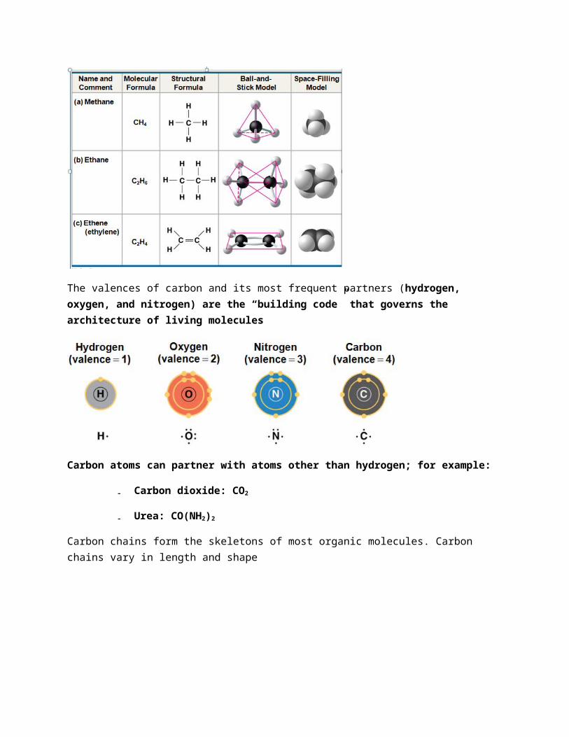
Carbon atoms can partner with atoms other than hydrogen; for example:
– Carbon dioxide: CO2
– Urea: CO(NH2)2
Carbon chains form the skeletons of most organic molecules. Carbon chains vary in length and shape
Hydrocarbons are organic molecules consisting of only carbon and hydrogen. Many organic molecules, such as fats, have hydrocarbon components. Hydrocarbons can undergo reactions that release a large amount of energy.
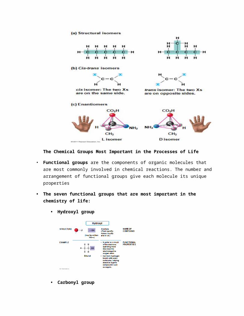
Isomers are compounds with the same molecular formula but different structures and properties
– Structural isomers have different covalent arrangements of their atoms
– Cis-trans isomers have the same covalent bonds but differ in spatial arrangements
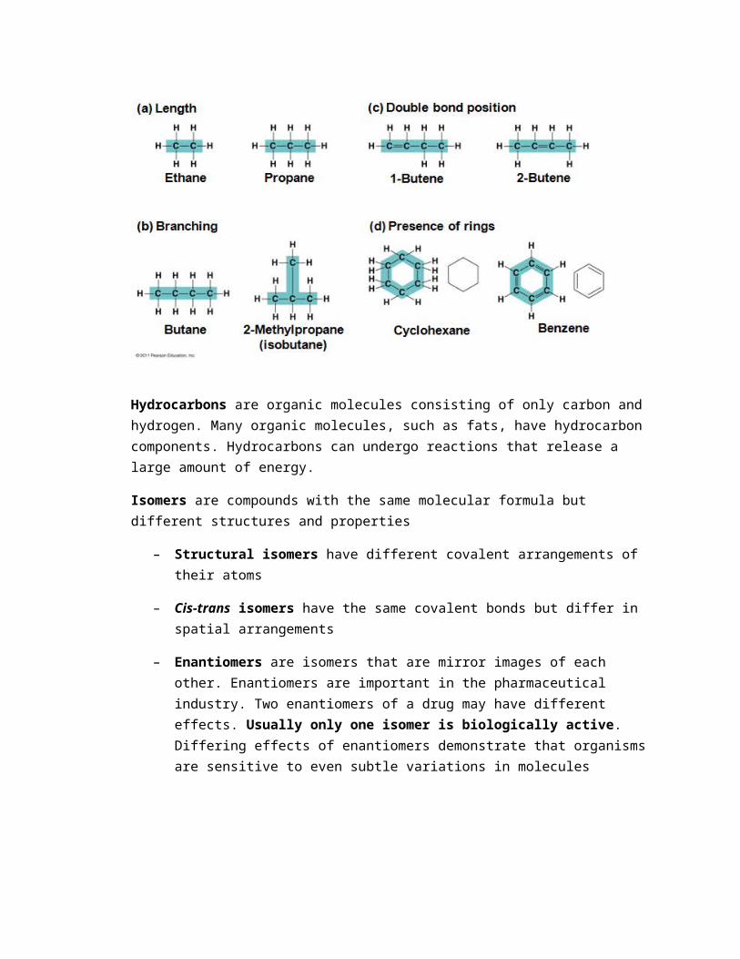
– Enantiomers are isomers that are mirror images of each other. Enantiomers are important in the pharmaceutical industry. Two enantiomers of a drug may have different effects. Usually only one isomer is biologically active. Differing effects of enantiomers demonstrate that organisms are sensitive to even subtle variations in molecules
The Chemical Groups Most Important in the Processes of Life
• Functional groups are the components of organic molecules that are most commonly involved in chemical reactions. The number and arrangement of functional groups give each molecule its unique properties
• The seven functional groups that are most important in the chemistry of life:
• Hydroxyl group
• Carbonyl group
• Carboxyl group
•
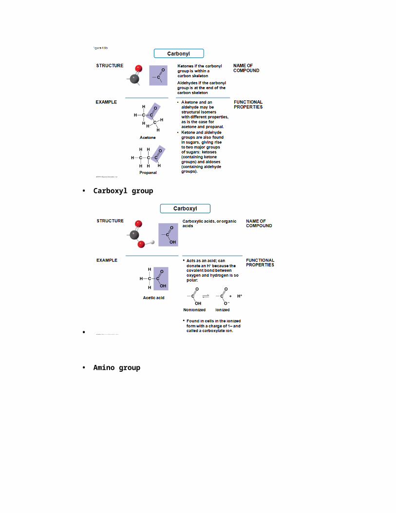
• Amino group
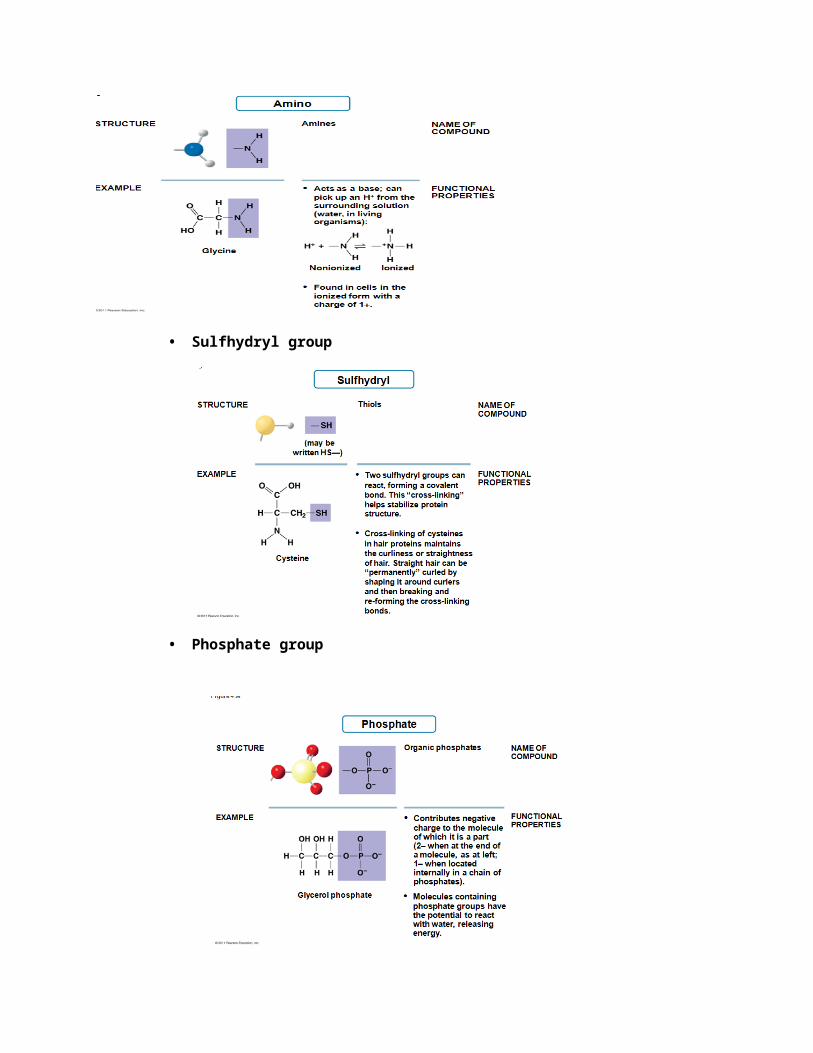
• Sulfhydryl group
• Phosphate group
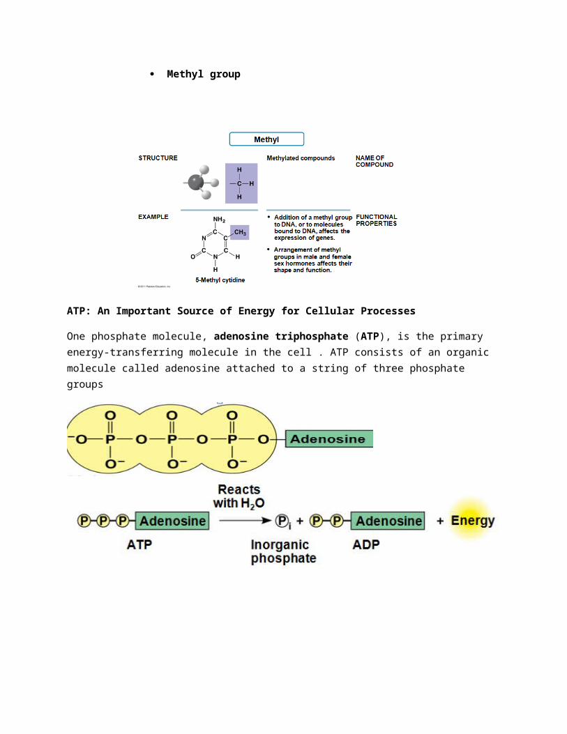
Methyl group
ATP: An Important Source of Energy for Cellular Processes
One phosphate molecule, adenosine triphosphate (ATP), is the primary energy-transferring molecule in the cell . ATP consists of an organic molecule called adenosine attached to a string of three phosphate groups
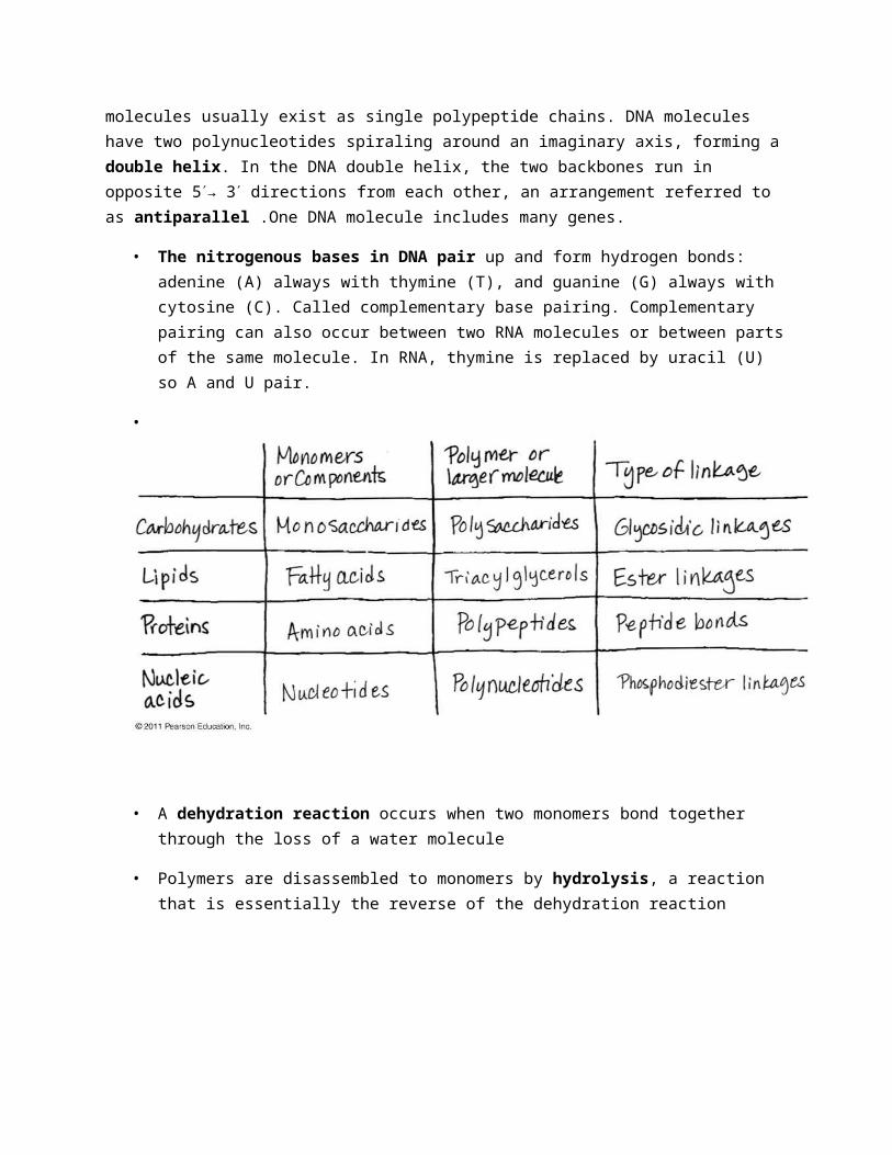
Chapter 5 (The Molecules of Life):All living things are made up of four classes of large biological molecules: carbohydrates, lipids, proteins, and nucleic acids. Macromolecules are large molecules composed of thousands of covalently connected atoms. Molecular structure and function are inseparable
A polymer is a long molecule consisting of many similar building blocks
• These small building-block molecules are called monomers
• Three of the four classes of life’s organic molecules are polymers
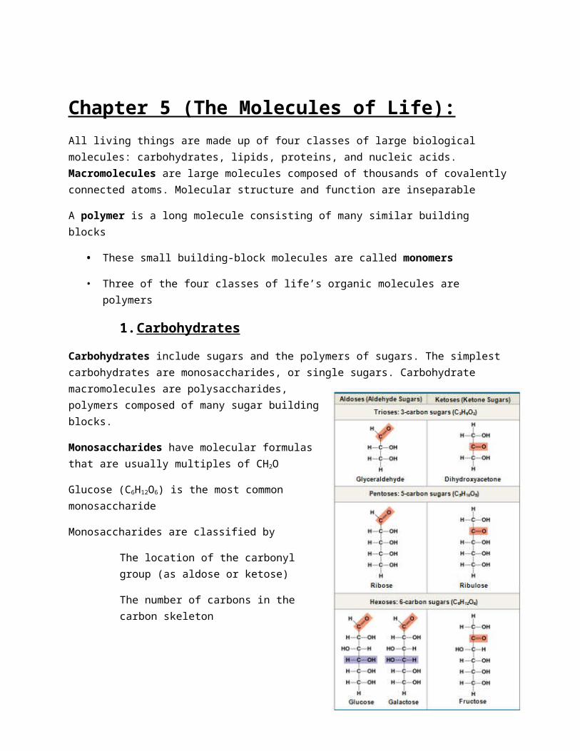
1. Carbohydrates
Carbohydrates include sugars and the polymers of sugars. The simplest carbohydrates are monosaccharides, or single sugars. Carbohydrate macromolecules are polysaccharides, polymers composed of many sugar building blocks.
Monosaccharides have molecular formulas that are usually multiples of CH2O
Glucose (C6H12O6) is the most common monosaccharide
Monosaccharides are classified by
The location of the carbonyl group (as aldose or ketose)
The number of carbons in the carbon skeleton
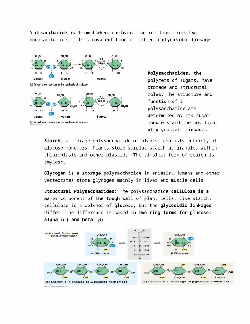
A disaccharide is formed when a dehydration reaction joins two monosaccharides . This covalent bond is called a glycosidic linkage
Polysaccharides, the polymers of sugars, have storage and structural
roles. The structure and function of a polysaccharide are determined by its sugar monomers and the positions of glycosidic linkages.
Starch, a storage polysaccharide of plants, consists entirely of glucose monomers. Plants store surplus starch as granules within chloroplasts and other plastids .The simplest form of starch is amylase.
Glycogen is a storage polysaccharide in animals. Humans and other vertebrates store glycogen mainly in liver and muscle cells
Structural Polysaccharides: The polysaccharide cellulose is a major component of the tough wall of plant cells. Like starch, cellulose is a polymer of glucose, but the glycosidic linkages differ. The difference is based on two ring forms for glucose: alpha (a) and beta (b)
• Polymers with a glucose are helical
• Polymers with b glucose are straight
• In straight structures, H atoms on one strand can bond with OH groups on other strands
• Parallel cellulose molecules held together this way are grouped into microfibrils, which form strong building materials for plants
Enzymes that digest starch by hydrolyzing a linkages can’t hydrolyze b linkages in cellulose
• Cellulose in human food passes through the digestive tract as insoluble fiber. Some microbes use enzymes to digest cellulose. Many herbivores, from cows to termites, have symbiotic relationships with these microbes. Chitin, another structural polysaccharide, is found in the exoskeleton of arthropods. Chitin also provides structural support for the cell walls of many fungi
2. Lipids are a diverse group of hydrophobic molecules
Lipids are the one class of large biological molecules that do not form polymers. The unifying feature of lipids is having little or no affinity for water. Lipids are hydrophobic because they consist mostly of hydrocarbons, which form nonpolar covalent bonds. The most biologically important lipids are fats, phospholipids, and steroids.
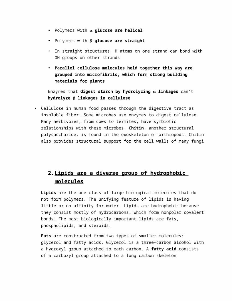
Fats are constructed from two types of smaller molecules: glycerol and fatty acids. Glycerol is a three-carbon alcohol with a hydroxyl group attached to each carbon. A fatty acid consists of a carboxyl group attached to a long carbon skeleton
Fats separate from water because water molecules form hydrogen bonds with each other and exclude the fats. In a fat, three fatty acids are joined to glycerol by an ester linkage, creating a triacylglycerol, or triglyceride.
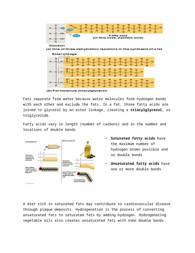
Fatty acids vary in length (number of carbons) and in the number and locations of double bonds
• Saturated fatty acids have the maximum number of hydrogen atoms possible and no double bonds
• Unsaturated fatty acids have one or more double bonds
A diet rich in saturated fats may contribute to cardiovascular disease through plaque deposits. Hydrogenation is the process of converting unsaturated fats to saturated fats by adding hydrogen. Hydrogenating vegetable oils also creates unsaturated fats with trans double bonds. These trans fats may contribute more than saturated fats to cardiovascular disease
Phospholipids:
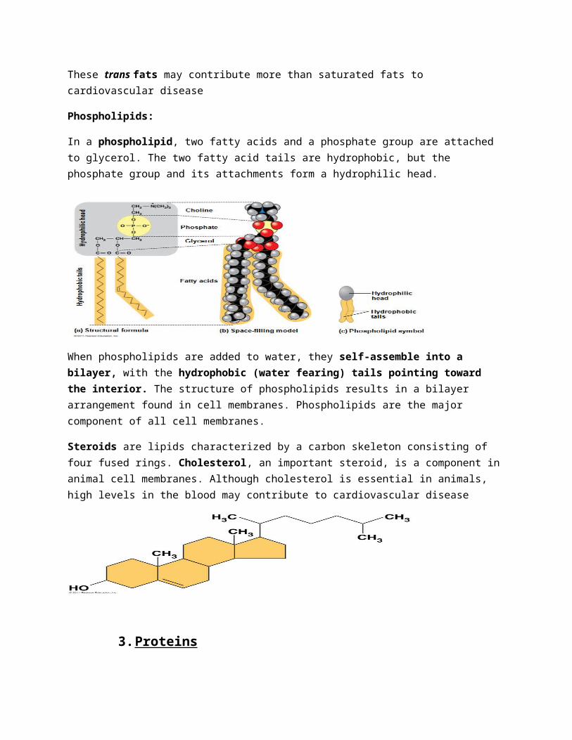
In a phospholipid, two fatty acids and a phosphate group are attached to glycerol. The two fatty acid tails are hydrophobic, but the phosphate group and its attachments form a hydrophilic head.
When phospholipids are added to water, they self-assemble into a bilayer, with the hydrophobic (water fearing) tails pointing toward the interior. The structure of phospholipids results in a bilayer arrangement found in cell membranes. Phospholipids are the major component of all cell membranes.
Steroids are lipids characterized by a carbon skeleton consisting of four fused rings. Cholesterol, an important steroid, is a component in animal cell membranes. Although cholesterol is essential in animals, high levels in the blood may contribute to cardiovascular disease
3. Proteins
Proteins account for more than 50% of the dry mass of most cells
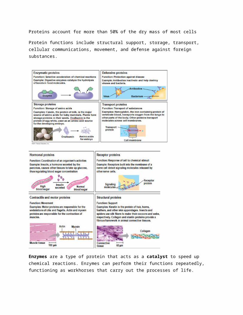
Protein functions include structural support, storage, transport, cellular communications, movement, and defense against foreign substances.
Enzymes are a type of protein that acts as a catalyst to speed up chemical reactions. Enzymes can perform their functions repeatedly, functioning as workhorses that carry out the processes of life.
Polypeptides are unbranched polymers built from the same set of 20 amino acids. A protein is a biologically functional molecule that consists of one or more polypeptides
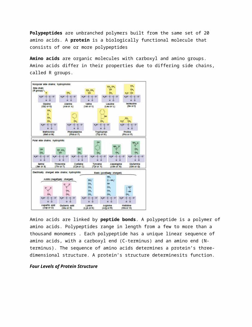
Amino acids are organic molecules with carboxyl and amino groups. Amino acids differ in their properties due to differing side chains, called R groups.
Amino acids are linked by peptide bonds. A polypeptide is a polymer of amino acids. Polypeptides range in length from a few to more than a thousand monomers . Each polypeptide has a unique linear sequence of amino acids, with a carboxyl end (C-terminus) and an amino end (N-terminus). The sequence of amino acids determines a protein’s three-dimensional structure. A protein’s structure determinesits function.
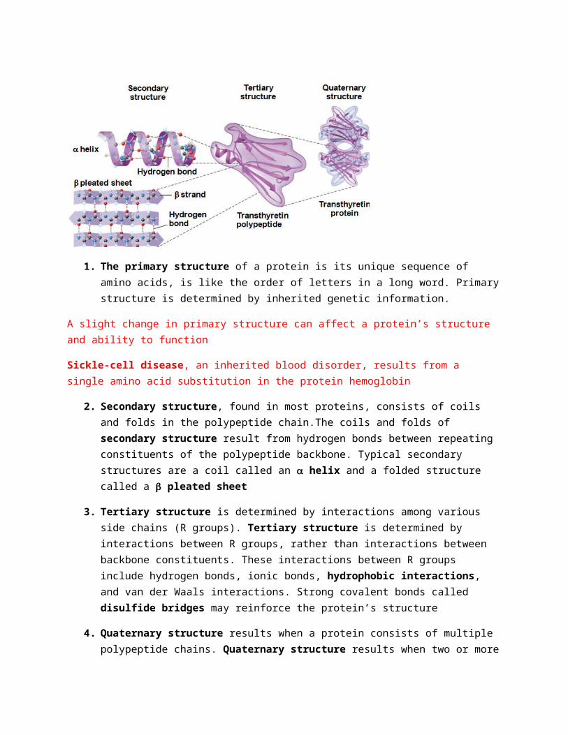
Four Levels of Protein Structure
1. The primary structure of a protein is its unique sequence of amino acids, is like the order of letters in a long word. Primary structure is determined by inherited genetic information.
A slight change in primary structure can affect a protein’s structure and ability to function
Sickle-cell disease, an inherited blood disorder, results from a single amino acid substitution in the protein hemoglobin
2. Secondary structure, found in most proteins, consists of coils and folds in the polypeptide chain.The coils and folds of secondary structure result from hydrogen bonds between repeating constituents of the polypeptide backbone. Typical secondary structures are a coil called an a helix and a folded structure called a b pleated sheet
3. Tertiary structure is determined by interactions among various side chains (R groups). Tertiary structure is determined by interactions between R groups, rather than interactions between backbone constituents. These interactions between R groups include hydrogen bonds, ionic bonds, hydrophobic interactions, and van der Waals interactions. Strong covalent bonds called disulfide bridges may reinforce the protein’s structure
4. Quaternary structure results when a protein consists of multiple polypeptide chains. Quaternary structure results when two or more polypeptide chains form one macromolecule. Collagen is a fibrous protein consisting of three polypeptides coiled like a rope. Hemoglobin is a globular protein consisting of four polypeptides: two alpha and two beta chains
In addition to primary structure, physical and chemical conditions can affect structure. Alterations in pH, salt concentration, temperature, or other environmental factors can cause a protein to unravel. This loss of a protein’s native structure is called denaturation .A denatured protein is biologically inactive.
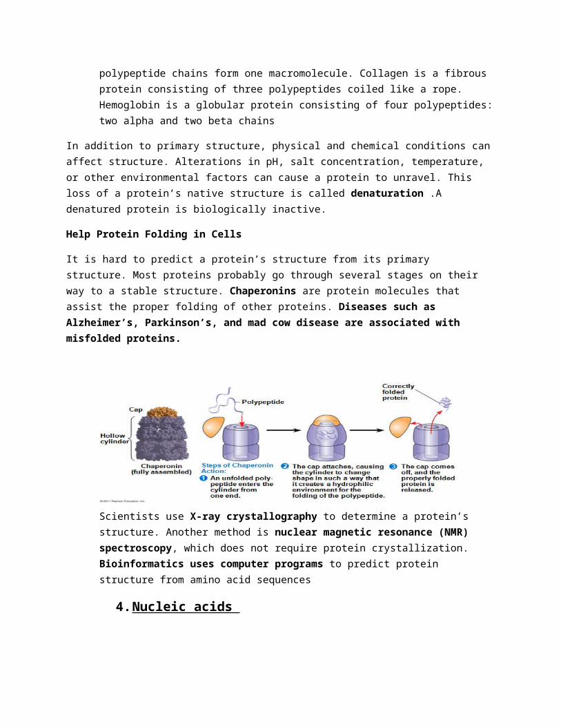
Help Protein Folding in Cells
It is hard to predict a protein’s structure from its primary structure. Most proteins probably go through several stages on their way to a stable structure. Chaperonins are protein molecules that assist the proper folding of other proteins. Diseases such as Alzheimer’s, Parkinson’s, and mad cow disease are associated with misfolded proteins.
Scientists use X-ray crystallography to determine a protein’s structure. Another method is nuclear magnetic resonance (NMR) spectroscopy, which does not require protein crystallization. Bioinformatics uses computer programs to predict protein structure from amino acid sequences
4. Nucleic acids
The amino acid sequence of a polypeptide is programmed by a unit of inheritance called a gene. Genes are made of DNA, a nucleic acid made of monomers called nucleotides.
There are two types of nucleic acids
– Deoxyribonucleic acid (DNA)
– Ribonucleic acid (RNA)
DNA provides directions for its own replication
DNA directs synthesis of messenger RNA (mRNA) and, through mRNA, controls protein synthesis
Protein synthesis occurs on ribosomes.
Nucleic acids are polymers called polynucleotides .Each polynucleotide is made of monomers called nucleotides. Each nucleotide consists of a nitrogenous base, a pentose sugar, and one or more phosphate groups. The portion of a nucleotide without the phosphate group is called a nucleoside.
Nucleoside = nitrogenous base + sugar
There are two families of nitrogenous bases
– Pyrimidines (cytosine, thymine, and uracil) have a single six-membered ring
– Purines (adenine and guanine) have a six-membered ring fused to a five-membered ring
In DNA, the sugar is deoxyribose; in RNA, the sugar is ribose
Nucleotide = nucleoside + phosphate group
Nucleotide polymers are linked together to build a polynucleotide. Adjacent nucleotides are joined by covalent bonds that form between the —OH group on the 3¢ carbon of one nucleotide and the phosphate on the 5¢ carbon on the next. These links create a backbone of sugar-phosphate units with nitrogenous bases as appendages. The sequence of bases along a DNA or mRNA polymer is unique for each gene. RNA molecules usually exist as single polypeptide chains. DNA molecules have two polynucleotides spiraling around an imaginary axis, forming a double helix. In the DNA double helix, the two backbones run in opposite 5¢→ 3¢ directions from each other, an arrangement referred to as antiparallel .One DNA molecule includes many genes.
• The nitrogenous bases in DNA pair up and form hydrogen bonds: adenine (A) always with thymine (T), and guanine (G) always with cytosine (C). Called complementary base pairing. Complementary pairing can also occur between two RNA molecules or between parts of the same molecule. In RNA, thymine is replaced by uracil (U) so A and U pair.
•
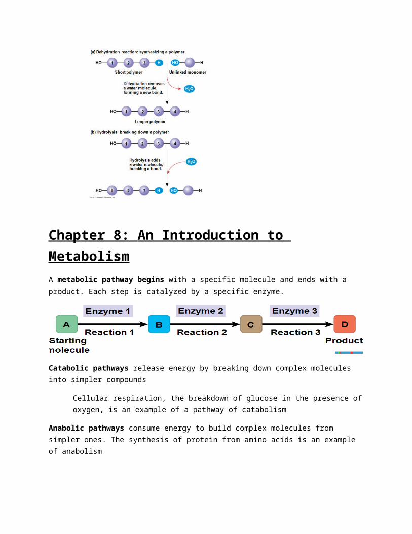
• A dehydration reaction occurs when two monomers bond together through the loss of a water molecule
• Polymers are disassembled to monomers by hydrolysis, a reaction that is essentially the reverse of the dehydration reaction
Chapter 8: An Introduction to Metabolism A metabolic pathway begins with a specific molecule and ends with a product. Each step is catalyzed by a specific enzyme.
Catabolic pathways release energy by breaking down complex molecules into simpler compounds
Cellular respiration, the breakdown of glucose in the presence of oxygen, is an example of a pathway of catabolism
Anabolic pathways consume energy to build complex molecules from simpler ones. The synthesis of protein from amino acids is an example of anabolism
Bioenergetics is the study of how organisms manage their energy resources
Forms of Energy
• Kinetic energy is energy associated with motion
• Heat (thermal energy) is kinetic energy associated with random movement of atoms or molecules
• Potential energy is energy that matter possesses because of its location or structure
• Chemical energy is potential energy available for release in a chemical reaction
*** Energy can be converted from one form to another
The Laws of Energy Transformation
Thermodynamics is the study of energy transformations. A isolated system, such as that approximated by liquid in a thermos, is isolated from its surroundings. In an open system, energy and matter can be transferred between the system and its surroundings. Organisms are open systems
1. According to the first law of thermodynamics, the energy of the universe is constant
– Energy can be transferred and transformed, but it cannot be created or destroyed
The first law is also called the principle of conservation of energy
2. During every energy transfer or transformation, some energy is unusable, and is often lost as heat. According to the second law of thermodynamics . Every energy transfer or transformation increases the entropy (disorder) of the universe
The evolution of more complex organisms does not violate the second law of thermodynamics. Entropy (disorder) may decrease in an organism, but the universe’s total entropy increases.
The free-energy change of a reaction tells us whether or not the reaction occurs spontaneously
Free-Energy Change, DG
A living system’s free energy is energy that can do work when temperature and pressure are uniform, as in a living cell.
The change in free energy, or Gibbs free energy, (∆G) during a process is related to the change in enthalpy, or change in total energy (∆H), change in entropy (∆S), and temperature in Kelvin (T)
∆G = ∆H – T∆S
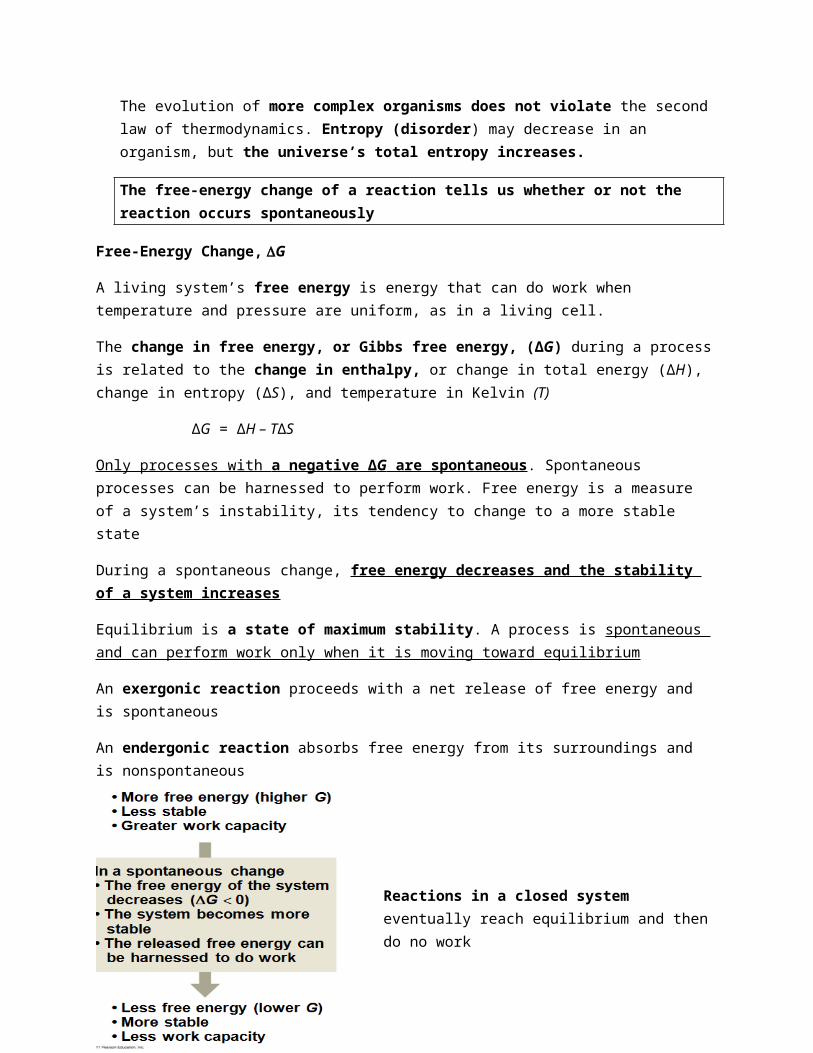
Only processes with a negative ∆ G are spontaneous . Spontaneous processes can be harnessed to perform work. Free energy is a measure of a system’s instability, its tendency to change to a more stable state
During a spontaneous change, free energy decreases and the stability of a system increases
Equilibrium is a state of maximum stability. A process is spontaneous and can perform work only when it is moving toward equilibrium
An exergonic reaction proceeds with a net release of free energy and is spontaneous
An endergonic reaction absorbs free energy from its surroundings and is nonspontaneous
Reactions in a closed system eventually reach equilibrium and then do no work
Cells are not in equilibrium; they are open systems experiencing a constant flow of materials
A defining feature of life is that metabolism is never at equilibrium
A catabolic pathway in a cell releases free energy in a series of reactions
Closed and open hydroelectric systems can serve as analogies
The Structure and Hydrolysis of ATP
ATP (adenosine triphosphate) is the cell’s energy shuttle
ATP is composed of ribose (a sugar), adenine (a nitrogenous base), and three phosphate groups
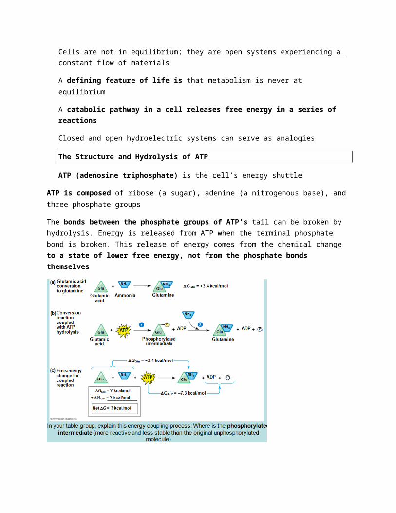
The bonds between the phosphate groups of ATP’s tail can be broken by hydrolysis. Energy is released from ATP when the terminal phosphate bond is broken. This release of energy comes from the chemical change to a state of lower free energy, not from the phosphate bonds themselves
ATP drives endergonic reactions by phosphorylation, transferring a phosphate group to some other molecule, such as a reactant. The recipient molecule is now called a phosphorylated intermediate (more reactive and less stable than the original unphosphorylated molecule)
The Regeneration of ATP
ATP is a renewable resource that is regenerated by addition of a phosphate group to adenosine diphosphate (ADP). The energy to phosphorylate ADP comes from catabolic reactions in the cell. The ATP cycle is a revolving door through which energy passes during its transfer from catabolic to anabolic pathways. SLIDE 44.
The Activation Energy Barrier
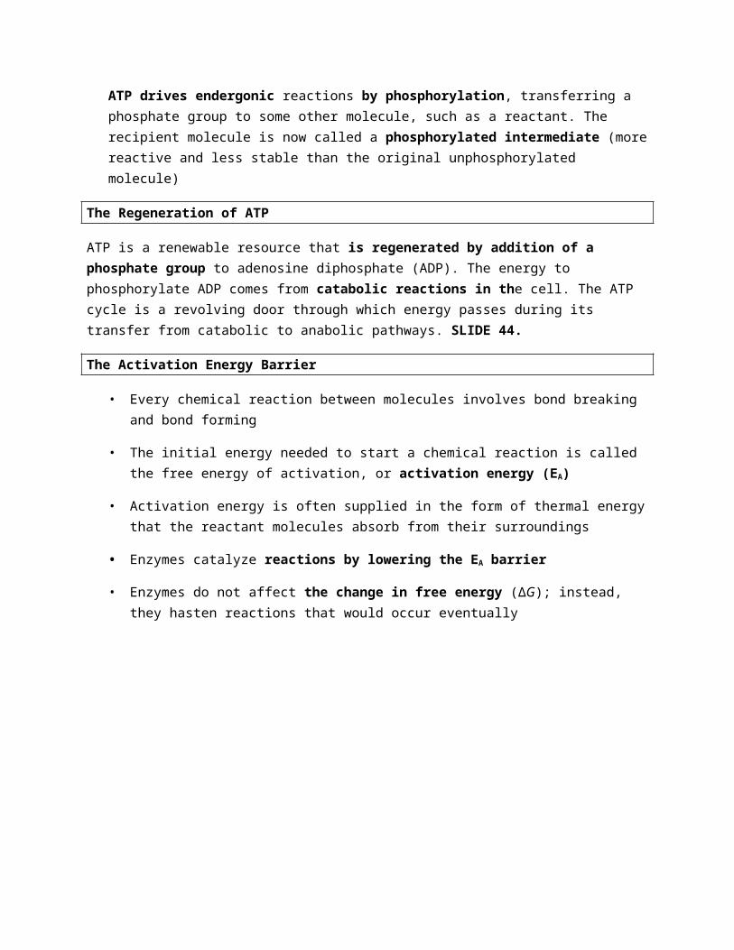
• Every chemical reaction between molecules involves bond breaking and bond forming
• The initial energy needed to start a chemical reaction is called the free energy of activation, or activation energy (EA)
• Activation energy is often supplied in the form of thermal energy that the reactant molecules absorb from their surroundings
• Enzymes catalyze reactions by lowering the EA barrier
• Enzymes do not affect the change in free energy (∆G); instead, they hasten reactions that would occur eventually
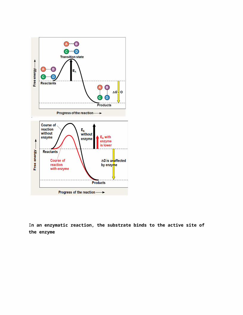
In an enzymatic reaction, the substrate binds to the active site of the enzyme
The active site can lower an EA barrier by
– Orienting substrates correctly
– Straining substrate bonds
– Providing a favorable microenvironment
(temp and PH) Each enzyme has an optimal temperature in which it can function. Each enzyme has an optimal pH in which it can function. Optimal conditions favor the most active shape for the enzyme molecule
Cofactors are nonprotein enzyme helpers. Cofactors may be inorganic (such as a metal in ionic form) or organic. An organic cofactor is called a coenzyme .Coenzymes include vitamins
– Covalently bonding to the substrate
Enzyme Inhibitors
• Competitive inhibitors bind to the active site of an enzyme, competing with the substrate
• Noncompetitive inhibitors bind to another part of an enzyme, causing the enzyme to change shape and making the active site less effective
• Examples of inhibitors include toxins, poisons, pesticides, and antibiotics
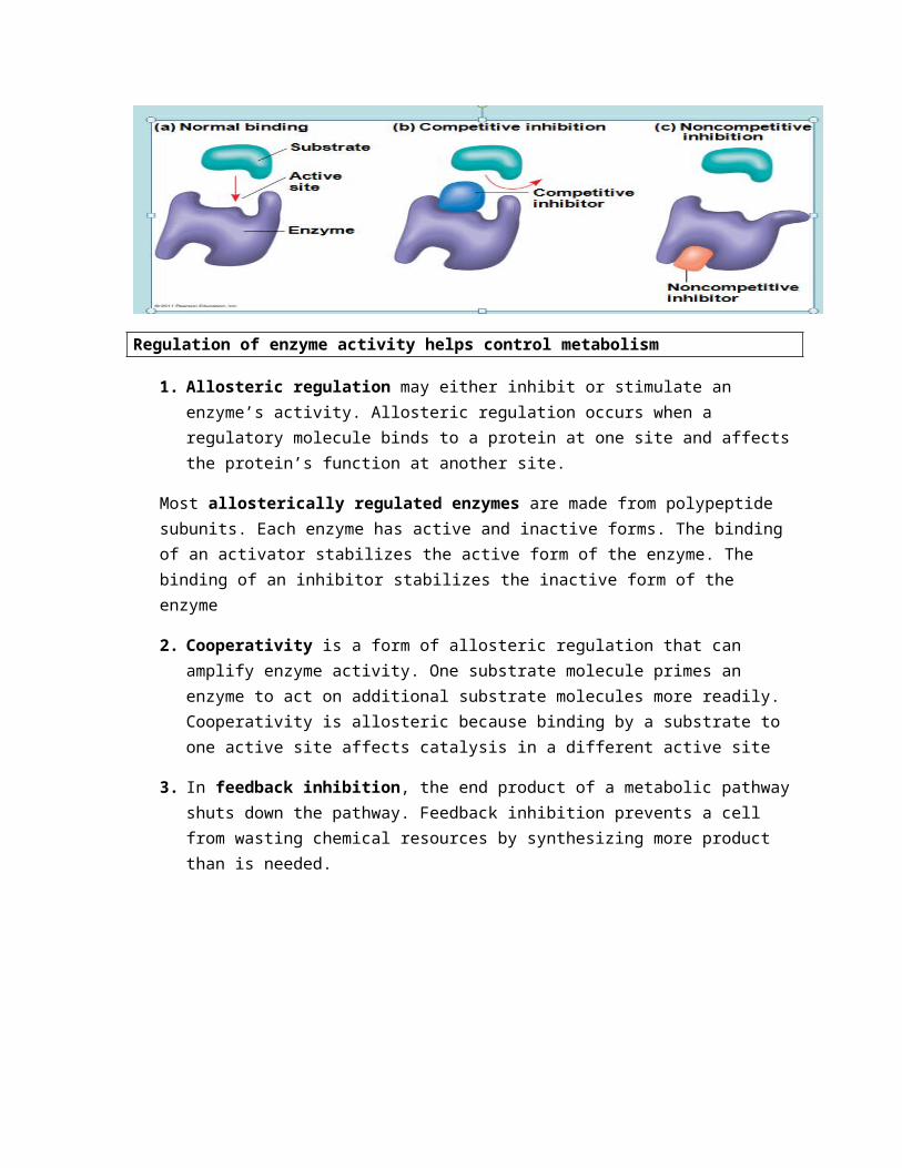
Regulation of enzyme activity helps control metabolism
1. Allosteric regulation may either inhibit or stimulate an enzyme’s activity. Allosteric regulation occurs when a regulatory molecule binds to a protein at one site and affects the protein’s function at another site.
Most allosterically regulated enzymes are made from polypeptide subunits. Each enzyme has active and inactive forms. The binding of an activator stabilizes the active form of the enzyme. The binding of an inhibitor stabilizes the inactive form of the enzyme
2. Cooperativity is a form of allosteric regulation that can amplify enzyme activity. One substrate molecule primes an enzyme to act on additional substrate molecules more readily. Cooperativity is allosteric because binding by a substrate to one active site affects catalysis in a different active site
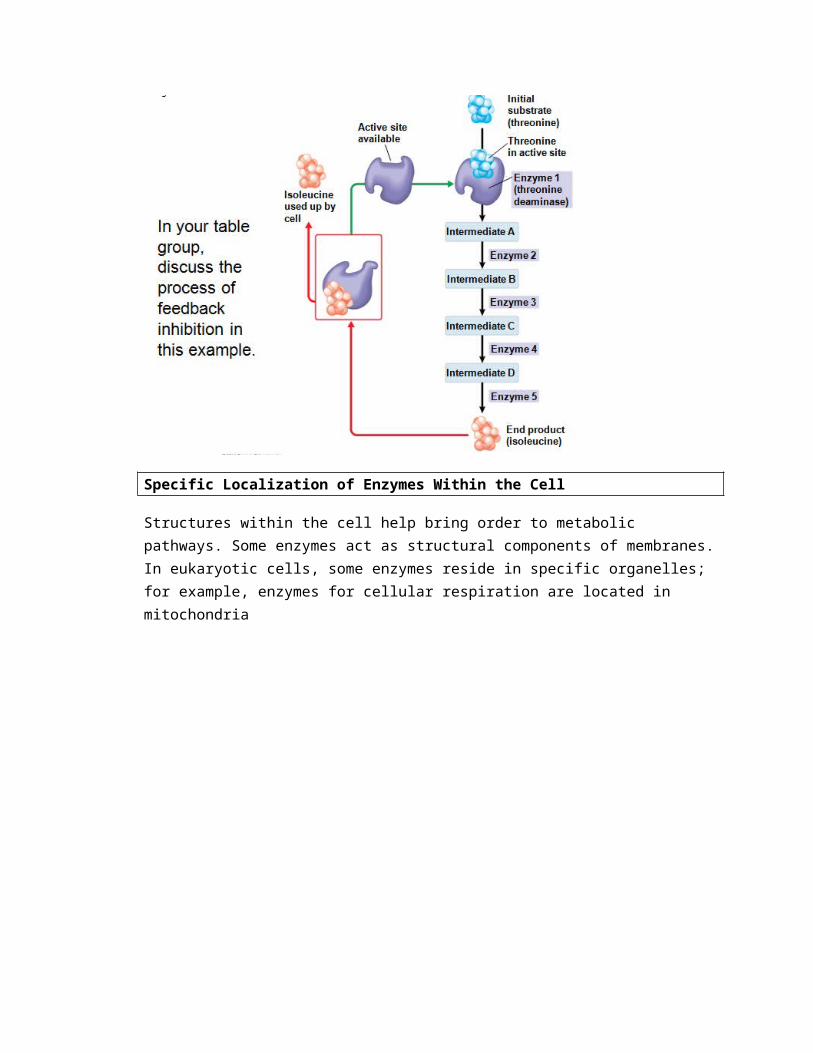
3. In feedback inhibition, the end product of a metabolic pathway shuts down the pathway. Feedback inhibition prevents a cell from wasting chemical resources by synthesizing more product than is needed.
Specific Localization of Enzymes Within the Cell
Structures within the cell help bring order to metabolic pathways. Some enzymes act as structural components of membranes. In eukaryotic cells, some enzymes reside in specific organelles; for example, enzymes for cellular respiration are located in mitochondria
Chapter 9: Cellular Respiration and Fermentation
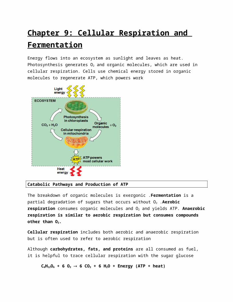
Energy flows into an ecosystem as sunlight and leaves as heat. Photosynthesis generates O2 and organic molecules, which are used in cellular respiration. Cells use chemical energy stored in organic molecules to regenerate ATP, which powers work
Catabolic Pathways and Production of ATP
The breakdown of organic molecules is exergonic .Fermentation is a partial degradation of sugars that occurs without O2 .Aerobic respiration consumes organic molecules and O2 and yields ATP. Anaerobic respiration is similar to aerobic respiration but consumes compounds other than O2.
Cellular respiration includes both aerobic and anaerobic respiration but is often used to refer to aerobic respiration
Although carbohydrates, fats, and proteins are all consumed as fuel, it is helpful to trace cellular respiration with the sugar glucose
C6H12O6 + 6 O2 ® 6 CO2 + 6 H2O + Energy (ATP + heat)
Redox Reactions: Oxidation and Reduction
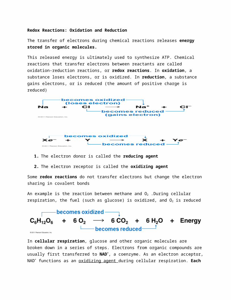
The transfer of electrons during chemical reactions releases energy stored in organic molecules.
This released energy is ultimately used to synthesize ATP. Chemical reactions that transfer electrons between reactants are called oxidation-reduction reactions, or redox reactions. In oxidation, a substance loses electrons, or is oxidized. In reduction, a substance gains electrons, or is reduced (the amount of positive charge is reduced)
1. The electron donor is called the reducing agent
2. The electron receptor is called the oxidizing agent
Some redox reactions do not transfer electrons but change the electron sharing in covalent bonds
An example is the reaction between methane and O2 .During cellular respiration, the fuel (such as glucose) is oxidized, and O2 is reduced
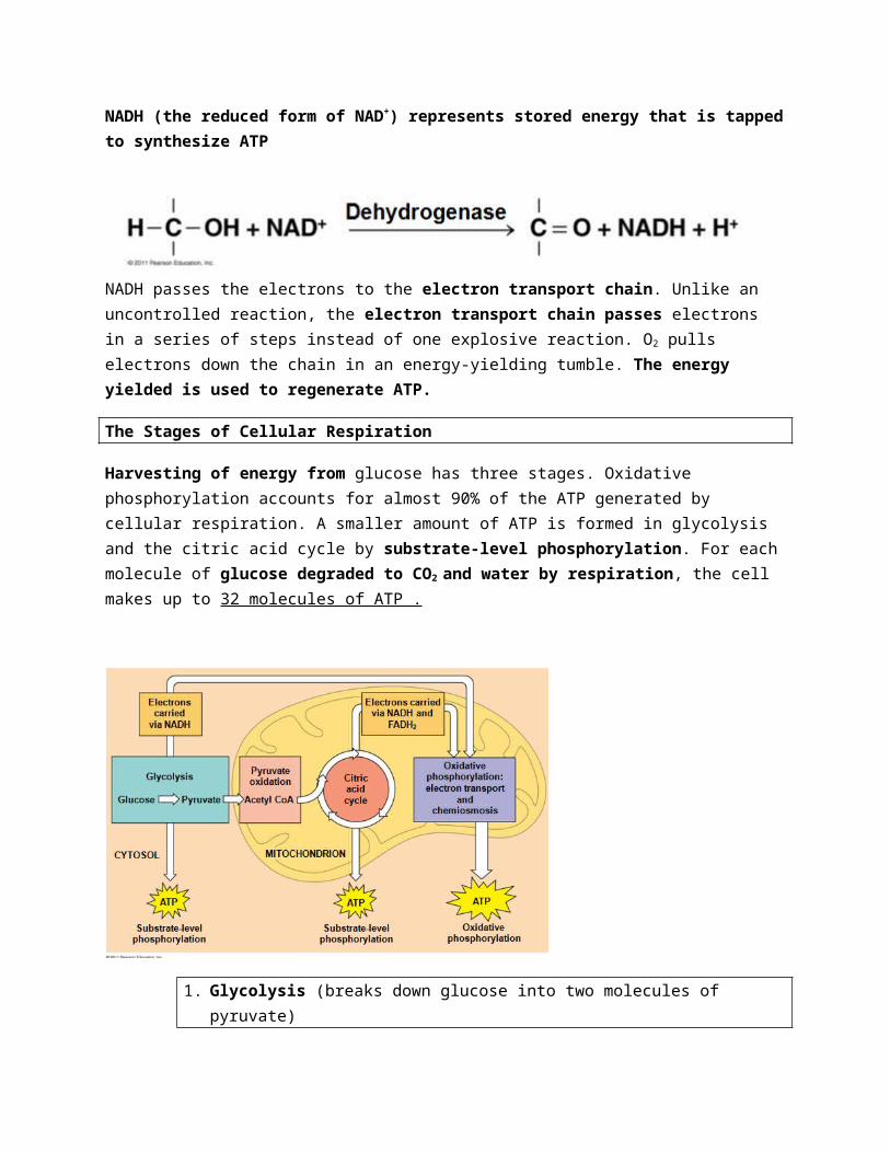
In cellular respiration, glucose and other organic molecules are broken down in a series of steps. Electrons from organic compounds are usually first transferred to NAD+, a coenzyme. As an electron acceptor, NAD+ functions as an oxidizing agent during cellular respiration. Each NADH (the reduced form of NAD+) represents stored energy that is tapped to synthesize ATP
NADH passes the electrons to the electron transport chain. Unlike an uncontrolled reaction, the electron transport chain passes electrons in a series of steps instead of one explosive reaction. O2 pulls electrons down the chain in an energy-yielding tumble. The energy yielded is used to regenerate ATP.
The Stages of Cellular Respiration
Harvesting of energy from glucose has three stages. Oxidative phosphorylation accounts for almost 90% of the ATP generated by cellular respiration. A smaller amount of ATP is formed in glycolysis and the citric acid cycle by substrate-level phosphorylation. For each molecule of glucose degraded to CO2 and water by respiration, the cell makes up to 32 molecules of ATP .
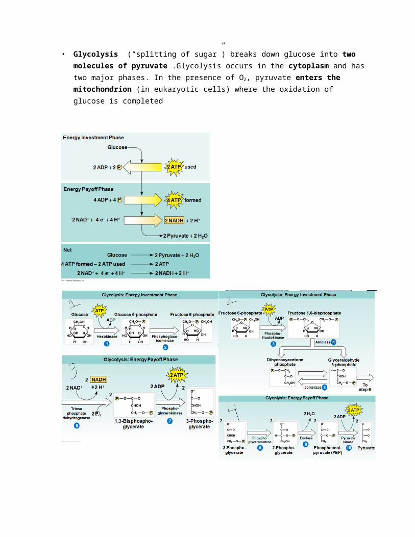
1. Glycolysis (breaks down glucose into two molecules of pyruvate)
• Glycolysis (“splitting of sugar”) breaks down glucose into two molecules of pyruvate .Glycolysis occurs in the cytoplasm and has two major phases. In the presence of O2, pyruvate enters the mitochondrion (in eukaryotic cells) where the oxidation of glucose is completed
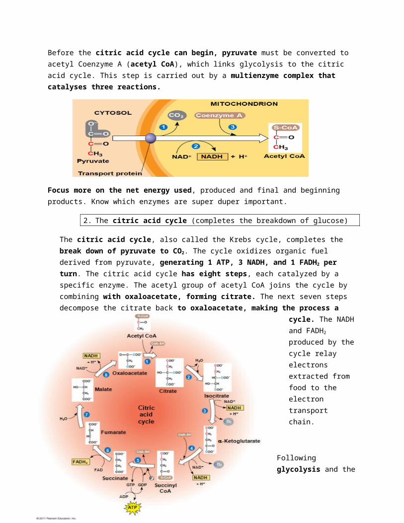
Before the citric acid cycle can begin, pyruvate must be converted to acetyl Coenzyme A (acetyl CoA), which links glycolysis to the citric acid cycle. This step is carried out by a multienzyme complex that catalyses three reactions.
Focus more on the net energy used, produced and final and beginning products. Know which enzymes are super duper important.
2. The citric acid cycle (completes the breakdown of glucose)
The citric acid cycle, also called the Krebs cycle, completes the break down of pyruvate to CO2. The cycle oxidizes organic fuel derived from pyruvate, generating 1 ATP, 3 NADH, and 1 FADH2 per turn. The citric acid cycle has eight steps, each catalyzed by a specific enzyme. The acetyl group of acetyl CoA joins the cycle by combining with oxaloacetate, forming citrate. The next seven steps decompose the citrate back to oxaloacetate, making the process a cycle. The NADH and FADH2 produced by the cycle relay electrons extracted from food to the electron transport chain.
Following glycolysis and the citric acid cycle, NADH and FADH2 account for most of the energy extracted from food. These two electron carriers donate electrons to the electron transport chain, which powers ATP synthesis via oxidative phosphorylation.
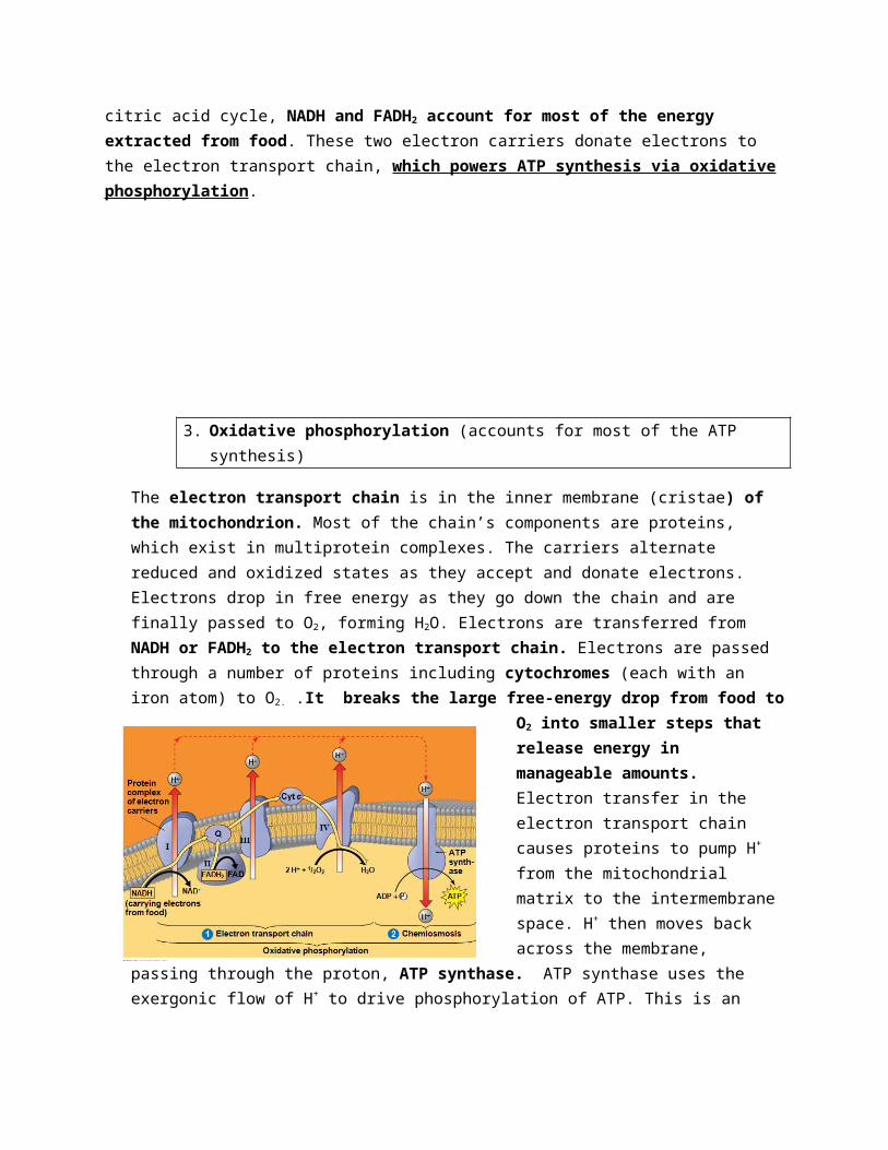
3. Oxidative phosphorylation (accounts for most of the ATP synthesis)
The electron transport chain is in the inner membrane (cristae) of the mitochondrion. Most of the chain’s components are proteins, which exist in multiprotein complexes. The carriers alternate reduced and oxidized states as they accept and donate electrons. Electrons drop in free energy as they go down the chain and are finally passed to O2, forming H2O. Electrons are transferred from NADH or FADH2 to the electron transport chain. Electrons are passed through a number of proteins including cytochromes (each with an iron atom) to O2. .It breaks the large free-energy drop from food to O2 into smaller steps that release energy in manageable amounts. Electron transfer in the electron transport chain causes proteins to pump H+ from the mitochondrial matrix to the intermembrane space. H+ then moves back across the membrane, passing through the proton, ATP synthase. ATP synthase uses the exergonic flow of H+ to drive phosphorylation of ATP. This is an
example of chemiosmosis, the use of energy in a H+ gradient to drive cellular work
The energy stored in a H+ gradient across a membrane couples the redox reactions of the electron transport chain to ATP synthesis
The H+ gradient is referred to as a proton-motive force, emphasizing its capacity to do work. Most cellular respiration requires O2 to produce ATP. Without O2, the electron transport chain will cease to operate. In that case, glycolysis couples with fermentation or anaerobic respiration to produce ATP
Anaerobic respiration uses an electron transport chain with a final electron acceptor other than O2, for example sulfate. Fermentation uses substrate-level phosphorylation instead of an electron transport chain to generate ATP.
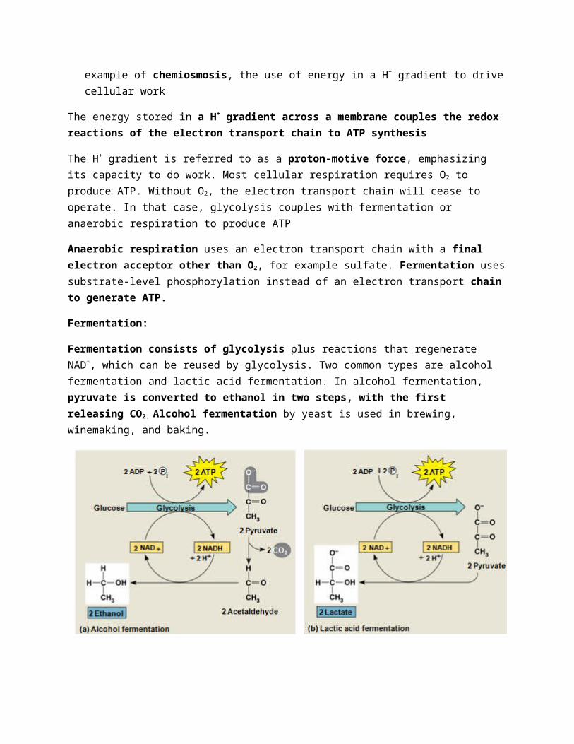
Fermentation:
Fermentation consists of glycolysis plus reactions that regenerate NAD+, which can be reused by glycolysis. Two common types are alcohol fermentation and lactic acid fermentation. In alcohol fermentation, pyruvate is converted to ethanol in two steps, with the first releasing CO2. Alcohol fermentation by yeast is used in brewing, winemaking, and baking.
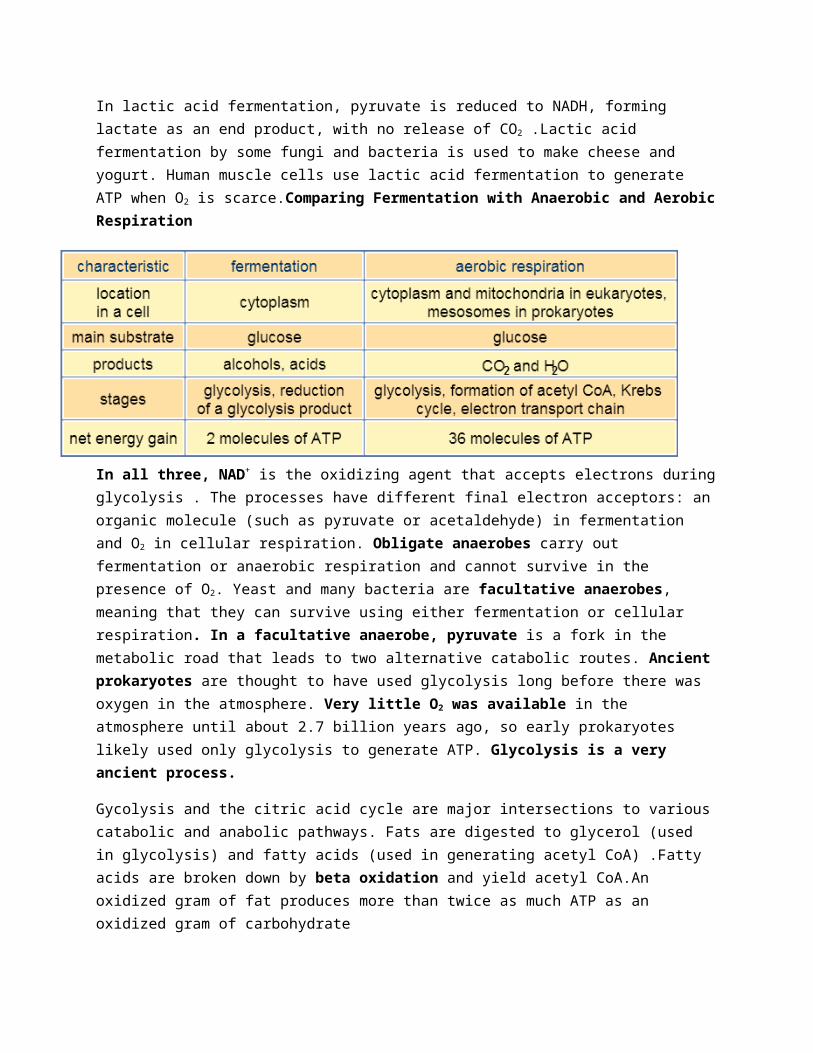
In lactic acid fermentation, pyruvate is reduced to NADH, forming lactate as an end product, with no release of CO2 .Lactic acid fermentation by some fungi and bacteria is used to make cheese and yogurt. Human muscle cells use lactic acid fermentation to generate ATP when O2 is scarce.Comparing Fermentation with Anaerobic and Aerobic Respiration
In all three, NAD+ is the oxidizing agent that accepts electrons during glycolysis . The processes have different final electron acceptors: an organic molecule (such as pyruvate or acetaldehyde) in fermentation and O2 in cellular respiration. Obligate anaerobes carry out fermentation or anaerobic respiration and cannot survive in the presence of O2. Yeast and many bacteria are facultative anaerobes, meaning that they can survive using either fermentation or cellular respiration. In a facultative anaerobe, pyruvate is a fork in the metabolic road that leads to two alternative catabolic routes. Ancient prokaryotes are thought to have used glycolysis long before there was oxygen in the atmosphere. Very little O2 was available in the atmosphere until about 2.7 billion years ago, so early prokaryotes likely used only glycolysis to generate ATP. Glycolysis is a very ancient process.
Gycolysis and the citric acid cycle are major intersections to various catabolic and anabolic pathways. Fats are digested to glycerol (used in glycolysis) and fatty acids (used in generating acetyl CoA) .Fatty acids are broken down by beta oxidation and yield acetyl CoA.An oxidized gram of fat produces more than twice as much ATP as an oxidized gram of carbohydrate
Biosynthesis (Anabolic Pathways)
The body uses small molecules to build other substances. These small molecules may come directly from food, from glycolysis, or from the citric acid cycle
Regulation of Cellular Respiration via Feedback Mechanisms
Feedback inhibition is the most common mechanism for control. If ATP concentration begins to drop, respiration speeds up; when there is plenty of ATP, respiration slows down. Control of catabolism is based mainly on regulating the activity of enzymes at strategic points in the catabolic pathway
Chapter 10: Photosynthesis Photosynthesis is the process that converts solar energy into chemical energy. Directly or indirectly, photosynthesis nourishes almost the entire living world.
Autotrophs sustain themselves without eating anything derived from other organisms. Autotrophs are the producers of the biosphere, producing organic molecules from CO2 and other inorganic molecules, using the energy of sunlight to make organic molecules. Photosynthesis occurs in plants, algae, certain other protists, and some prokaryotes
Heterotrophs obtain their organic material from other organisms. Heterotrophs are the consumers of the biosphere. Almost all heterotrophs, including humans, depend on photoautotrophs for food and O2
Photosynthesis converts light energy to the chemical energy of food
Chloroplasts are structurally similar to and likely evolved from photosynthetic bacteria . The structural organization of these cells allows for the chemical reactions of photosynthesis.
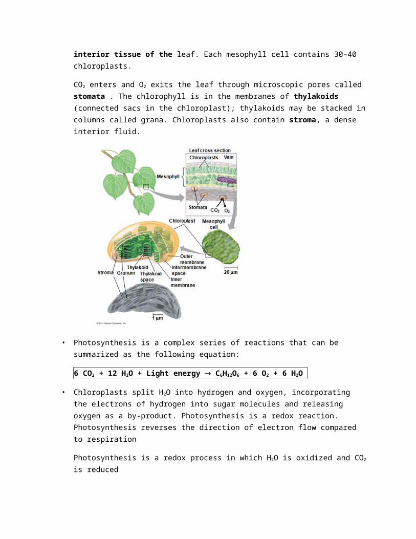
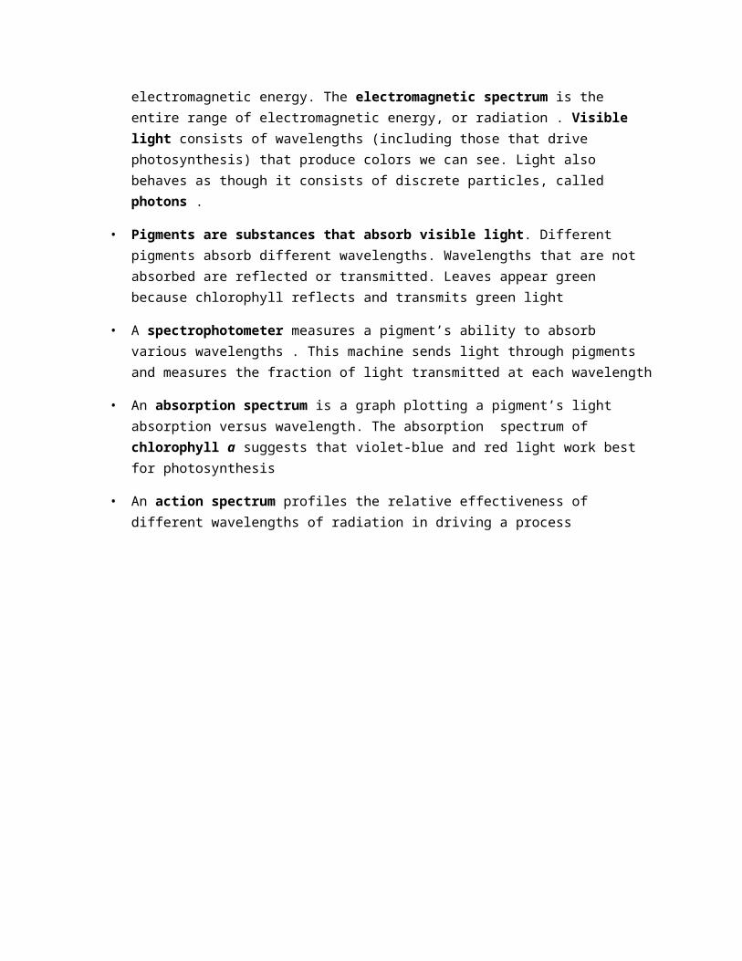
Leaves are the major locations of photosynthesis. Their green color is from chlorophyll, the green pigment within chloroplasts. Chloroplasts are found mainly in cells of the mesophyll, the interior tissue of the leaf. Each mesophyll cell contains 30–40 chloroplasts.
CO2 enters and O2 exits the leaf through microscopic pores called stomata . The chlorophyll is in the membranes of thylakoids (connected sacs in the chloroplast); thylakoids may be stacked in columns called grana. Chloroplasts also contain stroma, a dense interior fluid.
• Photosynthesis is a complex series of reactions that can be summarized as the following
equation:
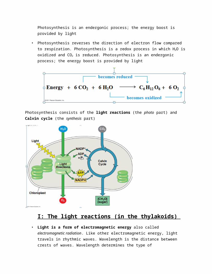
6 CO2 + 12 H2O + Light energy ® C6H12O6 + 6 O2 + 6 H2O
• Chloroplasts split H2O into hydrogen and oxygen, incorporating the electrons of hydrogen into sugar molecules and releasing oxygen as a by-product. Photosynthesis is a redox reaction. Photosynthesis reverses the direction of electron flow compared to respiration
Photosynthesis is a redox process in which H2O is oxidized and CO2 is reduced
Photosynthesis is an endergonic process; the energy boost is provided by light
• Photosynthesis reverses the direction of electron flow compared to respiration. Photosynthesis is a redox process in which H2O is oxidized and CO2 is reduced. Photosynthesis is an endergonic process; the energy boost is provided by light
Photosynthesis consists of the light reactions (the photo part) and Calvin cycle (the synthesis part)
I: The light reactions (in the thylakoids)
• Light is a form of electromagnetic energy also called electromagnetic radiation. Like other electromagnetic energy, light travels in rhythmic waves. Wavelength is the distance between crests of waves. Wavelength determines the type of electromagnetic energy. The electromagnetic spectrum is the entire range of electromagnetic energy, or radiation . Visible light consists of wavelengths (including those that drive photosynthesis) that produce colors we can see. Light also behaves as though it consists of discrete particles, called photons .
• Pigments are substances that absorb visible light. Different pigments absorb different wavelengths. Wavelengths that are not absorbed are reflected or transmitted. Leaves appear green because chlorophyll reflects and transmits green light
• A spectrophotometer measures a pigment’s ability to absorb various wavelengths . This machine sends light through pigments and measures the fraction of light transmitted at each wavelength
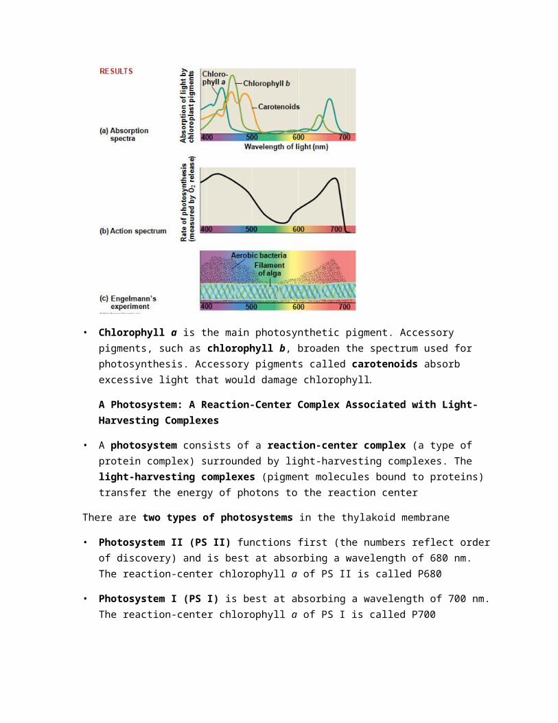
• An absorption spectrum is a graph plotting a pigment’s light absorption versus wavelength. The absorption spectrum of chlorophyll a suggests that violet-blue and red light work best for photosynthesis
• An action spectrum profiles the relative effectiveness of different wavelengths of radiation in driving a process
• Chlorophyll a is the main photosynthetic pigment. Accessory pigments, such as chlorophyll b, broaden the spectrum used for photosynthesis. Accessory pigments called carotenoids absorb excessive light that would damage chlorophyll.
A Photosystem: A Reaction-Center Complex Associated with Light-Harvesting Complexes
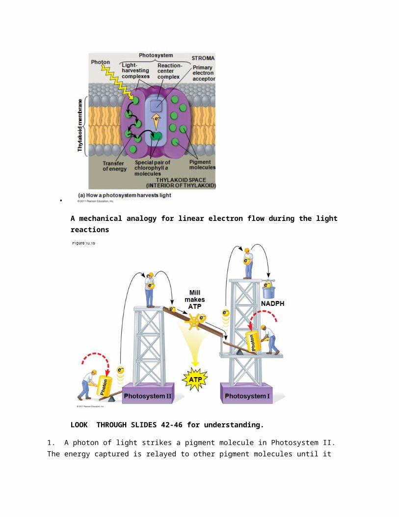
• A photosystem consists of a reaction-center complex (a type of protein complex) surrounded by light-harvesting complexes. The light-harvesting complexes (pigment molecules bound to proteins) transfer the energy of photons to the reaction center
There are two types of photosystems in the thylakoid membrane
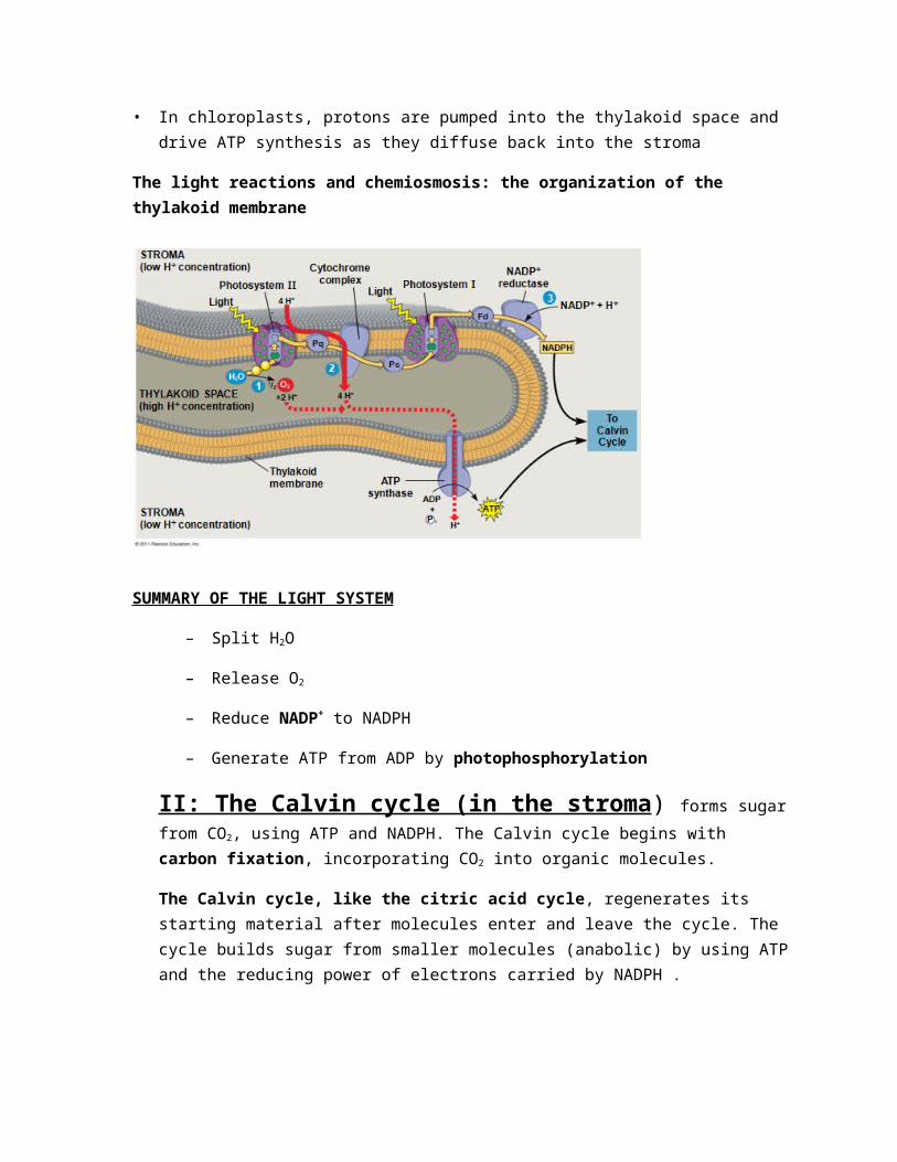
• Photosystem II (PS II) functions first (the numbers reflect order of discovery) and is best at absorbing a wavelength of 680 nm. The reaction-center chlorophyll a of PS II is called P680
• Photosystem I (PS I) is best at absorbing a wavelength of 700 nm. The reaction-center chlorophyll a of PS I is called P700
•
A mechanical analogy for linear electron flow during the light reactions
LOOK THROUGH SLIDES 42-46 for understanding.
1. A photon of light strikes a pigment molecule in Photosystem II. The energy captured is relayed to other pigment molecules until it reaches the P680 pair of chlorophyll a molecules. An electron in the chlorophyll a is excited to a higher energy state.
2. This electron is transferred from the excited P680 to the primary electron acceptor.
3. This leaves the chlorophyll with a +ve charge. An enzyme splits a water molecule into 2e-, 2 H+ and an oxygen atom. The electrons are used to fill the electron vacancy when the excited electrons are transferred from P680 to the primary electron acceptor. The oxygen will combine with another (the result of another water molecule splitting) to form O2.
4. Each photo-excited electron passes from the primary electron acceptor of PS II to PS I via an electron transport chain. It is made up of the electron carrier plastoquinone (Pq), a cytochrome complex, and a protein called plastocyanin (Pc).
5. The “fall” of electrons to a lower energy level provides the energy required for the synthesis of ATP. As electrons pass through the cytochrome complex, the pumping of protons (H+) builds a proton gradient that is used in chemiosmosis. (Explained later)
6. At the same time, light energy is absorbed by a pigment molecule in PS I exciting a pair of P700 chlorophyll a molecules. The photo-excited electrons are transferred to the primary electron acceptor for PS I. As with Photosystem II, this leaves an electron vacancy in the chlorophyll a. The P700+ will now accept the electrons that have reached the bottom of the electron transport chain from PS II.
7. Photo-excited electrons are passed in a series of redox reactions from the primary electron acceptor of PS I down a second electron transport chain through the protein ferredoxin (Fd).
8. The enzyme NADP+ reductase catalyses the transfer of electrons from Fd to NADPH+. Two electrons are required for its reduction to NADPH. This molecule is at a higher energy level that water and its electrons are more readily available for the reactions of the Calvin Cycle than were those of water.
In summary: Light reactions use: water and energy from the sun
Light reactions produce: atmospheric oxygen, ATP and NADPH which are both required to build the carbohydrates that result from the Calvin Cycle.
Cyclic electron flow uses only photosystem I and produces ATP, but not NADPH
• No oxygen is released
• Cyclic electron flow generates surplus ATP, satisfying the higher demand in the Calvin cycle
A Comparison of Chemiosmosis in Chloroplasts and Mitochondria
Chloroplasts and mitochondria generate ATP by chemiosmosis, but use different sources of energy. Mitochondria transfer chemical energy from food to ATP; chloroplasts transform light energy into the chemical energy of ATP. Spatial organization of chemiosmosis differs between chloroplasts and mitochondria but also shows similarities.
• In mitochondria, protons are pumped to the intermembrane space and drive ATP synthesis as they diffuse back into the mitochondrial matrix
• In chloroplasts, protons are pumped into the thylakoid space and drive ATP synthesis as they diffuse back into the stroma
The light reactions and chemiosmosis: the organization of the thylakoid membrane
SUMMARY OF THE LIGHT SYSTEM
– Split H2O
– Release O2
– Reduce NADP+ to NADPH
– Generate ATP from ADP by photophosphorylation
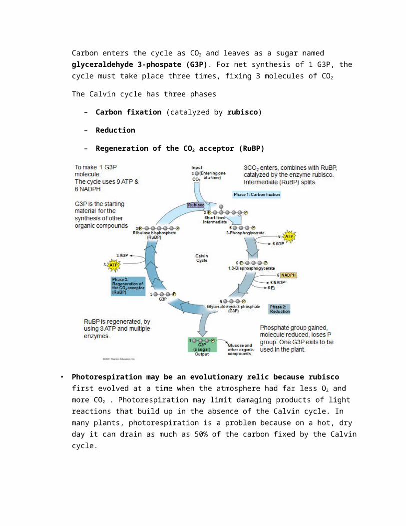
II: The Calvin cycle (in the stroma) forms sugar from CO2, using ATP and NADPH. The Calvin cycle begins with carbon fixation, incorporating CO2 into organic molecules.
The Calvin cycle, like the citric acid cycle, regenerates its starting material after molecules enter and leave the cycle. The cycle builds sugar from smaller molecules (anabolic) by using ATP and the reducing power of electrons carried by NADPH .
Carbon enters the cycle as CO2 and leaves as a sugar named glyceraldehyde 3-phospate (G3P). For net synthesis of 1 G3P, the cycle must take place three times, fixing 3 molecules of CO2
The Calvin cycle has three phases
– Carbon fixation (catalyzed by rubisco)
– Reduction
– Regeneration of the CO2 acceptor (RuBP)
• Photorespiration may be an evolutionary relic because rubisco first evolved at a time when the atmosphere had far less O2 and more CO2 . Photorespiration may limit damaging products of light reactions that build up in the absence of the Calvin cycle. In many plants, photorespiration is a problem because on a hot, dry day it can drain as much as 50% of the carbon fixed by the Calvin cycle.
• C4 plants minimize the cost of photorespiration by incorporating CO2 into four-carbon compounds in mesophyll cells . This step requires the enzyme PEP carboxylase . PEP carboxylase has a higher affinity for CO2 than rubisco does; it can fix CO2 even when CO2 concentrations are low. These four-carbon compounds are exported to bundle-sheath cells, where they release CO2
that is then used in the Calvin cycle
CAMP PLANTS
Some plants, including succulents, use crassulacean acid metabolism (CAM) to fix carbon. CAM plants open their stomata at night, incorporating CO2 into organic acids. Stomata close during the day, and CO2 is released from organic acids and used in the Calvin cycle
Chapter 11 Study ProductCells most often communicate with each other via chemical signals. For example, the fight-or-flight response is triggered by a signaling molecule called epinephrine
Evolutionary Terms of Cell Signaling
The yeast, Saccharomyces cerevisiae, has two mating types, a and a .Cells of different mating types locate each other via secreted factors specific to each type
A signal transduction pathway is a series of steps by which a signal on a cell’s surface is converted into a specific cellular response. Signal transduction pathways convert signals on a cell’s surface into cellular responses. Pathway similarities suggest that ancestral signaling molecules evolved in prokaryotes and were modified later in eukaryotes. The concentration of signaling molecules allows bacteria to sense local population density
Local and Long Distance Signalling
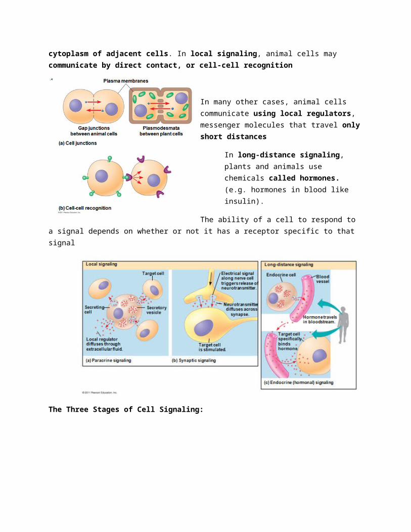
Cells in a multicellular organism communicate by chemical messengers. Animal and plant cells have cell junctions that directly connect the cytoplasm of adjacent cells. In local signaling, animal cells may communicate by direct contact, or cell-cell recognition
In many other cases, animal cells communicate using local regulators, messenger molecules that travel only short distances
In long-distance signaling, plants and animals use chemicals called hormones. (e.g. hormones in blood like insulin).
The ability of a cell to respond to a signal depends on whether or not it has a receptor specific to that signal
The Three Stages of Cell Signaling:
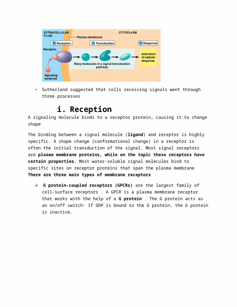
• Sutherland suggested that cells receiving signals went through three processes
i. ReceptionA signaling molecule binds to a receptor protein, causing it to change shape
The binding between a signal molecule (ligand) and receptor is highly specific. A shape change (conformational change) in a receptor is often the initial transduction of the signal. Most signal receptors are plasma membrane proteins, while on the topic these receptors have certain properties. Most water-soluble signal molecules bind to specific sites on receptor proteins that span the plasma membrane There are three main types of membrane receptors
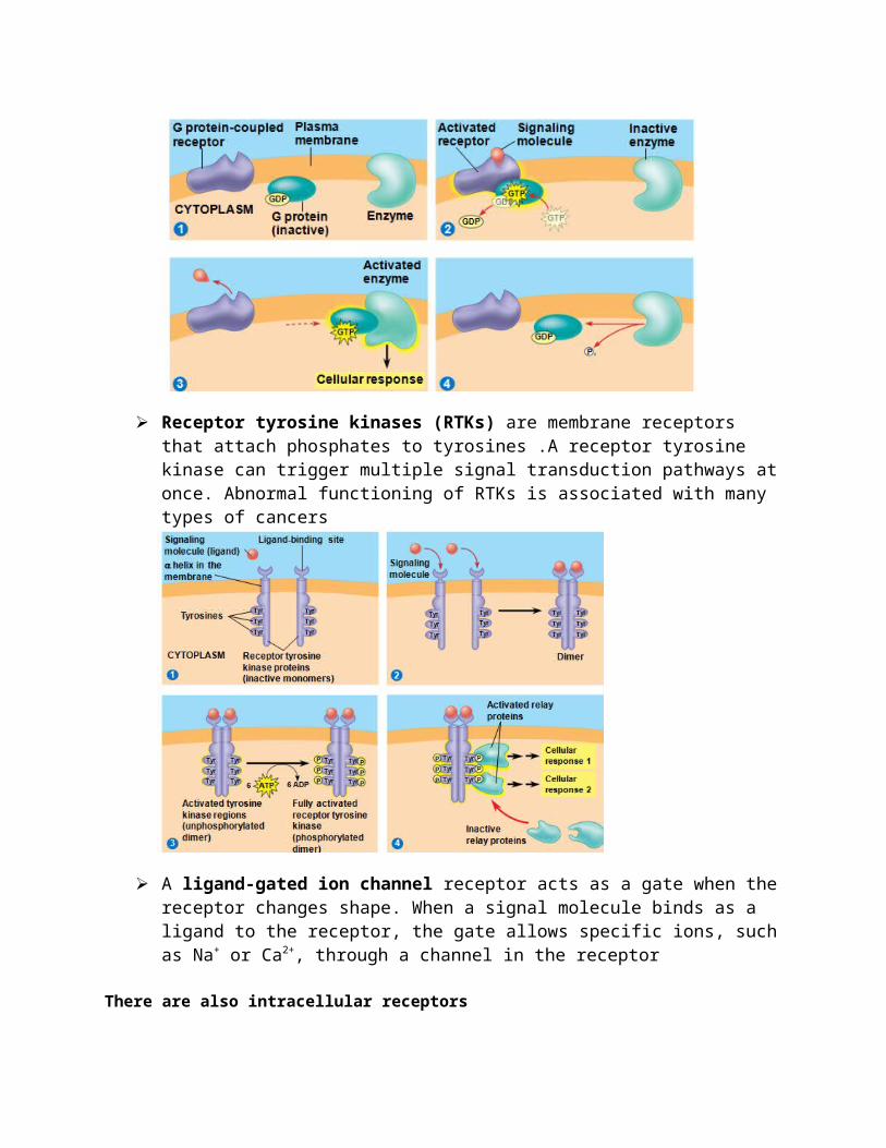
G protein-coupled receptors (GPCRs) are the largest family of cell-surface receptors . A GPCR is a plasma membrane receptor that works with the help of a G protein . The G protein acts as an on/off switch: If GDP is bound to the G protein, the G protein is inactive.
Receptor tyrosine kinases (RTKs) are membrane receptors that attach phosphates to tyrosines .A receptor tyrosine kinase can trigger multiple signal transduction pathways at once. Abnormal functioning of RTKs is associated with many types of cancers
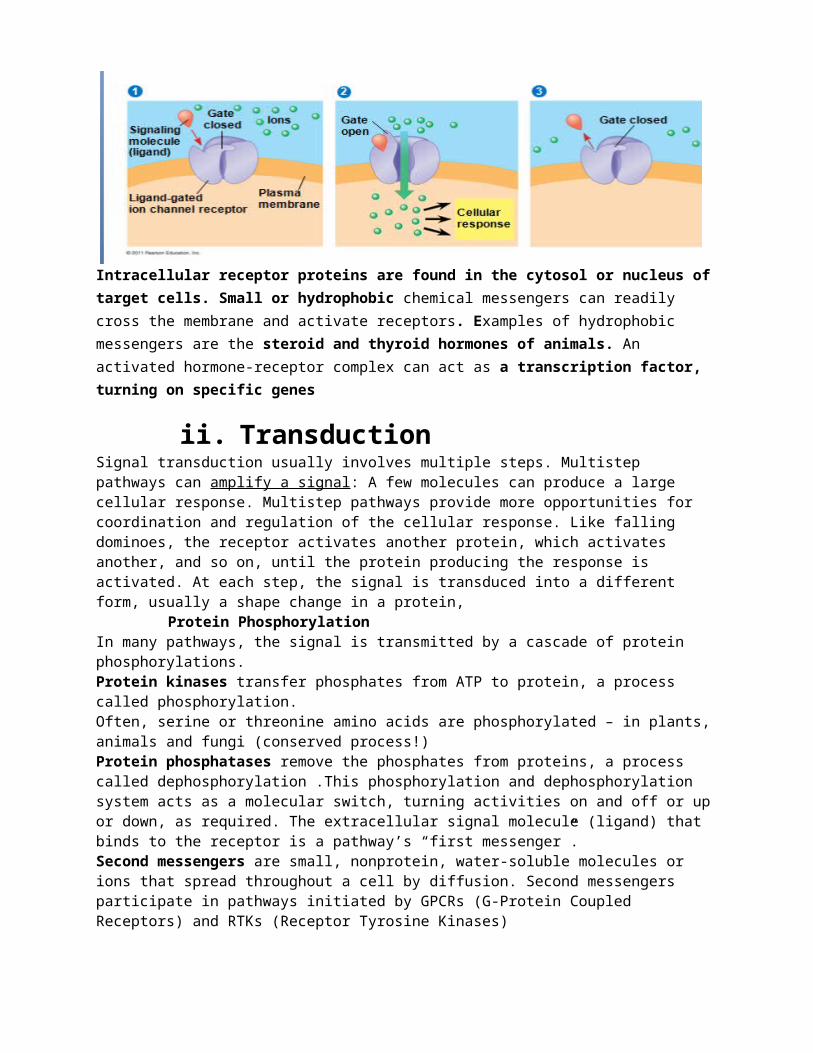
A ligand-gated ion channel receptor acts as a gate when the receptor changes shape. When a signal molecule binds as a ligand to the receptor, the gate allows specific ions, such as Na+ or Ca2+, through a channel in the receptor
There are also intracellular receptors
Intracellular receptor proteins are found in the cytosol or nucleus of target cells. Small or hydrophobic chemical messengers can readily cross the membrane and activate receptors. Examples of hydrophobic messengers are the steroid and thyroid hormones of animals. An activated hormone-receptor complex can act as a transcription factor, turning on specific genes
ii. TransductionSignal transduction usually involves multiple steps. Multistep pathways can amplify a signal: A few molecules can produce a large cellular response. Multistep pathways provide more opportunities for coordination and regulation of the cellular response. Like falling dominoes, the receptor activates another protein, which activates another, and so on, until the protein producing the response is activated. At each step, the signal is transduced into a different form, usually a shape change in a protein,
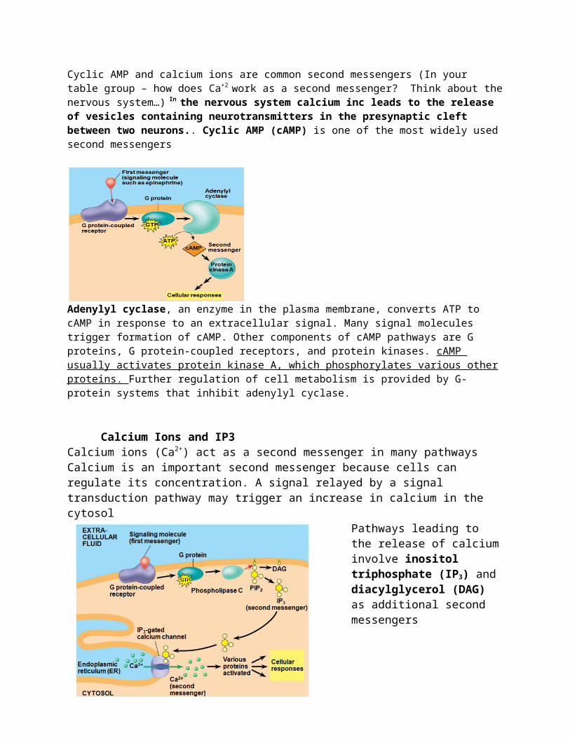
Protein PhosphorylationIn many pathways, the signal is transmitted by a cascade of protein phosphorylations. Protein kinases transfer phosphates from ATP to protein, a process called phosphorylation.Often, serine or threonine amino acids are phosphorylated – in plants, animals and fungi (conserved process!)Protein phosphatases remove the phosphates from proteins, a process called dephosphorylation .This phosphorylation and dephosphorylation system acts as a molecular switch, turning activities on and off or up or down, as required. The extracellular signal molecule (ligand) that binds to the receptor is a pathway’s “first messenger”.Second messengers are small, nonprotein, water-soluble molecules or ions that spread throughout a cell by diffusion. Second messengers participate in pathways initiated by GPCRs (G-Protein Coupled Receptors) and RTKs (Receptor Tyrosine Kinases) Cyclic AMP and calcium ions are common second messengers (In your table group – how does Ca+2 work as a second messenger? Think about the nervous system…) In the nervous system calcium inc leads to the release of vesicles containing neurotransmitters in the presynaptic cleft between two neurons.. Cyclic AMP (cAMP) is one of the most widely used second messengers
Adenylyl cyclase, an enzyme in the plasma membrane, converts ATP to cAMP in response to an extracellular signal. Many signal molecules trigger formation of cAMP. Other components of cAMP pathways are G proteins, G protein-coupled receptors, and protein kinases. cAMP usually activates protein kinase A, which phosphorylates various other proteins. Further regulation of cell metabolism is provided by G-protein systems that inhibit adenylyl cyclase.
Calcium Ions and IP3Calcium ions (Ca2+) act as a second messenger in many pathwaysCalcium is an important second messenger because cells can regulate its concentration. A signal relayed by a signal transduction pathway may trigger an increase in calcium in the cytosol
Pathways leading to the release of calcium involve inositol triphosphate (IP3) and diacylglycerol (DAG) as additional second messengers
iii. ResponseResponse: Cell signaling leads to regulation of transcription or cytoplasmic activities
Ultimately, a signal transduction pathway leads to regulation of one or more cellular activities. The response may occur in the cytoplasm or in the nucleus. Many signaling pathways regulate the synthesis of enzymes or other proteins, usually by turning genes on or off in the nucleus. The final activated molecule in the signaling pathway may function as a transcription factor
Fine Tuning SignalsThere are four aspects of fine-tuning to consider
1. Amplification of the signal (and thus the response) Enzyme cascades amplify the cell’s response. At each step, the number of activated products is much greater than in the preceding step
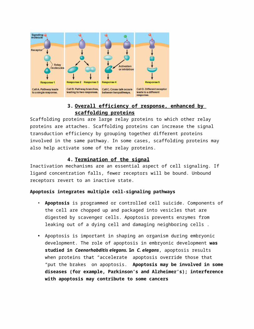
2. Specificity of the response Different kinds of cells have different collections of proteins. These different proteins allow cells to detect and respond to different signals. Even the same signal can have different effects in cells with different proteins and pathways. Pathway branching and “cross-talk” further help the cell coordinate incoming signals
3. Overall efficiency of response, enhanced by scaffolding proteins Scaffolding proteins are large relay proteins to which other relay proteins are attaches. Scaffolding proteins can increase the signal transduction efficiency by grouping together different proteins involved in the same pathway. In some cases, scaffolding proteins may also help activate some of the relay proteins.
4. Termination of the signal Inactivation mechanisms are an essential aspect of cell signaling. If ligand concentration falls, fewer receptors will be bound. Unbound receptors revert to an inactive state.
Apoptosis integrates multiple cell-signaling pathways
• Apoptosis is programmed or controlled cell suicide. Components of the cell are chopped up and packaged into vesicles that are digested by scavenger cells. Apoptosis prevents enzymes from leaking out of a dying cell and damaging neighboring cells .
• Apoptosis is important in shaping an organism during embryonic development. The role of apoptosis in embryonic development was studied in Caenorhabditis elegans. In C. elegans, apoptosis results when proteins that “accelerate” apoptosis override those that “put the
brakes” on apoptosis. Apoptosis may be involved in some diseases (for example, Parkinson’s and Alzheimer’s); interference with apoptosis may contribute to some cancers
Caspases are the main proteases (enzymes that cut up proteins) that carry out apoptosis
Apoptosis can be triggered by 1. An extracellular death-signaling ligand 2. DNA damage in the nucleus 3. protein misfolding in the endoplasmic reticulum
Chapter 15:
Chapter 25: The History of LifeChemical and physical processes on early Earth may have produced very simple cells through a sequence of stages:
1. Abiotic synthesis of small organic molecules
2. Joining of these small molecules into macromolecules
3. Packaging of molecules into protocells . Replication and metabolism are key properties of life and may have appeared together. Protocells may have been fluid-filled vesicles with a membrane-like structure. In water, lipids and other organic molecules can spontaneously form vesicles with a lipid bilayer. Adding clay can increase the rate of vesicle formation. Vesicles exhibit simple reproduction and metabolism and maintain an internal chemical environment.
4. Origin of self-replicating molecules
The first genetic material was probably RNA, not DNA. RNA molecules called ribozymes have been found to catalyze many different reactions. For example, ribozymes can make complementary copies of short stretches of RNA. Natural selection has produced self-replicating RNA molecules. RNA molecules that were more stable or replicated more quickly would have left the most descendent RNA molecules. Vesicles with RNA capable of replication would have been Protocells. RNA could have provided the template for DNA, a more stable genetic material.
Earth formed about 4.6 billion years ago, along with the rest of the solar system. Earth’s early atmosphere likely contained water vapor and chemicals released by volcanic eruptions (nitrogen, nitrogen oxides, carbon dioxide, methane, ammonia, hydrogen, hydrogen sulfide).
However, the evidence is not yet convincing that the early atmosphere was in fact reducing.
Instead of forming in the atmosphere, the first organic compounds may have been synthesized near volcanoes or deep-sea vents. Miller-Urey–type experiments demonstrate that organic molecules could have formed with various possible atmospheres.
RNA monomers have been produced spontaneously from simple molecules. Small organic molecules polymerize when they are concentrated on hot sand, clay, or rock
The fossil record documents the history of life
The fossil record reveals changes in the history of life on Earth.Sedimentary rocks are deposited into layers called strata and are the richest source of fossils
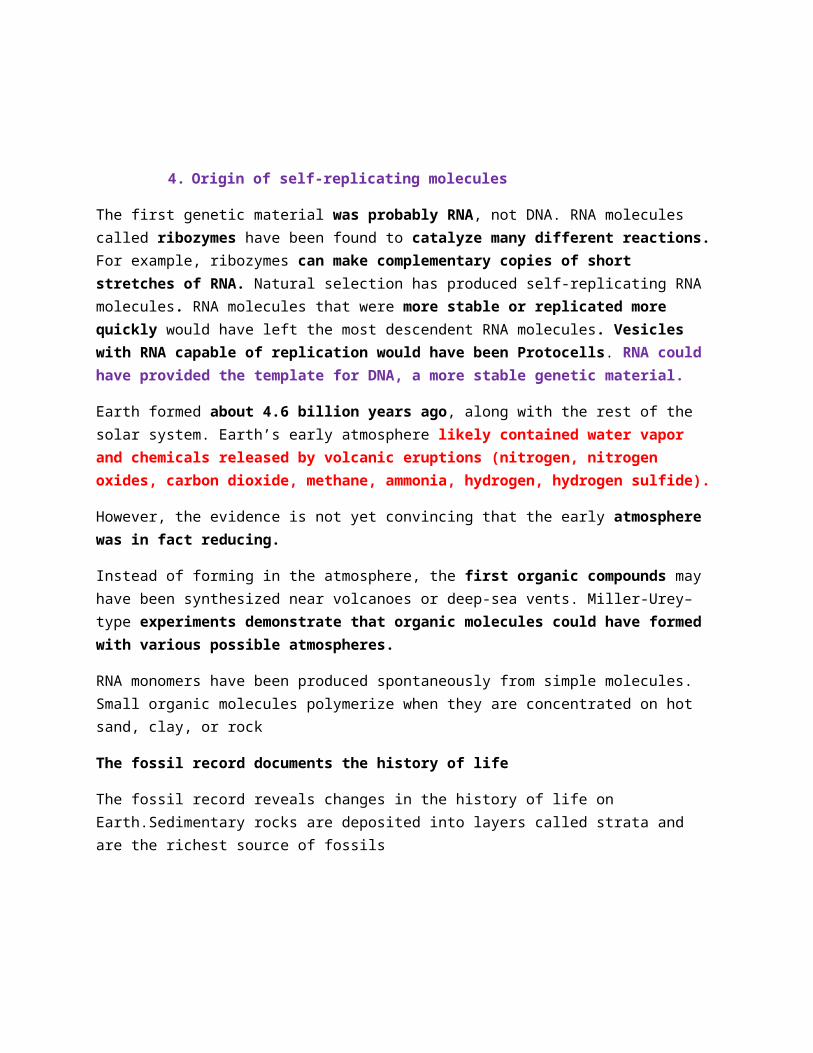
Few individuals have fossilized, and even fewer have been discovered.The fossil record is biased in favor of species that Existed for a long time. These were abundant and widespread and had hard parts. Fossil discoveries can be a matter of chance or prediction. For example, paleontologists found Tiktaalik, an early terrestrial vertebrate, by targeting sedimentary rock from a specific time and environment
How Rocks and Fossils Are Dated
Sedimentary strata reveal the relative ages of fossils. The absolute ages of fossils can be determined by radiometric dating. A “parent” isotope decays to a “daughter” isotope at a constant rate. Each isotope has a known half-life, the time required for half the parent isotope to decay
Radiocarbon dating can be used to date fossils up to 75,000 years old. For older fossils, some isotopes can be used to date sedimentary rock layers above and below the fossil
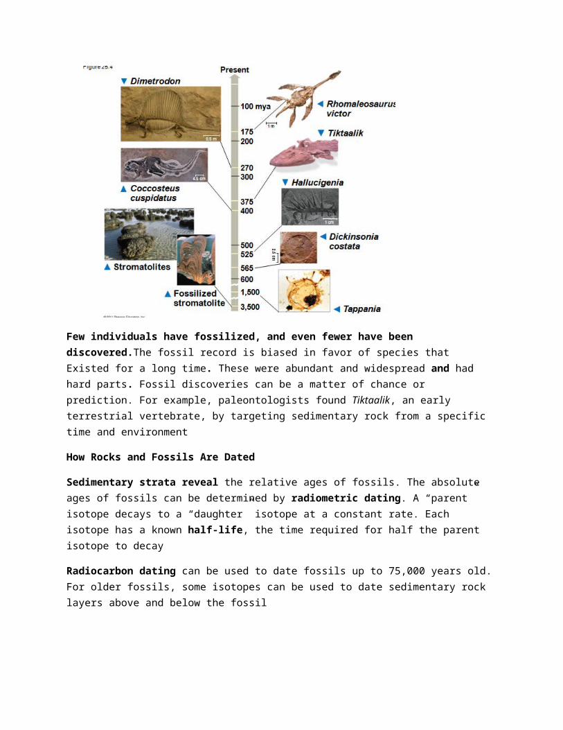
The geologic record is divided into the Archaean, the Proterozoic, and the Phanerozoic eons. The Phanerozoic encompasses multicellular eukaryotic life. The Phanerozoic is divided into three eras: the Paleozoic, Mesozoic, and Cenozoic
The First Single-Celled Organisms
The oldest known fossils are stromatolites, rocks formed by the accumulation of sedimentary layers on bacterial mats. Stromatolites date back 3.5 billion years ago. Prokaryotes were Earth’s sole inhabitants from 3.5 to about 2.1 billion years ago. Most atmospheric oxygen (O2) is of biological origin***
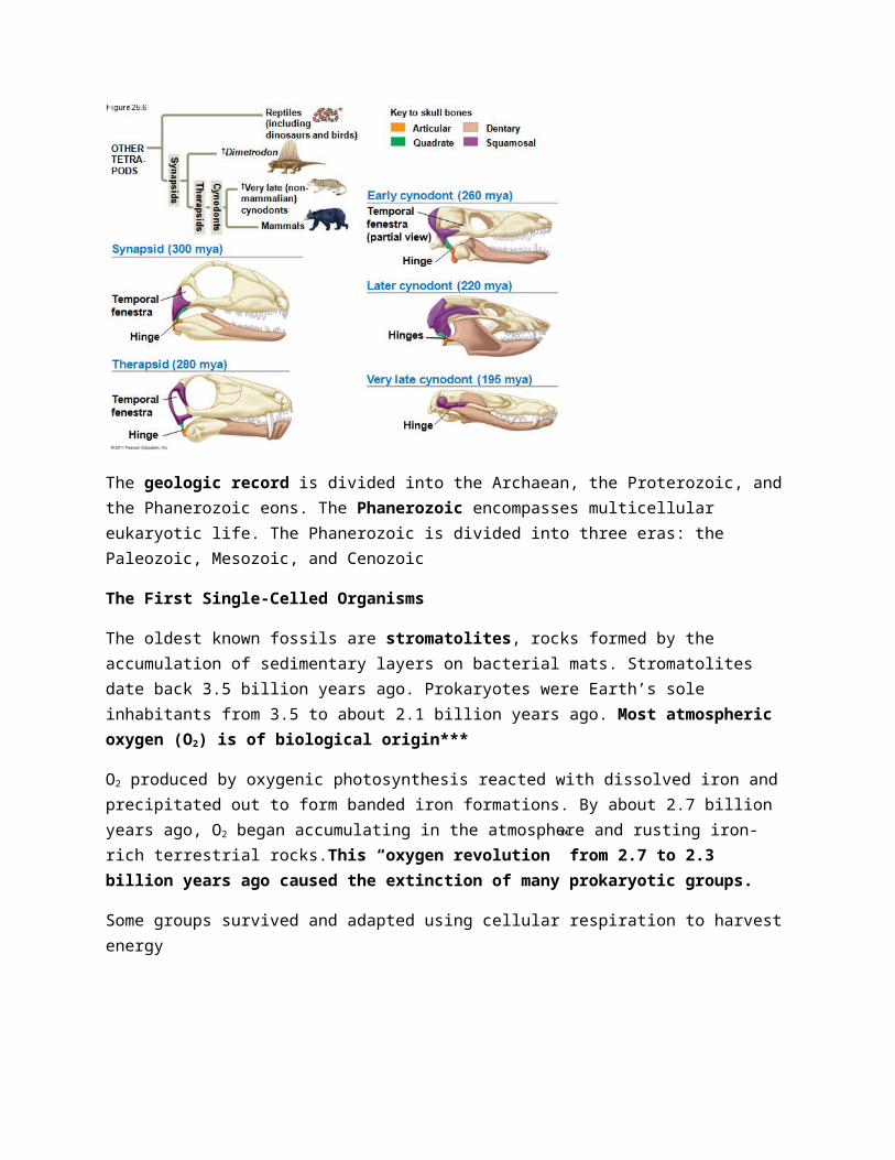
O2 produced by oxygenic photosynthesis reacted with dissolved iron and precipitated out to form banded iron formations. By about 2.7 billion years ago, O2 began accumulating in the atmosphere and rusting iron-rich terrestrial rocks.This “oxygen revolution” from 2.7 to 2.3 billion years ago caused the extinction of many prokaryotic groups.
Some groups survived and adapted using cellular respiration to harvest energy
The early rise in O2 was likely caused by ancient cyanobacteria . A later increase in the rise of O2 might have been caused by the evolution of eukaryotic cells containing chloroplasts
The First Eukaryotes
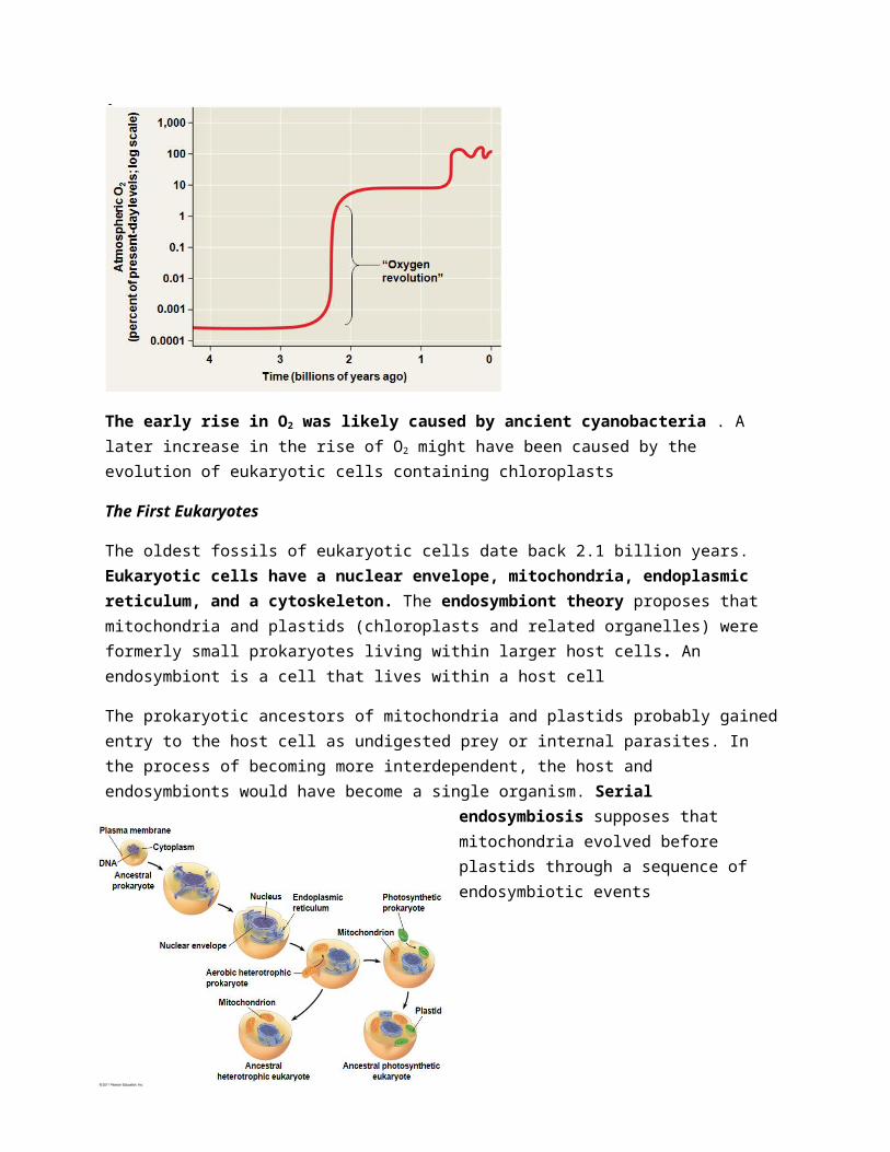
The oldest fossils of eukaryotic cells date back 2.1 billion years. Eukaryotic cells have a nuclear envelope, mitochondria, endoplasmic reticulum, and a cytoskeleton. The endosymbiont theory proposes that mitochondria and plastids (chloroplasts and related organelles) were formerly small prokaryotes living within larger host cells. An endosymbiont is a cell that lives within a host cell
The prokaryotic ancestors of mitochondria and plastids probably gained entry to the host cell as undigested prey or internal parasites. In the process of becoming more interdependent, the host and endosymbionts would have become a single organism. Serial endosymbiosis supposes that mitochondria evolved before plastids through a sequence of endosymbiotic events
Key evidence supporting an endosymbiotic origin of mitochondria and plastids: Inner membranes are similar to plasma membranes of prokaryotes. Division is similar in these organelles and some prokaryotes. These organelles transcribe and translate their own DNA. Ribosomes are more similar to prokaryotic than eukaryotic ribosomes . The evolution of eukaryotic cells allowed for a greater range of unicellular forms. A second wave of diversification occurred when multicellularity evolved and gave rise to algae, plants, fungi, and animals
The Earliest Multicellular Eukaryotes
Comparisons of DNA sequences date the common ancestor of multicellular eukaryotes to 1.5 billion years ago. The oldest known fossils of multicellular eukaryotes are of small algae that lived about 1.2 billion years ago
The “snowball Earth” hypothesis suggests that periods of extreme glaciation confined life to the equatorial region or deep-sea vents from 750 to 580 million years ago
The Cambrian explosion refers to the sudden appearance of fossils resembling modern animal phyla in the Cambrian period (535 to 525 million years ago). A few animal phyla appear even earlier: sponges, cnidarians, and molluscs .The Cambrian explosion provides the first evidence of predator-prey interactions
The Colonization of Land
Fungi, plants, and animals began to colonize land about 500 million years ago. Vascular tissue in plants transports materials internally and appeared by about 420 million years ago .Plants and fungi today form mutually beneficial associations and likely colonized land together.
Plate Tectonics
At three points in time, the land masses of Earth have formed a supercontinent: 1.1 billion, 600 million, and 250 million years ago .According to the theory of plate tectonics, Earth’s crust is composed of plates floating on Earth’s mantle. Tectonic plates move slowly through the process of continental drift. Oceanic and continental plates can collide, separate, or slide past each other. Interactions between plates cause the formation of mountains and islands, and earthquakes
Consequences of Continental Drift
Formation of the supercontinent Pangaea about 250 million years ago had many effects
– A deepening of ocean basins
– A reduction in shallow water habitat
– A colder and drier climate inland
Continental drift has many effects on living organisms. A continent’s climate can change as it moves north or south. Separation of land masses can lead to allopatric speciation.. The distribution of fossils
and living groups reflects the historic movement of continents. For example, the similarity of fossils in parts of South America and Africa is consistent with the idea that these continents were formerly attached
Mass Extinctions
The fossil record shows that most species that have ever lived are now extinct. Extinction can be caused by changes to a species’ environment. At times, the rate of extinction has increased dramatically and caused a mass extinction. Mass extinction is the result of disruptive global environmental changes. A number of factors might have contributed to these extinctions
1) Intense volcanism in what is now Siberia 2) Global warming resulting from the emission of large amounts of CO2 from the volcanoes 3)Reduced temperature gradient from equator to poles 4) Oceanic anoxia from reduced mixing of ocean waters
The presence of iridium in sedimentary rocks suggests a meteorite impact about 65 million years ago. Dust clouds caused by the impact would have blocked sunlight and disturbed global climate. The Chicxulub crater off the coast of Mexico is evidence of a meteorite that dates to the same time. Scientists estimate that the current rate of extinction is 100 to 1,000 times the typical background rate. Extinction rates tend to increase when global temperatures increase. Data suggest that a sixth, human-caused mass extinction is likely to occur unless dramatic action is taken
Consequences of Mass Extinctions
The percentage of marine organisms that were predators increased after the Permian and Cretaceous mass extinctions. Mass extinction can pave the way for adaptive radiations
Adaptive radiation is the evolution of diversely adapted species from a common ancestor. Adaptive radiations may follow : 1) Mass extinctions 2) The evolution of novel characteristics 3) The colonization of new regions. Mammals underwent an adaptive radiation after the extinction of terrestrial dinosaurs . The disappearance of dinosaurs (except birds) allowed for the expansion of mammals in diversity and size. Other notable radiations include photosynthetic prokaryotes, large predators in the Cambrian, land plants, insects, and tetrapods .Adaptive radiations can occur when organisms colonize new environments with little competition. The Hawaiian Islands are one of the world’s great showcases of adaptive radiation.
Effects of Development Genes
Genes that program development control the rate, timing, and spatial pattern of changes in an organism’s form as it develops into an adult
Changes in Rate and Timing
Heterochrony is an evolutionary change in the rate or timing of developmental events. It can have a significant impact on body shape. The contrasting shapes of human and chimpanzee skulls are the result of small changes in relative growth rates. Heterochrony can alter the timing of reproductive development relative to the development of nonreproductive organs. In paedomorphosis, the rate of reproductive development accelerates compared with somatic development. The sexually mature species may retain body features that were juvenile structures in an ancestral species
Changes in Spatial Pattern
Substantial evolutionary change can also result from alterations in genes that control the placement and organization of body parts. Homeotic genes determine such basic features as where wings and legs will develop on a bird or how a flower’s parts are arranged. Hox genes are a class of homeotic genes that provide positional information during development. If Hox genes are expressed in the wrong location, body parts can be produced in the wrong location
For example, in crustaceans, a swimming appendage can be produced instead of a feeding appendage. New morphological forms likely come from gene duplication events that produce new developmental genes. A possible mechanism for the evolution of six-legged insects from a many-legged crustacean ancestor has been demonstrated in lab experiments. Specific changes in the Ubx gene have been identified that can “turn off” leg development
Changes in morphology likely result from changes in the regulation of developmental genes rather than changes in the sequence of developmental genes
For example, threespine sticklebacks in lakes have fewer spines than their marine relatives
The gene sequence remains the same, but the regulation of gene expression is different in the two groups of fish
Evolution is not goal oriented
Evolution is like tinkering—it is a process in which new forms arise by the slight modification of existing forms
Evolutionary Trends
Extracting a single evolutionary progression from the fossil record can be misleading. Apparent trends should be examined in a broader context. The species selection model suggests that differential speciation success may determine evolutionary trends. Evolutionary trends do not imply an intrinsic drive toward a particular phenotype
Chapter 26: Phylogeny and the Tree of Life
Phylogeny is the evolutionary history of a species or group of related species
The discipline of systematics classifies organisms and determines their evolutionary relationships. Systematists use fossil, molecular, and genetic data to infer evolutionary relationships
Taxonomy is the ordered division and naming of organisms
Advantages of Binomial Nomenclature
Carolus Linnaeus published a system of taxonomy based on resemblances. Two key features of his system remain useful today: 1. two-part names for species and 2. hierarchical classification.
How Naming Works The two-part scientific name of a species is called a binomial
The first part of the name is the genus
The second part, called the specific epithet, is unique for each species within the genus
The first letter of the genus is capitalized, and the entire species name is italicized
Both parts together name the species (not the specific epithet alone)
The taxonomic groups from broad to narrow are domain, kingdom, phylum, class, order, family, genus, and species
A taxonomic unit at any level of hierarchy is called a taxon
******The broader taxa are not comparable between lineages (For example, an order of snails has less genetic diversity than an order of mammals)
Linnaean classification and phylogeny can differ from each other
Systematists have proposed the PhyloCode, which recognizes only groups that include a common ancestor and all its descendants
A phylogenetic tree represents a hypothesis about evolutionary relationships. Each branch point represents the divergence of two species. Sister taxa are groups that share an immediate common ancestor
A rooted tree includes a branch to represent the last common ancestor of all taxa in the tree
A basal taxon diverges early in the history of a group and originates near the common ancestor of the group
A polytomy is a branch from which more than two groups emerge
Phylogenetic trees show patterns of descent, not phenotypic similarity
Phylogenetic trees do not indicate when species evolved or how much change occurred in a lineage. It should not be assumed that a taxon evolved from the taxon next to it. ( KNOW THESE RULES, PERFECT FOR MC QUESTIONS).
Applying Phylogenies
Phylogeny provides important information about similar characteristics in closely related species
A phylogeny was used to identify the species of whale from which “whale meat” originated.
To infer phylogenies, systematists gather information about morphologies, genes, and biochemistry of living organisms. Phenotypic and genetic similarities due to shared ancestry are called homologies
Organisms with similar morphologies or DNA sequences are likely to be more closely related than organisms with different structures or sequences
When constructing a phylogeny, systematists need to distinguish whether a similarity is the result of homology or analogy
Homology is similarity due to shared ancestry. (we have similar bones, organs and DNA to monkeys because evolved from a common ancestor.)
Analogy is similarity due to convergent evolution. Convergent evolution occurs when similar environmental pressures and natural selection produce similar (analogous) adaptations in organisms from different evolutionary lineages. You could have 2 birds one in the middle east and one in South America that look very similar. However they do not share a common ancestor. They look similar because similar food and environmental conditions have selected for the survival of certain genes that resulted in them looking similar.
Bat and bird wings are homologous as forelimbs, but analogous as functional wings.
Analogous structures or molecular sequences that evolved independently are also called homoplasies. Homology can be distinguished from analogy by comparing fossil evidence and the degree of complexity. The more complex two similar structures are, the more likely it is that they are homologous.
It is also important to distinguish homology from analogy in molecular similarities. Mathematical tools help to identify molecular homoplasies, or coincidences. Molecular systematics uses DNA and other molecular data to determine evolutionary relationships.
Cladistics groups organisms by common descent
A clade is a group of species that includes an ancestral species and all its descendants . Clades can be nested in larger clades, but not all groupings of organisms qualify as clades.
A valid clade is monophyletic, signifying that it consists of the ancestor species and all its descendants
A paraphyletic grouping consists of an ancestral species and some, but not all, of the descendants
A polyphyletic grouping consists of various species with different ancestors
A shared ancestral character is a character that originated in an ancestor of the taxon . A shared derived character is an evolutionary novelty unique to a particular clade (organisms in that clade derived that character on their own). A character can be both ancestral and derived, depending on the context.

An outgroup is a species or group of species that is closely related to the ingroup, the various species being studied. The outgroup is a group that has diverged before the ingroup . Systematists compare each ingroup species with the outgroup to differentiate between shared derived and shared ancestral characteristics.
In some trees, the length of a branch can reflect the number of genetic changes that have taken place in a particular DNA sequence in that lineage.
Systematists can never be sure of finding the best tree in a large data set : They narrow possibilities by applying the principles of maximum parsimony and maximum likelihood
Maximum parsimony assumes that the tree that requires the fewest evolutionary events (appearances of shared derived characters) is the most likely
The principle of maximum likelihood states that, given certain rules about how DNA changes over time, a tree can be found that reflects the most likely sequence of evolutionary events
The best hypotheses for phylogenetic trees fit the most data: morphological, molecular, and fossil
Phylogenetic bracketing allows us to predict features of an ancestor from features of its descendants
– For example, phylogenetic bracketing allows us to infer characteristics of dinosaurs
• EXAMPLE: Birds and crocodiles share several features: four-chambered hearts, song, nest building, and brooding. These characteristics likely evolved in a common ancestor and were shared by all of its descendants, including dinosaurs. The fossil record supports nest building and brooding in dinosaurs.
GENOME AND EVOLUTION
*******Comparing nucleic acids or other molecules to infer relatedness is a valuable approach for tracing organisms’ evolutionary history. DNA that codes for rRNA changes relatively slowly and is useful for investigating branching points hundreds of millions of years ago. mtDNA evolves rapidly and can be used to explore recent evolutionary events.
Gene duplication increases the number of genes in the genome, providing more opportunities for evolutionary changes. Repeated gene duplications result in gene families. Like homologous genes, duplicated genes can be traced to a common ancestor.
Orthologous genes are found in a single copy in the genome and are homologous between species. They can diverge only after speciation occurs. (MARKERS)
Paralogous genes result from gene duplication, so are found in more than one copy in the genome. They can diverge within the clade that carries them and often evolve new functions.
Orthologous genes are widespread and extend across many widely varied species For example, humans and mice diverged about 65 million years ago, and 99% of our genes are orthologous.
1. Gene number and the complexity of an organism are not strongly linked
2. For example, humans have only four times as many genes as yeast, a single-celled eukaryote
3. Genes in complex organisms appear to be very versatile, and each gene can perform many functions.
A molecular clock uses constant rates of evolution in some genes to estimate the absolute time of evolutionary change. In orthologous genes, nucleotide substitutions are proportional to the time since they last shared a common ancestor. In paralogous genes, nucleotide substitutions are proportional to the time since the genes became duplicated.
• Neutral theory states that much evolutionary change in genes and proteins has no effect on fitness and is not influenced by natural selection. It states that the rate of molecular change in these genes and proteins should be regular like a clock. HOWEVER: The molecular clock does not run as smoothly as neutral theory predicts
Irregularities result from natural selection in which some DNA changes are favored over others. Estimates of evolutionary divergences older than the fossil record have a high degree of uncertainty . The use of multiple genes may improve estimates.
Phylogenetic analysis shows that HIV is descended from viruses that infect chimpanzees and other primates. HIV spread to humans more than once. Comparison of HIV samples shows that the virus
evolved in a very clocklike way. Application of a molecular clock to one strain of HIV suggests that that strain spread to humans during the 1930s. The tree of life suggests that eukaryotes and archaea are more closely related to each other than to bacteria
The tree of life is based largely on rRNA genes, as these have evolved slowly
3 Domains of Life
There have been substantial interchanges of genes between organisms in different domains
Horizontal gene transfer is the movement of genes from one genome to another. Horizontal gene transfer occurs by 1. exchange of transposable elements 2. plasmids,3. viral infection, and 4. fusion of organisms. Horizontal gene transfer complicates efforts to build a tree of life.
Chapter 36: Resource Acquisition and Transport in Vascular Plants
Adaptations for acquiring resources were key steps in the evolution of vascular plants.
The algal ancestors of land plants absorbed water, minerals, and CO2 directly from the surrounding water. Early nonvascular land plants lived in shallow water and had aerial shoots. Natural selection favored taller plants with flat appendages, multicellular branching roots, and efficient transport.
The evolution of xylem (water transporting vascular tissue) and phloem (sugar transporting vascular tissue) in land plants made possible the long-distance transport of water, minerals, and products of photosynthesis. Xylem transports water and minerals from roots to shoots. Phloem transports photosynthetic products from sources to sinks (to be used or stored).
Adaptations GOALS: compromises between enhancing photosynthesis and minimizing water loss.
Phyllotaxy, the arrangement of leaves on a stem, is specific to each species. Some angiosperms arranged their leaves in a spiral way with angles so the leaves do not create shade on each other.
Light absorption is affected by the leaf area index, the ratio of total upper leaf surface of a plant divided by the surface area of land on which it grows.
Total Upper Leaf Surface/Surface Area of Land
Leaf orientation affects light absorption. In low-light conditions, horizontal leaves capture more sunlight. In sunny conditions, vertical leaves are less damaged by sun and allow light to reach lower leaves. ( GOES BACK to the compromise between photosynthesis and losing water).
Roots and the hyphae of soil fungi form mutualistic associations called mycorrhizae . Mutualisms with fungi helped plants colonize land. Mycorrhizal fungi increase the surface area for absorbing water and minerals, especially phosphate.
The apoplast consists of everything external to the plasma membrane. It includes cell walls, extracellular spaces, and the interior of vessel elements and tracheids (non-living)
The symplast consists of the cytosol of the living cells in a plant, as well as the plasmodesmata (protoplast – internal to cell walls)
Three transport routes for water and solutes are
– The apoplastic route, through cell walls and extracellular spaces
– The symplastic route, through the cytosol
– The transmembrane route, across cell walls
Plasma membrane permeability controls short-distance movement of substances. Both active and passive transport occur in plants. In plants, membrane potential is established through pumping H+ by
proton pumps (creation of an electrochemical gradient). In animals, membrane potential is established through pumping Na+ by sodium-potassium pumps.
Osmosis determines the net uptake or water loss by a cell and is affected by solute concentration and pressure. Water potential (Ψ) is a measurement that combines the effects of solute concentration and pressure. Water potential determines the direction of movement of water.
Water flows from regions of higher water potential to regions of lower water potential. **Potential refers to water’s capacity to perform work. Both pressure and solute concentration affect water potential
This is expressed by the water potential equation: Ψ = ΨS + ΨP. The solute potential (ΨS) of a solution is directly proportional to its molarity (moles per liter = concentration!). Solute potential is also called osmotic potential. Pressure potential (ΨP) is the physical pressure on a solution
Turgor pressure is the pressure exerted by the plasma membrane against the cell wall, and the cell wall against the protoplast. The protoplast is the living part of the cell, which also includes the plasma membrane.
Water potential affects uptake and loss of water by plant cells. If a flaccid cell is placed in an environment with a higher solute concentration, the cell will lose water and undergo plasmolysis . Plasmolysis occurs when the protoplast shrinks and pulls away from the cell wall. SLIDE 39 & 40.
Efficient long distance transport of fluid requires bulk flow, the movement of a fluid driven by pressure. Water and solutes move together through tracheids and vessel elements of xylem, and sieve-tube elements of phloem.
Most water and mineral absorption occurs near root tips, where root hairs are located and the epidermis is permeable to water. Root hairs account for much of the surface area of roots. After soil solution enters the roots, the extensive surface area of cortical (cortex) cell membranes enhances uptake of water and selected minerals. The endodermis is the innermost layer of cells in the root cortex
It surrounds the vascular cylinder and is the last checkpoint for selective passage of minerals from the cortex into the vascular tissue. Water can cross the cortex via the symplast or apoplast
The waxy Casparian strip of the endodermal wall blocks apoplastic transfer of minerals from the cortex to the vascular cylinder. Water and minerals in the apoplast must cross the plasma membrane of an endodermal cell to enter the vascular cylinder
Xylem sap, water and dissolved minerals, is transported from roots to leaves by bulk flow. The transport of xylem sap involves transpiration, the evaporation of water from a plant’s surface. Transpired water is replaced as water travels up from the roots.
At night root cells continue pumping mineral ions into the xylem of the vascular cylinder, lowering the water potential (lowering yS) Water flows in from the root cortex, generating root pressure (increasing yP. According to the cohesion-tension hypothesis, transpiration and water cohesion pull water from shoots to roots. Xylem sap is normally under negative pressure, or tension.
Transpirational Pull
Water vapor in the airspaces of a leaf diffuses down its water potential gradient and exits the leaf via stomata. As water evaporates, the air-water interface retreats further into the mesophyll cell walls . The surface tension of water creates a negative pressure potential. (so more water could be pulled up). This negative pressure pulls water in the xylem into the leaf (into surrounding mesophyll cells and leaf space). The transpirational pull on xylem sap is transmitted from leaves to roots.
Water molecules are attracted to cellulose in xylem cell walls through adhesion (cellulose and H2O are both polar) .Adhesion of water molecules to xylem cell walls helps offset the force of gravity.
Water molecules are attracted to each other through cohesion (H – bonding) . Cohesion makes it possible to pull a column of xylem sap.
Thick secondary walls prevent vessel elements and tracheids from collapsing under negative pressure (similar to metal rings in a vacuum hose). (If you have negative pressure inside two thin walls, the walls will eventually collapse with the two sides attached to each other.)
Bulk flow differs from diffusion
– It is driven by differences in pressure potential, not solute potential
– It occurs in hollow dead cells, not across the membranes of living cells
– It moves the entire solution, not just water or solutes and It is much faster
Leaves generally have broad surface areas and high surface-to-volume ratios. These characteristics increase photosynthesis and increase water loss through stomata. Guard cells help balance water conservation with gas exchange for photosynthesis.
Changes in turgor pressure open and close stomata
SUPER DUPER UPER IMPORTANT
When turgid, guard cells bow outward and the pore between them opens
When flaccid, guard cells become less bowed and the pore closes
Function of K + and membrane potential in guard cells
This results primarily from the reversible uptake and loss of potassium ions (K+) by the guard cells. The [H+] creates a membrane potential (- charge inside cell), which causes K+ to move into the cell. (K+ is positive so it would be attracted to a negative environment).
Increased K+ inside the cell, causes y to decrease and water moves into the cell, making it turgid (pores open)
Generally, stomata open during the day and close at night to minimize water loss. Stomatal opening at dawn is triggered by
Light (stimulates guard cells to take up K+ - signal transduction cascade)
CO2 depletion (decrease of CO2 in leaf air-space triggers opening of guard cells)
An internal “clock” in guard cells
All eukaryotic organisms have internal clocks; circadian rhythms are 24-hour cycles
Drought, high temperature, and wind can cause stomata to close during the daytime
The hormone abscisic acid (ABA) is produced in response to water deficiency and causes the closure of stomata (reduces wilting and CO2 uptake…slowing photosynthesis and plant growth)
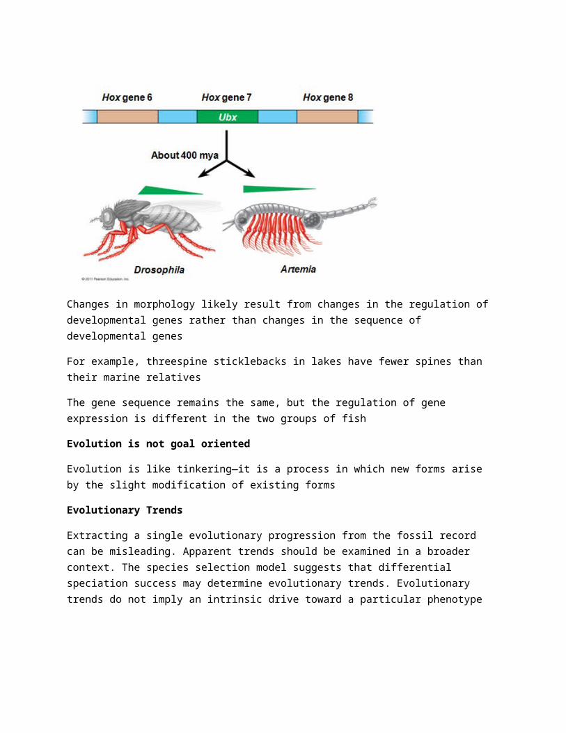
Sugars are transported from sources to sinks via the phloem. The products of photosynthesis are transported through phloem by the process of translocation. In angiosperms, sieve-tube elements are the conduits for translocation. Phloem sap is an aqueous solution that is high in sucrose. It travels from a sugar source to a sugar sink. A sugar source is an organ that is a net producer of sugar, such as mature leaves. A sugar sink is an organ that is a net consumer or storer of sugar, such as a tuber or bulb. In many plants, phloem loading requires active transport. Proton pumping and cotransport of sucrose and H+ enable the cells to accumulate sucrose. At the sink, sugar molecules diffuse from the phloem to sink tissues and are followed by water. Phloem is a “superhighway” for systemic transport of macromolecules and viruses. Systemic communication helps integrate functions of the whole plant
The phloem allows for rapid electrical communication between widely separated organs
– For example, rapid leaf movements in the sensitive plant (Mimosa pudica)
Chapter 40: Basic Principles of Animal Form and FunctionControl and coordination within a body depend on the endocrine system and the nervous system. The endocrine system transmits chemical signals called hormones to receptive cells throughout the body via blood. The nervous system transmits information between specific locations. The information conveyed depends on a signal’s pathway, not the type of signal
Animals manage their internal environment by regulating or conforming to the external environment
A regulator uses internal control mechanisms to moderate internal change in the face of external, environmental fluctuation. (e.g increasing your Hear Rate because you have to exercise and need more blood in your own body).
A conformer allows its internal condition to vary with certain external changes. Animals may regulate some environmental variables while conforming to others. Organisms use homeostasis to maintain a “steady state” or internal balance regardless of external environment. (you breathe faster because you are on top of a mountain where there is less oxygen. In humans, body temperature, blood pH, and glucose concentration are each maintained at a constant level.
Mechanisms of homeostasis moderate changes in the internal environment
For a given variable, fluctuations above or below a set point serve as a stimulus; these are detected by a sensor and trigger a response . The response returns the variable to the set point
The dynamic equilibrium of homeostasis is maintained by negative feedback, which helps to return a variable to a normal range. Most homeostatic control systems function by negative feedback, where buildup of the end product shuts the system off. Positive feedback amplifies a stimulus and does not usually contribute to homeostasis in animals.Set points and normal ranges can change with age or show cyclic variation In animals and plants, a circadian rhythm governs physiological changes that occur roughly every 24 hours. Homeostasis can adjust to changes in external environment, a process called acclimatization
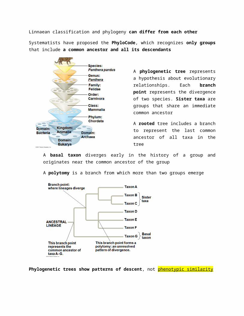
Thermoregulation is the process by which animals maintain an internal temperature within a tolerable range. Endothermic animals generate heat by metabolism (making ATP); birds and mammals are endotherms. Ectothermic animals gain heat from external sources; ectotherms include most invertebrates, fishes, amphibians, and nonavian reptiles. (this why people say reptiles are cold blooded and humans are warn blooded).
Organisms exchange heat by four physical processes: radiation, evaporation, convection, and conduction. Heat regulation in mammals often involves the integumentary system: skin, hair, and nails
Five adaptations help animals thermoregulate
1. Insulation
Insulation is a major thermoregulatory adaptation in mammals and birds. Skin, feathers, fur, and blubber (adipose tissue) reduce heat flow between an animal and its environment. Insulation is especially important in marine mammals such as whales and walruses
2. Circulatory adaptations
Regulation of blood flow near the body surface significantly affects thermoregulation. Many endotherms and some ectotherms can alter the amount of blood flowing between the body core and the skin. In vasodilation, blood flow in the skin increases, facilitating heat loss. In vasoconstriction, blood flow in the skin decreases, lowering heat loss. (example: when it is hot outside, your cheeks get red, your facial arteries are vasodilating, to let the heat out and bring back your body temperature vs when you are cold and you get so colorless.)
The arrangement of blood vessels in many marine mammals and birds allows for countercurrent exchange. Countercurrent heat exchangers transfer heat between fluids flowing in opposite directions and reduce heat loss. Some bony fishes and sharks also use countercurrent heat exchanges. Many endothermic insects have countercurrent heat exchangers that help maintain a high temperature in the thorax
3. Cooling by evaporative heat loss
Many types of animals lose heat through evaporation of water from their skin. Panting increases the cooling effect in birds and many mammals. Sweating or bathing moistens the skin, helping to cool an animal down
4. Behavioral responses
Many types of animals lose heat through evaporation of water from their skin. Panting increases the cooling effect in birds and many mammals. Sweating or bathing moistens the skin, helping to cool an animal down
5. Adjusting metabolic heat production
Thermogenesis is the adjustment of metabolic heat production to maintain body temperature. Thermogenesis is increased by muscle activity such as moving or shivering Nonshivering thermogenesis takes place when hormones cause mitochondria to increase their metabolic activity (heat production instead of ATP). Some ectotherms can also shiver to increase body temperature
In Humans
Thermoregulation is controlled by a region of the brain called the hypothalamus. The hypothalamus triggers heat loss or heat generating mechanisms. Fever is the result of a change to the set point for a biological thermostat
Bioenergetics is the overall flow and transformation of energy in an animal. It determines how much food an animal needs and it relates to an animal’s size, activity, and environment. Animals harvest chemical energy from food. Energy-containing molecules from food are usually used to make ATP, which powers cellular work. After the needs of staying alive are met, remaining food molecules can be used in biosynthesis. Biosynthesis includes body growth and repair, synthesis of storage material such as fat, and production of gametes
Metabolic rate is the amount of energy an animal uses in a unit of time. Metabolic rate can be determined by: 1. An animal’s heat loss 2. The amount of oxygen consumed or carbon dioxide produced
Basal metabolic rate (BMR) is the metabolic rate of an endotherm at rest at a “comfortable” temperature
Standard metabolic rate (SMR) is the metabolic rate of an ectotherm at rest at a specific temperature. Both rates assume a nongrowing, fasting, and nonstressed animal. Ectotherms have much lower metabolic rates than endotherms of a comparable size. Metabolic rate is proportional to body mass to the power of three quarters (m3/4)
Smaller animals have higher metabolic rates per gram than larger animals. The higher metabolic rate of smaller animals leads to a higher oxygen delivery rate, breathing rate, heart rate, and greater (relative) blood volume, compared with a larger animal. Activity greatly affects metabolic rate for endotherms and ectotherms .In general, the maximum metabolic rate an animal can sustain is inversely related to the duration of the activity.
Torpor is a physiological state in which activity is low and metabolism decreases . Torpor enables animals to save energy while avoiding difficult and dangerous conditions. Hibernation is long-term torpor that is an adaptation to winter cold and food scarcity (like those lazy ass polar bears).