


Languages
Pages
Legal

Two Human MYD88 Variants, S34Y and R98C, Interfere withMyD88-IRAK4-Myddosome Assembly□S
Received for publication, June 30, 2010, and in revised form, October 18, 2010 Published, JBC Papers in Press, October 21, 2010, DOI 10.1074/jbc.M110.159996
Julie George‡1, Precious G. Motshwene§2, Hui Wang‡1, Andriy V. Kubarenko‡1, Anna Rautanen¶3, Tara C. Mills¶4,Adrian V. S. Hill¶5, Nicholas J. Gay§2, and Alexander N. R. Weber‡1,6
From the ‡German Cancer Research Centre (DKFZ), Division Toll-like receptors and Cancer, Heidelberg, 69120 Germany, the§Department of Biochemistry, University of Cambridge, Cambridge CB2 1SG, United Kingdom, and the ¶Wellcome Trust Centre forHuman Genetics, University of Oxford, Oxford OX3 7BN, United Kingdom
Innate immune receptors detect microbial pathogens andsubsequently activate adaptive immune responses to com-bat pathogen invasion. MyD88 is a key adaptor molecule inboth Toll-like receptor (TLR) and IL-1 receptor superfamilysignaling pathways. This is illustrated by the fact that hu-man individuals carrying rare, naturally occurring MYD88point mutations suffer from reoccurring life-threateninginfections. Here we analyzed the functional properties of sixreported non-synonymous single nucleotide polymorphismsof MYD88 in an in vitro cellular system. Two variants foundin the MyD88 death domain, S34Y and R98C, showed se-verely reduced NF-�B activation due to reduced homo-olig-omerization and IRAK4 interaction. Structural modelinghighlights Ser-34 and Arg-98 as residues important for theassembly of the Myddosome, a death domain (DD) post-receptor complex involving the DD of MyD88, IRAK4, andIRAK2 or IRAK1. Using S34Y and R98C as functionalprobes, our data show that MyD88 homo-oligomerizationand IRAK4 interaction is modulated by the MyD88 TIR andIRAK4 kinase domain, demonstrating the functional impor-tance of non-DD regions not observed in a recent Myddo-some crystal structure. The differential interference of S34Yand R98C with some (IL-1 receptor, TLR2, TLR4, TLR5, andTLR7) but not all (TLR9) MyD88-dependent signaling path-ways also suggests that receptor specificities exist at thelevel of the Myddosome. Given their detrimental effect onsignaling, it is not surprising that our epidemiological anal-ysis in several case-control studies confirms that S34Y andR98C are rare variants that may drastically contribute tosusceptibility to infection in only few individuals.
In mammals, innate immunity relies on Toll-like receptor(TLR)7 pathways for the detection of invading pathogens (1).TLRs are a family of germ line-encoded pattern recognitionreceptors endowed with the capacity to recognize differentpathogen-associated molecular patterns (1, 2). Upon patho-gen-associated molecular pattern recognition, TLRs triggerthe production of proinflammatory cytokines and interferonsby transcriptional regulation and, thus, initiate and shapeadaptive immune responses (3). Different TLRs possess speci-ficity for diverse structural classes of pathogen molecules. Forexample, TLR2 is the receptor for bacterial lipopeptides, andTLR4 detects bacterial lipopolysaccharide (LPS), TLR7 viralsingle-stranded RNA, and TLR9 bacterial and viral DNA (1).TLRs engage pathogen-associated molecular patterns in theextracellular space or endosomes. Their cytoplasmic Toll/IL-1receptor (TIR) domains relay signals intracellularly wherethey are integrated and diversified by four adaptor molecules(4); that is, myeloid differentiation primary response gene 88(MyD88), MyD88-adaptor-like (Mal), TIR-domain-containingadaptor protein inducing IFN-� (TRIF), and TRIF-relatedadaptor molecule (TRAM).MyD88, the first TLR adaptor to be discovered, transduces
incoming TLR signals emanating from all TLRs except TLR3(4). Additionally, MyD88 is required for intracellular signalingin response to IL-1 and IL-18 (5). MyD88-dependent signal-ing involves three functional domains; that is, a C-terminal,evolutionarily conserved TIR domain (residues 159–296)present in all TLR adaptors that allows for upstream homo-typic interactions with stimulated TLR and IL-1 receptorcomplexes, a central so-called intermediate domain (ID; resi-dues 110–154) of unknown protein architecture that is absentin an inhibitory, shorter splice variant, MyD88s (6), and anN-terminal domain belonging to the death domain (DD; resi-dues 19–109) superfamily required for recruitment and acti-vation of downstream DD-containing kinases of the IL-1 re-ceptor-associated kinase (IRAK)-family. DDs are �-helicaland engage in homotypic and heterotypic protein-proteincomplexes (7–9). In MyD88, ID and DD are required for therecruitment and activation of IRAK4, which subsequently
Author’s Choice—Final version full access.□S The on-line version of this article (available at http://www.jbc.org) con-
tains supplemental Tables S1 and S2 and Figs. S1–S4.1 Supported by Deutsche Forschungsgemeinschaft Grant We4195-1
(to A. N. R. W.) and by the German Cancer Research Centre.2 Funded by grants from the UK Medical Research Council, the Wellcome
Trust, and the Biotechnology and Biological Sciences Research Council.3 Funded by the European Union FP6 GRACE grant and the Academy of
Finland.4 Funded by the Wellcome Trust.5 A Wellcome Trust Principal Fellow.6 To whom correspondence should be addressed: Toll-like Receptors and
Cancer, German Cancer Research Centre (DKFZ), Im Neuenheimer Feld580, 69120 Heidelberg, Germany. Tel.: 49-6221-424958; Fax: 49-6221-4958; E-mail: [email protected].
7 The abbreviations used are: TLR, Toll-like receptor; AP, activating protein;DD, death domain; ID, intermediate domain; IL-1R, IL-1 receptor; IRAK,IL-1 receptor-associated kinase; Mal, MyD88-adaptor like; MyD88, mye-loid differentiation primary response gene 88; ns, non-synonymous; SNP,single nucleotide polymorphism; TIR, Toll/interleukin 1 receptor; d(H),hydrodynamic diameter; FL, full-length.
THE JOURNAL OF BIOLOGICAL CHEMISTRY VOL. 286, NO. 2, pp. 1341–1353, January 14, 2011Author’s Choice © 2011 by The American Society for Biochemistry and Molecular Biology, Inc. Printed in the U.S.A.
JANUARY 14, 2011 • VOLUME 286 • NUMBER 2 JOURNAL OF BIOLOGICAL CHEMISTRY 1341
by guest on March 18, 2018
http://ww
w.jbc.org/
Dow
nloaded from
triggers the association and phosphorylation of IRAK1 orIRAK2 (4). Activated IRAK1 has been shown to then dissoci-ate from the MyD88 signaling complex to subsequently acti-vate tumor necrosis factor (TNF) receptor-associated factor 6(TRAF-6) (10, 11). This process culminates in the activationof the nuclear factor (NF)-�B and activating protein (AP)-1transcription factors and leads to the expression of a multi-tude of inflammatory downstream genes (for review, see Ref.4). In mice, IRAK1 appears to be recruited during early NF-�Bresponses; IRAK2 plays the same role in late NF-�B responses(12) and is involved in LPS-mediated post-transcriptionalcontrol (13). The relative contributions of IRAK1 or -2 in hu-mans have yet to be defined, but it has been suggested thatIRAK2 may be the most relevant in humans (14). Upon recep-tor recruitment of MyD88 via TIR-TIR interactions (15),MyD88 orchestrates the assembly of a post-receptor complextermed the Myddosome (16). A recently reported MyD88-IRAK4-IRAK2 Myddosome crystal structure strikingly eluci-dated the architecture and stoichiometry of such DD com-plexes (17). Myddosome assembly is nucleated by theoligomerization of six MyD88 DDs. Through an unknownmechanism, instead of additional MyD88 DD, four IRAK4DDs are subsequently incorporated into the structure. Thisforms a docking site for another four IRAK2 DDs. The affinityof the MyD88 DD oligomer for IRAK4 DDs exceeds that forIRAK2 DDs, apparently due to charge complementarity.The functional importance of MyD88 as a signaling adaptor
in innate immune defenses was recently illustrated by rareMYD88 point mutations. Individuals carrying these mutationswere found to be highly susceptible to infection with pyogenicbacteria (18). Although single nucleotide polymorphisms(SNPs) in other TLR signaling molecules have been associatedwith infectious, inflammatory, or autoimmune diseases (forreview, see Ref. 19), sequence variants in MyD88 have notbeen the subject of systematic investigation.In this study we used naturally occurring non-synonymous
(ns) SNPs as probes to study the molecular biology of theadaptor molecule MyD88 in detail with a view of identifyingprotein phenovariants whose corresponding nsSNPs might beof epidemiological interest due to an interesting functionalphenotype. The molecular basis for the observed functionaleffects were subsequently investigated and led to some novelinsights into the molecular interactions of MyD88 in the con-text of the Myddosome.
EXPERIMENTAL PROCEDURES
Reagents and Cells—Reagents were from Sigma unless oth-erwise stated. Human embryonic kidney (HEK) 293 cells (A.Dalpke, University of Heidelberg, Germany) were cultured inDMEM supplemented with 10% fetal calf serum, L-glutamine,and penicillin/streptomycin (Invitrogen) at 37 °C and 5% CO2.MyD88-deficient IA3 HEK293 cells (G. Stark, Department ofMolecular Genetics, Cleveland, OH; Ref. 20) were cultured asabove. Pam3CSK4 and R848 were from Axxora, LPS was fromInvivogen, flagellin Salmonella typhimurium was fromBiomol, and CpG-ODN was fromMWG Biotech. The follow-ing expression plasmids were used: TLR2-FLAG, untaggedTLR7, and TLR9 (I. Bekeredjian-Ding and A. Dalpke, Univer-
sity of Heidelberg, Infectious Disease Department, Heidel-berg, Germany); untagged TLR4, MD-2, and TLR5 (P. Ah-mad-Nejad, University of Mannheim, Mannheim, Germany).Cloning and Site-directed Mutagenesis—A template plas-
mid containing human MyD88 (NCBI accession no.AAC50954) was a gift from U. Hasan, IARC Lyon. N-terminalhemagglutinin (HA)- and Myc-tagged MyD88 variants werecloned to pcDNA3.1 (�) as described (21). N-terminal taggedMyD88 DD-ID truncations were generated by introducing astop codon at amino acid position 157. MyD88 and IRAK4DD constructs for bacterial purification were described in Ref.16. Mutations corresponding to MyD88 nsSNPs were intro-duced with a QuikChange II kit (Stratagene). Gateway-com-patible entry clones for Mal and IRAK4 full-length (FL; ob-tained from the German Cancer Research Centre Genomicsand Proteomics core facility) or DD-only (16) were trans-ferred via LR reaction (Invitrogen) to a pT-Rex-Dest30-basedplasmid containing N-terminal Renilla or Protein A tags. Al-ternatively, the IRAK 4 DD construct was transferred to apcDNA5/FRT/TO-based plasmid to add an N-terminalStrep-HA tag (T. Burckstummer, CeMM, Vienna. Austria).N-terminal-tagged Mal-Myc was described in George et al.(21). PCR and mutagenesis primers are listed in supplementalTable S2.Signaling Assays and Quantitative Real-time PCR—
HEK293 cells in 24-well plates (7.5 � 104 cells/well) weretransfected using the calcium phosphate method with a fireflyluciferase reporter (100 ng; Stratagene), pRL-Tk Renilla lucif-erase control reporter (10 ng; Promega), and 30 ng of pC1-EGFP (Clontech) to monitor transfection, incubated for 48 h,lysed, and measured using the Dual Luciferase Assay system(Promega) on a Fluoskan (Thermo Fisher Scientific) or Flu-ostar Optima (BMG LabTech) luminometer. Differentamounts of MyD88 were transfected as described. For titra-tion experiments the transfected plasmid amount was ad-justed to an equal amount with empty pcDNA3.1(�) vector.Luciferase measurements were performed in triplicate(�S.D.). For receptor stimulation experiments, stimuli wereadded 16 h before lysis. For quantitative real-time PCR,2.25 � 105 HEK293 cells were transfected with 240 ng of therespective plasmids. 48 h later, total RNA was extracted(RNeasy kit, Qiagen), cDNA was synthesized (Superscript III,Invitrogen), and quantitative PCR was performed in duplicateusing the Lightcycler 480 Probes Master Mix (Roche AppliedScience). Glycerinaldehyde-3-phosphate dehydrogenase(GAPDH) was used as a reference gene. For primers andprobes see supplemental Table S2.Co-immunoprecipitation and Expression Analysis—Immu-
noprecipitation experiments were essentially described (21).In brief, for MyD88 interactions cells were lysed in 50 mM
HEPES, pH 7.5, 150 mM NaCl, 1% Nonidet P-40, 20 mM
�-glycerophosphate, 2 mM DTT, 1 mM Na3VO4, and proteaseinhibitors. For MyD88-IRAK4 DD interactions cells were ly-sed in 20 mM Tris, pH 7.5, 250 mM NaCl, 1% Triton X-100, 10mM EDTA, 10 mM DTT, and protease and phosphatase inhib-itors (Roche Applied Science). Lysis was performed for 20min on ice. Immunoprecipitations were performed fromcleared cell lysates with monoclonal mouse �-Myc (Sigma)
MYD88 Variants Interfere with Myddosome Assembly
1342 JOURNAL OF BIOLOGICAL CHEMISTRY VOLUME 286 • NUMBER 2 • JANUARY 14, 2011
by guest on March 18, 2018
http://ww
w.jbc.org/
Dow
nloaded from
and Protein A/G beads (Pierce). Immunocomplexes werewashed three times with lysis buffer and analyzed by immu-noblot. For expression analysis epitope tags were detectedwith rabbit �-Myc or �-HA (Cell Signaling). �-Tubulin ex-pression was monitored with mouse �-�-tubulin (Sigma).
LUMIER was described earlier (22) and performed withProtein A and Renilla luciferase-tagged proteins. In brief thismethod relies on an affinity purification of Protein A-taggedproteins via magnetic IgG-beads (Dynabeads M-280, sheepanti-rabbit IgG, Invitrogen). Co-purified interacting proteinswere detected via Renilla luciferase activity. HEK293 cellswere seeded in 96-well plates (10,000 cells/well) and trans-fected with 20 ng of Protein A- and 20 ng of Renilla-encodingplasmids using Lipofectamine 2000 (Invitrogen). 48 h post-transfection, cells were lysed for 15 min on ice in 10 �l ofbuffer containing 20 mM Tris, pH 7.5, 250 mM NaCl, 1% Tri-ton X-100, 10 mM EDTA, 10 mM DTT, 0.0125 units/�l Ben-zonase (Novagen), protease and phosphatase inhibitors(Roche Applied Science), and 1% IgG Dynabeads. The lysateswere diluted with 100 �l of PBS. To assess differences intransfection efficiencies, 10 �l of the diluted lysate were mea-sured for raw Renilla activity. The remaining lysate waswashed 5 times with ice-cold PBS on a magnetic plate washer(Tecan). The washed samples remained in 20 �l of PBS andwere measured for bound luciferase activity. An interactionsignal was processed by normalizing the activity of bound Re-nilla luciferase to the amount of raw Renilla activity. The wellestablished interaction of Protein A-tagged Jun and Renilla-tagged Fos (23) served as a positive control and was includedin each experiment. As a negative control Renilla-tagged pro-teins were co-transfected with plasmids encoding for ProteinA alone. The LUMIER interactions are represented as nor-malized signal to noise (S/N) ratios in which the normalizedinteraction signal was divided by its respective negative con-trol. Interaction analysis was performed in triplicate in eachexperiment.Recombinant Protein Production and Gel Filtration—The
DDs of both IRAK4 and MyD88 wild type (WT) and mutantswere expressed from pETG-30 (A. Geerlof, EMBL, Heidel-berg) expression vectors in BL21 Codon Plus (Novagen;IRAK4 DD) and BL21-DE3 (Novagen; hMyD88) as describedin Motshwene et al. (16). Briefly, bacterial cells expressingIRAK4 DDs or MyD88 DDs were resuspended in 20 mM Tris,pH 8.0, and lysed. The lysate was centrifuged at 1050 � g for1 h, and the supernatant was loaded onto a glutathione-Sepharose column and eluted with 20 mM Tris with or with-out 50 mM NaCl, pH 8.0, 40 mM reduced glutathione. Aftercleavage of the GST tag, the solution was loaded onto a nickel-nitrilotriacetic acid column to remove cleaved GST. Pro-tein solutions were concentrated, and the individual DDswere purified using ion exchange followed by Superdex-75size exclusion column (GE Healthcare) with the same buffersupplemented with 5 mM DTT. The MyD88�IRAK4 com-plexes were prepared by adding a slight molar excess ofIRAK4 DD, and the complex was purified using a Superdex-200 column. Purity at all stages was assessed by SDS-PAGE.Dynamic light scattering was performed using a Zetasizer
Nano-S (Malvern) instrument. Protein samples (1.1–2 mg/ml)
were centrifuged for 10 min at 13,000 rpm before beingloaded on a 12.5 � 12.5 � 45-mm UV-transparent disposablecuvette (Sarstedt) in 20 mM Tris, pH 8.0, 50 mM NaCl. Scat-tering data were collected at 20 °C at a measurement positionof 4.65 mm over a time course of 60–80 s per cycle. The totalnumber of cycles per sample ranged between 13 and 18, anddata were averaged across cycles.Sequence Alignments, Structural Analysis, and Homology
Modeling—Protein sequences were from NCBI, alignmentswere performed with ClustalX and color-formatted accord-ing to amino acid physicochemical properties (Joy server).The coordinates for the Myddosome crystal structure (pdbcode 3MOP, Ref. 17) were obtained from the Protein DataBank and analyzed in PyMOL (Schrodinger). Homologymodels for MyD88 S34Y and R98C were generated usingtheMODELLER package (24) as described (25, 26) based onthe structure of MyD88 M5.Polymorphism Information, Analysis, and Genotyping—A
list of reported SNPs in humanMYD88 (Gene ID 4615) wasobtained from NCBI (supplemental Table S1). HapMap datawere from the International HapMap Project (27), and geno-typing was performed using the Sequenom iPLEX assay (Se-quenom Inc.) under standard conditions.Statistical Analysis—p values were determined using Stu-
dent’s t test and designated with p � 0.01 (**) and p � 0.05 (*)throughout.
RESULTS
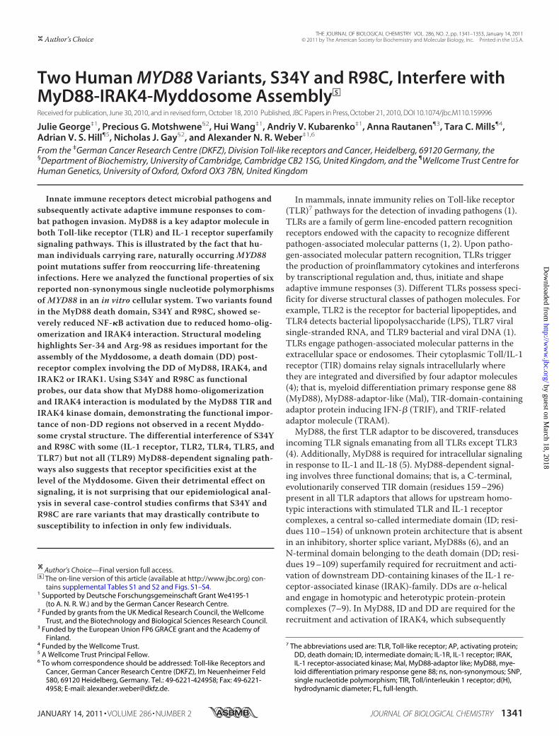
Two Naturally Occurring Mutations in the MyD88 DeathDomain Are Dysfunctional in Downstream Signaling—In thehumanMYD88 gene more than 40 SNPs have been reported,six of which result in amino acid substitutions (nsSNPs) indifferent MyD88 domains: S34Y, L35V, L74M, R98C (DD),K115N (ID), and M178I (TIR) (see Fig. 1A and supplementalTable S1). Generally, nsSNPs have a high potential impact onprotein function (28), and in MyD88 most of the affectedamino acid positions are highly conserved in a range of spe-cies (supplemental Fig. S1). Although little is known about thefrequencies of these naturally occurring variants, we wereintrigued to investigate if any of these reported variants wereassociated with an altered phenotype compared with WTMyD88. To functionally characterize the genetic variants re-ported for MyD88, Myc-tagged expression constructs corre-sponding to the nsSNP-associated amino acid changes(“MyD88 mutants”) were generated. The expression levelswere similar (Fig. 1B), and the MyD88 mutants were testedfor their ability to activate the NF-�B signaling pathway usinga dual luciferase assay in HEK293 cells. Fig. 1C shows thatoverexpression of WTMyD88 results in NF-�B activationand that most MyD88 mutants induce signaling to a levelcomparable to WT. However, the mutants S34Y and R98Cshowed a complete loss of function or a reduction to �20% ofthe WT signal, respectively. This reduction was also observedat the level of cytokine induction, as IL-8 and TNF-� tran-scription levels measured by real-time PCR were reduced forMyD88-S34Y and -R98C (Fig. 1D). To rule out the possibilitythat our results could be influenced by the presence of endog-enous MyD88, we checked NF-�B activation in MyD88-defi-
MYD88 Variants Interfere with Myddosome Assembly
JANUARY 14, 2011 • VOLUME 286 • NUMBER 2 JOURNAL OF BIOLOGICAL CHEMISTRY 1343
by guest on March 18, 2018
http://ww
w.jbc.org/
Dow
nloaded from
cient HEK293 IA3 cells (20). In agreement with Fig. 1C,MyD88 S34Y and R98C revealed the same pattern of reducedNF-�B activation (supplemental Fig. S2). In two other celllines, Huh7 hepatocytes and C33A keratinocytes, NF-�B-driven luciferase production was also strongly reduced forS34Y and R98C compared with WTMyD88 (data not shown).Taken together, these data suggest that the mutants S34Y andR98C interfered with WTMyD88 downstream signaling.S34Y and R98C Differently Modify MyD88-dependent Re-
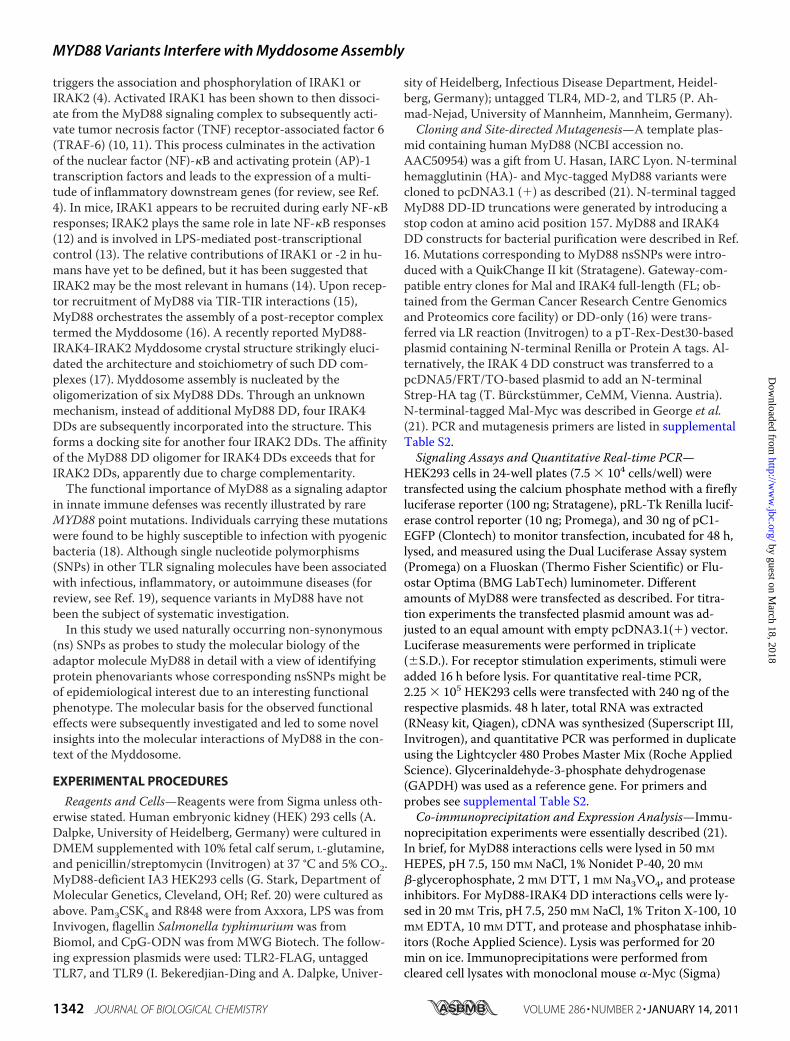
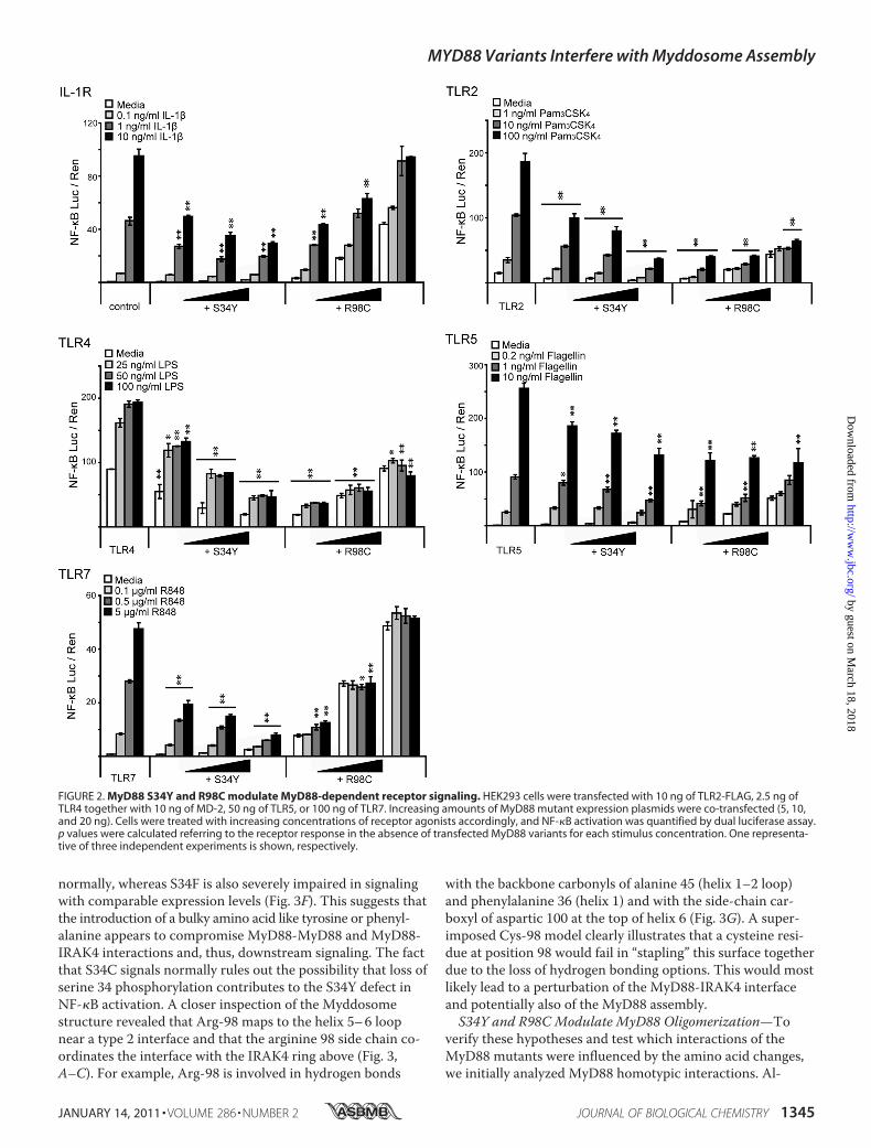
ceptor Activation—We investigated whether the expression ofthese mutants interfered with signaling initiated upon activa-tion of TLR2, TLR4, TLR5, TLR7, TLR9, or IL-1R, all ofwhich require MyD88 for downstream signal transduction (1).MyD88 mutants were transiently co-transfected with differ-ent TLRs in HEK293 cells, and NF-�B-linked luciferase pro-duction was measured upon treatment with the appropriatestimuli. Alternatively, the effect of overexpressed mutants onthe stimulation of endogenous IL-1R and TLR5 was analyzed.Whereas overexpression of MyD88 WT increased the overallamount of activated NF-�B as expected (data not shown),both S34Y and R98C exerted a dominant negative effect onmost MyD88-dependent receptor signaling pathways in adose-dependent and stimulation-dependent manner (Fig. 2),with the exception of TLR9 stimulation (supplemental Fig.S3A). S34Y dose-dependently interfered with the inducedNF-�B response in the case of all of these receptor pathwayswhen titrating stimulation concentration and the transfectedplasmid amount (Fig. 2). However, different TLRs appeareddifferentially sensitive to this effect; in the case of TLR2, -4,and -7, a dominant negative effect was already observed at thelowest plasmid and stimulation concentrations applied (seeFig. 2). In contrast, in the case of endogenous IL-1R and TLR5(both endogenous and transfected), signaling was not inhib-ited in the presence of S34Y at the lowest ligand concentra-tions (see supplemental Fig. S3B). For R98C, overexpressionresulted in a dose-dependent basal level of NF-�B activation(cf. Fig. 1C and see Fig. 4D). In case of IL-1R, TLR2, andTLR5, this signal was further increased in a ligand concentra-
tion-dependent way. Nevertheless, the presence of R98C sup-pressed ligand-induced activation below the level of control-transfected, stimulated cells, especially when low plasmidamounts were transfected and high stimulation concentra-tions were applied. Interestingly, this effect was even strongerfor TLR4 and TLR7 signaling where R98C effectively blockedactivation above the level of R98C alone (see Fig. 2, comparewhite and shaded bars). In summary, our data imply that bothS34Y and R98C are able to interfere with MyD88-dependentreceptor stimulation in a dominant-negative way across arange of ligand concentrations but with differences in the ef-fectiveness of inhibition between different TLRs (see“Discussion”).Ser-34 and Arg-98 Play Important Structural Roles in the
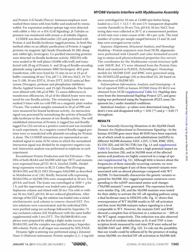
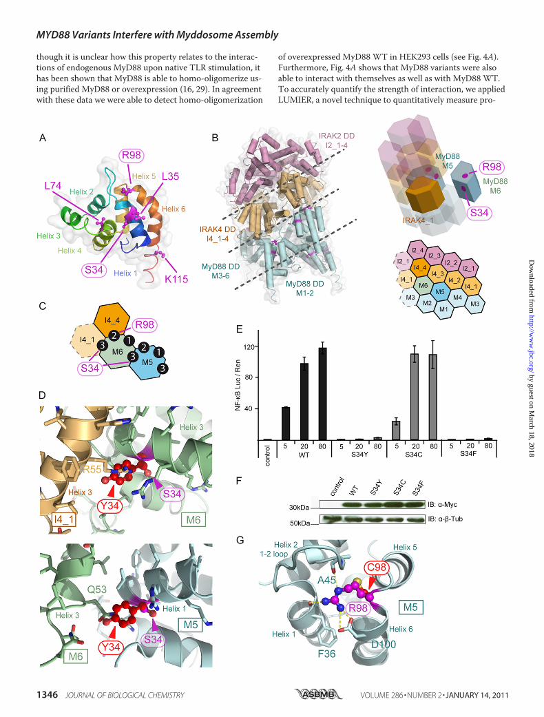
Myddosome—We next sought to determine the molecularbasis for the effects observed for MyD88 S34Y and R98C. Be-fore investigating which protein-protein interactions might beaffected by MyD88 S34Y and R98C, their location was ana-lyzed in the three-dimensional framework in which MyD88has been proposed to operate, the Myddosome (16). Ser-34 isfound on helix 1, and Arg-98 is located in the loop connectinghelix 5 and 6 (Fig. 3A) in a recently published MyD88-IRAK4-IRAK2 DDMyddosome crystal structure (17) (Fig. 3B). Ser-34is part of a type 3 helix3 helix 3 interaction interface of con-secutive MyD88 molecules (e.g.M5 and M6) and of MyD88M6 with the first IRAK4 DD (I4_1�, see Fig. 3C and Ref. 17).Using homology modeling, a Tyr-34 structure of MyD88 DDwas generated and superimposed onto M5 to assess its impacton MyD88-MyD88 interactions (Fig. 3D, lower panel) and onM6 for MyD88-IRAK4 DD interactions (Fig. 3D, upperpanel). Evidently, due to its larger size, Tyr-34 would protrudefrom helix 1 and, thus, clash with the neighboring M6 (gluta-mine 63) or I4_1 (arginine 55) amino acids. To assess whethersteric clashes provide a structural explanation for the ob-served loss of function, serine 34 was also mutated to the ser-ine-isosteric residue cysteine (S34C) and to phenylalanine(S34F), which is of a similar size as tyrosine. NF-�B activationwas subsequently assessed. Fig. 3E shows that S34C signals
FIGURE 1. MyD88 S34Y and R98C show reduced NF-�B activation. A, shown is a schematic overview of MyD88 domains with non-synonymous SNPshighlighted. B, expression of all MyD88 mutants is comparable with WT. HEK293 cells were transfected with Myc-tagged expression constructs and analyzedby immunoblot (IB). �-Tubulin served as a loading control. C, S34Y and R98C fail to induce NF-�B activation. HEK293 cells were transiently transfected withthe indicated amounts of Myc-tagged MyD88 constructs. NF-�B activation was quantified by dual luciferase assay. D, IL-8 and TNF-� mRNA induction is re-duced for S34Y and R98C. HEK293 cells were transiently transfected with MyD88 variants, and induced cytokine mRNA levels were quantified and normal-ized to GAPDH using LightCycler Realtime PCR. One representative of three independent experiments is shown.
MYD88 Variants Interfere with Myddosome Assembly
1344 JOURNAL OF BIOLOGICAL CHEMISTRY VOLUME 286 • NUMBER 2 • JANUARY 14, 2011
by guest on March 18, 2018
http://ww
w.jbc.org/
Dow
nloaded from
normally, whereas S34F is also severely impaired in signalingwith comparable expression levels (Fig. 3F). This suggests thatthe introduction of a bulky amino acid like tyrosine or phenyl-alanine appears to compromise MyD88-MyD88 and MyD88-IRAK4 interactions and, thus, downstream signaling. The factthat S34C signals normally rules out the possibility that loss ofserine 34 phosphorylation contributes to the S34Y defect inNF-�B activation. A closer inspection of the Myddosomestructure revealed that Arg-98 maps to the helix 5–6 loopnear a type 2 interface and that the arginine 98 side chain co-ordinates the interface with the IRAK4 ring above (Fig. 3,A–C). For example, Arg-98 is involved in hydrogen bonds
with the backbone carbonyls of alanine 45 (helix 1–2 loop)and phenylalanine 36 (helix 1) and with the side-chain car-boxyl of aspartic 100 at the top of helix 6 (Fig. 3G). A super-imposed Cys-98 model clearly illustrates that a cysteine resi-due at position 98 would fail in “stapling” this surface togetherdue to the loss of hydrogen bonding options. This would mostlikely lead to a perturbation of the MyD88-IRAK4 interfaceand potentially also of the MyD88 assembly.S34Y and R98C Modulate MyD88 Oligomerization—To
verify these hypotheses and test which interactions of theMyD88 mutants were influenced by the amino acid changes,we initially analyzed MyD88 homotypic interactions. Al-
FIGURE 2. MyD88 S34Y and R98C modulate MyD88-dependent receptor signaling. HEK293 cells were transfected with 10 ng of TLR2-FLAG, 2.5 ng ofTLR4 together with 10 ng of MD-2, 50 ng of TLR5, or 100 ng of TLR7. Increasing amounts of MyD88 mutant expression plasmids were co-transfected (5, 10,and 20 ng). Cells were treated with increasing concentrations of receptor agonists accordingly, and NF-�B activation was quantified by dual luciferase assay.p values were calculated referring to the receptor response in the absence of transfected MyD88 variants for each stimulus concentration. One representa-tive of three independent experiments is shown, respectively.
MYD88 Variants Interfere with Myddosome Assembly
JANUARY 14, 2011 • VOLUME 286 • NUMBER 2 JOURNAL OF BIOLOGICAL CHEMISTRY 1345
by guest on March 18, 2018
http://ww
w.jbc.org/
Dow
nloaded from
though it is unclear how this property relates to the interac-tions of endogenous MyD88 upon native TLR stimulation, ithas been shown that MyD88 is able to homo-oligomerize us-ing purified MyD88 or overexpression (16, 29). In agreementwith these data we were able to detect homo-oligomerization
of overexpressed MyD88 WT in HEK293 cells (see Fig. 4A).Furthermore, Fig. 4A shows that MyD88 variants were alsoable to interact with themselves as well as with MyD88 WT.To accurately quantify the strength of interaction, we appliedLUMIER, a novel technique to quantitatively measure pro-
MYD88 Variants Interfere with Myddosome Assembly
1346 JOURNAL OF BIOLOGICAL CHEMISTRY VOLUME 286 • NUMBER 2 • JANUARY 14, 2011
by guest on March 18, 2018
http://ww
w.jbc.org/
Dow
nloaded from
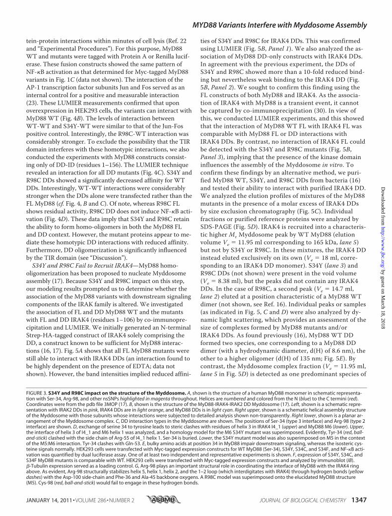
tein-protein interactions within minutes of cell lysis (Ref. 22and “Experimental Procedures”). For this purpose, MyD88WT and mutants were tagged with Protein A or Renilla lucif-erase. These fusion constructs showed the same pattern ofNF-�B activation as that determined for Myc-tagged MyD88variants in Fig. 1C (data not shown). The interaction of theAP-1 transcription factor subunits Jun and Fos served as aninternal control for a positive and measurable interaction(23). These LUMIER measurements confirmed that uponoverexpression in HEK293 cells, the variants can interact withMyD88 WT (Fig. 4B). The levels of interaction betweenWT-WT and S34Y-WT were similar to that of the Jun-Fospositive control. Interestingly, the R98C-WT interaction wasconsiderably stronger. To exclude the possibility that the TIRdomain interferes with these homotypic interactions, we alsoconducted the experiments with MyD88 constructs consist-ing only of DD-ID (residues 1–156). The LUMIER techniquerevealed an interaction for all DD mutants (Fig. 4C). S34Y andR98C DDs showed a significantly decreased affinity for WTDDs. Interestingly, WT-WT interactions were considerablystronger when the DDs alone were transfected rather than theFL MyD88 (cf. Fig. 4, B and C). Of note, whereas R98C FLshows residual activity, R98C DD does not induce NF-�B acti-vation (Fig. 4D). These data imply that S34Y and R98C retainthe ability to form homo-oligomers in both the MyD88 FLand DD context. However, the mutant proteins appear to me-diate these homotypic DD interactions with reduced affinity.Furthermore, DD oligomerization is significantly influencedby the TIR domain (see “Discussion”).S34Y and R98C Fail to Recruit IRAK4—MyD88 homo-
oligomerization has been proposed to nucleate Myddosomeassembly (17). Because S34Y and R98C impact on this step,our modeling results prompted us to determine whether theassociation of the MyD88 variants with downstream signalingcomponents of the IRAK family is altered. We investigatedthe association of FL and DDMyD88WT and the mutantswith FL and DD IRAK4 (residues 1–106) by co-immunopre-cipitation and LUMIER. We initially generated an N-terminalStrep-HA-tagged construct of IRAK4 solely comprising theDD, a construct known to be sufficient for MyD88 interac-tions (16, 17). Fig. 5A shows that all FL MyD88 mutants werestill able to interact with IRAK4 DDs (an interaction found tobe highly dependent on the presence of EDTA; data notshown). However, the band intensities implied reduced affini-
ties of S34Y and R98C for IRAK4 DDs. This was confirmedusing LUMIER (Fig. 5B, Panel 1). We also analyzed the as-sociation of MyD88 DD-only constructs with IRAK4 DDs.In agreement with the previous experiment, the DDs ofS34Y and R98C showed more than a 10-fold reduced bind-ing but nevertheless weak binding to the IRAK4 DD (Fig.5B, Panel 2). We sought to confirm this finding using theFL constructs of both MyD88 and IRAK4. As the associa-tion of IRAK4 with MyD88 is a transient event, it cannotbe captured by co-immunoprecipitation (30). In view ofthis, we conducted LUMIER experiments, and this showedthat the interaction of MyD88 WT FL with IRAK4 FL wascomparable with MyD88 FL or DD interactions withIRAK4 DDs. By contrast, no interaction of IRAK4 FL couldbe detected with the S34Y and R98C mutants (Fig. 5B,Panel 3), implying that the presence of the kinase domaininfluences the assembly of the Myddosome in vitro. Toconfirm these findings by an alternative method, we puri-fied MyD88 WT, S34Y, and R98C DDs from bacteria (16)and tested their ability to interact with purified IRAK4 DD.We analyzed the elution profiles of mixtures of the MyD88mutants in the presence of a molar excess of IRAK4 DDsby size exclusion chromatography (Fig. 5C). Individualfractions or purified reference proteins were analyzed bySDS-PAGE (Fig. 5D). IRAK4 is recruited into a characteris-tic higher Mr Myddosome peak by WT MyD88 (elutionvolume Ve � 11.95 ml corresponding to 165 kDa, lane 5)but not by S34Y or R98C. In these mixtures, the IRAK4 DDinstead eluted exclusively on its own (Ve � 18 ml, corre-sponding to an IRAK4 DD monomer). S34Y (lane 3) andR98C DDs (not shown) were present in the void volume(Ve � 8.38 ml), but the peaks did not contain any IRAK4DDs. In the case of R98C, a second peak (Ve � 14.7 ml,lane 2) eluted at a position characteristic of a MyD88 WTdimer (not shown, see Ref. 16). Individual peaks or samples(as indicated in Fig. 5, C and D) were also analyzed by dy-namic light scattering, which provides an assessment of thesize of complexes formed by MyD88 mutants and/orIRAK4 DDs. As found previously (16), MyD88 WT DDformed two species, one corresponding to a MyD88 DDdimer (with a hydrodynamic diameter, d(H) of 8.6 nm), theother to a higher oligomer (d(H) of 135 nm; Fig. 5E). Bycontrast, the Myddosome complex fraction (Ve � 11.95 ml,lane 5 in Fig. 5D) is detected as one predominant species of
FIGURE 3. S34Y and R98C impact on the structure of the Myddosome. A, shown is the structure of a human MyD88 monomer in schematic representa-tion with Ser-34, Arg-98, and other nsSNPs highlighted in magenta throughout. Helices are numbered and colored from the N (blue) to the C termini (red).Coordinates were from the pdb file 3MOP (17). B, shown is the structure of the MyD88-IRAK4-IRAK2 DD Myddosome (17). Left, shown is a schematic repre-sentation with IRAK2 DDs in pink, IRAK4 DDs are in light orange, and MyD88 DDs is in light cyan. Right upper, shown is a schematic helical assembly structureof the Myddosome with those subunits whose interactions were subjected to detailed analysis shown non-transparently. Right lower, shown is a planar ar-rangement of the Myddosome complex. C, DD interaction types in the Myddosome are shown. The positions of Ser-34 (type 3 interface) and Arg-98 (type 2interface) are shown. D, exchange of serine 34 to tyrosine leads to steric clashes with residues of helix 3 in IRAK4 I4_1 (upper) and MyD88 M6 (lower). Upper,the interface of helix 3 of I4_1 and M6 helix 1 was analyzed, and a homology model for the M6 S34Y mutant was superimposed. Evidently, Tyr-34 (red, ball-and-stick) clashed with the side chain of Arg-55 of I4_1 helix 1. Ser-34 is buried. Lower, the S34Y mutant model was also superimposed on M5 in the contextof the M5:M6 interaction. Tyr-34 clashes with Gln-53. E, bulky amino acids at position 34 in MyD88 impair downstream signaling, whereas the isosteric cys-teine signals normally. HEK293 cells were transfected with Myc-tagged expression constructs for WT MyD88 (Ser-34), S34Y, S34C, and S34F, and NF-�B acti-vation was quantified by dual luciferase assay. One of at least two independent and representative experiments is shown. F, expression of S34Y, S34C, andS34F MyD88 mutants is comparable with WT. HEK293 cells were transfected with Myc-tagged expression constructs and analyzed by immunoblot (IB).�-Tubulin expression served as a loading control. G, Arg-98 plays an important structural role in coordinating the interface of MyD88 with the IRAK4 ringabove. As evident, Arg-98 structurally stabilizes helix 5, helix 1, helix 2, and the 1–2 loop (which interdigitates with IRAK4) through hydrogen bonds (yellowdashes) with the Asp-100 side-chain and Phe-36 and Ala-45 backbone oxygens. A R98C model was superimposed onto the elucidated MyD88 structure(M5). Cys-98 (red, ball-and-stick) would fail to engage in these hydrogen bonds.
MYD88 Variants Interfere with Myddosome Assembly
JANUARY 14, 2011 • VOLUME 286 • NUMBER 2 JOURNAL OF BIOLOGICAL CHEMISTRY 1347
by guest on March 18, 2018
http://ww
w.jbc.org/
Dow
nloaded from
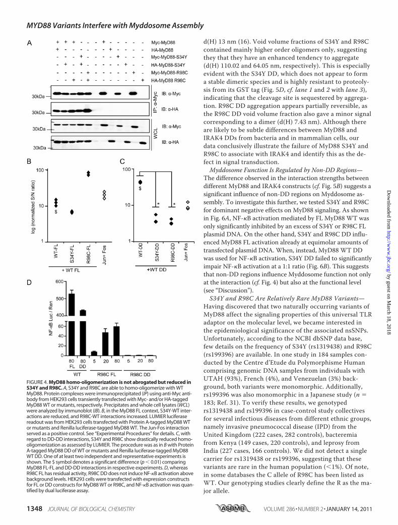
d(H) 13 nm (16). Void volume fractions of S34Y and R98Ccontained mainly higher order oligomers only, suggestingthey that they have an enhanced tendency to aggregate(d(H) 110.02 and 64.05 nm, respectively). This is especiallyevident with the S34Y DD, which does not appear to forma stable dimeric species and is highly resistant to proteoly-sis from its GST tag (Fig. 5D, cf. lane 1 and 2 with lane 3),indicating that the cleavage site is sequestered by aggrega-tion. R98C DD aggregation appears partially reversible, asthe R98C DD void volume fraction also gave a minor signalcorresponding to a dimer (d(H) 7.43 nm). Although thereare likely to be subtle differences between MyD88 andIRAK4 DDs from bacteria and in mammalian cells, ourdata conclusively illustrate the failure of MyD88 S34Y andR98C to associate with IRAK4 and identify this as the de-fect in signal transduction.Myddosome Function Is Regulated by Non-DD Regions—
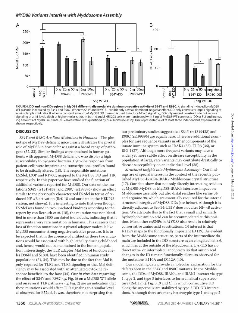
The difference observed in the interaction strengths betweendifferent MyD88 and IRAK4 constructs (cf. Fig. 5B) suggests asignificant influence of non-DD regions on Myddosome as-sembly. To investigate this further, we tested S34Y and R98Cfor dominant negative effects on MyD88 signaling. As shownin Fig. 6A, NF-�B activation mediated by FL MyD88 WT wasonly significantly inhibited by an excess of S34Y or R98C FLplasmid DNA. On the other hand, S34Y and R98C DD influ-enced MyD88 FL activation already at equimolar amounts oftransfected plasmid DNA. When, instead, MyD88 WT DDwas used for NF-�B activation, S34Y DD failed to significantlyimpair NF-�B activation at a 1:1 ratio (Fig. 6B). This suggeststhat non-DD regions influence Myddosome function not onlyat the interaction (cf. Fig. 4) but also at the functional level(see “Discussion”).S34Y and R98C Are Relatively Rare MyD88 Variants—
Having discovered that two naturally occurring variants ofMyD88 affect the signaling properties of this universal TLRadaptor on the molecular level, we became interested inthe epidemiological significance of the associated nsSNPs.Unfortunately, according to the NCBI dbSNP data base,few details on the frequency of S34Y (rs1319438) and R98C(rs199396) are available. In one study in 184 samples con-ducted by the Centre d’Etude du Polymorphisme Humancomprising genomic DNA samples from individuals withUTAH (93%), French (4%), and Venezuelan (3%) back-ground, both variants were monomorphic. Additionally,rs199396 was also monomorphic in a Japanese study (n �183; Ref. 31). To verify these results, we genotypedrs1319438 and rs199396 in case-control study collectivesfor several infectious diseases from different ethnic groups,namely invasive pneumococcal disease (IPD) from theUnited Kingdom (222 cases, 282 controls), bacteremiafrom Kenya (149 cases, 220 controls), and leprosy fromIndia (227 cases, 166 controls). We did not detect a singlecarrier for rs1319438 or rs199396, suggesting that thesevariants are rare in the human population (�1%). Of note,in some databases the C allele of R98C has been listed asWT. Our genotyping studies clearly define the R as the ma-jor allele.
FIGURE 4. MyD88 homo-oligomerization is not abrogated but reduced inS34Y and R98C. A, S34Y and R98C are able to homo-oligomerize with WTMyD88. Protein complexes were immunoprecipitated (IP) using anti-Myc anti-body from HEK293 cells transiently transfected with Myc- and/or HA-taggedMyD88 WT or mutants, respectively. Precipitates and whole cell lysates (WCL)were analyzed by immunoblot (IB). B, in the MyD88 FL context, S34Y-WT inter-actions are reduced, and R98C-WT interactions increased. LUMIER luciferasereadout was from HEK293 cells transfected with Protein A-tagged MyD88 WTor mutants and Renilla luciferase-tagged MyD88 WT. The Jun-Fos interactionserved as a positive control. See “Experimental Procedures” for details. C, withregard to DD-DD interactions, S34Y and R98C show drastically reduced homo-oligomerization as assessed by LUMIER. The procedure was as in B with ProteinA-tagged MyD88 DD of WT or mutants and Renilla luciferase-tagged MyD88WT DD. One of at least two independent and representative experiments isshown. The $ symbol denotes a significant difference (p � 0.01) comparingMyD88 FL-FL and DD-DD interactions in respective experiments. D, whereasR98C FL has residual activity, R98C DD does not induce NF-�B activation abovebackground levels. HEK293 cells were transfected with expression constructsfor FL or DD constructs for MyD88 WT or R98C, and NF-�B activation was quan-tified by dual luciferase assay.
MYD88 Variants Interfere with Myddosome Assembly
1348 JOURNAL OF BIOLOGICAL CHEMISTRY VOLUME 286 • NUMBER 2 • JANUARY 14, 2011
by guest on March 18, 2018
http://ww
w.jbc.org/
Dow
nloaded from
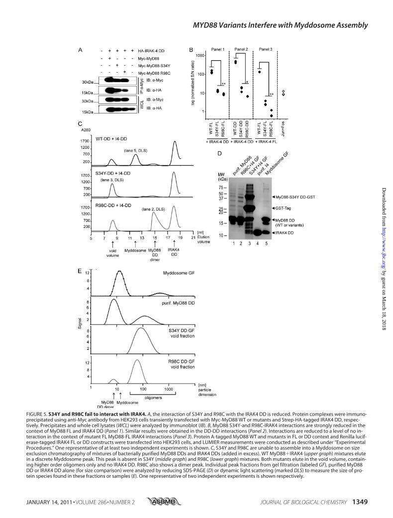
FIGURE 5. S34Y and R98C fail to interact with IRAK4. A, the interaction of S34Y and R98C with the IRAK4 DD is reduced. Protein complexes were immuno-precipitated using anti-Myc antibody from HEK293 cells transiently transfected with Myc-MyD88 WT or mutants and Strep-HA-tagged IRAK4 DD, respec-tively. Precipitates and whole cell lysates (WCL) were analyzed by immunoblot (IB). B, MyD88 S34Y-and R98C-IRAK4 interactions are strongly reduced in thecontext of MyD88 FL and IRAK4 DD (Panel 1). Similar results were obtained in the DD-DD interactions (Panel 2). Interactions are reduced to a level of no in-teraction in the context of mutant FL MyD88-FL IRAK4 interactions (Panel 3). Protein A-tagged MyD88 WT and mutants in FL or DD context and Renilla lucif-erase-tagged IRAK4 FL or DD constructs were transfected into HEK293 cells, and LUMIER measurements were conducted as described under “ExperimentalProcedures.” One representative of at least two independent experiments is shown. C, S34Y and R98C are unable to assemble into a Myddosome on sizeexclusion chromatography of mixtures of bacterially purified MyD88 DDs and IRAK4 DDs (added in excess). WT MyD88�IRAK4 (upper graph) mixtures elutein a discrete Myddosome peak. This peak is absent in S34Y (middle graph) and R98C (lower graph) mixtures. Both mutants elute in the void volume, contain-ing higher order oligomers only and no IRAK4 DD. R98C also shows a dimer peak. Individual peak fractions from gel filtration (labeled GF), purified MyD88DD or IRAK4 DD alone (for size comparison) were analyzed by reducing SDS-PAGE (D) or dynamic light scattering (marked DLS) to measure the size of pro-tein species found in these fractions or samples (E). One representative of two independent experiments is shown respectively.
MYD88 Variants Interfere with Myddosome Assembly
JANUARY 14, 2011 • VOLUME 286 • NUMBER 2 JOURNAL OF BIOLOGICAL CHEMISTRY 1349
by guest on March 18, 2018
http://ww
w.jbc.org/
Dow
nloaded from
DISCUSSION
S34Y and R98C Are Rare Mutations in Humans—The phe-notype of MyD88-deficient mice clearly illustrates the pivotalrole of MyD88 in host defense against a broad range of patho-gens (32, 33). Similar findings were obtained in human pa-tients with apparent MyD88 deficiency, who display a highsusceptibility to pyogenic bacteria. Cytokine responses frompatient cells were impaired and transcriptional profiles foundto be drastically altered (18). The responsible mutationsE52del, L93P and R196C, mapped to the MyD88 DD and TIR,respectively. In this paper we have studied the function ofadditional variants reported for MyD88. Our data on the mu-tations S34Y (rs1319438) and R98C (rs199396) show an effectsimilar to the previously characterized E52del in terms of re-duced NF-�B activation (Ref. 18 and our data in the HEK293system, not shown). It is interesting to note that even thoughE52del was found in two unrelated kindreds according to thereport by von Bernuth et al. (18), the mutation was not identi-fied in more than 1800 unrelated individuals, indicating that itrepresents a very rare mutation in humans. This suggests thatloss of function mutations in a pivotal adaptor molecule likeMyD88 encounter strong negative selective pressure. It is tobe expected that in the absence of antibiotics these two muta-tions would be associated with high lethality during childhoodand, hence, would not be maintained in the human popula-tion. Interestingly, the TLR adaptor Mal loss of function alle-les D96N and S180L have been identified in human studypopulations (21, 34). This may be due to the fact that Mal isonly required for TLR2 and TLR4 signaling or that Mal defi-ciency may be associated with an attenuated cytokine re-sponse beneficial to the host (34). Our in vitro data regardingthe effect of S34Y and R98C (cf. Fig. 6) on a MyD88 WT alleleand on several TLR pathways (cf. Fig. 2) are an indication thatthese mutations would affect TLR signaling to a similar levelas observed for E52del. It was, therefore, not surprising that
our preliminary studies suggest that S34Y (rs1319438) andR98C (rs199396) are equally rare. There are additional exam-ples for rare sequence variants in other components of theinnate immune system such as IRAK4 (35), TLR3 (36), orRIG-I (37). Although more frequent variants may have awider yet more subtle effect on disease susceptibility in thepopulation at large, rare variants may contribute drastically todisease susceptibility on an individual level (38).Structural Insights into Myddosome Assembly—Our find-
ings are of special interest in the context of the recently pub-lished MyD88-IRAK4-IRAK2 Myddosome crystal structure(17). Our data show that not only directly interacting residuesat MyD88-MyD88 or MyD88-IRAK4 interfaces impact onMyddosome assembly but also distal residues like serine 34and arginine 98, which are essentially required for the internalstructural integrity of MyD88 DDs (see below). Although it isdirectly adjacent to Ser-34, L35V does not alter NF-�B activa-tion. We attribute this to the fact that a small and similarlyhydrophobic amino acid can be accommodated at this posi-tion. Most other nsSNPs inMYD88 also result in relativelyconservative amino acid substitutions. Of interest is thatK115N maps to the functionally important ID (39). As evidentfrom the Myddosome structure, parts of the intermediate do-main are included in the DD structure as an elongated helix 6,which lies at the outside of the Myddosome. Lys-115 has nodirect intra- or intermolecular contacts so that amino acidchanges in the ID remain functionally silent, as observed forthe mutations E110A and D112A (40).Our modeling data provide a molecular explanation for the
defects seen in the S34Y and R98C mutants. In the Myddo-some, the DDs of MyD88, IRAK4, and IRAK1 interact via type1, type 2, and type 3 interfaces to form a helical superstruc-ture (Ref. 17; cf. Fig. 3, B and C) in which consecutive DDalong the superhelix are stabilized by type 3 DD-DD interac-tions. Although there are many heterotypic type 1 and type 2
FIGURE 6. DD and non-DD regions in MyD88 differentially modulate dominant-negative activity of S34Y and R98C. A, signaling induced by MyD88WT plasmid is reduced by S34Y and R98C. Whereas S34Y and R98C FL exhibit only a weak dominant-negative effect, DD-only constructs impair signaling atequimolar plasmid ratio. B, when a constant amount of MyD88 DD plasmid is used to induce NF-�B signaling, DD-only mutant constructs do not reducesignaling at a 1:1 level, albeit at higher molar ratios. In both A and B HEK293 cells were transfected with 5 ng of MyD88 WT constructs (DD or FL) and increas-ing amounts of MyD88 mutants. NF-�B activation was quantified by dual luciferase assay. One representative of at least three independent experiments isshown, respectively.
MYD88 Variants Interfere with Myddosome Assembly
1350 JOURNAL OF BIOLOGICAL CHEMISTRY VOLUME 286 • NUMBER 2 • JANUARY 14, 2011
by guest on March 18, 2018
http://ww
w.jbc.org/
Dow
nloaded from
DD-DD interactions between the constituent subunits of theMyddosome, there is only one type 3 interaction between theMyD88 and the first IRAK4 protomer to be incorporated (Fig.3C). This type 3 interface occurs between helix 1 residues inM6 and helix 3 of I4_1 (Fig. 3D, upper panel). A very similarstructural configuration is observed at the type 3 interfacebetween M5 and M6 (Fig. 3D, lower panel). It is clear from themodeling study we present that substitution of serine 34 inhelix 1 by tyrosine would cause steric hindrance in both thehomotypic (M5:M6) and the single heterotypic type 3 inter-faces (M6:I4_1), leading to reduced affinity of homotypic (Fig.4C) and heterotypic (Fig. 5B, panel 2) interactions. It is con-ceivable that a weakened type 3 interface additionally de-creases the stability of type 1 and/or 2 interactions and, thus,helical assembly. The reason why MyD88 dimers are seen forR98C (Fig. 5C) and WT (not shown, Ref. 16) but not S34Ymay be due to the fact that initial MyD88 dimer formationdepends exclusively on a type 3 interface. For the R98C mu-tant the molecular basis of the defect appears to be different.Arg-98 is located close to (but not part of) a type 2 interface.Several hydrogen bonds formed by the Arg-98 side-chain co-ordinate helix 1, 5, 6, and the 1–2 loop, which contacts anysubsequent ring (MyD88 or IRAK4) above. Substitution ofArg-98 for cysteine would cause perturbations in this type 2interface. Whereas dimer formation would not be affected (asevidenced by gel filtration of purified R98C, cf. Fig. 5C), fullMyddosome assembly is probably compromised. The residuallevel of R98C activity observed in the FL but not DD context(Fig. 4D) may be due to the fact that the TIR domain recoversinteraction with WTMyD88 (Fig. 4B) through an unknownmechanism (see also below). In conclusion, this identifies theformation of the heterotypic type 2 and 3 interfaces betweenIRAK4 and MyD88 and, by implication, helical assembly ofthe Myddosome complex as essential steps in signaltransduction.Influence of Non-DD Regions on Myddosome Function—An-
other important insight from our work is the influence ofnon-DD regions on Myddosome assembly. The crystal struc-ture of the Myddosome obviously does not include non-DDregions like the MyD88 TIR domain or the IRAK kinase do-mains (17). We found that DD-DD interactions in general arestronger than looking at the same interactions in an FL con-text as assessed by LUMIER, a technique providing a morequantitative measurement of protein-protein interactionsthan co-immunoprecipitation. For example, the presence ofthe MyD88 TIR domain significantly modulated MyD88 ho-mo-oligomerization (cf. Fig. 4, B versus C,WT FL versus WTDD) and heterotypic interaction with IRAK4 DDs (cf. Fig. 5B,Panel 1 versus Panel 2,WT FL versus WT DD). This is inkeeping with earlier immunoprecipitation studies whichfound that the TIR domain plays a role in MyD88 homodimerformation independently of the DD. Unfortunately, thestrengths of interaction for different MyD88 (FL, DD or TIR-only) constructs was not quantified (29). The MyD88 TIRdomain also seems to play an important role in the dominant-negative effect of MyD88 (cf. Fig. 6). For example, MyD88 FLis only inhibited significantly by DD-only S34Y and R98C atequimolar ratios of transfected plasmid (right part of Fig. 6A).
Based on our LUMIER data, this may be due to a strongerinteraction upon removal of the TIR domain of S34Y orR98C. Surprisingly, when TIR domains are absent in bothMyD88 WT and mutants, this inhibition is again lost (left partof Fig. 6B). Due to the fact that S34Y DD and R98C DD inter-act with WT DD far less strongly than WT DD; cf. Fig. 4C),inactive S34Y or R98C DD would be excluded from activeWT DDMyddosomes. In the case of WT FL-WT FL versusWT FL-mutant DD interactions, the difference in affinity maybe less, so that signaling-deficient mutant DDs are also incor-porated into Myddosomes, leading to an overall decrease indownstream signal activation. We also found that the IRAK4kinase domain influences MyD88-IRAK4 DD interactions (cf.(Fig. 5B): for instance, S34Y and R98C FL interacted, albeitweakly, with the IRAK4 DD (Panel 1). The interaction withIRAK4 FL for the same MyD88 constructs was however abro-gated (Panel 3). Because the structural assembly shown in thecrystal structure appears to form spontaneously from purifiedDDs (16, 17), in a cellular context TIR domain and kinase do-mains may, thus, modulate Myddosome assembly. Althoughat this stage the precise mechanisms of Myddosome regula-tion in vivo remain elusive, our data suggest that additionalwork both structural and biochemical is required at the levelof FL proteins. We introduce LUMIER as a technique able toassess even transient IRAK4 FL interactions. Additionally, thespatiotemporal framework of Myddosome assembly in livingcells should be addressed to complement the information de-rived from isolated crystallized proteins.Receptor-specific MyD88 Post-receptor Complexes—Our
data suggest that the mutants S34Y and R98C negatively in-fluence MyD88-dependent receptor stimulation and that theMyD88 requirement for different TLR pathways may vary.We would have expected that, if at all, a loss of function mu-tation in the MyD88 DD would interfere with all MyD88-de-pendent signaling pathways in a similar way, as receptor-spe-cific coupling is assumed to be mediated exclusively by therespective TIR domains. Notwithstanding the limitations ofthe HEK293 system, according to the data shown in Figs. 2and supplemental Fig. S3, S34Y and R98C only affected IL-1R,TLR2, TLR4, TLR5, and TLR7 (not TLR9), and differences inthe effectiveness of inhibition between the different receptorswere repeatedly observed: whereas S34Y inhibited TLR2,TLR4, and TLR7 ligand-induced levels of activation, alreadyat low ligand concentrations, and TLR5 and IL-1R were onlysensitive to S34Y at high ligand concentrations (cf. supple-mental Fig. S3B). In contrast, the analysis of the mutant R98Crevealed distinct dominant negative effects between differentreceptor pathways; R98C attenuated IL-1R, TLR2, and TLR5signals significantly but only entirely blocked TLR4 and TLR7ligand-induced effects (cf. Fig. 2B). These results might indi-cate differences in MyD88-dependent receptor-proximaldownstream events. Given that Mal-MyD88 interactions werecomparable with WT in S34Y and R98C (cf. supplemental Fig.S4), upstream coupling to TLR2 and 4 via Mal and, thus, TIR-mediated TLR2/4-Mal-MyD88 associations are presumablyintact. Consequently, the dominant-negative effect wouldmanifest itself at the level of the DD assembly. Targeting dif-ferent interfaces of DD assembly (S34Y, type 3; R98C, type 2)
MYD88 Variants Interfere with Myddosome Assembly
JANUARY 14, 2011 • VOLUME 286 • NUMBER 2 JOURNAL OF BIOLOGICAL CHEMISTRY 1351
by guest on March 18, 2018
http://ww
w.jbc.org/
Dow
nloaded from
in a dominant-negative fashion appears to affect specific TLR-induced Myddosomes differently. These assemblies may,therefore, show greater receptor-specific differences than pre-viously thought, and a model of signal transduction in whichall MyD88-dependent pathways are converted to the samedownstream signal at the level of MyD88 is likely to be over-simplified. A certain degree of specificity is potentially main-tained from incoming signals via different TLRs at and be-yond the level of post-receptor complexes. Reconstitutionexperiments into MyD88-deficient cells endogenously ex-pressing TLRs may be ways to confirm this hypothesis. Addi-tionally, studies involving LUMIER assays between differentTLR TIR domains and MyD88 mutants or proteomics ap-proaches involving mass spectrometry may help to unravelthe structural framework for the specificity of differentMyD88 TLR interactions.In conclusion, we present here a detailed functional and
biochemical analysis of two rareMYD88 loss of function vari-ants whose significance for human disease waits to be ex-plored more fully. Our study provides valuable insights intothe assembly of MyD88-IRAK4 Myddosomes. Our data pointto the fact that this process is regulated by non-DD regions inboth MyD88 and IRAK4 and presumably also IRAK1 or -2.The precise contribution of these domains will need to beclarified functionally, biochemically, and cell biologically tounderstand and therapeutically interfere with, the function ofthe Myddosome as a post-receptor TLR signaling complex.
Acknowledgments—We gratefully acknowledge J. Meckler, M. Scheuer-mann, T. Schmidt, M. Kogl, and the other members of the GermanCancer Research Centre Proteomics core facility for technical assis-tance. We thankM. Frank and T. Holz for support regarding moleculardynamics and computers. J. C. Crook, K. Knox, and the Oxford Pneu-mococcal Surveillance Group, A. Berkley, K. Marsh, N. Peshu, A. G.Scott, T. Williams (Kilifi, Kenya), S. Roy, S. K. Hazra, and B. Saha(Kolkata, India), were involved in the collection of samples of the differ-ent study populations investigated. We thank R. Jerala for critical read-ing of this manuscript and helpful comments.
REFERENCES1. Kawai, T., and Akira, S. (2008) Ann. N.Y. Acad. Sci. 1143, 1–202. Rock, F. L., Hardiman, G., Timans, J. C., Kastelein, R. A., and Bazan, J. F.
(1998) Proc. Natl. Acad. Sci. U.S.A. 95, 588–5933. Iwasaki, A., and Medzhitov, R. (2004) Nat. Immunol. 5, 987–9954. Watters, T. M., Kenny, E. F., and O’Neill, L. A. (2007) Immunol. Cell
Biol. 85, 411–4195. O’Neill, L. A. (2008) Immunol. Rev. 226, 10–186. Burns, K., Janssens, S., Brissoni, B., Olivos, N., Beyaert, R., and Tschopp,
J. (2003) J. Exp. Med. 197, 263–2687. Weber, C. H., and Vincenz, C. (2001) Trends Biochem. Sci. 26, 475–4818. Scott, F. L., Stec, B., Pop, C., Dobaczewska, M. K., Lee, J. J., Monosov, E.,
Robinson, H., Salvesen, G. S., Schwarzenbacher, R., and Riedl, S. J.(2009) Nature 457, 1019–1022
9. Park, H. H., and Wu, H. (2007) Acta Crystallogr. Sect F Struct. Biol.Cryst. Commun. 63, 229–232
10. Kobayashi, K., Hernandez, L. D., Galan, J. E., Janeway, C. A., Jr., Medzhi-tov, R., and Flavell, R. A. (2002) Cell 110, 191–202
11. Brikos, C., Wait, R., Begum, S., O’Neill, L. A., and Saklatvala, J. (2007)Mol. Cell. Proteomics 6, 1551–1559
12. Kawagoe, T., Sato, S., Matsushita, K., Kato, H., Matsui, K., Kumagai, Y.,Saitoh, T., Kawai, T., Takeuchi, O., and Akira, S. (2008) Nat. Immunol.
9, 684–69113. Wan, Y., Xiao, H., Affolter, J., Kim, T. W., Bulek, K., Chaudhuri, S., Carl-
son, D., Hamilton, T., Mazumder, B., Stark, G. R., Thomas, J., and Li, X.(2009) J. Biol. Chem. 284, 10367–10375
14. Conner, J. R., Smirnova, II, and Poltorak, A. (2009) J. Exp. Med. 206,1615–1631
15. Wesche, H., Henzel, W. J., Shillinglaw, W., Li, S., and Cao, Z. D. (1997)Immunity 7, 837–847
16. Motshwene, P. G., Moncrieffe, M. C., Grossmann, J. G., Kao, C., Ay-aluru, M., Sandercock, A. M., Robinson, C. V., Latz, E., and Gay, N. J.(2009) J. Biol. Chem. 284, 25404–25411
17. Lin, S. C., Lo, Y. C., and Wu, H. (2010) Nature 465, 885–89018. von Bernuth, H., Picard, C., Jin, Z., Pankla, R., Xiao, H., Ku, C. L., Chra-
bieh, M., Mustapha, I. B., Ghandil, P., Camcioglu, Y., Vasconcelos, J.,Sirvent, N., Guedes, M., Vitor, A. B., Herrero-Mata, M. J., Arostegui, J. I.,Rodrigo, C., Alsina, L., Ruiz-Ortiz, E., Juan, M., Fortuny, C., Yague, J.,Anton, J., Pascal, M., Chang, H. H., Janniere, L., Rose, Y., Garty, B. Z.,Chapel, H., Issekutz, A., Marodi, L., Rodriguez-Gallego, C., Banchereau,J., Abel, L., Li, X., Chaussabel, D., Puel, A., and Casanova, J. L. (2008)Science 321, 691–696
19. Misch, E. A., and Hawn, T. R. (2008) Clin. Sci. 114, 347–36020. Li, X., Commane, M., Burns, C., Vithalani, K., Cao, Z., and Stark, G. R.
(1999)Mol. Cell. Biol. 19, 4643–465221. George, J., Kubarenko, A. V., Rautanen, A., Mills, T. C., Colak, E.,
Kempf, T., Hill, A. V., Nieters, A., and Weber, A. N. (2010) J. Immunol.184, 3025–3032
22. Barrios-Rodiles, M., Brown, K. R., Ozdamar, B., Bose, R., Liu, Z., Dono-van, R. S., Shinjo, F., Liu, Y., Dembowy, J., Taylor, I. W., Luga, V., Przulj,N., Robinson, M., Suzuki, H., Hayashizaki, Y., Jurisica, I., and Wrana,J. L. (2005) Science 307, 1621–1625
23. Chiu, R., Boyle, W. J., Meek, J., Smeal, T., Hunter, T., and Karin, M.(1988) Cell 54, 541–552
24. Sali, A., and Overington, J. P. (1994) Protein Sci. 3, 1582–159625. Kubarenko, A., Frank, M., and Weber, A. N. (2007) Biochem. Soc. Trans.
35, 1515–151826. Kubarenko, A. V., Ranjan, S., Colak, E., George, J., Frank, M., and
Weber, A. N. (2010) Protein Sci. 19, 558–56927. (2003) Nature 426, 789–79628. Ramensky, V., Bork, P., and Sunyaev, S. (2002) Nucleic Acids Res. 30,
3894–390029. Burns, K., Martinon, F., Esslinger, C., Pahl, H., Schneider, P., Bodmer,
J. L., Di Marco, F., French, L., and Tschopp, J. (1998) J. Biol. Chem. 273,12203–12209
30. Li, S., Strelow, A., Fontana, E. J., and Wesche, H. (2002) Proc. Natl. Acad.Sci. U.S.A. 99, 5567–5572
31. Suzuki, H., Suzuki, Y., Narita, I., Aizawa, M., Kihara, M., Yamanaka, T.,Kanou, T., Tsukaguchi, H., Novak, J., Horikoshi, S., and Tomino, Y.(2008) J. Am. Soc. Nephrol. 19, 2384–2395
32. Hoebe, K., Du, X., Georgel, P., Janssen, E., Tabeta, K., Kim, S. O., Goode,J., Lin, P., Mann, N., Mudd, S., Crozat, K., Sovath, S., Han, J., and Beut-ler, B. (2003) Nature 424, 743–748
33. Kawai, T., Adachi, O., Ogawa, T., Takeda, K., and Akira, S. (1999) Im-munity 11, 115–122
34. Khor, C. C., Chapman, S. J., Vannberg, F. O., Dunne, A., Murphy, C.,Ling, E. Y., Frodsham, A. J., Walley, A. J., Kyrieleis, O., Khan, A., Aucan,C., Segal, S., Moore, C. E., Knox, K., Campbell, S. J., Lienhardt, C., Scott,A., Aaby, P., Sow, O. Y., Grignani, R. T., Sillah, J., Sirugo, G., Peshu, N.,Williams, T. N., Maitland, K., Davies, R. J., Kwiatkowski, D. P., Day,N. P., Yala, D., Crook, D. W., Marsh, K., Berkley, J. A., O’Neill, L. A., andHill, A. V. (2007) Nat. Genet. 39, 523–528
35. Picard, C., Puel, A., Bonnet, M., Ku, C. L., Bustamante, J., Yang, K., Sou-dais, C., Dupuis, S., Feinberg, J., Fieschi, C., Elbim, C., Hitchcock, R.,Lammas, D., Davies, G., Al-Ghonaium, A., Al-Rayes, H., Al-Jumaah, S.,Al-Hajjar, S., Al-Mohsen, I. Z., Frayha, H. H., Rucker, R., Hawn, T. R.,Aderem, A., Tufenkeji, H., Haraguchi, S., Day, N. K., Good, R. A., Goug-erot-Pocidalo, M. A., Ozinsky, A., and Casanova, J. L. (2003) Science299, 2076–2079
36. Zhang, S. Y., Jouanguy, E., Ugolini, S., Smahi, A., Elain, G., Romero, P.,
MYD88 Variants Interfere with Myddosome Assembly
1352 JOURNAL OF BIOLOGICAL CHEMISTRY VOLUME 286 • NUMBER 2 • JANUARY 14, 2011
by guest on March 18, 2018
http://ww
w.jbc.org/
Dow
nloaded from
Segal, D., Sancho-Shimizu, V., Lorenzo, L., Puel, A., Picard, C., Chapgier,A., Plancoulaine, S., Titeux, M., Cognet, C., von Bernuth, H., Ku, C. L.,Casrouge, A., Zhang, X. X., Barreiro, L., Leonard, J., Hamilton, C.,Lebon, P., Heron, B., Vallee, L., Quintana-Murci, L., Hovnanian, A., Ro-zenberg, F., Vivier, E., Geissmann, F., Tardieu, M., Abel, L., andCasanova, J. L. (2007) Science 317, 1522–1527
37. Pothlichet, J., Burtey, A., Kubarenko, A. V., Caignard, G., Solhonne, B.,Tangy, F., Ben-Ali, M., Quintana-Murci, L., Heinzmann, A., Chiche,
J. D., Vidalain, P. O., Weber, A. N., Chignard, M., and Si-Tahar, M.(2009) PLoS One 4, e7582
38. Schork, N. J., Murray, S. S., Frazer, K. A., and Topol, E. J. (2009) Curr.Opin. Genet. Dev. 19, 212–219
39. Janssens, S., Burns, K., Tschopp, J., and Beyaert, R. (2002) Curr. Biol. 12,467–471
40. Loiarro, M., Gallo, G., Fanto, N., De Santis, R., Carminati, P., Ruggiero,V., and Sette, C. (2009) J. Biol. Chem. 284, 28093–28103
MYD88 Variants Interfere with Myddosome Assembly
JANUARY 14, 2011 • VOLUME 286 • NUMBER 2 JOURNAL OF BIOLOGICAL CHEMISTRY 1353
by guest on March 18, 2018
http://ww
w.jbc.org/
Dow
nloaded from
WeberRautanen, Tara C. Mills, Adrian V. S. Hill, Nicholas J. Gay and Alexander N. R. Julie George, Precious G. Motshwene, Hui Wang, Andriy V. Kubarenko, Anna
MyD88-IRAK4-Myddosome Assembly Variants, S34Y and R98C, Interfere withMYD88Two Human
doi: 10.1074/jbc.M110.159996 originally published online October 21, 20102011, 286:1341-1353.J. Biol. Chem.
10.1074/jbc.M110.159996Access the most updated version of this article at doi:
Alerts:
When a correction for this article is posted•
When this article is cited•
to choose from all of JBC's e-mail alertsClick here
http://www.jbc.org/content/286/2/1341.full.html#ref-list-1
This article cites 40 references, 16 of which can be accessed free at
by guest on March 18, 2018
http://ww
w.jbc.org/
Dow
nloaded from
Top Related