


Languages
Pages
Legal


THROMBOCYTOPENIA IN INFECTIONS

THES I S
SUBMITTED FOR THE DEGREE OF
DOCTOR OF MEDICINE
IN THE
UNIVERSITY OF .ADELAIDE
BY
RICHARD GRAHAM PEMBREY
ADELAIDE, I973

DECLARATION
ACKNOWLE DGEMENTS
INTRODUCTION
CHAPTER I
CHAPTER I I
CHAPTER I I I
I NDEX
A REV I EI/'l OF THE NATURE'r' STRUCTURE,
METABOLISM AND FUNCTION OF PLATELETS
'A REVIEV\, OF STUD'tES OF PLATELËTS IN
TNFECTIONS AND PLATELET INTERACTIONS
li'/ITH MI.CROORGAN'I'SMS AND THEIR TOXTNS
COLLECTION OF BLOOD-SAMPLES AND THE
.EVAI,UATI.ON OF METHODS USED FOR THE
COUNTING OF PLATELETS
INTRODUCTION
THE COLLECTION OF .BLOOD SAMPLES
METHODS OF PLATELET COUNTING
STATISTICAL METHODS
RESULTS
EVALUATTON OF THE METHODS USED
AND OF THE ACCURACY AND
REL,IABILITY OF THE PLATELET
COUNTS OBTAINED
SUMMARY
1
4
33
69
70
70
73
74
75
78

CHAPTER IV
CHAPTER V
A SURVEY OF THE CAUSES OF THROMBO-
CYTOPENIA IN A. HOSPITAE, POPULATION
WITH SPECIAL REFERENCE TO THE
OCCURRENCE OF LOW PLATELET COUNTS
IN INFECTIONS
' I"NTRODUCT I ON
. A. PR,OSPECTIVE SURVEY,OF PLATELET
COUNTS IN T25 'PAT I'ENTS
.RESULTS
.COMMENT
: ,-- :: THE .RE.LATI,VE ,FREQUENCY 'OF THE
CAUSES OF TI,I.ROMtsOCYTOPENIA
"] I "'IN'A HOSFITAL POPULATION
I i': RESULTS
"'"' 1 DI,SCUSS I'ON 'OF Tl-1E CAUSES OF
TI.lROMtsOCYTOPEN IA ENCOUNTE RED
' : ¡ '' ,I'N .THE S IX MONTH S.URVEY
SUMMARY
'' THE. CASE H.ISTORIES .OF .18.,'PAT'IENTS
. . W ITH. THROMBOCYTOPENl.I A- ASSOC I ATED
' 'l^,ITH'fNFECTIONS
., . . INTRODUCTION,.. ' CASE REPORTS.. DTSCUSSION
SUMMARY
80
BO
81
82
83
87
89
101
105
105
t3r140

CHAPTER VI STUDIES OF THE RESPONSE OF FLATELETS
AND THE COAGULATION SYSTËM TO
EXPERIMENTALLY INDUCED BACTERAEMIAS
AND ENDOTOXAEMIA IN RABBITS
I NTRODUCT I ON
MATERIALS AND METHODS
' NORMAL VALUES IN RABBITS' CHANGES IN PLATELET NUMBËRS IN
RESPONSE' TO INTRAVENOUS
I NFUS I ON OF Mf CR.OORGANT SMS
AND ENDOTOXIN
CONCLUS IONS
THE CONSUMPTION OF CIOTTING. FACTORS IN RESPONSE TO
INFUSIONS OF SMALL. AMOUNTS
OF BACTERIAL ENDOTOXIN
CONCLUSIONS
THE RESPONSE IN PLATELET COUNT
I N RABB ITS TREATED l,'/I TH HEPARI N
PRIOR TO THE INFUSION OF ENDO_
TOXIN AND ORGANISMS
CONCLUS I ONS
S UMMAR Y
142
r42144
145
154
156
162
1.63
165
166
CHAPTER VI I S UMMARY
RE FE REN CE S
ERRATA
Page numbering errors: Page 55 and 164 omit,ted,
page 78 duplicated. Text is in correct, sequence.
169
180
DE CT'ARAT TON
I herebg declare that this thesis is of
mg own composÍtion and is a ttue tecotd
of original work which has not been
submitted for the awatd of anv degtee or
dipTona in ang universitg. To the best
of mg knowled.ge and beTief the thesjs
contains no materiaJ- pteviouslg pubTÍshed
or written except where due teference to
such maëerial is made in the text.
June , L973. R. G. Pembrey.

ACKNO\^/LEDGEMENTS
The studies reported.ín this thesís hrere carried out .inÈhe Division of Haematology of the Institute.of Medícal andVeterinary Science, Adelaide. I v¡orked under the supervísíonof Dr. Richard Kimber -and Dr. ..Harry -Lander .who -gave,meconsiderable encouragement and helpful.críËícism. I was alsogreat.ly assisted by Dr" .Donald llandley,whose continued-ínterest .
in the problerns involved.and technical:experience in the .area ofblood coagulatíon proved to be of inestímable value.
I gratefully acknowledge the..assistance -given.hy -members .ofÈhe Staf f of the Instítute -of "Medical .and -Ve.terínary Scíence ínperformíng the rout.ine haematological and coagulatíon,studíes.Thís work would also not have been possíble wiÈhout the kindcooperatíon of Professor D"J" Deller and Prof,essor J. Ludbrookand members of the Visitíng -Medícal .Staff .of the Royal AdelaídeHospítal, who permitted studíes of patients under their care.
I am índebted Èo -Mrs.- .Iralda Abolins and Míss -ChrístíneHolzer for the illustraÈions and to Miss J. Devaney for rnakíngthe text legible.
R. G. PEMBREY.
Institute of Medícal and Veterínary Scíence, Adelaíde.June, L973.

INTRODUCTION AND AIMS OF THE STUDY

The platelet, a smal-l structure circulating in the b1ood,
has, in the past two decades, become an object which has
attracted interest and research activity of impressíve
proportions. l4ost of this research has been directed towards
answering some of the many complex questions underlying the
functions of platelets in coagulation, haemostasis and their
role in the pathogenesis of occlusive vascular disease and
thrombosis.
Haemorrhagic and thrombotic complicatíons can arise in
association with a wide varíety of pathologícal processes of
different aetiology. In pestilential fevers, fot example,
the appearance of purpura and bleeding have, since the time of
Hippocrates in the fifth century 8.C., been recognized as
grave signs usually foreshadowing death. Towards the end of
the nineteenth century changes in the numbers of platelets
circulating in the blood were noted in acute febríle íllnesses.
A reduction ín the number of circulating platelets was noted in
some infections but frequently the platelet numbers remaíned at
normal l-evels or \^Iere even increased. This led to the belief
that most of the haemorrhagic manifestations seen in fevers
were the result of injury to the vascular endothelium caused
directly by the micro-organisms of their toxins.

2
More recently it. has been recognized that when micro-
organisms or their toxins enter the blood stream they can
interact with platelets and other coagulation factors. This
may initiate clotting of the circulatíng blood. Mul-tiple
deficiencies of círculating haemostatíc components may
follow due to the consumption of platelets and other
coagulation factors. If this is associated with the
deposition of fibrin thrombi ín the microvasculature it willcause ischaemic necrosis of the more distal tissues. This
consumption of clotting factors can, if severe, result in a
generalised bleeding diathesis. "Disseminated intravascular
coagulation" or "consumption coagulopathy", terms which are
generally accepted to describe this process, is now recognísed
as a complication of sepsis.
However, in addítion to their haemostatic properties,
platelets interact both in vitro and in vivo with particui-ate
matter such as carbon particles, thorotrast, lipid emulsíons
and also with bacteria, endotoxíns and viruses. These
reactions in many \^rays resemble those of leucocytes. The
coagulation system as a whole may not be significantlyinvolved. This has led some authors to suggest the possibility
that platelets may, along with other cells of the body, play a
part in resisting infections. Doubt stitl remains, howeveç,

3
as to the exact mode and sphere of activity of platelets ín
this respect.
It is the purpose of this thesis to review the research
made in this field to the present time, and to attempt todefine more clearly the effect of bacterial infections on
platelet number, and the relationship of t.hese changes tothe abnormalitíes in the coagulatíon system sometimes assoc-
iated with infectious diseases.
To this aim changes in platelet numbers and some testsof haemostasis are examined in a number of patients admitted
to the Royal Adelaide Hospital in whom infection \^/as diagnosed
as the primary or predominant disorder. Alterations inplatelet numbers and coagulation system are also examined inan experimental animal, the rabbit, ín response toinoculations with various organisms and bacterial endotoxin.

CHAPTER I
THE NATURE, STRUCTURE, METABOLISM
AND FUNCTiON OF PLATELETS

4
The Discovetg of Pl-ate-Z,eËs
Although Donne is frequently credited with the first
observation of platelets in 1842, it appears that there were
a number of ínvestigators just before, and about this time,
who suspected the presence of smal1 formed elements in the
blood apart from erythrocytes and leukocytes. Donne,
however, label1ed these particles "les globulins du chyle"
believing them to be derived from lymph (Donne, J.B42l . This
term, "les globulins", \^/as used by many French workers to
describe platelets until as recently as the 1920's.
There can be litt1e doubt that the particles ín the
blood described by Osler in ï874, who mistook them forbacteria (Osler, LB74) and by Hayem in 1878, who thought
they vlere precursors of red cells and called them "haemato-
blasts" (Hayem, 1B7Ba, 1878b) \^rere in fact platelets . Both
these observers appreciated that these particles were in
some rvüay assocíated with the coagulation of the blood.
It remained however, for Bizzozero in 1881 to lay the
foundation of present day concepts concerning platelets which
he cal-Ied "piastrini" (Bizzozero, IBBI) . As well as describ-
ing platelets as they appeared in the mesenteric vessels of
rabbits and guinea pigs he recognised, more accurately than

5
hís predecessors, their association with haemeistasis and
thrombosis. Bizzozero \^/as also the f irst to recognise the
presence of giant cells in the bone marrow (Bizzozero, 1869),
later to be called "megakaryocytes" by Howell (1890) .
Another twenty years were to elapse before Wright was
to present the hypothesis that platelets are derived by frag-
mentation of the megakaryocytes (Wright, 1910). This hypo-
thesis concerning the origin of platelets is accepted to the
present day.
Since 1910 the term "platelet" has been generally
accepted, although in French literature the term "globin"
persisted as late as L927. Also in current usage is the
term "thrombocyte", which was first used by Dekhuyzen in 1901
to describe the nucleated cel1s associated with haemostasis
seen in the btood of birds (Dekhuysen, 190I) .
In a recent review entitled "Why the platelets were
discoverêd", Robb-Smith traces the history of the controversy
which surrounded the discovery of platelets for more than
eíghty years and illustrates how it coul-d be related, in
part, to the increasing sophistication of the liEht microscope
and improvement in staining techniques occurring during the
latter half of the last century (Robb-smith, 1967).

6
The Formation and Structure of PTateTets
The small size of platelets (1 5 u in diameter) imposes
some limits upon the detail that can be seen wíth the light
microscope. Until the advent of the electron microscope,
the appearance of platelets when stained with Wright's stain
is no better described than by Wright hímself in 1910 (V'lright'
Ier0).
"Theg consjst of a hgaLine bLue staining
substance in which are embedded cToselg set,
minute, ted to purpTe staining granu-l.es " The
red to purpTe sta ining granufes maV be aggtegated
in a more ot Less sharpLg outTined mass in the
centraf part of the plateTet so as to suggest a
nucLeus surrounded bg hgaTine cgtopTasm. Ïn
some pLatef ets a c7ear, unsÈ ained t more ox -7.ess
sharplg outTined vacuof e-7 Íke area maV be 'seen
in the midst of the gtanul es. "
It was also appreciated that this separatíon ínto two
zones, the clear hyalomere and the chromomere made up of
granules, probably occurred after the platelet left the
vessel, whereas in the circulatíng intact platelet the
granular material is distributed finely and evenly through-

7
out the cytoplasm.
Platelets are formed by fragmentation of the megakaryo-
cyte in, for the most part, the bone marrow, al-though thismay also occur in the lungs and to some extent ín the spleen
(Kaufman, 1965). Each platelet is bounded by a triple layerplasma membrane similar to the "unit membranes" of other cells
"
This membrane which is composed of phospholipid micelles and
sulphated mucopolysaccharides appears to be derived from the
membrane of the megakaryocyte by a process of invagínations of
this membrane into the cytoplasm of the megakaryocyte (Behnke,
1968; Nakao and Angrist, 1968) a process often referred to as
"platelet budding". The platelets, so formed, become
detached from the megakaryocyte and are released into the
circulating blood.
Vüithin the lumen of normal vessels the platelets are
usually randomly distributed amongst the other formed elements
of the blood. Circulating platelets have an elJ-ipticalprofile and their outline is more or less smooth and free of
the spikes and projections so frequently seen when plateletsare separated from blood and fixed in vitro (French , 1967;
Hovig, 1-970). .

B
Before describing the visible structures contained
within the platelet, mention should be made to the presence
of a "plasmatic atmosphere" over its surface whích was first
recognised by Roskam in 1922 (Roskam, 1922) . It consists of
plasma proteins, includíng coagulation factors and gamma
globulins, absorbed onto the surface of the platel-et.
Indeed, because of the concentration of these proteins which
can be removed by repeated washings ín saline, some authors
have likened the platelet to a "sponge" (Adelson et a1ii,1961). It does appear that this surface coat of plasma
proteins is of great importance in many of the adhesive
reactions of platelets.
Mammalian platelets have no nucleus, contaín no deoxy-
ribonucleíc acid (ona¡ and contain only trace amounts of ribo-nucleic acid (RNA). They do, however, contain a number of
organelles. These can, with the electron microscope, be
separated into several distínct categories.
l. Dense granul,es. These are the largest organelles
(0.2 0.3 u in diameter) and correspond with the azurophil
granules seen with the light microscope. These granules,
bounded by a "unit membrane", contain a number of substances
which are important to platelet function. They appear
similar to the lysosome granules Ín leukocytes, the contents

9
of which are lost during platelet aggregatíon and when plate-
lets phagocytose small particulate matter (Marcus et alii,
1966 ) . Other granules appear to contaín an abundance of
glycogen (French, 1967) .
Highly osmiophilic granules (aIso known as "dense
bodies") have been identified. Although these dense bodies
are relatively sparse in human platelets (oavis and White,
1967 ) tney are conìmon in rabbit platelets (Vùurzel et al-ii,1965) . They contaín serotonin (5-hydroxytryptamine) which
is absorbed with great avídity, even agaínst a concentration
gradíent, f rom the medium surrounding the pJ-ateJ-et and
concentrated ín these dense bodies (Maynert and Isaac, 196B).
Structural or biochemical disturbance of the platelet by
thrombin or bacterial ehdotoxins causes a rapid disappearance
of these bodíes and the liberation of serotonín into the
surrounding medium (Des Prez, 1961).
2 " trtitochond.tia. These are smaller (0.15 0.2 u in
diameter) in platelets than in most mammalian cel1s, but
readily recognised in electron micrographs by their double
boundary membrane and christae mitochondriales " This
correlates with biochemical evidence of mitochondrial enzyme
activity which has been shown to be present (Marcus et alii,1966).

10
i" Vesicular el.ements. These are of a variety ofshapes and sizes, they are bounded by a smooth surfaced
membrane and are distinguishable from other cytoplasmíc
inclusions by the relative translucency of their eontents "
When particulate matter, such as thorotrast or ferritin, isinjected into the circulation it can, within a few minutes,
be seen inside these platelet vesicles (French, L967) . Thís
has been confirmed by a number of observers using a varietyof small particles (Movat et alii , l-965¡ Schul_2, 1961) .
4. Microtubul-es. Microtubules (200 - 250 A in diam-
eter), arranged in bundles of five to ten, circumscribe the
platelet disc in the equatorial plane and 1íe just below the
prasma membrane. These microtubules are simí1ar to those
of other animal and plant cells. rt has been suggested thatthe microtubules are rigid and under tension in the íntactplatelet and, as such, act as a cytoskeleton for the maint-
enance of its normal discoid shape (Behnke and Zelander,
1967\ "
Associated with the mícrotubules are finer microfilaments("microfibrils"). There is some speculation as te¡ whether
there may be a relationship between the microtubules, micro-
fibrils and the contractile protein, thrombasthenin, wtrích
platerets have been shown to contain (Zucker-Franklin, L96g\ "

11
There appear to be two forms of thrombastheníni the first isfound within the cytoplasm (C-thrombasthenin) and the second
is found on the external surface of the platelet (s-thromn-
asthenin). It is the S-thrombasthenín which appears to be
important in forming molecurar bridges between adjacent plaLe-lets in response to aggregating agents (i.e. thrombin), which
in turn can contract drawing the platelets into a tight mass
or thrombus (Booyse and Rafelson, l.}Tl-ll .
The platelet populati-on is not homogeneous; it can be
separated into two density populations, a large-heavy platelet.populatíon with a s.c. > r.042 and a light-sma11 populationwith a s.c. < 1.032. Each represent 15-20å of the totalpopulation of pratelets with the volume of each being 12 and
5 cu.u respectively. The heavy-large platerets contain more
glycogen, adenosínetriphosphate (ATp), adenosinemonophosphate
(AMP), glutathione (GSH) and protein than the light-smali_platelets. rt is postulated that the heavy-rarge ptateretsare metabolícally more active and that they are younger thanthe light-small platelets (xarpatkin, 1971).
Regulation of pLatelet. Numbers
The number of platelets in the normal human circulationwas accurately determined by Hayem in rgzg. since then normal

levels of 140r000
observers using a
to 3501000 cu mm have
variety of techniques
L2
been confirmed by many
(Brecher et alii, 1953;
BuII et alii, 1965¡ Ï{introbe, 1967) "
Under normal circumstances the platelet count. remains
remarlcably constant and, in the past few years, consj-derable
attention has been directed towards elucidating the mechanisms
by which blood platelet numbers are regulated. ff t.hrombo-
cytopenía is induced by anti-platelet serum or by exchange
transfusion it is fol-lowed, after a delay of a few days, by a
transient thrombocytosis (Craddock et alii, 1955; t4atter etalii' 1960). Thrombocytopenia induced stimulation of thrombo-
poiesis results in increased entry of precursor ce11s into the
megakaryocyte poolr ârr increased maturation of megakaryocytes
and an increase in megakaryocyte size and numbers in the bone
marro\^/ (Levin, 1970). on the other hand transfusion induced
thrombocytosis is forlowed by a transient thrombocytopenia
(De Gabriele and Peníngton, J,967) . Studies using 75S"
Methione, which is incorporated into the megakaryocytes and
is subsequently present in platelets, show an increased rate
of incorporation of 75s" Methione in the platerets of normal
recipients infused with the plasma of thrombocytopenic donors
(Levin, 1970). These observations do suggest the presence ofsome humoral factor controlling the rate of production of

I3
platelets, l_.e thrombopoietin.
Other factors such as destruction and the sequestration
of circulating platelets also influence the platelet count"
The role of the spleen in this reoard j-s not clear; there is,
however, evidence of a "splenic-pool"of plaLelets and, in
conditions associated v¡ith splenomegaly, the increase in the
size of the spleen alone may substantially influence platelet
numbers in the circulation (Penny et a1ii, L966) .
Despite the considerable manipulation involved ín the
tagging of platelets with Chromium 51, which are then re-
infused, this method has yielded much useful information
concerning platelet survival and destruction. Vlhen platelets
of a normal subject are labelled with Chromium 51, the radj-o-
activity disappears in 8 to 11 days, possibly by both random
dísappearance and senescence. The spleen appears to be the
primary site of accumulation of transfused and non-viable
platelets (Davey, 1966) .
Bioahemistrg and MetaboTism of Pfate-Zets
The metabolic activity of platelets was' for a long
time, considered to be insignifícant despite Aynaudrs
observations in 1909 which indicated that platelets consumed

L4
oxygen (Aynaudr1909). It is now realised that platelets
contain all the enzymes required for glycolysis and oxidative
metabolism necessary for the maíntenance of a constant supply
of adenosíne triphosphate (ATP) needed for their metabolic
activity (Doery et alii, L97 0b) .
As with most other cells of mammalian origin, g,lucose
seems to be the primary source of energy for platelets,although a substantial ability to oxidase long chain fattyacids still exists (Doery et alíi, L970a¡ Cohen and Wittels,1970). Glycolysis, the metabolísm of gtucose vía the glyco-
lytic pathway, is more active in the platelet than is oxidativemetabolism via the citric acid cycle (l4arcus and Zucker, 1965¡
Karpatkin, L967; Haslam, 1968). The platelet enzyme
phosphofructokinase, which has one of the lowest measurable
in vitro activities, is probably the site of regulation ofplatelet glycolysis. It is likely that¡ âs ín other ceIIs,the overall control of the various metabolic cycles in the
platelet is mediated by the ADPIATP ratio acting at this step
(Doery et alii, L970a) .
Energy
pathways can
derived
support
normal
from the pentose phosphate or glycolyticplatelet reactions but bot.h appear to be
platel-et function. To some extentnecessary for

15
either pathway may compensate for a decrease in actj-vity ofthe other (Doery et a1ii, I97 0a) . Oxidative metabolism viaan intact citric acid cycle is required for the synthesis ofplateret phosohat.ides, amino acids and for the process of de-
granulation associated with the "release react.ion" (stej-ner
and Kuramoto, 1971) which will be described in the followingsection. Tnterference to either glycolysis or oxidativemetabolism can inhíbit platelet functions such as aggregation(including the changes in shape preceding ít) induced by
adenosínediphosphate (ADp), adhesion to glass and clotretraction (l4urer et atii , 1967) .
Aggregating agents which cause degranulation of theplatelet such as thrombin, epinephrine, colragen and ADp (ínappropriate concentrations) íncrease glucose and oxygen
consumption and increase the production of lactat.e. This
increased rate of breakdown of glucose occurs as a result ofstimulation of oxidative metabolism via the cit.ric acid cycle.Glycolysis is stimulated to a lesser degree while the activityof pentose phosphate shunt does not apnear to be alteredsigníficantly (Steiner and Kuramoto , LgTI; Warshaw et alii,1966) . Glycogen, which is stored in abundance in the granules
of fresh platelets, constitutes one of the principal substratesfor this increased metabolism (Karpatkin, Lg67\ . Aggregating

16
agents also cause accelerated incorporat.ion of glucose carbon
into glycogen, lipids and proteins but only ADP appears to
enhance the rate of oxidation of fatty acids (steiner and
Kuramoto, L97L) .
In contrasù to erythrocytes and leukocytes' platelets
contain enzymes of fatty acid synthesís including acetyl-CoA
carboxylase. Studies of the patterns of incorporation of
C14 acetate into platelet fatty acids indicate both a de novo
synthetic pathway and a chain elongatíon pathway (Deykin and
Desser, 1968; l{ajerus et alii, J-969) . It is probable that
this fatty acid synthesis and metabolism in platelets is
specially related to their role in haemostasis since the
availability of platelet phospholipíd is normally essential
in the process of blood coagulat,ion (Majerus et alii, 1969¡
Davie et alii, 1969¡ Marcus, 1969).
Platelets have also been shown to synthesize proteíns
(Warshaw et a1ií, 1967) and, in addition to the specificplatelet proteins (thrombasthenin and platelet factor IV,
the anti-heparin factor) , they also contain plasma proteins
whích include albumin, gamma-globulins, plasminogenr fibrin-
ogen and other coagulation factors (Davey and Luscher, L967¡
Horowitz and Futimoto, L965¡ Kiesselbach and Warner, 1966) .

L7
Platelets, however, cannot synthesize adenine nucleotides de
novo but do so from some preformed purines, í"e, adenine and
adenosine. Platelet adenine nucleotides are compartmental-
ized, one pool participating in metabolism, and another,
metabolically active, is stored in specific a'ranules. During
energy-requiring platelet functions, such as the "releasereaction", there is a rapid net depletion of metabolicallyactive ATP and the extrusion of stored ADp ínto the
environment surrounding the platelet (Holmsen and Day, I97l-) .
PJateLet Adhesive, ReTease and Aggtegation Reactions
Hayem (1B7Bb) , Bizzozero (1881) and Eberth and schimmel-
bush (1886) recognised that platelets differed from otherelements in the blood by their abilíty to adhere to surfaces
and to form clumps, but not until 1958 following Helrem's
observations of the biochemical and physical response ofplatelets to ADP and of those induced by collagen observed
by Bounameaux in 1959, did the comprexity of the biochemical
and metabolic changes in platelets associated with adhesive,
release and aggregation reactrons induced by collagen become
apparent.
This response of platelets to collagen is central to itsprim4ry role in the process of haemostasis. However, many

18
other chemíca1 and physical agents can induce a similar chain
of reactions.
A summary of the biochemical changes associated with
these reactions ís presented as they are pertínent to some
aspects of the response of platelets to particulate matter,
micro-organisms and their endotoxins. With reference to the
haemostatic process, these reactions can be divided into five
steps (Hellem and Stormorken, 1969) :-
Adhesion of platelets to injured vessels,
particularly to collagen fibres.
Reiease reaction phase of adhered platelets.
ReversibTe aggtegation, an aggregation of platelets
which is loose and still permeable to the blood
stream.
rrreversibl-e aggregation, the transformatíon of the
permeable haemostatic plug into the impermeable
plug ("viscous metamorphosis") .
Reinforcement of the platelet plug by the formatj-on
of fibrin from plasma coagulatíon factors.
1
2
3
4
5
The
collagen
first reaction of platelets coming
ís one of adhesion. The reaction
into contact wíth
appears to be

19
highIy
adhere
specific as
to collagen
bl-ood cel1s
(Hellem and
other than platelets do not
Stormorken, 1969) .
The adhesion of platelets to collagen is independent ofcalcium ions and of metabolic functions but ít can be
inhibíted by the pre-treatment of the platelets with proteo-lytic enzymes. Thís suggests that chemicar or physicalsurface property of the platelet membrane ís involved(He11em, 1958; Hovig, ]-965).
The second phase is the "release reaction", a term
introduced by Grette in 1962 to describe the explosíve releaseof adenine nucleotides, serotonin, fibrinogen and otherproteins induced by thrombin. A year later, Hovig showed
that when platelets adhered to collagen they rost theírgranules and that the aggregation which followed was secondary
to the release of ADp from the platelets themselves (Hovig,
1963a, l-963b) .
During these early thrombin and collagen induced reactionssurprisingly few breaks, as judged by electron microscopy, are
apparent in the platelet membrane (Hovig, 1963a; Kjaerheim and
Hovig, 1962) . This has led to the concept thaÈ the releasereaction constitutes a rapid discharge of the contents of the

20
dense bodies and granules from the platelet. to the extra-
cellular environment which occurs as a result of a sudden
change in membrane permeability. The release process is
selective; only granule associated mat.erial is specifically
released (Iysís has to be excluded under experimental
conditions by measuríng markers for cytoplasm and mito-
chondria) (Day and Holmsen, 1971) "
Components released from platelets in addition to ADP
include ATP (Holmsen I l-967) , serotonin (Buckíngham and
l{aynert, ]-964) , potassium (Zíeve et alii , l-964) , fibrinogen
and other proteins (Grette, L962) , platelet factor 3 (Spaet
and Cintron, 1965), platelet factor 4 (Farbiszewski et alii,
1968), and a material with a "tissue thromboplastic-like
activity" (Biggs et alíí, 1968). Other substances include
enzlumes, one at least is a protease which resembles cathepsin
A of tissues and which can digest fibrinogen but. is distinct
from plasmin (Nachman and Ferris, 1968), and a substance(s)
which in some \^ray can alter vascular permeabitity and
possibly muscle tone (Packham et alii, L968¡ Donaldson,
1970) . Histamine, contained but not concentrated in plate-
lets except those of the rabbit, ad.renalin and noradrenalin
are also released (V'Iaa1kes and Coburn, 1959; I{eil-Malherbe
and Bone, 1954) .

2L
The number of both chemical and physical stimuli, apart
from thrombin and collagen, shown to provoke the release
reaction has been extended to include snake venoms (Davey and
Lllscher, 1967) , aggregated gamma-globulins (Ishizaka and
Ishizaka, L962) , long chain fatty acids (Haslarn, L964) ,
antigen-antibody complexes (Movat et a1ií, l-965), latexparticles (Glynn et a1ii, 1-965) , and bacterial endotoxins (Des
Prez, 1961) .
Contact with non-enzymic stimuli will cause changes inplatelets which bear a close resemblance to those seen in
other phagocytic cells. Like phagocytosis these changes, and
the release reactíon, are energy dependent since they can be
inhibited by blocking platelet glycolytic and oxidative meta-
bolic activity (Packham et alii, 1968; l4urer et alii, 1967¡
Kuramoto et alii, 1970) .
The release of ADP from the platelet Ís of pri-mary
ímportance in the formation of the loose and reversible
aggregates of platelets which follows the release reaction.
ADP, as wel-1 as appearíng to specifically aggregate platelets,
also causes them to become more sticky which gi-ves them the
ability to adhere to foreign surfaces such as glass. Several
other substances will convert non-sticky platelets to sticky

22
platelets. It has been suggested that this effect j-s mediated
through ADP released from the platelets themselves (Has1am,
L967) ¡ however, this explanation does not satisfy the effects
observed with all platelet aggregatíng substances, i.e.adrenalin (Hellem and Stormorken, 1969¡ Haslam, 1968) .
Low concentrations of ADP from sources external to the
platelet wí11 also induce aggregation, but only at relativelyhigh concentrations will it cause the release reaction. The
mechanism by which ADP induces this reversible aggregation ofplatelets has, and continues, to receive considerable
attention. No attempt will be made to summarise the work ín
this field. This has been amply reviewed by a number of
investigators (He11em and Stormorken, 1969¡ ¡4arcus, 1969¡
OrBrien I 1966; .l4ustard, 1968; Brinkhous, J-967) .
After the release reaction the platelets then become
íreversibly aggregated, a phase which corresponds to the
process of "viscous metamorphosis" observed by light micro-
scopy. The presence of thrombín is requíred for this phase
which entails the rupture of the membrane and lysis of the
platelets (He1lem and Stormorken, 1969; l4arcus, 1969) .

23
And finally, this mass of irreversibly aggregated plate-
l-ets is reinf orced by the laying dov¡n of f ibrin. The release
of platelet factor 3 plays a part in this process. There is
also some evidence t.o suggest that the breakdovrn of platelets
can supoly a "tissue thromboplastic-1ike activj-ty" (Biggs et
alii, 1968), although the primary trigger for coagulation ín
the normal haemostatic process is predominantly "tissuethromboplastin" (Marcus, I969J .
Functions of PLatel-ets
Hayem (1878b; 1BB2) and Bizzozero (1881) first appreciated
that platelets vrere involved in haemostasis and thrombosís.
They both noted that platelets stuck to the edges of cut
vessels and to each other to form a plug which prevented
further bleeding. Eberth and Schimmelbusch extended these
observations. They noted that not all the platelets which
aggregated at the site of vessel injury remained but that some
became detached and passed on apparently intact, while the
others which remained lost their individual identity to form
a fused mass " This phenomenon they termed "viscous meta-
morphosis" (Eberth and Schimmelbusch, 1886) .
Hayem suggested that a deficiency of platelets may resulta bleeding díathesis (Hayem, 1882) . Thji-s was soon confrrmedl_n
24
by reports of abnormal bleeding in patients with low plate-
let counts by Krauss (1883), Denys (1889) , and by Hayem
himself (1891). Hayem also noted that the blood from
patíents with low platelet counts showed reduced clotretraction (f896) . In 1910 Duke reported the finding of a
prolonged bleeding time in patient.s wíth purpura and low
platelet counts but in whom the whole blood coagulation time
was normal. This led him to suggest that platelets may insome way be involved ín the maintenance of normal vascul-ar
integrity, a suggestion that has since been reaffirmed by
several investigators although Èhe mechanism of this action
is still unknown (Van Horn and Johnson, 1966, L968¡ Robson
and Duthie, 1950).
Observations concerning the structure of thrombÌ reported
(1887) and it
Za]nn (1875) and
by Hayem and Bj-zzozero were confirmed by Welch
became clear that white thrombi, the linesthe granular collections within blood clots(1873) were platelets.
noted by Ranvj-er
Renewed interest in the plateletrs role in coagulation
began in the 1930's (Chargaff et aliit L936), but only in the
last fifteen years has there been a general reawakening of
interest j-n the role the platelet plays in "Þrimary haemo-
of

25
stasis", thrombus formation and the conBlex series of bio-
chemical and physical events occurring within the platelet
during these processes.
The RoLe of the PLatelet in the CoaguLation of the Bl-ood
Thrombus formation, prevention of bleeding and mainten-
ance of the fluidity of the blood rely for the most part oÍì a
complex series of interactions between various factors
circulating in the plasma v¡hj-ch comprise the coagulation
system. This system consists of a number of factors which
promote the formation of a fíbrin clot from the soluble pre-
cursor fibrl-nogen and others which are involved in the break-
down or lysis of fibrin once it is formed. The firstlpro".="
is knov¡n as coagulation and the latter as fibrinolysis.
The theory now generally accepted is that there are "two
systems" involved in the clotting of blood, the "extrinsic"and "intrinsic" systems. The extrinsic system is actívated
by tissue factor v¡hich enters the blood from damaged ce1ls,
with calcium and the plasma protein clotting factofs,
Factors VIl, X and V thromboplastin is generated whj-ch
converts prothrombin to thrombin. Thrombin then converts
circulating fibrinogen to an insoluble ge1, fibrin. The
intrinsic system is activdted by contact of Hageman Factor

26
(Factor XfT) with a foreign surface or material. Thís is
followed in the presence of calcium by activation of Factor
XI, IX (Factors VfII and V appear to act as co-factors) in a
cascade progression with the formation of thrombin and the
conversion of fibrinogen to fibrin. Once formed the
insoluble fibrin is stabilized by activation of Factor XIII.A schematic representation of thís Ðrocess is illustrated inFigure 1.
Activation of fibrinolysis possibly occurs simultaneous-
ly with actívated Factor XII initiating changes in circulat-ing plasminogen resulting in the formation of pl-asmin which
then primarily breaks down fibnin.
Not until 1936 \^rere the earlier suggestions that plate-
l-ets may promote blood coagulation confirmed (Chargaff et
aIii, 1936) . Lipoprotein isolated from platelet membranes
has been found to be an essential catalyst for the inter-
actions of coagulation factors reguired for the formation of
thrombin (oavie et alii, 1969¡ l{arcus, 1966) . Although
crude mixtures of phospholipid and even certarn índivrdual
phosphatides can serve as platelet substitutes in in vitrocoagulation tests (l!{arcus and Spaet, 1958) , platelet membranes
remain the most potent clot oromoting substance tested to date
(l4arcus et alii, 1966).

INTRINSIC SYSTEM
ACTTVATt0f{ 0F tAcToR xllI
EXTRINSIC .SYSTEM
TISSUE TACTf)R
Xlla
xt
FIGURE I :
Xla
C a++
lXa V lla vllvlllca*+PF3
x Xa x
V
ca+ +
PF3il THROMBIII¡
FIBRIfI¡OGEl{ FIBRIN MOi¡OMERCa++I
S()TUBIE FIBRIN CLf)T
ca++ | r,'"I1{SOLUBLE TIBRIN
Schemat i c representat i on of lhe lwosystems involved ìn the clotti.ng ofb I ood.
+ac
lx

27
This platelet lipoprotein is now designated platelet
factor 3 or platelet phospholipid. Platelet factor 3 isprobably not a biochemical entity that can be isolated but
reflects rather a property of the surface structure of the
lipoprotein in platelet membranes whích promotes the ínter-
action of clotting factors (Marcus, L969) "
Platelet factor 3 activity can be detected in vitroafter platelets have been aggregated with ADP or other agents
(Hardísty, 1968). It is known to functíon at two stages in
the intrínsic coagulation system and probably acts as a
catalyst surface for Èhe interaction of clotting factors.
The interactions involved are those between factor VIII and
activated factor IX in the activation of factor X, and the
conversion of prothrombin to thrombin by a complex of factor
V, activated factor X and platelet factor 3 (Davie et a1íi,L969; l{arcus, 1969) .
There is some evidence to suggest that platelets can
also supply a "tissue thromboplastic-like" material for the
activation of the extrinsic clottíng system (Biggs et alíi,le6B).
The Rol-e of PTatefeÉs in HaemosÈasis
Platelets are involved in "primary haemostasis"; this

28
refers to the events occurríng immediately after the severance
or injury of a blood vessel. Within seconds platelets adhere
to the traumatised area. Electron microscope studies have
shown that the inítial layer of platelets attach themselves tothe exposed collagen fibres of the connective tissue beneath
the damaged endothelium (prench et alii, L964; Deykin, 1967) .
The complex sequence of biochemical and physical events set ínmotion by this platelet-collagen contact have already been
referred to in a previous section.
As more platelets arrive in the circulating blood, these
also adhere to those already present. The lumen or vessel
defect then becomes rapidly occluded by a mass of aggregated
platelets ín what is frequently termed the "plateïet haemo-
st.atic plug". Thrombin, formed as a result. of activation ofthe clotting system, then causes this plug to consolidate.
This is brought about by the deposition of fíbrin prímarílyover the periphery of the plug and the action of thrombin inthe platelet contractile protein, thrombasthenin (¡{arcus,
1969; Booyse and Rafelson, l-97I) . If insufficient fibrinís formed the platelet ptug will be unstable and lcreak down
(Hovig et alii, 1967) .
Much of the recent information obtained with regard to

29
platelets has been gleaned from research directed towards
answering Some of the many complex quest,íons underlying the
pathogenesis of occlusive vascular disease and thrombo-
embolism, and has been publíshed in a number of reviews -
However, this is not of ímmediate relevance to the present
thesis (Mustard, L96B¡ Deykin, L967 ¡ Brj-nkhous, L967 ¡
Marcus, 1969¡ Packham and Mustard, 1971).
PlateLet Interactions apart from those associated with
Coagulation and Haemostasjs
For many years it has been recognised that following
the intravenous injection of bactería there is a rapid fall
in the number of circulating platelets (Levaditti' 1901-;
Aynaud, 1911). A number of studies have demonst.rated that
platelets adhere to bacteria in vitro (Govaerts, L92I¡
Houlihan and Copley, 1946; Aynaud, 1911; Houlihan, L947).
There is even evidence that platelets or an extract of
platelets (plakin) is capable of kíIling some micro-
organisms (Amano , fisz) .
Bacteria in the presence of ganìma 91obu1in, viruses and
bacterial endotoxin have been shown to cause the release
reaction and to aggregate platelets (Packham et alii, 1968¡
Jerushalmy, 1961) . V,lhen given intravenously, endotoxin ís

30
rapidly and selectively accumulated in the platelets rather
than the other formed elements of the blood (Herring et alii,1963). A símílar response is seen following the intravenous
infusion of colloidal carbon (Stehbens and Florey, 1960¡
Cohen et alíi, 1965) . Platelets have also been shown to
phagocytose small particles in a manner which closely resembles
that seen in other phagocytic ce11s (Movat et alii, 1965;
Mustard and Packham, 1968) .
These interactions of platelets with particulate matter
have led a number of authors to suggest that platelets may wellplay an intermediary role in the removal of such particles from
the blood stream (Stehbens and Florey, 1960¡ Cohen et alii,1965; Van Aken et alii, 1968¡ TanJ-guchi et alii, 1930) .
However, studies of the mechanisms involved in the response of
platelets to bacteria and other micro-organisms, with respect
to this, are incomplete (Packham and }4ustard, J-97L),
These interactions, it is suspected, may contribute to
the genesis of the generalised Shwartzman reaction. This
reaction, the Sanarelli-Shivartzman phenomenon described in
laboratory animals, is frequently taken as a model to explain
similar changes sometimes seen in humans with septicaemic
infectíons (Hjort and Rapaport, 1965; Rodriguez-Erdmann,L965\ .

31
Tn the animal model' spontaneous bleeding and, in the
rabbit, acute haemorrhagic necrosis of the renal cortex
occurs following two intravenous injections of microbial
products spaced 24 to 48 hours apart. This is accompanied
by a reduction in the number of circulating platelets and a
fall in the leve1 of other clotting factors. Histological
examination of tissues of animals dying as a result of, or
killed during, this reaction show the presence of widespread
platelet and fibrin thrombi in smal1 vessels (Stetson, 1951;
Rodriguez-Erdmann, 1964; Hardawây, 1966; McKay, 1965).
Platelets possess, in addition to prívate antigens, the
same antigens as the leucocytes, i.e. the HLA system or the
so-called transplantation antigens. They share both with
the leucocytes and the red cells the ABO system of antigens.
Platelets may be responsible, at least i-n part , far the
rejection of homographs because they adhere non-specifically
to antigen-antibody complexes and this can ínitiate Lhrombus
formation (Svejgaard,L969; Stargl et alii' 1970) . There is
some evidence to indicate that bacterial endotoxin and virus
induced platelet injury is also mediated through the effect
of an antigen-antibody complex (Des Prez and Bryant, L966¡
layl1y1ä et alii , 1969\ .

32
These interactíons with micro-organisms, endoLoxin and
antigen-antibody complexes are reviewed j-n more detail in
the next chapter.
S umma rq
Blood platelets are smal1 structures derived from bone
marrovr megakaryocytes. They have a distinct and complex
ultrastructure and their biochemistry, metabolism and bio-
logical functions are as diverse as any other ce1l. The
principal function of the platelet is the maintenance of
haemostasis and vascular integrity. They do, howeverr appear
to have other capabilities which include the abilíty to phago-
cytose and clear small particles from the blood stream, whích
may be an important intermedíary mechanísm for the removal of
micro-organisms and microbial toxins.
The role of the platelet in the genesis of consumptíon
coagulopathy assocíated with sepsis has been the subject of
current interest and research because of its possible bearing
on the incidence of septic shock and the accompanying acute
renal failure. However, the role of the platelet in resist-
ing infection and the mechanisms of platelet interactíons
with mi-cro-organisms has received much less attention.

CHAPTER I I
A REVIEW OF STUDIES OF PLATELETS IN iNFÊCTIONS
AND PLATELET iNTERACTIONS WITH MICRO-
ORGANISMS AND THEIR TOXiNS

33
Introduct ion
The apparent general una\^/areness in the early 1960rs nf
the reports of changes in the platelet count during the course
of infectious diseases is exemplified in a letter to the
Journal of the American }4edical Association by Reimann as
recently as 1968. In it he cites a number of publications
appearing after l-964 describíng thrombocytopenia, occasionally
with purpura, during viral infections as if it were a new
observation. Speculation that thrombocytopenia may be a
rather frequent characteristíc of, for example, rickettsialinfections, he suggested could be resolved by reference toearlier literature on the matter of typhus alone (Reimann11968).
Indeed, Hayem was amongst the first to note changes inthe number of circulating blood plat.elets in "aeute febrilei11nesses". In 1882 he described a rise in platelet numbers
with the onset of fever with a faIl to normal- levels during
the perÍod of recovery (Hayem, 1882). Two years later,Afanassiew reported the occurrence of thrombocytonenia intyphus (Afanassiew, 1884) . Between 1900 and 1940 the litera-ture concerning platelets contained a number of references to
thrombocytopenia, often followed by a transient thrombocyt.osis
in the recovery period, in acute infections in general. This

suggested that this may not be an uncornmon,
findíng. Then, for twenty years from 1940
phenomenon was largely neglected.
34
albeit a transient,
to 1960 this
Commenting on thís in 1966, Cohen and Gardner suggested
that the finding of thrombocytopenia had not received
sufficient emphasis as an indícator of sepsis in the then
recent textbooks of haematology, infectious díseases and
medicine. They went further to suggest that thrombocyto-
penia probably occurs more frequently in bacteraemíc infect-ions, and in Gram-negative bacteraemíc infections in partic-ular, than could be suspected from readíng the more recent
literature of infectious diseases. Tn a review of seven
large series of cases of Gram-negative bacillary sepsis since
1956 they found only two out of a total of 1,114 patients inwhom thrombocytopenia was documented" It j-s noteworthy t.hat
there was no mention of platelet counts in most of these
reviews (Cohen and Gardner, L966) .
Within the past ten years, hov¡ever, reports of 1ow plate-1et counts, with or without associated abnormalities in haemo-
stasis, in patients with bacterial, protozoal, rickettsial and
viral infections have been appearing with increasing
frequency. This has been prompted in great measure by the

35
recognition that sepsis may be complicated by the syndrome
of consumption coagulopathy, and by the increasing use of
laboratory tests in the management of al1 patients.
Data have no\^/ accumulated to indicate that platelets are
invol-ved in the bodyrs response to infectj-on. The extent and
signíficance of this involvement remains in doubt; the
literature has therefore been revíewed in order to co1lect.
the information already available concerning the mode and
sphere of activity of the platelet in this respect.
Changes in Pl-atel-et Numbers observed durìng lnfections in Man
The most characteristic feature of the changes in plate-let numbers seen during the course of an ínfection, and one
which tends to distinguish it from the behaviour of plateletinvolvement in many other pat.hological processes, is the
Iabilíty, rapidity and variabilíty with which they occur.
There can be either a fal1 or rise ín platelet count, changes
which may occur within the space of a few hours or develop
over a period of days or weeksr or there may be no change inplatelet numbers at all. I{hether fluctuations occur or not,
whether this be a fall or rise in platelet numbers, and the
timing of onset and duration of these changes appear to
depend on several factors. These include the type of

36
infecting micro-organism, whether the infection j-s acute or
chronic, localised or disseminated, on the severity of the
infection and on the development of complicatíons such as
bone marro\^r depression or consumption coagulopathy. Despíte
these variations certain patterns have emerged.
The most consistently reported pattern is that of an
early and rapíd fall in the number of circulating platelets
which may persist for several days. This is then followed by
a rapid rise in platelet numbers to levels exceedíng the
normal límit, within six to nine days, when the patient is
recovering. The thrombocytosis may be mild, persisting only
a few days t oy marked with the platelet count sometimes rising
in excess of 750,000 cu.nm. between the eighth to fourteenth
day after the onset of the infection. The platelet count
thereafLer falls to normal levels during the convalescent
period.
In prolonged severe sepsis and in septicaemic states
thrombocytopenia is relatively common. Ind.eed the degree of
thrombocytopenÍa may reflect the severity of the disease and,
in those infections with a fatal terminatíon, the platelets
virtually disappear from the peripheral blood (Tocantins,
1938). The persist.ence of thrombocytopenia in septicaemíc

37
states can also
bacteraemia and
Hendry,1955;
an indicator of continued
(Cohen and Gardner, L966i
be useful as
endotoxaemia
I'lackay , 19 31) .
On the other hand a persistent thrombocytosis can be a
prominent feature in localised infections. An immediate
rise in platelet numbers at the onset of infection is
associated with localised infections of the kidney, abscess
formation, chronic lung ínfections and localised suppurative
lesions (Tocantins, 1938; Brock, L934¡ Wright and Kinnicutt,
1911; Vüebb et alii, l-9:-.4¡ Bannerman, L924) .
Different responses, with respect to the time of onset
of infection and thrombocytopenia, have been noted in some
ínfections, in particular those caused by viruses" From the
onset the platelet count may fall progressively but slowly
with the lowest counts, frequently of the order of 50,000 cu"
mm. to I00r000 cu.mm., being recorded at the height of the
infection. When the patient recovers the platelet count
slowly rises to normal levels usually without a subsequent
thrombocytosis (Myllylä et alii, 1969; Oskí and Naiman,
1966).
Other reports
in the acute phase
record an increase in the
of infections followed by
platelet
a return
count
to

38
normal level-s during the convalescent period (Hayem, L882¡
Tocantins, 1938), but when such a response has been observed
a preceding transíent thrombocytopenia cannot be excluded
(Cohen and Gardner, f966) . In some viral infections, the
thrombocytopenia has been noted during the incubation period
with a sudden rise in platelet numbers occurring with the
advent of fever (Stahl, 1923) .
Thrombocytopenia, often followed by a thrombocytosis is,
however, the most consistently reported finding. ft has
been documented ín pneumonia (Reimann, 1924; Wright and
Kinnicutt, 1911; Stah1, 1923, Mackay, 1931), miliary tuber-
culosis (Brock, 7934¡ Rahal et alii, 1968; Bannerman, 1924¡
Hendry, 1955), typhoid fever (Raybaud and Scarpellíni, 1933;
Vüright and Kinnicutt, 1911; Banatvala et alii, 1965), other
salmonella and shigella infections (Shimamoto et alii, 1958;
Freiman and Super, 1966) , meningococcal septicaemia (l4cGehee
et alii , L967 ¡ HiLzig et atíi , 1968¡ Rosner and Rítz , 1966) ,
bacterial endocarditis (Pepper, 1927; t{ackay, 1931); scarlet
fever (Bonciu, L925) , syphilis (Freiman and Super, L966) ,
clostridial, staphylococcal, streptococcal and Gram-negative
bacillary septicaemic infections (Cohen and Gardner, 1966i
Raybaud and Scarpellini, 1933; Rubenberg et a1ii, 1967¡
Rosner and Ritz, 1966¡ Rahal et a1ii, 1968), malaria

39
(Degkwitz, 1920; Darling, 1911; Hill et alii, 1964¡ Dennís
et alii, 1967) , typhus (Afanassiew, 1884; Reimann et a1ii,1926) , Rocky Mountain spotted fever (schaffner et a1ii, L96s) |
chicken-pox (Welch, 1956¡ Clarkes, 1961) , ínfectious mono-
nucleosis (Schumacher and Barcluy, Lg62; Clarke and Davis,
1964; Angle and A1t, 1950) , influenza (Stahl, L923¡ Gram,
1920; Rinsella and Brown, 1920) , measles (Fisher and Kras-
zewski, 1952¡ Oski and Naiman, l-966],, mumps (Fama et aIií,1964; Kolars and Spink, 1958), rubella (Banatvala et atii,1965; Bayer, L965; Ackroyd, L949; taylly1ä et alií , L969¡
Morse et alii , T966) , smallpox (Xeda , L926 i Nícolau I I92g)
and a number of other infections (Tocantins, l93B; McKay
and Margaretten, 1967¡ Rubio et alii, l-968¡ Mackay, l-93I).
The PossÍb7e Meçhanisms TnvoTved jn the Causation of theVatiations in the Pl-atelet Count Seen in lLan during Infections
The rapídity and complexity of the changes in homeo-
stasis and the clinical course in patients often criticallyill \,ùith infections are sufficient to explain the sparseness
of procedures dj-rected towards elucidating the mechanisms
invorved in the causation of the accompanying fluctuatíons inplatelet count. Several factors appear to be ínvolved.Depression of production of platelets in the bone marrow,
sequestration and destruetion of circulating platelets may be

40
responsible to varying degrees even in the same individual,
A reduction in the number of megakaryocytes in the bone
marrow has been noted in some viral infections (Osfi and
Naiman, 1966¡ Howie and Crosby, 1961) . The replacement of
the bone marrow by granulomatous infiltration can occur j-n
tuberculosis (Hendry, 1955) . And the anaemia, leucopenia
and thrombocytopenia not uncommonly observed in severe or
prolonged sepsis also suggests that interference with bone
marro\^r function may be responsible for the fall in platelet
count in some cases (Vaughan, ]-948) .
Studies of the survival time of infused Chromium 51
labeIled platelets with infections and thrc¡mbocytopenia are
few and the results contradictory. It ís reported in a
viral infection that the survival time is normal (Bhanchet
et alii, 1968) which suggests depression of production;
others have reported a shortening of the platelet survíval
time both in viral and bacterial infections (Cohen and
Gardner, L966; Morse et alii, 1966) .
Human platelets have been shown to clump about some
micro-organisms and to be aggregated, in the presence of
factor V, by bacterial endotoxin in vÍtro (Houlihan, 1947¡
Copley and Houlihan , L947 i Ream et a1ii, 1965) , And, in

4I
human subjects, a rapid and profound fall- in platelet count
has been observed following the intravenous injection of
T.A"B. vaccine. The reduction in platelet count is maximal
30 minutes after the injection, thereafter it rises to normal
levels within a few hours (Mackay, 1931) " A temporary
sequestration of clumped platelets (rather than complete
destruction of the circulating platelets) probably in the
capíllaries of the lung, spleen and liver is the most. plausible
explanation for such a response, and is similar to that
observed in experimental anímals.
The thrombocytopenia seen in acute infection5, usually
persists for several days and is only then followed by a
rapid rise to levels often in excess of normal. This suggests
that, ín addition to sequestration, there is also destruction
of platelets" Indeed, in a single thrombocytopenic subject
suffering with a Gram-negatj-ve sepÈÍcaemic infection, Cohen
and Gardner demonstrated a shortened life span for Chromium 51
labelled platelets j-nfused at the height of the infection" In
the recovery period, when the platelet count was rising, the
life span of infused Chromium 51 labe1led platelets returned
to normal (Cohen and Gardner, 1966).
There are
destruction of
a number of
circulatrng
factors which could cause thisplatelets. The direct ínteraction

42
of platel-et.s with the invading micro-organisms and their
toxins probably accounts for the early and most immediate
fal1 in platelet count.
Antigen-antibody reactions, v¡here antibodies are formed
and directed against a platelet-vj-rus complex, is a possible
explanation for the fall in platelet count seen in the later
stages of some viral infections. The formation of antibodies
to such a platelet-virus complex appears to operate Ín some
cases of post-rubella thrombocyt.openic purpura (Myllylå,
1969; Ferguson and Cantab, 1960). Such a mechanism could
explain the occurrence of the self-limiting idiopathícthrombocytopenic purpura seen in younger people who often give
a history of an ínfection in the preceding weeks (Hardisty and
Ingram, 1965) .
"Hypersplenism" or the poolíng of platelets in an
enlarged spJ-een has also been suggested as a factor contribut-
ing to the thrombocytopenia seen in infectious mononucl-eosis
(Angle and Alt, 1950) .
Platelets may also be "consumed" in a process of intra-
vascular coagulation. Direct injury to the vascular endo-
thelium or to the platelets themselves and the release of

43
"tissue factors" into the blood stream may all be invol_ved ininitiating coagulation of the circulating b1ood. The consump-
tion of praterets in this process is only one of the manifest-ations of this complicationi the clinical features o.f septic"shock" which include hypotension, renal failure and, in some,
a generalised bleeding diathesis may also be related in some
way to the occurrence of intravascular coagulation which issometífies seen in severe infections.
A haemorrhagic diathesis, however, can occuï wi-thout any
apparent involvement of the circulating haemostatic compon-
ents. conversely, significant alterations in circulatingcoagulation factors can be observed without the appearance ofa haemorrhagic diathesis.
rhe ReJ.ationship of Thrombocgtopenia to the HaemotrhagÍeManifestations of Tnfections
Long before the discovery of the platel-et, purpura and
other bleeding phenomena hrere accepted as a manifestation ofpestilential fevers. rn the fifth century B.c. Hippocrates
recognised the unfavourable prognosis of patients with feverswho b1ed, particurarly when bleeding occurred from the gastro-intestinal tract (Hippocrates, in "A Theory and practice ofl4edj-cine'! , Phílosophíca1 Library , l.,964) . Centuries 1ater,

44
Giovanni Boccacío presented a graphic account of purpura in
victims of the "Black Death" which swept through northern
Italy and hís home city of Florence in 1349" He described
the appearance of rbuborin the groins and armpits and how the
"form of the malady began to change, black spots or livid
making their appearance in many cases on the arms and thighs
or elsewhere, now few and J-arge, no\^r mínute and numerous"
(Bocaccío, in "Decameron", Navaare Society, London) .
Similar descriptions of victims of the 'Great Plague of
London' which occurred in 1665 appear in Daniel Defoers
'Journal of the Plague Year' (Defoet 1969t Oxford University
Press) .
Within the past fifty years it has been appreciated that
a reduction in platelets may or may not occur j-n association
with these haemorrhagic manifestations but when it. does it is
recognised to be a poor prognostic sign (Tocantins, 1938) .
There is, however, some controversy concerning the relation-
ship of platelet involvement in these manifestations.
I. BTeeding in the absence af thrombocgtopenta" The
rash of scarlet fever, measles, typhoid fever and other
infectious diseases not uncommonly becomes haemorrhagic,
especially over areas of skin subject to trauma. This is

45
due to local injury to the smal1 blood vessels by the direct
action of micro-orgranisms and their toxins (Hardisty and
Ingram, 1965) .
Alternativelyr Þurpura may appear several weeks after
the onset of the j-nfection when the patient is recovering.
It is not associated with thrombocytopenia or any other
defect in circulating coagulation factors. This syndrome'
known as Schönlein-Henoch purpura, anaphyl-actoid or allergicpurpura, has the clinical features and the vascular lesions
which indicate a hypersensitivity phenomenon (Gairdner, :-.94B¡
Hardisty and Tngram, L965) . The bleeding appears to be the
result of local injury to the vascular endothelium by antigen-
antibody complexes induced by the preceding infection. In
animals, antisera against vascular endothelium wi1l produce a
similar type of bleeding (Clark and Jacobs, 1950).
2. BTeeding manif estations ass ociated with thromboctJto-
penia " A rare complication sometimes occurring after acute
infections, at about the same time as anaphylactoid purpura,
is so-called "purpura fulminans". The lesions in this
condítion are primarily gangrenous rather than haemorrhagíc,
and are caused by local occlusion of smal1 blood vessels. In
about half the cases a more generalised bleeding state super-
venes and this is characterised by a fal1 in the platelet

46
count and levels of plasma clotting facLors. Skin biopsy
reveals numerous masses of platel-ets, leukocytes and fibrinwithin the sma1l vessels of the dermis over the affected
areas. Thís localised intravascular coagulation is not
reflected by a reduction j-n the levels ín circulating clottingfactors, including platelets, unless the process ís extensive
and severe or unless it becomes 'dísseminated' (Hjort et
alii, L964) .
Similar clinical and laboratory features have been noted inpatients with bacillary and viral infections. They díffer,however, ín that there is usually no latent period; the
haemorrhagic manifestations occur at the height of the
infection. Again it seems that micro-organisms or theirtoxins injure the vascular endothelium directly (¡,tartin and
Shore, L92B) . Either excessíve local consumption, or loss
of coagulation factors or the supervention of disseminated
intravascular coagulation may then cause a fall in the plate-let count (Bayer, 1965) .
3. Disseminated intravascuTar eoaquLation: Coneum¡:tion
coaguTopathg.'Consumption coagulopathy' or'díssemínated
intravascular coagulation' (the term'defibrination syndrome'
is inadequate as the level of fibrinogen may be normalr or

47
even raised) is an intermediary mechanism of disease. Tissue
extracts, products from haemolysis of red ce11s, proteolyticenzymes, colloidal matter, damaged vascular endothelium,
micro-organisms and endotoxin can all initiat.e coagulation of
the flowing blood if they enter the circulation. The changes
ín the haemostatic mechanism which follow initially show a
reduction in the number of platelets and the plasma coagul-
ation factors V and VITI. In the more severe cases
depletion of fibrinogen and factors IT, VIf, IX, X, XI, XIIand XIIT may also occur (Hardaway, 1966; McKay, L965¡ Hjortand Rapaport, 1965).
Almost simultaneously, secondary activation of the
fibrinolytic system is reflected by a decrease in the levelof circulating plasminogen and the appearance of fibrin splitproducts in the serum. In some patients, in particular those
with inflammatory conditions, the coagulation process pre-
dominates and activation of fibrinotysis ís slow or minimal
(McKay, 1965¡ Corrigan and Jord.an, L970, Fearnley, 1969) .
Platelet and fibrin thrombi then appear in the arterioles,capillaries and venules of many viscera. If these thrombí
persist for any length of time, ischaemic and, frequently,
haemorrhagic necrosis of organs will occur.

4B
The whole spectrum of reduced platelet numbers, coagul-
ation factors, haemorrhagic and ischaemic necrosis of organs
and a generalised bleeding diathesis is fortunately a
relatively rare occurrence but has been documented in Gram-
negative bacillary infectíons (Corrígan et alii, L968¡
Goldenfarb et alii, L970; Rapaport et alii, 1964) , meningo-
coccal (Abildgaard et alii, 7967 t lrlinkelstein, 1-969¡ Rosner
and Ritz, 1966i HíLzíg et alii, L968) , clostridial (Ruben-
berg et alii , L967) , other bacteraemic (Rosner and Ritz,
L966; Hjort and Rapaport, L965¡ Goldenfarb et atii, 1970) ,
protozoal (Dennis et a1ii, 1967) and viral ínfections in man
(McKay and l.,largaretten , L967) .
The Incidence of Thrombocgtopenia, Thrombocgtosis and otherAbnormalities of Haemostasis Ín Human Infections
There are no\^r a number of reports describíng changes
which occur in the haemostatic mechanism in septicaemic
states. These sugçfest that thrombocytopenia is not an
uncommon finding, albeit a transient phenomenon, in severe
sepsi s .
In a survey of 36 children with septicaemia, Corrigan
and co-workers report Èhrombocytopenia t.o be the most
frequent single abnormality of the haemostatic mechanísm"
49
It was noted in 6Ie" of all cases; associated abnormalities
of plasma coagulation were less common. Bleeding epísodes,
notably gastrointestinal bleeding, were recorded in eight
patients (222) (Corrigan et alii' 1968). In another study
of 26 children with septicaemic shock, Corrigan and Jordan
documented thrombocytopenia in 23 (1970) . Thrombocytopenia
and other abnormalities indicative of intravascular coagul-
ation have also been reported in eight of ten cases of
meningococcaemia observed over a period of six years (Hitzig
et alii¿ 1968). In a group of adults with septicaemia,
Goldenfarb et alii recorded thrombocytopenía in t6 of 34
cases. A thrombocytosis at some time during the course of
these patients' illness hras noted in 17 , in 12 the platelet
count was in excess of 7501000 cu mm and in three greater
than 1r0d0,000 cu mm (Goldenfarb et atii, L970).
Desþite these reports, the relative frequency of
infectio¡i as a cause of thrombocytooenia, or the incidence
of thrombpcytopenia, thrombocytosis and abnor¡palities in
haemostaÈicimechanism in infections in general is not
known

50
SÈudies jn Experimental- Animafs of the,l?espon,se of Pl-ateTetsto Micto-otganisms and Endotoxirt
The irtj'ection of bacteria ' endotoxin and particulate
matter into the circulation of experimental animals almost
invariably causes a rapid and sudden fal1 in the number of
circulating platelets (Aynaud, 1911; Cohen et alii, :-.965¡
Taniguchí et alii, 1930; Delrez and Govaerts' 1918; Saluídio
and Crosby, l-96I¡ Tait and Elvidge , L926; Dudgeon and Goaby'
1931) . Several interactíons between platelets and the
injected material are involved; adhesion' aggregation,
lysis, phagocytosis and particípatíon in intravascular coag-
ulation may all occur to varying degrees.
1. Adhesion and aggtegation, Platelets rapidly adhere
and aggregate about injected particles, a phenomenon which in
the past was often referred to as 'platelet loading'. It
was first described by l,evaditti in 1901. He noted, after an
intravenous injection of cholera vibrio into rabbíts, that in
addition to the phagocytosis of organisms by the leukocytes'
some became adherent to platelets (Levaditti, 1901). In 19lt
Aynaud extended this observation by showj-ng that 'plateletloading' could be induced by several different species of
bacteria and a variety of other materials such as colloj-ds and
peptones (Aynaud, 1911) .

51
A few years later Delrez and Govaerts described clumping
and adherence of platelets to bacteria almost 5-mmediatel-y
after the latter were injected into the blood stream of a dog
or rabbit. The bacteria and platelet aggregates so formed
rapidly disappeared from the circulating blood but could
then be found in the capillaries of the lung and liver (Delrez
and Govaerts, 1918) . These observations have since been
confirmed by several investigators (Dudgeon and Goaby, 1931;
Taniguchi et alii, 1930).
Platelets will also adhere to bacteria in vitro. However,
the affinity with which they do so varies considerably from one
animal specíes to the next. Rabbit and dog platelets raoidlyadhere and clump about a wide variety of different bacterialspecies, whereas human and pig platelets show far less tendency
to do so (Houlihan and Copley, 1946¡ Houlíhan, 1947; Cooley
and Houlihan, 1947\ .
This ability of platelets to adhere to bacterj_a is greatlyfacilitated by the presence of plasma or serum. Roskam
postulated that this was a property of the 'atmosphere plasma-
tique' which 'opsonised' the bacterj-a, for if the plasma
proteins are removed from the surface of the platelet by
repeated washings in saline this reduces their ability to adhere

52
to bacteria (Houlihan and Copley, L946i Govaerts , I92I;Roskam, 192J-) . Direct evidence as to the nature of the
factor responsible for platelet adhesiveness and aggregation
was not forthcoming but it was differentiated from bacterialagglutinins, complement and opsonins by various means
(Houlihan, 1947) .
In the light of more recent research it seems probable
that the release of ADP from platelets coming into contact
with bacteria or endotoxin may be partly responsible,
although it has been shown that the presence of ptasma
proteins, or the coating of the surface of particulatematter, greatly enhances the release reaction and subsequent
phagocytosis of smal1 particles by platelets (Glynn et alii,196s).
The adhesion and aggregation of platelets about particu-late matter is a basic response of the platelet as evidenced
by the wide variety of agents which cause this phenomenon.
In some instances, however, it appears to be selective. Ifpneumonococci and staphylococci are ínjected together intra-venously, only the staphylococci form aggregates with plate-lets (Tocantins, 1938) .

53
This aggregat.ion is, for the most part, reversible but a
proportion of ptatelets are injured and undergo lysis. It
has been .statedby some investigators that platelet lysis
correlates better with the thrombocytopenia seen after the
injection of bacteria or endotoxin than aggregation and that
it is also associated with a rise in circulating heterophil
antibody titre (Tocantins, 1938) .
2. Phagocatosis. Platelets can phagocytose small
particles, including viruses, although there is no evidence
available to indicate that bacteria are ingested (G1ynn et
alii ¡ l-965¡ Mustard and Packham, 1968; David-Ferriera, 1961).
Despite certain differences' the response of platelets coming
in contact with parLiculate matter is similar in many respects
to that shown by leukocytes (Glynn et alii, L965i Mustard and
Packham, 1968) .
tf colloida1 carbon is injected into the blood stream
there is an immediate faIl in platelet count. Furthermore,
the platelets become 'loaded' with carbon particles and
aggregate about themselves. Ctumps of carbon-laden platelets
then appear in the capillaríes of possibly all the organs of
the body (Stehbens and Florey, 1960¡ Dudgeon and Goaby,
1931; Roskam, I92L). Leukocytes accumulate round these

54
clumps and may ingest both platelets and carbon particles,although most of the platelets appear to return to tLre
circulation (Stehbens and Florey, 1960) . I{ith the returnof the plateletsr.no\¡r free of carbon, the process can be
repeated, but with each successive injection of col_loídal
carbon the number of platelets which return to the circul-ation gradually diminishes (Cohen et alii, 1965). It has
also been demonstrated that the rate of clearance of colloid-a1 carbon injected into thrombocytopenic animals can be
increased by the transfusion of platelets (Van Aken et alií,1968).
A response similar to that induced by carbon occurs
after the intravenous injection of endotoxin" The fa1l inplatelet count is, however, more orolonged and the return ofplatelets to the circulatíon less complete. (Cohen et a1ii,1965). Like carbon particles, the observation that chrom-
ium 51 labelled'endotoxin is rapídly taken up by platelets,and that almost all of a dose remaining in the circulation(within 5 minutes of injection) can be found concentrated inthe platelets does suggest that platelets frây, in some way,
be involved in its clearance from the blood stream (Herring
et a1ii, 1963).

56
Phagocytosis of endotoxin by platelets appears to be
the mechanism by which this is achieved. There are two
phases, the first consisting of an interaction between endo-
toxin and antibody to form immune complexes, and the second
is the phagocytosís of these complexes by the platelet (Des
Prez and Bryant, 1966; Des Prez, 1964).
3. lntravascul-ar coagulation. The more profound and
persistent fall in platelet count in response to the intra-
venous injection of bacterial endotoxin canr to some extent,
be explained on the basis of endotoxin-induced platelet
injury and intravascular coagulation.
E. coli endotoxin causes rabbit platelets to aggregate
in vitro \,rith the release of platelet phospholipid. It
also shortens the coagulation tíme of recalcified plasma or
whole blood in siliconised tubes; this acceleration of coag-
ulation only occurs when platelets are present (Des Prez eL
a1ii, 1961) .
This transfer of platelet factor 3 activity from plate-
lets to plasma can also be demonstrated in 'vivo after the
intravenous ínjection of a sublethal dose of endotoxin. At
the same time the whole blood clotting tíme is significantly
shortened. The effect reaches a maximum one to five minutes

57
after the injection and becomes more pronounced rvith increasing
doses of endotoxin. The clotting time then returns to normal
within an hour. A fal1 in the platelet count occurs in all
animals but usually there is no reduction in the plasma level
of fibrinogen; on the contrary there can be a rise, and no
prolongation of the one-stage prothrombin time (Horowitz eE
alii , 1962¡ Kliman and l/tcKay, 1958) .
With the administration of a larger dose, endotoxin-
índuced shock in dogs results in an immediate disappearance
of platelets followed by a gradual decrease in plasma levels
of fibrinogen, factor V and factor VII and activatíon of an
endogenous heparin-like activíty. Prior heparinization of
the animals prevents the fal1 in fibrinogen and signifícantly
decreases the rate of mortality (Hardaway and Johnson, 1963).
The enzymic and physiological events caused by the intra-
venous ínjection of endotoxin are complex. Endotoxin can,
for example, initiate coagulation by its action on factor xII.
Activated factor XII may also activate the plasma kallikrein
and f ibrinol-ytic systems (Erdös and l{iwa , 1968\ . The f orma-
tion of platelet aggregates and micro-thrombi also appear to
be important in the genesis of the clinical manifestations and
abnormalities of septic shock, and the Shwartzman reaction.

This lattera model for
58
phenomenon has been used by many investígators as
endotoxin-induced intravascular coagulation"
4. The shwartzman reaction. This can be either a
localised or a generalísed phenomenon. Classically it Ís
elicited in rabbits by the intravenous injection of two
appropriately spaced sub-lethal doses of bacterial endotoxin.
The firstr or 'preparation', dose is ínjected intradermally
for the localised reaction, and intravenously for the general-
ised reaction. In the localised reaction the capillaries and
small veins in the area of prepared skin become occluded with
platelets and neutrophils. In the generalised reaction the
first dose of endotoxin causes tninor changes in the haemo-
static systemn referred to in the previous section, wíth the
deposition of a few fibrin thrombi in the liver, Iungs and
spleen. These thrombi do not persist but undergo rapíd
di ssolution .
The secondt ot 'provoking' dose has to be administered in
general between 24 and 4B hours after the 'preparationt dose,
intravenously in both the localised and gTeneralised reactions.
In the localised reaction, in the area of prepared skin,
haemorrhage and. necrosis occurs about the involved vessels
(Stetson, 1951) . In the generalised reaction there is a rapid

59
and marked fal1 in platelets together with other coagulation
factors and the deposition of fibrin thrombi in the capill-
aries of the renal glomeruli with the ultimate development
of bilateral haemorrhagic renal cortical necrosis (Stetson,
1951; Rodriguez-Erdmann, L964a¡ Hardaway, L966i l4cKay'
I965; McKay, 1963) .
other known \^Iays of 'preparing' the animal, be it a
rabbit or rat, for the Shwartzman reaction include the
induction of pregnancy (Ktiman and McKay, 1958; Roclrigvez-
Erdmann, L964b) , administration of cortisone (Thomas and
Good , L95I) , reticulo-endothelial blockade (Good and Thomas,
1952; Rodriguez-Erdmann, L964b) , the inhibition of fibríno-
lysis (l,ee, 1962) , and the production of obstructive jaundice
(Wardle and Vüright, 1970) . A single intravenous dose of
endotoxin will cause death with associated consumption coagulo-
pathy in any of these situations.
lntravascular coagulation and ímpaired fibrinolysis are
both facets of this phenomenon. To lend emphasis to this the
pre-treaÈment of animals with antícoagulants before' but not
after the second dose of endotoxin (Good and Thomas' 1953;
Shapiro, 1958; Corrigan, L970) and activation of fíbrinolysis
with streptokinase after the second dose effectively prevents

60
the tissue damage attributed to the deposition of micro-
thrombi (Condie, L957 ¡ Rodriguez-Erdmann, 1964b) .
It has been suggested that platelets are not essential
for the genesis of the Shwartzman reaction since animals
rendered neutropenic but not thrombocytopenic with nitrogen
mustard do not develop the characteristic features following
the injection of endotoxín (Forman et alii ' 1969) .
Conclusions drawn from such experiments may not be entirely
valid since drug induced impaírment of platelet function is
sufficient to prevent the Shwartzman reaction (Evans and
Mustard, 1968) . Although experiments in which animals
rendered thrombocytopenic with antiplatelet serum are also
suspect because antigen-antibody reactions can induce a state
of 'preparedness', Margaretten and l4cKay (1969) have devised
models which suggest that platelets are essentíal for the
genesis of the Shwartzman reaction.
Even though there is stí1I considerable speculation as
to the mechanisms, it does appear that the platelet is
intimately involved in the haemorrhagic and tissue injury
manifestation induced by endotoxin.

61
Bactericidal PîopetËjes of PLatl-ets
Substances formed or released during the coagulation of
whole blood may impart to the serum bactericidal activities
not present in the circulating plasma. The presence of
platelets appears to be an important factor in contributing
to this enhanced activíty as significant differences have been
noted between the ability of whole blood serum to kill
organisms; this is serum obtained after clotting has occurred
in the presence of red ce1ls, leukocytes and platelets, and
plasma serum which is obtained from the clotting of plasma in
the absence of formed elements of the blood.
Small quantities of rabbit whole blood serum has a lethal
effect on several Gram-negatíve organisms, whereas hígh concen-
trations of plasma serum does not kill the organisms. Serum
collected from plasma rich in platelets is, howeverr âs lethal
as whole blood serum, the effect being proportional to the
number of platelets contained in the plasma. Serum collected
after clotting has occurred in the presence of red cells or
leukocytes does not have this effect (Hírsch, 1960; Des Prez
et alii, 196l).
A substance, designated 'plakin', has been extracted from
horse platelets which is bactericidal to a variety of

62
organísms. i,¡Iithin ten minutes of adding plakín to a suspen-
sion of organisms, the bacteria have been shown by electron-
microscopy to become 'ghost ce11s'" Plakin appears to attack
constituents of the surface membrane of the bacteria. There
is loss of the normal staining characterístics and semi-
permeabílity properties of the cell wall. The latter results
in an outpouring of cell contents when plakin-treated organisms
are exposed to hyoertonic saline solutions. Concurrent with
these changes, oxygen uptake by the bacteria ceases and there
is complete inactivation of the cytochrome C oxidase actívity
within the cell (Amano et alii, 1953a, 1953b, I953c, f954).
Human serum is found to contain low levels of bactericid-
a1 activity against Gram-positive organisms when compared with
that of the horse or rabbit. Human plasma serum is also
capable of killing a number of Gram-positive and Gram-negative
organisms as effectively as serum collected from whole blood
(girsch I L960) .
Although controversy still exists concerning the origin'
nature and significance of bacterícidal substances, for there
are many, found in the plasma and sera of anímals and man the
release of platelet components during aggregat.i-on, lysis and
clotting do appear to contribute to an increase in the leve1

63
of circulating antíbacterial substances in some species.
Summarg of Theories Proposed Concerning the Rol-e of Pl-ateletsin the Bodgts Defence Against Ínfections
Govaerts in 1921 was among the fj-rst to suggest thatplatelets may play a role in the body's reaction to invasion
by micro-organisms. He did not observe any evidence of
destruction of bacteria in the platelet-bacteria aggregates
seen in the blood stream and capillaries of the lung and
liver, but postulated that the interaction between bacteria
and platelets was an important mechanism for the rapíd
removal of organisms from the blood stream, and that the
accumul-ation of bacteria and platelet aggregates in the
capillaries was the means by which the organisms were
anchored thereby preventing their dispersion and continued
presence in the circulation (Govaerts, I92L\ .
Platelets aggregate about bacteria and particular matter
in general. Accumulatíon of these aggregates ín close
proximity to ce11s of the reticulo-endothelial system then
occurs in the capillaries of possíbly all the organs of the
body. This may facilitate the phagocytosis of foreign
matter by leukocytes and macrophages. This concept has been
reiterated and extended by a number of investigators over the
past fifty years (Popesco-Combiesco, L928; Taniguchi et alii,

1930; Dudgeon and GoadbY, 1931;
and Florey, 1960; Cohen et alií'
1968; Packham et alii' 1968).
Taniguchi ascribed
greater significance in
64
Tocantins, 1938; Stehbens
1965; l{ustard and Packham,
Doubt was cast on the signifícance of platelet adhesion
by Bull and Ì{cKee. They found that bacteria \^lere removed
more rapidly from the circulation of both immune and non-
immune rabbits which had been previously depleted of plate-
lets by an infusion of antiplatelet serum (Butl and l4cKee'
Ig22) . The observations of later workers that lysis of
platelets in vivo increased the level of ganuna-globulins
in the plasma together with the release of other platelet
components such as plakin have been offered as an explanation
of this phenomenon.
Popesco-Combiesco, while supporting these early observ-
ations, went further to suggest that platelets were capable
of destroying adherent micro-organisms by undergoing lysis
with the liberation of haemolysins, opsonins or agglutinins
condensed about the surface of the platelet (Popesco-
Combiesco, L928, 1930) .
to the platelet a role of even
the body's defence against invasion

65
by micro-organisms. He observed, as other investigators had
noted, that following an intravenous injection of bacteria the
same rapid disappearance and associated fa1I ín number of
circulating platelets which hras, within five minutes of the
infusion, followed by the appearance of aggregates of
bacteria and platelets in the capillaries of the lung. He
then described a process by which the bacteria were digested
by secretions elaborated by the adherent platelets. With
the destruction of the bacteria most of the platelets returned
to the circulation. Designating this process 'pepticytosis',he uses by way of an illustration the analogy that this has
the appearance of 'a swarm of insects devouring a fruit' .
He maintained that ohagocytosis by leukocytes appeared slower,
reaching its maximum activity in about thirty minutes and
continuing f or more than an hour, whereas pepticytosis \^ras
complete within fifteen to twenty minutes.
These observations led Taniguchi to conclude that plate-
lets serve as the first line of defence against foreign
particulate matter and that leukocytes form the second, since
the numbers of micro-organisms acted on by platelets \^ras so
much greater and more rapid than by leukocytes (Taniguchi et
alii, I930) .

66
Little interest was shown in the platelets response to
infection over the next twenty years; it could be speculated
that the advent of effective antibiotics for the treatment of
infectj-ons had much to do with thís decline. Only recently
has there been renewed interest in this field with the
apparently overstated views of Taniguchi receiving partial
endorsement with the confirmation that platelets are involved
in the removal of colloidal particles (Stehbens and Florey,
1960; Cohen et alii, 1965) and endotoxin from the circulation
(Cohen et alii, L965¡ Herring et alíi, L963¡ Des Prez,
J-964) and the reiteration that the platelet may olay a role
in the body's reaction to infections (l{ustard and Packham,
1968) . The mode and significance of the platelets reactions
with respect to this have not yet been defíned.
Summaîg
Thrombocytopenia in the acute phase of many infections
may not be an infrequent finding, albeit a transient and
variable phenomenon. Despite the many reports over the past
seventy years to suggest that this is so ín both man and
animals, ínfections have only recently been accepted as a
significant cause of thrombocytopenia. The frequency with
whích this occurs in human infections has not been fuIly
established.

67
It is clear that thrombocytopenia in severe infectíons
can be accompanied by other abnormalities of the haemostatic
system. There is much evidence to suggest that damage to
many organs, in particular the kidneys, occurs at the same
time due to the deposition of micro-thrombi deposited in the
capillaries. This process appears to be important as one of
the factors in the genesis of the acute renal failure and
other manifestations seen in patients with septicaemia.
Only occasionally will Èhese changes cause an overt thrombo-
haemorrhagic diathesis. The platelet is intimately involved
in these processes.
Platelets also interact with micro-organisms and endo-
toxins both in vivo and in vitro. A number of investigators
have suggested that the platelet may act as a 'scavenger' ,
removing particulate matter from the circulation and trapping
it in capillaries in the vicinity of cells of the reticulo-
endothelial system. The plasma proteins condensed about the
platelet surface and the release of bactericídal factors may
also facilitate the death and phagocytosis of micro-organisms
by macrophages. The platelet appears to be even more intim-
ately ínvolved in the removal of bacterial endotoxins from the
circulation. Just how important the role of the platelet is

inof
respect to this and its sígnificance in
the body against infection, however, is
68
the overêll defence
not yet known,

CHAPTER I I I
COLLECTION OF BLOOD SAMPLES AND THE
EVALUATION OF METHODS USED FOR THË
COUNTING OF PLATELETS

69"#
Platelet counts are invaluable in the diagnosis of
bleeding disorders and other haematological problems and.,
in general, the main causes of 1ow platelet counts in man
are well recognizeð.. The use of platelet counts, hourever,
has been limited by the errors existÍng in present. day
techniques.
The accuracy of standard manual techníques is known not
to be good. Assuming perfect technical manipulation with
phase contrast microscopy, there is an inherent variation of
11 per cent from the Poisson distribution effect. The method
is also time consuming and the resultant eye strain, even in
the hands of a skilled technician, can increase the risk of
error. Electronic methods for counting platelets are more
rapid and precise but are associated with many technícal
variables. In addition, poor venepuncture techníque and
partial clotting of the blood due to inadequate mixing with
anticoagulant have always to be considered as a cause for a
low platelet count.
In this chapter the methods of collection and the
technical manipulation of the blood samples carried out
routinely for diagnostic purposes in the haematology
laboratory of the Tnstitute of Medical and Veterinary Science

70
ale described and evaluated.
The CoLLection of BTood Samples
Blood samples \^rere collected, in the great majority ofcases, by the investigator or by a team of nursing sisterswhose sole duty was the routine collection of blood specimens.
The remaining samples were collected by the medical officerscaring for the patients attending the Royal Adelaide
Hospital.
Blood for routine haematological procedures \^/as obtained
by standard venepuncture techniques using disposable plasticsyringes (Jintan Terumo Co. Tokyo t or Pharma-Plast (Australia)Pty. Sydney) through a 0.80 x 25 mm (2Ic) Terumo steríledisposable needle. For routine haematorogical procedures
which included estimation of haemoglobin, cell counts, bloodsmear and platelet count, 5 ml of blood was then ímmediatery
delivered into a polypropylene container with 1 mg dry di-potassium ethylenedíamine tetraacetate (EDTA) and gently mixed.
PLATELET COUNTS
Pl-atel-et counts bg a visuaL method. The method used was
slightly modified from that described in 'Practical Haematology'
by Dacie and Lewis. whole blood was diluted 1:l0o by pipetting40 ul into 4 ml of a sol-ution of ammonium oxalate, lG in 100 ml
7L
distilled water. After mixingr ân aliquot of this dilution
\^ras transferred to a double sided Neubauer counting chamber by
means of a capil1-ary tube. Using a Leitz phase contrast
system, aI1 platelets in both sides of the chamber vlere count-
ed. The mean of these trnro counts was taken as the whole
blood platelet count.
PLateTet counts using the CouTter counter, model- F " The
method described by BulI et atii (1965) was modified slightly.
Platelet-rich plasma was obtained by centrifuging the blood in
t.he Institute of Medical and Veterinary Science-designed EDTA
container for five minutes in a MSE Vfintrobe haematocrit
centrifuge at 1r000 rpm.
An inítia1 estimate of platelet numbers was made by
inspection of the lclood fí1m or from knowledge of previous
counts. The following dilutions were made with saline with
respect to these estimates:
20
20
20
20
u1
u1
ul
u1
l_n
fn
r-n
Ln
20 m1 for counts
40 m1 for counts
100 ml for counts
200 mI for counts
10,000-70,000 cu mm
50,000-150,000 cu fltm
100,000-300 r000 cu mm
300,000-1,000,000 cu mm

72
A counter setting of 8 for the lower threshold and 70 for
the upper threshotd was used. The crude counts were then
corrected for coincidence error and the whole blood platelet
count derived by correcting for the packed cell volume using
factors listed by Bu11 et alii for platelet.-rich plasma
obtained by sedimentation.
Vi sua-7, es tÍmation of pTateTet counts in bTood f ilms. All
plat.elet counts were checked, and frequently instituted' by
inspection of the blood film. In an evenly smeared blood
film a single platelet in a high po\^7er field was found to
roughly correspond to a count of 10'000 cu mm. Inspection of
the film also obviated errors due to gross clumping of the
platelets and the presence of fibrin strands índicated in
which specimens clotting might have occurred.
To assess the reproducibility and reliability of plate-
let counts obtained in the routine haematology laboratory,
courlts were performed in patients and normal subjects by
both the investigator and laboratory technícians using the
visual- and Coulter counter methods.
Blood was coltected from 46 normal subjects' al1 members
of the staff of the Institute of Medical and Veterinary
Science. Visua1 counts were performed in duplicate by the

73
investigator; blood was sampled both prior to centrifuging
for counts by the Coulter counter method and after remixing
the centrifuged blood subsequent to the sampling of the plate-
let-rich plasma for the Coulter counter dilutions. A further
50 specimens from patients in the wards of the Royal Adelaide
Hospital were treated in the same manner. Platelet counts
were also repeated on 27 samoles by technicians using the
Coulter counter in the I.M.V.S. laboratory, and a further
count was performed by a technícian at the Queen Elizabeth
Hospital, Woodville, South Australj-a, using the same
procedure on a different Coulter counter.
STATTSTTCAL METHODS
The analysis of data was carried out by the methods
contained in the Geigy Scientific Tables (J.R" Geigy, S.4.,
Bas1e, Switzerland, 6th edition, L962\ with the assistance
of the mathematical and statistical tables in the same
publication. rn reporting results, the form A t B refers
to the arithmetical mean, the standard (root mean square)
deviation orr âs in the animal experiments, the standard
error. Probabitíty values, quoted a p 4 Xr are the two
sided limits for deviation from the null hypothesis.
Correlations betvreen variables have been evaluated by
calculation of the regression equations and correlation

74
coefficients, quoted as r, and by the student t test"
RE SULTS
Using the visual method, the mean platelet count in 46
normal subjects is 264,000 t 511000 cu mm. This is in
close agreement with the results obtained by other workers
(Brecher et a1ii, 1953; Fry and Hoak, 1969).
The mean platelet count by the Coulter counter method is
322t000 S.D. ! 72,000 cu mm. This count is significantly
higher (p < 0.00I) than the value obtained by the visual
method.
A comparison of 96 duplicate counts on samples from
normal subjects and patients reveals satisfactory correlation
between counts made by the same method, i.e. visual against
visual (Figure 2, r 0.707, t 6.63 coefficient of varíatíon
133) and Coulter counÈer against Coulter counter (Figure 3,
r 0.959 and t L7 coefficient of variation 10.73). Correlation
between the two fnethods is less satísfactory. Over a wide
range of values the platelet counts by the Coulter counter
method are higher than those obtained by the visual method.
Shown in Figure 4 are the regression lines for values between
0 - 150,000 cu mm: Iine A (r 0 .7 46 , E 4 .48) and 150 ,000

80o
o
EE=C)
cl
9x(f,Þl¡¡.Jt¡JÞ
Jo-
700
60
500
¡l o0
30
20
FIGURE 2 Comparisonthe visual
o
a o
oo . IOITAL3U¡JECI8
o ?atlEll¡oa
a
ot
o 1 00 200 300 4 00 500 600 700 800
o
100
o
o
PLATELETS x lO3 cumm
of dup I i cate P I ale I et counls bY
method.

l-u¡
dE]-EJ.Ðo-o,--
xÉ,Ycnt-_¡F
=Zgr()O
oØ
Ë¿
80
700
600
500
¡l O0
300
200
100
o
o
a
a
a
o 1 00 200 300 4 00 500 600 700 800
aa
a
oa
FIGURE 3
QUEEN ELIZABETH HOSPITALCOULTER F PLATELET COUNTS
x lO3 cumm
Compar Î son of dup I i cale p I ate I el countsby the Cou I le r counle r method .

800,,E
E(J
r)
9x
CN
ÞzDo(J
¡!
É,t¡JÞJDo(J
700
600
500
¡lo
300
200
/
line for valuescu mm.
B
counts by the Cou I terp I ate I et counts by
Comparison of plalelefcounte r- melhod aga instthe visual method.
L i ne A Reg ress i on0 - t50r000
//
o
o
//
"/. o
ve
a ltotxAlsu¡JEcl8
o ?AflctaTsa
0 100 200 300 4 00 500 600 7 o 800
ooo
av
{.t-o oa
oa
/100
o
F IcURE 4 z
MANUAL COUNTS x lO3cumm
Regress i on I i ne for va I ues I 50,0001,000,000 cu mm.
L ine B

75
1,000,000 cu mm; line B (r 0.585, t 4"79) .
correlation but it is not linear throughout
of counts.
There is
the whole range
EVALUATTON OF THE METHOÐS USED AND OF THE ACCURACY ANDRELTABTLTTY OF THE PLATELET COUNTS OBTATNED
The accuracy of platelet counts obtained by visualmethods is not good. I¡üith phase contrast microscoÞy there
is an inherent variatíon of 1I per cent from the Poisson
distribution effect. Assuming perfect technical manipul-
ation, this will give a coefficient of variation of six per
cent. In practíce the coefficient of variation for manual
counts has been shown to be between eleven and thírteen per
cent (Brecher et alii, 1953; Fry and Hoak I 1969) . The
coeffícient of variation of 13 per cent obtained when 96
duplicate counts are compared is therefore within the
accepted practical limits of accuracy for visual methods.
The platelets are counted electronically by the Coult.er
counter. Atthough this procedure is quicker and more precise
than visual methods, platelets cannot be counted in the
presence of red ce11s and preparatíon of platelet-rich. plasma
is required to determine the whole blood platelet count.

76
lvith centrifugation very high plasma platelet counts
can be obtaíned, especially from the upper layers of the
plasma. This observation of a predictable excess concen-
tration of platelets in the supernatant plasma has invited
speculation. It has been postulated that there ís a fíxed
amount of platelet-free plasma surrounding each red ce11
which is carried down with the red cell during centrífug-
ation leaving a relatíve1y higher concentration of plate-
lets in the supernate. The excess concentratíon of
platelets in the supernatant plasma is significantly
inf luenced by the haematocrit of the blood sample. T.{hen
the haematocrit is high, more red cel1s and therefore more
platelet-free plasma will be carried down during centrifug-
ation. This then leaves less supernatant plasma in which
the platelets can disperse and thus lead to a greater excess
of platelets.
The mechanism by which the red cells retain a plate-
let-free zone of plasma round them is not clear. It is
related most probably to the stat.ic electrical charge on the
surface of the red cell as substances which reduce the
surface charge, such as aluminíum chloride, decrease the
amount of platelet-free plasma the red cell can retain
during centrifugation. under these conditions the

77
platelet concentration in the supernatant plasma isdecreased. Conversely, substances which increase the
surface charge íncrease the amount of platelet-freeplasma surrounding the red cel1 and thereby increases the
concentration of r:late1ets in the supernate.
There is accentuation also of the negative charge on
the surface of platelets when they are suspended in a
solution of electrolyte in water. This causes the
platelets to repel one another with the smaller lighterplatelets tending to rise relative to other larger and
heavier particles.
Although it is not possible to obtain an accurate
whole blood platelet count by the Coulter counter method,
this excess of platelets in the supernatant plasma is, inpractíce, of little consequence and can be overcome by
making a correction from an empirically derived table(Eastham, L963¡ Bu11 et alii, 1965).
As size is used to discriminate between platelets and
red cells this can also be a source of error in those
condítions in which the platelets are larger than normal;
this can result in counts lower than the true value (Fry
and Hoak, L969) .

78
When the Coulter method is compared with other methods
some authors claim that the errors associated with elect-
ronic counting of platelet-rích plasma as a means for
assessing the whole blood platelet count are of the same
order as those associaLed with vísual methods (Eggleton
and Sharp, 1963) .
However, the discrepancy between counts obtained by
the Coulter counter and visual method in this study does
suggest that there are fundamental differences in the
counting by the two methods since the coefficients of
variation between duplicate counts by the same method are
within the accepted limits of accuracy. An explanation
for this could be the result of the modification in the
method described by Bull et alii (1965) where centrifug-
ation is substituted for sedimentation and the use of
polyvinylpyrrolidone a substance which decreases the
surface charge on the red cells and thereby minimising the
tendency towards the excess concentration of platelets inplasma is omitted.
Other varíables also have to be considered; the vj-sual
counts were all performed by the investigator v¡hereas the
counts by the Coulter counter method were carried out by a

78
nurnlcer of the laboratory staff treating the specimens as
part of the normal- workload of the laboratory" The degree
of variation between duplicate counts by the Coulter
counter method is more than can be achieved under ideal
circumstances but is acceptable when viewed as a procedure
performed in the course of the routine work of the
laboratory.
rhe procedures ror .,':"::Ïl.tio' or specimens in the
Royal Adelaide Hospital and the methods used to count
platelets in the haematology laboratory of the Institute
of Medical and Veterinary Science are described. For
routine diagnostic purposes platelets are counted by an
electronic method with a Coulter counter, model F, using
a modífication of a previously described procedure.
The platelet count obtained by this method in normal
subjects was 322,000 S.D. ! 72,000 cu mm, a value sígnif-
icantly higher than the count obtaíned by phase cont.rast
microscopy of 264,000 t 51,000 cu mm. Al-though the degree
of varíation between duplicate counts by the same method
are just within the accepted practical limits of accuracy,
correlation between the two methods is not good. The

79
counts obtaÍned by the Coulter counter method are consist-
ently higher than those obtained by the visual method.
The possible reasons for this discrepancy have been
discus sed.
As the intent of this thesis is to examine thrombo-
cytopenia in infections, a platelet count of less than
150r000 cu mm by the Coulter counter method ""1 therefore
be regarded to reflect a significant thrombocytopenia.

CHAPTER IV
A SURVEY OF THE CAUSES OF THROMBOCYTOPENIA IN A
HOSPITAL POPULATION WITH SPECIAL REFERENCE
TO THE OCCURRENCE OF LOW PLATELET COUNTS
IN INFECTTONS

BO
Although the main causes of a low platelet. count are
generally recognized the difficulties associated with the
accurate assessment of platelet numbers on a routine basis
probably accounts for the few reports concerned with the
incidence of thrombocytopenia and its causes in the
community ín general. Indeed, the frequency with which
thrombocytopenia occurs in many disorders and the degree ofseverity in each do not appear to be widely known.
It was thought, therefore, that a survey of the
frequency of the causes of thrombocytopenia detected by
routine laboratory methods would be of value in assessing
the significance of a Iow platelet count.
As the provisional diagnoses attached to patients
admitted to hospital were frequently modified during the
course of the íllnesses, it was decided that the incidence
of thrombocytopenia in infections and other conditions
could be assessed most accurately if platelet counts hrere
performed on all patients within a day or two of theiradmission to one medical and surgical unit.
A Prospective Suryeg of PTatelet Counts in 725 Patients
Platelet counts, using the Coulter counter in the
manner previously described, were performed on blood samples

81
collected by the investigator from L25 patients within 48
hours of their admission to the wards of the Professorial
Medical- and Surgical Units in the Royal Adelaide Hospital
between lst July 1-969 and 12th August L969.
Res uJ Ès
The initíal platelet counts in the different clinical
conditions are shown in Table r. Nine patients r ot 7.2eo ¡
had low initial counts. Of these, two patients wíth myo-
cardial infarction died within 48 hours of admission to
hospital as did one patient with multiple pulmonary embolí
and a haemorrhagic pulmonary infarct. The platelet count
returned to normal values in all three patients who had
suffered major trauma or had undergone major surgery. One
patient of Mediterranean origin had large platelets. The
occurrence of thrombocytopenía in these conditions will be
discussed more fully in the following sectíon.
the
I7
An infective process vüas the final diagnosis in L7 of
I25 patients. The pertinent clinical details of the
patients are summarÍsed in Table II.
In only two was there a low platelet count and in both
the count rose to the upper limits of normal on recovery.
In patients present.ing with a history of two days or more,

TABLE I" TIIE RELATIVE FREQIIENCY OF FACTORS IüIICH APPEARED TO BE THE
PRIMARY CAUSE OF LOI{ PLATELET COUNTS
PRIMARY DISORDER PIATELET COI]NTmean/cu nm
NO. OF
PATIENTSPATIENTS I^IITHLO}ü PLATELETCOIINTS (lessthan 150,000/
cu mm)
No" ofpatients
Meanlowest
counÈ
Myocardial infarctíon 266,OOOs.D. t 86,000
15 2
1 r24,OOO
103 ,000
2 10 3 ,000
117,000
Ot.her thromboembolícand vasculardisorders
Infection
Surgical casest cold t
Trauma and surgícalemergencíes
Malígnant disease
Míscellaneousdísorders
294,000s.D. t 84,000
315,000s.D. t 125,000
306 ,000s.D. t 67,000
270,000s.D" t 108,000
410 ,000s"D. t 281,000
322,000s. D. 129 ,000
2L
L7
T7
L6
13
26
3
1
Total L25
73, ooo
9

TABLE II. CLINICAL DETAILS 0F THE 17 PATIENTS I^IITH LOI^I PLATELET COIINTSIN I^IIIOM THE FINAL DIAGI{OSIS I^IAS AN INFECTIVE PROCESS
INFLAMMATORY DISORDERPLATELET
coUNT( cu mrn)
REMARKS
1. Renal Ëract ínfection 303'000
2. Renal tracË ínfectíon 410,000
3. Renal tract infectíon 88,000
4, Renal tract infecËion 4511000
5. Renal tract ínfectíon 237,000
6. Renal ÈracÈ infection 446,000
7. Pneumonia 119 ,000
Fever, pus cel1s ín uríne,Proteus species isolated.Fever, pus cel1s in urine,Coliform bacillus isolated"Fever, pus cells in urine,Coliform bacíllus isolated.Feverr pus cells ín urine,no organism j.solaÈed.
Feverr pus ce1ls ín urÍne,abdominal pain, duplexkídney.
Fever, pus cells in uríne,no organism ísolated.Strep.pneumoniae and Haemo-philus influenzae.No organism ísolated"No organism ísolaËed.
No organism isolated.No organísm isolated"No organism ísolat.ed"
SÈreptococcus isolated.
Abdominal pain & tenderness,fever, no organísm isolaÈed.Proteus species ísolated.Streptolysin titre 6d4.
No organism isolated.
8.
9.
10.
11.
12.
Pneumonia
Pneumonía
Pneumonia
Chronic bronchitísUpper respíraËoryËract ínfection
ThroaÈ infectíon andmesenteric adenit.is
Cholecys titís
Buttock abscess
Erys ipelasEpídídymo-orchit is
282,000
416 ,000
325 ,000
316 ,000
310 ,000
13
r4
270,000
277 ,000
5 86 ,000
217,000
402,000
15.
16.
17.

B2
the platelet counts tended to be in the upper limits of the
normal range. In one patient wíth a buttock abscess from
which a Gram-negative organism (a Proteus species) was
isolated there was a distinct thrombocytosis.
Comment
Although the number of patients involved is sma1l, the
platelet counts obtained do confirm the variability of
counts seen in infections noted by earlier investigators.The platelet count may be normal t ot low in the early phase
of an infection followed by a transient thrombocytosis in
the recovery periodr or in localised infectíons there may
be a thrombocytosis.
Speculation as to whether a transient fall ín the
platelet count may have occurred prior to admission in
those patients presenting with high normal or increased
platelet counts t ot that the changes in platelet
numbers could in some way be related to the increase in
leukopoietic activity of the bone marro\,v associated with
infections, or that there may be a release of platelets
from the spleen or from the megakaryocytes in the lung,
would be difficult to confirm. As the incidence of thrombo-
penia was relatively low and because this line of study

appeared to give information of limitedto make a study of all patients in whom
were recorded in samples routinely sent
Haematology.
83
va1ue, it was decided
low platelet counts
to the Division of
äThe Relative Frequencg of the causes of Thtombocatopenia inHospitaT popuTation
As the incÍdence of thrombocytopenia in infections isrelatively low it was decided to undertake a survey of therelative frequency of the causes of thrombocytopenia in a
hospital population detected by routine methods over a
period of six months.
Duríng the period of the survey from the lst July 1969
to the 31st December 1969, haemogrlobin estimations and
leukocyte counts were carried gut and blood smears made on
all blood samples received in the laboratory from patientsattending the Royal Adelaide Hospital. The blood was
collected and handled in the manner described in chapter Trr.A minority of the specimens (r2g) were received from sourcesother than the Royal Adelaide llospítal.
Laboratorg Methods
Haemoglobin, leukocyte and red cell numbers were
estimated with a coulter counter, Model s (coulter

84
Electronics, Hileah, Florida, U"S,A") and blood films hrere
made and stained with Jenner-Giemsa by standard methods.
Where discrepancies were noted between the appearance of
the blood film and the Coulter counts, these estimations
were repeated and confirmed, if necessary, by manual
methods (Dacie and Lewis, 1968).
Vüith the initial haematological investigation the
blood smear was, in most instances, examined by a pathol-
ogist. If there was any indication that the platelet
count was reduced, a platelet count was carried out on the
same specimen using the Coulter counter. Samples contain-
ing small clots or strands of fibrin \^/ere discarded.
Platelet counts, with examination of the blood smear, were
also performed when requested by the referring physician.
Whenever possible, the platelet count was repeated on
another blood sample taken Èhe same day or wj-thin the next
48 hours to confirm low counts. In most cases, subsequent
changes in platelet numbers \^/ere recorded by f urther counts "
Platelet counts of less than I501000 cu mm using the
Coulter counter were designated as indicating thrombocyto-
penia. The patient and.lor the hospital notes \,vere examined
to ascertain the most probable cause of the thrombocytopenia

85
in each case.
In sever'atr instances, more extended laboratory
investigations of the patientf s haemostatic mechanism
\^rere undertaken" For the routine studies of coagulatíon,
blood was collected in the standard manner and 4 " 5 mI was
delivered into 0"5 ml of a solution of 3.8? sodium citratein distilled water. For studies requiring serum and forroutine biochemical tests, l0 ml of blood was delívered
into containers with ballotini beads added to initiate and
accelerate coagulation.
Routine coagulation screening procedures for detecting
abnormalities in plasma coagulation included:
Prothrombin activity by the Quick one-stage method,
expressed as a percentage based on previously
constructed dilution curves.to assess the extrinsicsystem (Dacie and Lewis, 1968).
A modified Hd-cks=Pítney, screening thromboplastin
generat,ion test, hereafter referred to as the T"S.T",
to assess the intrinsic system (MacPherson and
Hardisty, 196I).
I
2

4
86
3. A fibrinogen estimation by a clot densíty method
(Ellis and Strensky, 1961). The normal range forthe fibrinogen assay is 180-450 mg/I00 mI plasma.
Thrombin clotting time (T.C.T.) \4/as done with two
dilutions of thrombín in Owren's buffer previously
adjusted to give clotting times of 10 seconds and
20 seconds when 0.2 m1 of Èhe thrombin dilutÍons$¡ere added to 0.4 mI high spun normal platelet-poor plasma. This test was used to assess inter-ference of fibrin pollmerization (due to the presence
of fibrin degradation products) and heparin activityin the plasma.
VrÏhen abnormalities in these screening tests were
detected, the haemostatic mechanism was frequently investí-gated further. This entailed, in some patientsr âssays offactor II (Owren, 1949) , factor V (Hoffman, L967) , plasmin-
ogen (Berg et alii, T966) and, as a measure of the actívityof plasminogen activators in the plasma, the euglobulin
clot lysís time (euckel1, 1958).
The sera of these patients \trere also examíned for the
presence of f ibrin degradatj-on products (F. D. p. ) using the
'FÍ testr (Hyland Laboratories, Los Angeles, U.S.A'.). The

87
'Fi test' r¡/as used in the following manner. Blood (10
mI) hras specifically colleeted for this purpose into 0.1
ml thrombin ,(concentratíon 1,r000 unitslml)'to ensure
adequate clotting and 0.1 ml trasylol (concentration 500
units/ml) to inhibít fibrinolysis " Serial dilutions of
the test sera were made and one drop of the rFi test'reagent was then mixed with one drop of serum at each
dilution on a s1ide. After standíng for 5 mínutes at
room temperature the slides were inspected for evidence of
agglutination of t.he latex particles. The result is
expressed as the highest dilution at which agglutination
\^ras observed. A titre of- less than l:16 was considered to
be normal. Caut.ion in interpreting the significance of
these titres was required in Èhose patients who had estab-
lished líver disease in whom there is commonly increased
fibrinolytic activity in- the plasma and those with para-
proteinaemias'in whom the abnormal proteins ínterfere with
normal polymerization of. fibrinogen, In both conditions
abnormally high titres are obtained.
Resu-Zts
Detween the 1st JuIy and 31st December L969 the blood
films of 7,57 0 patients were examined" A platelet count
of less than 1501000 cu mm was recorded in 393 patients.

88
fn only 23 (5.83)'were the: blood samples .reeeived from
sources other than the RoyaL Adelaide ÏIospital. A low
platelet count, was confirmed by repeated counts on samples
taken on-more than'one occasion in 320 (81.4%) patients"
Death, discharger from hospital. or the outpatients depart-
ment'and specimens received from sources other than the
Royal Adelaide Hospitalr lrere. the'reasons which prevented
confirmat.ion of the reduced platelet counts in a second
sample ín 52 ,(I3.38) patients.. And in 2t (S"St), instances
the initial platelet count was found to be in error.
The clinical conditions which appeared to be primaríIy
responsible for the low platelet counts are listed in order
of frequency^in-Tables III and IV" The range and arith-
metical mean of the'counts obtained are listed for each
condition. A1I pencentagêsr'unless otherwise stated, ín
the text and tables relate to the total number of patients(393) in the survelz" The 52 patients in whom the low
platelet counts were'not confirmed have been included inthe dífferential causes as. most had cliníca1 condítions
compatible with Èhe platelet counts obtained and it seemed
reasonable to assume tfrát the majority of the counts \^rere
not erroneously low.

TABLE TTT.
Malignant d.isease (including haemo-poietic disorders)(a) presenting prior to therapy(b) presenting after therapy
(radiotherapy, cytotoxicdrugs)
Infection (bacterial, protozoal,rickettsial and viral)
Massive Èrauma, surgery and post-blood transfusion
Patients of Mediterranean originBone marro\4r hypoplasia, drugor toxic agent-induced
Hepatic diseaseErroneous countsIdiopathic thrombocytopeniaVitamin 812 and folate deficiencyDisseminated lupus erythematosusMiscellaneous causes
THE RELATIVE FR.EQUENCY OF'FACTORS !{HICH APPEAR.EÐ TO BE T}IE PRTI{ÄRY
CAUSE OF LOVü PT,ATELET COUNTS
Factor Plate1et No. patientscount Z of. total
(x ro3)Range and
mean
,10-147 m 44 30 (7 "62\
10-148 m 75 93 (24"3e"',)
11-148 m 80 56 (14.8ã)
No. unconfirmedplatelet counts
T2
7
10
5
7
44-141 m 97
55-14 4 m 96
(t I.2Z)e .72\
(6.e2)(6.62)(5.33)(4"1å)(2.22)(1.03)(5.8a)
10-14563-14810-12310- 83
28-]-4238-146t0-14s
m69m91m40m25m 74
m 106'
m90
48
38
27
26
2L
16
9
4
23
4
4
3
Total 393 52

TABLE IV. MISCELLANEOUS CAUSES OF TI{ROMBOCYTOPENIA NOT ITEMISEDIN TASLE I
Prímary Condition
P1aÈe1etcount
( cu nm)Range and
mean
No. ofPaËíents
AssociaÈed fíndíngs andoutcome
Chronic renaldisease
Myocardialínfarction
Congestive cardiacfailure
Cerebral ÈhromboËicepisodes
Congenital Èhrombo-cytopenia
Apparently normal
Mu1-Èiple pulmonaryernboli (wtÈh haem-orrhagÍ.c pulmonaryinfarct)
Abdomínal aorÈ1caneurysm
Eosinophili cleukaemoídreactíon (possiblyclue Èo Sulthiarne)
Overdose of eth-chlorvynol
6-L45m86
77-116m91
L24-r45m 134
100-1 20m 110
LO-23m11115-143m 136
93
L26
108
3 Multiple Èransfuslons inall, wíth a fall from.145,000 cu nm tp 6,000 ln1 follor^ríng transfusíon.Low platelet counts persís-ted in 2 (1 noÈ conflrmedby second count)
"
Features of dissemlnatedínÈravascular coagulationin 1" 2 died.
2 recovered.
2 1 díed, 1 recovered.
2
2 1 pregnancy.
1 Died (count not confirmed)
Díed.
Díed
Recovered.
3
2
l_
I
I109
. " " contínued

TABLE IV (ConÈinued)
Prlmary CondítionPl,atelet
counÈ(cu nm)
gange andmean
No, of Assoeíated findíngs andPatients outcome
Hypoxíc coma
Sodium chlorideinÈoxÍcation
Ante-parËumhaemorrhage
L02 1 Díed.
3B Features of disseminatedintravascular coagulatlon.Díed.
L24 Recovered (count noËconfirmed) .
110 1 Polycythaemla, recovered"
92 1 Minor trauma, reco$çire'd,
1
1
Following Í:r: Sfínfuslon of Cr"1abel1ed red cells
Post tetanus ÈoxoÍdvaccine

89
Details concerning the number of patients known to be
thrombocytopenic before the survey period; the number in
whom the reduced platelet count was not confirmed on a
second blood sample, the number in whom the platelet count
subsequently rose'to level-s in excess of 1501000 cu mm,
the number who died. and the number in whom the platelet
count did'not. rise above:1501000 cu mm by the end of the
survey period in each group of conditions, are set out inTable V. Individual patients are frequently included in
the figures of several columns in any one group. For
example, a patient who was known to be thrombocytopenic
before the start of the survey period and died during
survey is included in the figures of both column 2 and
column 5. The result.s have been tabulated in this
manner to give some indication of the sequential changes
in the number of platelets in each of the conditions
listed.
Djscussjon of the Causes of Thrombocgtopenia Encounteredin the Six Month Surveg
MaLignant disease. Not unexpectedly, thrombocyto-
penia was encountered most frequently in patíents reeeíving
cytot.oxic drugs or radiotherapy for the treatment of

TABLE V
q-.r
o+jÞÉ 0€€5r{ É OO O O"Flu -o t,t,[J C){J€ cd ÊooFl É.1 >'d¡ ,r{ Rt 0j+r(ÚÉ>CdÉHþFIOOJÊ{ltÉo
ÐÉ ò0€5 ÊoO O "F{ "rlO+J t{ t{
9(u+rÉ 'd o.ooFl çi rl O àot'¡d -d oÐ5É ¡r >d+rt{ qt t{rlOO 0) JÈtrÉ AØ
ÉoÉ+r (uÉr El5 O'r{oÉcJut{o
"rl ÀÐq{ øoÉr{ OÉOUÉ+JOd+r(Jrl Oq¡ê{Êo
U"r{ hÉo)o>À¡lo5+lohooo+J,a€Et{oO O 'r'llr .É{ l{,C¡rOH p.À
tlo,ôE
H
F{qt]Jots
Condítion
Malignant dísease
Haemopoiet.Íc disordersa. Chronic lymphatíc leukaemiab. Acute leukaemíac. Multiple myelomad. MyeloproliferaÈÍve disorders
r2s 56 T2 24 53
1146
10
36
2
102
2
18
16I4
820
T446
L4
:
71
Infections 56
Medíterranean subjects 3B
Pancytopenia 27
Líver dísease 26
Erroneous counts 2L
Massive Èrauma 19
I. T.P " T6
Post, blood transfusíon 15
PosÈ surgery 14
Pernicious anaemia 5
Folate defíciency 4
Díssemínated lupus eryÈhematosus 4
Miscellaneous 23
7
1
11
10
L2
10
4
4
2L
3
4
4
3
30
3
6
5
2L
I4
3
11
7
5
4
25
2L
L4
L2
I
3
5
2
1
6
2
6
4
3 68

90
malignant dj-sease. Of the l-25 patients with malignant
disease only 32 presented with low platelet counts. Of
these 14 had disseminated.malignancy with bone and/or
bone marrow invo,Ivement confirmed by radiology or bone
marrohl biopsy. Malignant diseases of haemopoietic origin
accounted for the remainder.: Thrombocytopenia was present
in L4 of 16 patients with acute leukaemia.at the time of
diagnosis,. It was notr howeveri, a,common finding at the
time of the, patient's presentati.on with" other malignant
disorders of'haemopoietic origin. OnIy 2 of 16 patients
presenting with chronic lymphatic leukaemia and 2 of the Ipatients with multiple myeloma had low platelet counts
before the commencement. of therapy with cytotoxic drugs. A.
fa1l in platelet count occurred only after therapy in
another 20 patients with chronic myeloid leukaemia, poly-
cythaemia vera or other myeloproliferat.ive disorders.
Thrombocatopenia in,persons of Mediterranean descent" In
the past two years r, the finding of stomatocytes, a mild. haemo-
lytic anaemiar ârr increased. incidence: of"splenomegaly, and
mild. thrombocytopenia: associated with abnormally large plate=
lets in. persons of'Mediterranean descent has been commented
on either singly' or in association'by several' cenÈres in
Australia'(Ducrou and Kimber, ]-969 i von Behrens et a1ii, L970¡

91
Jackson et alii, 1970) .
There were 65 persons (16.6å) of Mediterranean descent
included in this survey ¡ 24 (6 "32) had definite clinicaland pathological causes for the low platelet count. fn
AdeLaide, persons of Mediterranean (IÈalian and Greek)
descent account for approximately 6Z to 7Z of the populat-
ion, and therefore this latter figure of 6.32 is not unex-
pected. However, in 38 patients (9.72) of Mediterranean
descentr ro we1l-defined clinical or pathological cause could
be elicited. Five were symptomless, three being relatívesof pat.ients under investígation because of a 1ow plateletcount and/or stomatocytosis and other features associated
with this group. In six subjects the only complaint was of
intermittent abdominal pain; in another five, stomatocytosis
of the red cel1s was the only abnormality. A mild anaemia
(the lowest haemoglobin level was 9 gmll}O mt), a slightlyraised reticulocyte count, a varying percentage of ce1ls
showing the configuration of stomatocytes, splenomegaly and a
history of abdominal pain h/ere seen in a variety of combinations
ín the remaíning subjects. The microscopic morphological
finding of abnormally large platelets together with a marked
degree of anisocytosis of the platelets r¡ras, however, a
constant feature.

92
An explanation for these findings is at present not
forthcoming. An awareness that persons of Mediterranean
descent may have low platelet counts without any other
distinct abnormality or in association with a mild haemo-
tytic anaemia and splenomegalyr.the mechanísm of which is
not clear (Ducrou and Kimber, 1969) should be borne in
mind, since some of these patients may not necessarily
require treatment. Indeedr.in only six cases was there
any suggestion of increased bleeding; four of these
patíents were women with menorrhagia, and two were suffer-
ing from epistaxes of minor degree. The clinical picture
in these cases corresponds verlz closely with the descript-
ion of chronic idiopathic' thrombocytopenic purpura
(Hardisty and Ingram I 1966\ .
Pancgtopenia" No precipitatíng factor as a cause for
bone marrow depression or thrombocytopenia was elicited in
13 of these 27 cases. Only in two did the platelet count
at any time rise above 1501000 cu mm.
Exposure to drugs or'toxic agents reported to cause
bone marror^r depression or thrombocytopenia (Wintrobe, L966)
appeared to be responsible in 14 cases. Phenylbutazone
\^/as incriminated in four (three of these patients had a

93
generalised bleeding t.endency in the form of purpura and
bruising), and in none did the platelet count rise tonormal. Other agents which caused a'persistent. thrombo-
cytopenia were chlorampheni-co1 (one case), antitubercul-osis therapy combined.wi-thr tolbutamide and digoxin (one case)
and chlorpropamide' (one case). The platelet count returned
to normal with the withdrawa.l of the toxic stimulus aftercarbon monoxide poisoning (one case) , gold (one case) and.
organophosphate poisoning (one case) , and after excessive
alcohol intake, in two cases in which there'was'no evídence
of liver disease.
Liver disease. Thrombocytopenia is a-wel1 recognd-sed
feature of liver disease with portal hypertension and
splenomegaly. Of the 26 patient.s presenttng in'this group,
L4 had alcoholic liver disease, and in three the plateletcount returned to normal'during the period in hospital.Chronic active, hepatitis being treated with azothioprine
accounted for another seven cases. Cirrhosis with portalhypertension was diagnosed in the remainder.
A generalised bleeding. di-athesis in this group \^/as
predominantly associated. wi-th" deficiencies of plasma coag-
ulation factors and íncreased fibrinolysis rather than with
a low platelet count.

94
Erroneous coun¿s" The total number of platelet
counts below 1501000 cu mm not confirmed by a count on a
second specimen.\^ras 73 (18.6?) . , In 2l instances (5.62) ,
the initiatr tr-ow platelet count was'shown definitely to be
erroneously low by repeated counts on furtherspeci-mens;
how many of the remaining count,s not conf irmed'were erron-
eously low is not, possible, to estimate. However, it seems
reasonable to assume' that the majority'\^Iere.correct, since
in most cases the clinical conditions !^/ere compatible with
the count.
Fau1ty' technique during venepuncture and inadequate
mixing'of the blood with anticoagulant were the prímary
reasons for the erroneously low. platelet counts" Also, in
many instances in which there \^rere reduced numbers of plate-
Iets on the blood smear, the need for a platelet count was
obviated when inspection revealed a clot-in the blood
sample received.
i{assiye trauma. Multiple injuries (usually sustained
in road traffi.c accidents) , massive transfusion, surgery,
fat embolism and the multiplicity of drugs used in these
cases could all contribute to the fall in platelet count.
Disseminated intravascular coagulation is not an uncommon

95
f inding' (Hardaway, 1'970) , and. suggestive' features of 'this
were seen- in five cases: : . Numerous-microscopíc'fibrin
thrombi.\^rere demonstrated in: ¡¡s 1ungs, kidney; líver,
spleen and other.organs of one patient'at autopsy (Pembrey
et alii, 1970) "
Idiopathic thrombocgtapenic putputd" In this
condition-the platelet count is,generally very lowi the
mean in this group \^/as 161000'cu mm, and"spontaneous bleed=
ing was frequently troublesome. (10 of the L6 cases). How-
ever , this condition accounted for less than 53 of aIl
causes of thrombocytopeniaT. which.does emphasise that idio-
pathic thrombocytopenic. purpura should not be accepted as
the primary diagnosis: until a number of other causes have
been excluded.
BTood transfusion " Stored blood is deficíent in
platelets. The transfusion.of"more than five or six units
of stored blood over a períod of: several hours almost
invariably causes a falI in the platelet count as a result
of dilution with platelet-poorblood (Mol1ison, 1967). The
lowest count of 441000 cu mm.vras recorded ín a woman with
post-partum bleeding (without evidence of defibrination) 'who received 12 units of blood within the space of 8 hours.

96
The transfusion of' only two units.of blood to one
patient' with chronie renal failure, already thrombocytopenic
with a platelet counÈ of 145,000 cu mm; caused a further
precipitous fa1l to 6,000 cu mm'. ' "The platelet'count
remained below 20',000- cu mm. for several-days' before' slowly
rising to pretransfusion. levels: 'An antigen'antibody
reaction against the recipient' s platelets r¡7as suspected in
this instance.
surgerg. After surgery, the occurrence of a transíent
mild reduction in the platelet count, frequently followed by
a transient thrombocytosis, is well recognised (Ygge, L970) .
The comparatively low detection rate of this fall in plate-
let count after surgery generally results from the few
routine haematological investigations requested and carried
out in this group of cases.
However, a persistently 1ow platelet count in patients
who have undergone major abdominal surgery in which sepsis
has also been a factor is a particularly poor prognostic
sign. In some cases it may have been an indication of
Gram-negatíve bacteraemia or endotoxaemiar âs in a number
of these cases the clinical manifestations resembled those
seen with proven Gram-negative septicaemia.

97
ViEamin B lZ and folate deficienag. Pancytopenia is
not an infrequent presentation in cases of vitamin B, and
folate deficiency, as these factors are essential for the
normal maturation of the myeloid, megakaryocytic and erythro-cyte series. Thrombocytopeniar âs part of this picture, can
be moderately severe" Platelets of 341000 and 281000 cu mm
respectively \4rere noted in this laboratory. The degree of
thrombocytopenia produced by deficiency of either factor was
comparable.
MisceJ-7aneous. In thís group two patients exhibitedfeatures of disseminated intravascular coagulation" A fallin all coagulation factors, followed by an episode of
increased spontaneous fibrinolysisr wâs seen in associatíon
with an extensive anterior myocardial infarction in one. In
the second, these features \^/ere seen after sodium chloride
int.oxication, which resulted from gastric lavage wíth normal
saline after an overdose of barbiturates.
Infection" An infective process appeared to be
responsible for thrombocytopenia in 56 patients. Aftermalignant disease, untreated and treated, this was the second
most conìmon cause for a low platelet count in this survey.
The thrombocytopenía was confirmed by a further count in 44
of the 56 patients. Only one count was performed in the

98
remaining 12 patients who presented \^/ith clinical historiesof an infective process; death as a result of overwhelming
infection or discharge from hospital because of the minor
nature of the presenting illness \^rere the reasons for not
obtaining a second sample.
Details of the types of infection in the 56 cases are
summarised in Tab1e vr. These have been divided into three
groups; the first represents infections proven by posítiveblood cultures or by the visualisation of parasites in the
blood smears.
Gram-negative bacteraemic infections \^rere those most
commonly associated with episodes of thrombocytopenia.
Indeed low platelet counts \^rere recorded in 18 of 54
patients from whom Gram-negative organisms \^¡ere isolatedby blood culture in the Division of Microbiology during the
same six month period. On the other hand thrombocytopenia
u/as recorded in only 7 of. 71 patíents from whom Gram-
positive organisms \^/ere isolated, and two of these had con-
current Gram-negative bacteraemia. One patient, who also
had chronic lymphatic leukaemia (untreated), had a normal
platelet count before a Gram-negative bacteraemic episode
which returned to normal leve1s after treatment of the

Systemic ínfection provenposiÈive blood cultures,parasitaemia
Plateletcount /cu ntm
meanlowes tcounts
No" ofpaEienËsín whom
low plate-1eÈ countconfirmed
No. ofpatíentsín whom
low plate-let count
noËconfírmed
Gram-negative septicaemia
Gram-posítíve septícaemia
SepÈicaemía - míxed organisms
Malaría
77,000
100 ,000
20,000
65,000
L6 (8 díed)
5 (1 díed)
2 (2 died)
3
2 (2 died)
Other infect.íons where therewas evídence of systemícínvolvement
Q feverInf ectíous mononucleosís
Víral ínfections
2 3,000
140,000
101,000
1
1
1 1
Localised infections:blood cultures negativeor not performed
Pneumonía
Bilíary tracË ínfectionsRenal tract infectionsOther sítes
Otogenic abscess
Infected psoríasis
79 ,000
81,000
73 ,000
66 ,000
101,000
8 (2 died)
4
1
1
1
(4 died)5
3
I
Total 44 I2
TABLE VI. Analysís of the types of ínfectíon ín the 56 patients in whominfecÈíon was belíeved Èo be the primary cause of the thrombocyÈopenfa.

infection.
ium vivax)
99
Three of four patients with malaria (Plasmod-
enoountered in the six months had thrombocytopenia.
In the second group with rickettsial and vira'I infect-ions there was other evidence to suggest systemic infectíon,for instance, the patient with Q fever had an endocarditis
and hepatic involvement, the latter proven by biopsy.
Atypical lymphocytes, a positive Paul-Bunnell test, hepato-
splenomegaly and mild abnormalities in the biochemical
screening tests which suggested hepatic dysunfction were
seen in the case of infectious mononucleosis.
In the third group, systemi-c infect.ion was not proven,
that is'blood cultures.were negatíve or were not taken.
The most frequent cause of thnombocyt,openía in this group
was pneumonia; this was usually severe and often had a
fatal termination. Indeed. a falling platelet count inthese patients was a poor prognosti-c sígn.
Vüith the appreciation that abnormalities of haemostasis
do occur in septicaemic states, coagulation studies were
performed in 30 of the 56 patients. The results obtained
together with other clinical and. laboratory findings are
summarised ín Table VIf.

TABI,E VII,. SUMMARY OF .CLTNTCAL AI{D LA3OMTORY.-FINDÌNGS TN PATIENTSI{ITII II(TECTIONS ASSOEIATED I,.IITH, THR.OMBOCYTOPENTA
TEMPERATI]RE1. Pyrexia2. Normothermíc3. HypoÈhermic
BLOOD PRESSURN1. Blood pressure < 110 mg Hg
sysÈo1ie
LEUCOCYTE COI]NT1. Leucocytosis2. LeucocyËe counË wlthin
normal range (4,000-11,000cu nnn)
3. Leucopenia
COAGI]LATION STUDIES1. Prolonged l-stage pro-
thrombínÈime>2secs2, Prolonged TST > 2 secs3" Prolonged TCT > 3 secs
BIOCHEMICAI SCREENING TESTS1. Bílirubin > 1.5 mg/100 ml2. A1kalíne phosphaÈase > 16
KA uníts/100 nl3" SGOT (transamínase)
> 50 KU/ml4. Blood urea nítrogen
> 2L mg/L00 m1
No. of patíentsin whom deÈaílswere reeorded
56
45
56
30
4T
No. of patients.Inítíal findings
5411
18
32
186
I61316
2I
1B
26
2T

100
Abnormalities in coaguration tests were detected in 16
patients. rn three, liver dysfunction, and in another,vitamin K deficiency, appeared to be the primary cause forthe abnormalities. rn the remaining 12, despite evidence
of hepatic dysfunction in most cases, the presence ofconsumption coagulopathy of varying degrees was suspected.
The criteria used for suspecting this complícation were
the sudden onset of hypotension, oliguria or anuria, and
respiratory distress taken i-n association with the labora-tory findings of a falling platelet count, a prolongationof prothrombj-n time (pr), thromboplastin screening test (TST)
and the thrombj-n clotting time (TCT). Fibrinogen leve1swere usually raised; only in two patients during the surveyperiod did thís falt below I00 mg/100 mt.
The diagnosis of consumption coagulopathy was frequentlydifficurt to make r or to prove by laboratory t.esting. Thís
\¡/as especially so when there was associated evidence ofhepatÍc dysfunction. A rise in the platelet count and
improvement in other parameters of coagulation in the face ofincreasing liver dysfunction fotlowing the use of heparin did,however' support this suspicion in six patients. rn thefollowing chapter a number of these cases will be described

in detail to illustrate the variabílit.y of the clinical and
laboratory coagulation status.
SUMMARY
Apart from the finding of a reduced platelet count in a
proportionally larger group of patients of ì'Iediterranean
descent than would be expected, the conditions causing
thrombocytopenia recorded in this laboratory can be found
listed in most textbooks of haematology and medicine.
Not unexpecÈedIy malignant disease, including haemopoietic
malígnant disorders, in patients receivíng chemotherapy or
radiotherapy ' rûIaS the most commonly encountered cause of a
reduced platelet count. In fact, only 32 of L25 patients
with malignant disease actually presented with thrombocyto-
penia, and the majority of these had eíther disseminated
malignant disease with bone marro\^/ involvement or acute
Ieukaemia.
In this survey of the relative frequency of causes of
thrombocytopenia, infection was i-mplicated as a factor in 56
patients. Bacterial sepsis \^ras implicated as the primary
cause in 23 patients in whom posj-tive blood cultures were
obtained. In 16 Gram-negatíve organisms, in five Gram-
positive organisms and in two both a Gram-negative and a

r02
Gram-positive organism $rere.isolated. During the same
period Gram-negative organisms were isolated from blood
cultures taken from 54 patients and Gram-positive organisms
from 71. Low platelet counts \^Iere, therefore, encountered
in 372 of patients with proven Gram-negatÍve septicaemia and
t0å of those patients with proven Gram-posítive septicaemia
detected during the period of the survey.
Thrombocytopenia was also noted in three of four
patients with malaria, in two with viral infections and one
with Q fever. In the remaining 13 patients systemic
infection could not be proved; however, marked falls in
the platelet count \^rere noted Ín patients with severe and
frequently fatal pulmonary infections and in patíents with
ínfections of the biliary and renal tracts.
Abnormal haemostasis was noted in 16 patients with
infections; in three this could be ascribed primarily to
Iiver disease, and in another to vitamin K deficíency" The
evidence favoured disseminated intravascular coagulation in
the remaining L2 cases. A prolongation of the one-stage
prothrombin tíme, the thromboplastin screening test tíme and
the thrombin clotting time, together with the clinical
picture of hypotension, oliguria and respiratory dístressr

103
was taken as evj-dence of the pnesence of this complication.
Patients with Gram-negative bacteriaemic infections were
particularly susceptible to these changes.
Although infections \^/ere the second most common cause
of thrombocytopenia, the 56 patients in whom it was recorded
represent less than lå (0.742) of the 7t570 patients examined
in the larger survey.
In the prospective survey, infection was the primary
diagnosis in L7 or 13.6? of the 1-25 patients studíed. Only
2 or 11å of the 17 patients with infections had thrombocyto-
penia. These data are insuffícient to accurately assess the
frequency of thrombocytopenia in infectíons, but if the
results of the two surveys are taken in conjunction, ít ispossible to calculate a very approxímate incidence.
If it is as'sumed that the patients in the prospective
survey vùere representative. of all admissions, then 1r020
(i.e. 13.63) of, the 7,570 patients ínvestigated during the
six monÈh period could. be expected to have an ínfectiveprocess. There were 56 patients with infections and
thrombocytopenia during this period; thís represents 5.5å
of the L,020 patients which it is calculated could be
expected to have infection as a primary diagnosís.

104
It therefore seems readonable ùo speculate thaÈ the
frequency of Èhrombocytopenía' as' a, complication of infections
in a hospital environment is.probably'of t,hê order of 5 to
108.
In praotice the d.etection rate-ig. eonsiderably lower.
The reasons for this and the' sj-gnificance of the low plate-
let count in infections'will be elaborated on throughout
this þhesis.

CHAPTER V
THE CASE HISTORIES OF 18 PATIENTS WITH
THROMBOCYTOPENIA ASSOCIATED WITH INFECTlONS

r05
The extremely variable pattern of response in platelet
numbers in infections appeared to be influenced by a number
of factors. This variability in the platelet'response can
best be illustrated by a detailed description of the
clinical and laboratory manífestations encountered in a
number of patients with thrombocytopenia and ínfections.
The 18 patients under observation were admítted to the
Royal Adelaide Hospitat betweenr June 1968 and August I970.
The collection of blood samples and methods of laboratory
investigation were performed in the manner described in the
previous chapter. Whenever possible, autopsies were
performed on patients who died and samples from various
organs taken for histological examination. The sections
\^rere routinely'stained with haematoxytin and eosin (n & E)
and, to demonstrate fibrin, with Martius Scarlet Blue (MSB) .
CASE REPORTS
Case 7 (Figure 5): A 2l year old known diabetic
\^roman hras admitted in severe keto-acidotic coma. On the
day of admissíon the leucocyte count was 9,2001cu mm (78s"
granulocytes) and the platelets appeared to be within the
normal range on the Smear. She responded adequately to
treatment over the next 48 hours. Blood cultures and urine
106
cultures \,\rere done on day 3 and both were sterile.
On the morning of day. 5 the leucocyte count was 4,2001
cu mm, the neutrophils ,(884) showed marked toxic granulatíon
and some vacuolation, and the platelet,count was reduced to68,000/cu mm. Four hours later the"patíent had'a mild
rigor and a fever of 102or. A further blood picture atthis time confirmed the thrombocytopenia, platelet count
66,000,/cu mm, but the leucocyte count had fallen to 2,900/
cu mm. A coagulatj-on screening study was normal apart
from a raised fibrinogen leve1 of 520 mg ?. Blood cultures
taken at the same time gre\^r a coagulase positive staphgTo-
coccus aureus" The source, of infection was a cellulitisof the forearm whích had developed consequent on íntravenous
therapy.
The patient was started on penicillín G, but this \^las
changed to Lincomycin 48 hours later. The pyrexia settledand the platelet count rose, reaching a recorded maximum
count of 449,000/cu mm on day L4. Subsequent plateletcounts up to four weeks after the recorded períod were allwithin the normal range.

lro
o,
UJÞ
rl9xÞzDou]-u¡Ju¡
kJo.
tôIxl-3oo¡¡¡l-uou3u¡J
to4ro2
9srooo
t5
50
3
5
loo
F I GURE 5: CASE I
DAYS
SlaphY I ococca I
BC+ Blood cuBC- Blood cu
septicaemia.ture positìve.lure negative.
¡mcm

107
case 2 (Figure 6) t A 53 year old man was admitted
with a two day history of headache; diarrhoea and malaise.
Clinical examination suggested a diagnosis of acute
bacterial endocarditis. A blood picture revealed a
Ieucocyte count of 6,700/cu'mm with the'neutrophils (788)
showing slight toxic-granul-ati-on and shift to the left,
and a marked thrombocytopenia of 30r000/cu mm. The
bacteraemia was confirmed with the growth of a beta haem-
olytic streptococcus in the blood cultures.
On penicillin therapy the patient made a gradual
recovery and the platelet count rose'to 587 ,000/cu mm by
day L2. The thrombocytosis continued until day 18
(platelet count 623 t00O/cu, mm) , when the patient had a
second febrile episode, during which the platelet count
fel1 to 279,0001cu mm, but blood cultures taken on days 19
and 21 were sterile. As this pyrexial episode, which was
attributed to several haematomata associated wíth penicillin
t.herapy, settled the platel-et count again rose to 436,000/
cu nìm. The patient made:a complete recovery and just prior
to discharge from hospital six weeks later a blood picture
showed a normal leucocyte and platelet count (186'000,/cu mm).

l!o
o,
u¡t-
t04l02
98
looo
3
F I GURE 6: Case 2
DAYS
Beta-hemo I yt i c streptococca I
septicaemia.
Ixt-zÐIÞl¡¡Ju¡kd
r.laxÞzÐIl¡¡ÞuoIl¡¡J
5
20loo

108
case 3: A 72 year old woman \^/as admitted with a
history of dysuria, haematuria, a productive cough and
chest pain. She rdas þyrexic, but normotensive and had a
satisfactory renal output. There lrere chest signs
suggesting bronchopneumonia and Haemophilus influenzae was
isolated from the sputum. Urine cultures \^lere negative.
The blood picture showed a leucocyte count of 8,300/
cu mm (792 granulocytes) and a platelet count of 42,000/
cu mm. The prothrombin activity was 100? and the TST
normal. The fibrinogen was raised (704 mg å) and the
TCT slightly prolonged (patient 20 seconds, normal 15
seconds) and the 'Fi-test' was positive to a titre of J-264.
She \^ras treated with penicillin. On day 3 the plate-
let count was 53,000/cu mm; thereafter it rapidly rose to
I61r000lcw mm by day 8 during which tÍme she made a complete
recovery.
Case 4 (Figure 7): A 70 year o1d man was admitted
wíth a history of severe upper abdominal pain for one day.
Serum chemistry (bilirubin 4.0 mg/I00 ml, raised transamin-
ase and normal amylase) and the blood picture suggested a
biliary tract infection. Twe1ve hours after admission the

109
leucocyte count was 48,000,/cu mm (90å granulocytes) and a
platelet count of 64 t000/cu mm. Treatment with penicillin
and streptomycin was started without taking blood cultures.
Although the patient was clinically improved the
following day the platelet count had fa11en to 34,000/cu mm.
Coagulation studies revealed no abnormalíty apart from a
raised 1evel of fibrinogen (680 mg/100 ml) . The plateletcount then rose steadily despite the patient having a rigoron day 6. Blood cultures taken on days 4 and I were sterile.
At operation on day 14 the gall bladder, which showed
signs of chronic cholecystitis, \^ras removed and a culturetaken which grew a coliform bacillus. During the recovery
period the platelet and leucocyte counts remained normal.
Case 5: A 78 year o1d woman with a one week historyof dizziness, failing vision and weakness, and fever forone day was admitted pyrexic (IOIop) and had the signs of a
right lower lobe pneumonia. She was normotensive and renal
output was satisfactory.
A blood picture showed a leucocyte count of 7 ,L00,/cu mm
(8SA granulocytes) and a platelet count of 88r000/cu mm. She
hras treated with penicillin.

to4102
9
looo
to
oDAYS
Gram-negalive baci I lary infectîonof the biliary tract.
5
5
lro
o.
¡¡lt-
tôIxÞzfout-u¡Ju¡kJÀ
l.ìaxt-zÐouu¡ÞuouÐu¡J
20
FIGURE J: Case 4

110
on day 5 bruising and a morbiliform rash was noted over
the abdomen. The platelet count, however, had risen t.o
I23,000r/cu mm and, although the prothrombin activity and
the TST were normal, the fibrinogen \^/as reduced to 64 mg eo.
By day 7 the prothrombin activity had fallen to 55% ' the TST
was prolonged (patient L2 seconds, normal I seconds) and the
TCT (patient 17 seconds, normal 10 seconds) and the fibrin-
ogen remaíned 1ow (94 mg ?) . There was evidence of
increased fibrinolysis, the euglobulin lysis time was
sIíghtIy shortened (110 minutes) and the rFi-test' was
positive to a titre of L264.
There was no evidence to suggest hepatic dysfunction
and at this Èime the patient appeared to be makíng a
satisfactory recovery. Thís episode of increased fibríno-
lysis resolved over the next two days with the coagulation
parameters returning to normal, the fibrinogen rose to 439
mg ? by day 18 and the platelet count to 344,0001cu mm.
The patient was then
home after investigations
proved negative"
Case
diabetic
discharged to
for an occult
an old people's
malignancy had
6 (F igure 8):
had
A 62 year o1d bedridden elderly
the butt.ocks andwoman who a bed sore over

sacrum \^/as admitted with suspected bronchopneumonia.
was pyrexic and her renal output was 'poor'. She was
normotensive.
case 7: A 63 year old man with a past, history
chronic bronchitis was admitted with upper abdominal
The blood picture showed a leucocyte count of L4,200/
cu mm (953 granulocytes) and a platelet count of 56,000/cu
nìm. Treatment with ampicillin was commenced but when a
Coliform bacillus was isolated from blood cultures on day
2, this was changed to kanamycin.
On day 2, the prothrombin activity was 55 per cent, the
TST was normal, the TCT prolonged (patient 16 seconds,
normal 10 seconds) , the fibrinogen raised to 972 mg ? and
the "Fi-test' positive to a titre of L:64. Serum biochem-
ist.ry indicated no abnormality in hepatic function (bifi-
rubin 0.9 mg/L00 ml); however, the blood urea nitrogen was
73 ng/L00 ml.
The patient made an uneventful recovery with a rise in
platelet count to 423t0001cu mm on day 13, the coagulation
test returned to normal and the blood urea nitrogen fell to
27 mg/T00 m1. The bed sore hlas skin grafted on the 34th day.
r11
She
of
pain.

lto
o-
=t¡JÞ
l0¿l
102100
98I OOO
500
350
150
100
50
10
Þzfoc)
c)
l-o¡¡J -Jt¡J xl-Jo-
6
Fz:)oo
01,
u¡o
6xoC)f¡¡¡
5 10 15 20
DAYS25 30
FIGURE B: Case 6. Gram-negalive baci I lary septicaemia.

TL2
A laparotomy revealed a pseudocyst of the pancreas and
evidence of previous pancreatitis. No further action was
taken.
Blood pictures on the third and eighth post-operative
days showed a normal leucocyte count and platelets appeared
normal on the smear. On day 14 a leakage of sero-sanguin-
ous fluid from the abdominal wound was noted. On the
following day there was a platelet count of 64,000/cu mm
and a leucocyte count of 6,900/cu mm (942 granulocytes) .
Twenty-four hours later the platelet count had fallen
to 47,000 cu mm. The same day the patient suffered a
burst abdomen, and during resuturing it was noted that his
blood did not c1ot. Coagulation studies showed the
features of an acute defibrination syndrome, prothrombin
activity I0%, TST 43 seconds (normal 8 seconds) and fibrino-
gen 30 mg/100 mI. The patient received an infusion of 15 g
of fibrinogen and epsilon-aminocaproic acid in addition to
blood transfusion with no effect on the bleeding. Heparin
\¡/as then given in a single intravenous dose of 10'000 uníts
and this was followed by the administration of factor II 'VII, fX and X concentrate and cryoprecipitate (factor VIfI).
The patient by this time was markedly hypotensive, anuric and
comatose. He died two hours later.

113
Autopsy revealed massive haemorrhage into both pleural
cavities and into the peritoneal cavity. A ruptured pseudo-
cyst of the pancreas connected with a large abscess lying
under the left lobe of the liver from which a Coliform
bacillus was isolated. There \^ras massive antemortem
thrombosis of both intra- and extrahepatic portal vessels.
The intestines were congested with blood but not infarcted.
Thrombi occluded several medium sized and peripheral pulmon-
ary vessels in both lungs. Microscopically there were
numerous small thrombi, especially in the lung' with fewer
seen in the liver, kidneys and pancreas.
case I (Figure 9) : A 63 year old man was admitted with
a three day history of a febrile illness. On admission he
was hypotensive and there was marked peripheral vascular
constrictíon. A blood picture showed platelets 83r000lcu
mm and a leucocyte count of 24t600,/cu mm (922 granulocytes).
A diagnosis of a Gram-negative bacteraemia ínfection \^Ias
confirmed by blood cultures.
Coagulation studies revealed a slight decrease in pro-
thrombin activity (57%), borderline prolongation of the TST
(10 seconds, normal 8 seconds), a fibrinogen level of 938
mg/I00 ml and a fíbrinogen breakdown product titre of1;128.

rt4
There was hypotension, anuria (the blood urea nitrogen was
88 mgl100 ml) and a further fall in platelet count to 44,0001
cu mm, and it was felt that these findings indícated the
presence of disseminated intravascular coagulation.
Treatment with heparin, in a dose of 1'500 uníts hourly, was
started.
As there was no improvement in renal output, the blood
urea nitrogen continued to rise, he was transferred to
a renal unit for dialysis on day 4. The platelet count
had risen slightly (75,000/cu mm), the fibrinogen had fallen
to 470 ng/L00 mI and fibrinogen breakdown products Ì^rere
present in the serum to a titre of IzI2B ('Fi-test') "
Dialysis was continued into day 6 but it was not until day
7 that the patient entered a diuretic phase. The subsequent
decrease in blood urea nitrogen is indicated in Fígure 9.
The rise in platelet count \^/as. associated wi
ín the serum fibrinogen breakdown prôducts to l:8
Heparin therapy was stopped on day 20, although a
in fibrinogen breakdown products was recorded the
day. This rapidly fell to 1:8 by day 25.
AnÈibiotic
ínit1a1 dose of
therapy during this patientrs
chloramphenicol, followed by
th a falt
on day 19.
slight rise
following
illness was an
Thiskanamycin.

r04r02
98
550
t5
to
o
FIGURE 9z Case I
30
DAYS
Gram-negalive baci I lary seplicaemi¿w i th assoc i ated acute rena I fa i I u reand dissemînaled înlravascularcoagulalion trealed wîlh heparin.
¡ro
o.
u¡t--
tooo
5
r|Ixl-zÐI]-u¡Jl¡¡
kJo.
ño;t-zloul¡¡l-uouDu¡
20to
î/L"'o--",-'-tO,*o
t:128 1:6ó 1:8 1:32
BCr
t50 68 35 21 BþOD UREA NITROGENmgm/100mts
I.8 FBRINOGEN BREAI(DOWNPRODUCTS
NTNNNNN HEPARIN

1ts
was changed to methicillín and gentamycin on day 7
Case 9 (Figure l-0) : Four days after an appendicectomy,
a man of 56 years developed symptoms and signs of pulmonary
embolism. He was treated with heparin which was given
intravenously as a dose of 11500 uníts every hour. The
next 13 days \^rere uneventful, then on the ITth postoperative
day the patient manifested a swingíng pyrexia. Blood
cultures were taken and the patient was immediately started
on a course of kanamycin and tetracycline. A Coliform
bacillus was isolated from the blood cultures.
The patient's pyrexia persisted for the next few days.
On the morning of day 23 it was decided to stop the heparin
therapy which had continued since the fourth post-operative
day. Vüithin 4 hours of cessatíon of the heparin the
patient developed the features of 'septic shock' " He was
shortly afterwards transferred to the intensive care unit of
the Royal Adelaide Hospital.
On admission he was hypotensive (systolic pressure 65
* Hg) and oliguric. The platelet count had fallen from
57,000/cu mm in the specimen from the private hospital at
9 a.m. to 25,000rlcu mm in the one taken at 1p.m. shortly
after admission. The leucocyte count was 9r001/cu mm and

116
the neutrophils showed toxic changes. The prothrombín
activity was I93 (29 seconds) and the TST prolonged to 13"5
seconds (normal I seconds) . There was slight prolongation
of the TCT to 13 seconds (normal 10 seconds) , fibrin degrad-
ation products in the serum \¡/ere increased to a titre of
Lz64 ('Fi test') and the fibrinogen level was 635 mg Z.
A single dose of methedrine (10 mg) rn/as given intra-
venously and heparin at a dose of 2,000 units every hour
rtras restarted. The,antibiotic therapy \^Ias changed to chlor-
amphenicol 250 mg 6 hourly.
Within the next 3 hours the patient's blood pressure
rose to L30/80 and he began to pass urine to a total of 450
ml in the first 12 hours. The following day the urinary
output, returned to normal (2 t385 mll24 hours) , he remained
normotensive, the platelet count rose to 47 1000 cu mm and
the prothrombin activity increased to 592 even with the
presence of heparin. There was only a slight rise in bili-
rubin to I.4 mg/100 mI which felI to normal limits withín
48 hours.
The following day (day 25) there was a conÈinued
increase in the platelet count and prothrombin activity and,

LL7
after an initial rise, the blood urea nitrogen began to
decrease. Over the next 5 days there was a steady improve-
men!, with a continued rise in platelet count and a fall in
the blood urea nitrogen.
Although the dose of heparin remained unchanged from
day 24 to day 32, a progressive increase in heparin activity,
reflected by increased prolongation of the TCT, was seen.
This was associated with a fall in the PT after an initial
rise to 88S. When a haematoma developed at the site of
the sphygmomanometer cuff on day 32 the heparin was stopped;
this was followed immediately by a shortening of the pro-
thrombin time. The patient then proceeded to a complete
recovery. The sequence of events, treatment and changes
in some coagulation test parameters are summarised in Table
VIII.
Case 70 (Figure 1l-): A 73 year o1d man who had. four
days previously visited a doctor complaining of a sore
throat and fever was found in a delerious state in a hotel
room. There was also a hi-story of cirrhosis of the liver.
On admission he was semi-comatose, pyrexic and exhibited
the signs of a left lower lobe pneumonia. He was normo-
tensíve.

BC ++
rtto ¡¡ ¡¡ rt ta il !u¡
þ--...- --4r.¿
OtooIl¡¡
c
2o¡tÀ
taE?altta
LLoo-
=r¡Jt-
104
102100
981 000
t-zfo(),-otol¡¡ Xt-Jo-
5
l-zfo()
u¡ c)
l-o(Jxo()fl¡¡J
500
350
150
100
50
10
F I GURE I 0: Case 9
5 10 15 20 25 30 35
DAYS
Gram-negative baci I lary seplicaemi a
occurri ng Ì n a man be i ng treatedwith heparin and the onset ofd i ssem i nated i nlravascu I ar coagu I at i onw i lh i nte rrupt i on of the raPY.

TABLE VIII. A SUMI,IARY OF THE CLINICAI AND LABORATORY FINDTNGS IN CASE 9.
Progress Treatment P1atelets PTDay cu Ítm z
AppendicectomyPulmonary embolus Heparin 1,500
units í.v. /hr1-y
0
4
47 ,00071,0009 I ,000
152,000180 ,000148,000158,000
B . U.N.mglI}O m]-
8070553022l719I6
I718
tr'everBlood cultures
grernr a coliformbacillus
Heparin ceasedBP 70/-'Septic shock'Heparin re-
commencedBP 130/80
Urinary output450 mlUrinary output2,385 mI
KanamycinTetracycline
Heparin 2,000units i.v. /hrlyChloramphenicol250 mg q.i.d.
Vitamin K 10 mg
Heparinstopped
239am
lpm
4pm
24
25262728293032
57 ,0 oo
25,000 19 56
598855
48595155
33
Bleed into arm
175,000 79 17

1lB
A blood picture showed a leucocyte count of 5,7TOlcu
mm (904 granulocytes) and a platelet count of 42,000/cu mm.
The prothrombin activity was 38å, the TST showed a border-
rine prolongation (patient 11 second.s, normar B seconds) ,
the fibrinogen was raised (500 mg U ) and the euglobulin clotlysis time greater than 5l hours, indicating there was no
increase in fibrinolytic activity. The bilirubin hras
slightly raised (2.4 mgll0o ml) and the transaminase marked-
Iy elevated (in excess of r25o K units/ml) . The blood urea
nitrogen was 26 mg/r00 ml. No alcohol was detected in the
blood. No organisms were isolated from blood curtures.
He was treated with kanamycin and his conditionimproved. The platelet count returned to normar on day g,
as did the blood urea nitrogen and bilirubin, and the
transaminase reduced to 80 K uníts/ml.
Case 11 (Figure i2): A 29 year old man presented
fever and i-n a state of mild confusion. There was a
history of coryza and a sore t.hroat one week previousry,
of abdominal pain, diarrhoea, vomiting and headache two
prior to admission.
with
and
days
A diagnosis of meningitis or encephalitisAlthough there were l-8 polymorphs/cu mm in the
was made
cerebral

ö rr
r
O<
oOO
OO
oOps
(¡)
Ur
oìo
ooO
<¡l
oo(¡ o
o
(rl
0 Ø
f c) c Ð m c) OJ (/} o O @ I o = o J o -0 = o E 3 o l. o,
T-E
MP
Fo
LEU
CO
CY
TE
CO
UN
T
x lo
3
('r
PLA
TE
LET
CO
UN
T
x to
3
(¡

119
spinal fluidr rro organism was isolated. A Staph. pyogenes
hras, however, isolated from a throat swab and from five
consecutive blood cultures.
On day 2 Line blood picture showed a mild leucocytosis
(white blood count L2,300/cu mm with 892 granulocytes) and
a platelet count of 82 t000/cu mm. There was a mild
decrease in prothrombin activity (55?) and the fibrinogen
\^ras raised to 658 mg Z. The TST and TCT were normal. The
bilirubin hras mildly elevaÈed at 2.4 mg/I00 ml. The
patient was treated with parenteral lincomycin and given
vitamin Kr.
On day 4 the platelet count was 54,000/cu mm but the
prothrombin activity had risen to 888.
There was a rise in temperature on day 8; this was
associated with a decrease in prothrombin actíviÈy (558) and
prolongation of the TCT (pati-ent 31 seconds, normal 22 sec-
onds; there was no difference between the patient and the
normal at the higher concentratÍon of thrombin which gave a
clotting tíme of 13 seconds). There was no hypotension or
oliguria. Blood cultures were positive for Staph. pyogenes
on one occasion only at this time.

-Tl
G) c Ð m ) o) (n o U1 -+ O
J
! J o o o o o 0) a o -o l o o) o :. o,
LEU
CO
CY
TE
CO
UN
Tx
lo3
(rä
PLA
TE
LET
xl03
- ¿
qo(,
|oo
o
CO
UN
T
TE
MP
?O
r¿¿
o(D
ooo
OO
O'Ù
à
l,ol
('lo oo
0 (t
o| o (¡ N o N (¡ q) o o) (rl
5 o 5 (,
a a I a I a ¡ a ¡ ¡ a I I a t Þ J f a a I I I ¡ I I I a a ï I I I
o
a a a a a
ct
a t aa

L20
Within two days the coagulatíon studies returned to
normal values, and thereafter the patient'made a complete
recovery" A mild compensatory thrombocytosis was noted
in the convalescent period.
case 12 (Figure 73) : A 62 year old woman \^Ias admitted
with a history suggestive of acute cholecystitis. A
Iaparotomy on day 1 revealed no positive findings apart
from a few gaIl stones. The gaII bladder was not removed,
because of continued hypotension.
Blood cultures taken post-operatively were sterile, but
there was a thrombocytopenia (90r00O/cu mm) and'a leucocytosis
(16,000/cu mm). The patient remained. hypotensive and became
oliguric with the blood urea nitrogen risíng to 115 mg/100 ml
on day 5 when she entered a diuretic phase and then appeared
to improve.
On day 9 she suffered a burst abdomen and following re-
suturing she had bouts of rigors and hypotension. Blood
cultures \^/ere sterile. Biochemical evidence of liver
damage and renal insufficiency became increasingly apparent
by day 13 when a platelet count of 22,000/cu rnm was recorded"
Purpura was first noted on day 14. From day 15 to day 19
there was some clrnical improvement and the platelet count

T2L
rose to 68r000/cu mm.
On day 19 she had further bouts of rigors and became
hypotensive, and the following day the platelet count had
fallen to 19r000/cu mm. Coagulation studies, which were
repeated until the patj-entrs death, are summarised ín
Table IX. A bone marrow biopsy on day 23 showed megakaryo-
cytes to be present in normal numbers. Blood cultures
taken 2 days prior to the patient's death gre\^I Staphylo-
coccus aureus and a coliform bacillus. A Staphylococcus
aureus wiÈh a similar antibiotic sensitivity pattern had
been isolated from bronchial secretions on day L4"
Autopsy revealed a suppurative cholangitis and multiple
liver abscesses. In addition there was also a subphrenic
abscess. A haemorrhagic pulmonary infarct was noted in
the right lung and the myocardium and kidneys \^Iere described
as toxic. Swabs taken from the líver, subphrenic abscess
and the gall bladder aL1 gre\^r a coliform bacillus.
Case 13 (FÍgure 74): A 33 year old man was admítted in
a state of severe shock (blood pressure 60/40) , jaundice and
oliguria. A diagnosis of Gram-negative septicaemia was
suspected. Blood cultures \^rere taken which gre\^I a coliform
bacillus. Treatment wit,h kanamycin and ampicillin was

TABLE IX. CHANGES TN THE TESTS OF COAGULATTON RECORDED
IN THE TERMINAL STAGES OF THE ILLNESS OF CASE L2
Day
20 2INormal
19 23 26
Prothrombin activity I
TST screen (sec)
Fibrinogen mgl100 mI
Factor II Z
Factor V Z
Plasminogen v/ml
r131 fibrinogen rh(hours)
Fibrinogen breakdownproducts (index)
47
10. s
435
70
100
0 .25 0 .44
60
9
350
50
l-20
0.74
43
330
60
50
0.58
80-100
8-10
120-380
80- 12 0
50-150
0 .6-1.6
70-90
0-8
53
310
54
30
32
6.7 2.9

122
started.
The blood picture showed a leucocytosis (white blood
count 35,20O/cu mm, 89? granulocytes) and a platelet count
of 70,000/cu mm. Eighteen hours later the platelet count
had fallen to less than 10r000lcu mm. Although the TCT
was slightly prolonged, the prothrombin activity was not
significantly reduced (653). The fibrinogen was 524 mg Z.
During the first two days he passed only 570 mI of
urine, and the blood urea nitrogen rose from 56 mgll-}O ml
to 105 mgrl100 ml. In view of the fa1I in the platelet
count and the acute renal failure, heparin 100 units/hour
was commenced.
Despite the evidence of severe hepatic dysfunction
(bilirubín 9.5 mg/100 lnl, alkaline phosphatase 80 KA
units/100 ml and SGOT of 305 K unitslml) and the presence
of marked heparin activity (TCT - patient in excess of
I20 seconds, normal 9 seconds) , the prothrombin activity
was not markedly reduced (553) .
It was assumed, because of continued fever and
episodes of hypotension, that the septicaemia was due to a
cholangitis consequent to biliary obstruction. By day 5
the patient had shown sufficient improvement to allow a

123
Iaparotomy to be performed. A stenosed choledocho-
duodenal anastomosis and an enlarged fibrosed knobbly
Iiver \^Iere found. The anastomosis was refashioned.
Following the operatj-on, the heparin therapy was
continued and within 24 hours there was a diuresis (3'705
ml in 24 hours) and the blood pressure was sustained at
about ILO/60. The platelet count rose to 38'000lcu mm
and the prothrombin activity was 792.
The patient passed melaena stools on day '7 | and the
heparin was therefore stopped. At the Same time there was
a falt in blood pressure, renal output, prothrombín
activity (51 Z), prolongation of the TCT (patient L7
seconds, normal 11 seconds) and a fall in the platelet
count to t8r0O0/cu mm on day 9, 24 hours before he died.
case l-4: A 78 year old man was admitted with a
history of painless jaundice. A laparotomy revealed chole-
lithiasis and a chronically fibrosed ga11 bladder, whích was
removed. There \¡/aS transient postoperative hypotension.
On day 2 the leucocyte count was 3r500,/cu mm (6fa
granulocytes) and t.he platelets \^7ere noted to be clumped.
On day 3 there developed cettulitis and gangrene of the
anterior abdominal watl" Blood cultures grew a coliform

l.Lo
o-
=r¡¡Þ
104102100
98r ooo
5003so
l so
100
so
10
Þz:)o(J
(ÐÞot¡¡J¡¡¡ xÞJo-
Þ2fo(J
(Ð
r¡¡ oÞ
oxo(JDt¡¡J
5
FIGURE l4: Case ll.
s 10
DAYS
Gram-negative baci I I ary sepTicaemiawith associated acute renal fai lure,jaundice and abnormalities incoagu I al ion.
5
III
BC+
DÍE?^trì
t

l-24
bacillus and Streptococcus faecalis. A platelet count
of 61r000lcu mm \^/as recorded on day 4. Coagulation
studies showed a prothrombin activity of 642 | a prolonged
TST (patient 12 seconds, normar B seconds) with a decrease
in factor V (92) and factor Ir (504). The fibrinogen,however, was 635 mgllOO ml. The platelet count rose
slightly to 81r000/cu mm and there was a leucocytosis(I3,4001cu mm) on day 6. The patient at this stage was
severely jaundiced (bilirubin 12 mgll}O ml) and oliguric.Although a reucocytosis was maintained untít death a few
days laterr rro further platelet counts were recorded.
Autopsy revealed a large abscess under the right lobe ofthe Iiver. No histologj-caI details were available.
Case l-5: A 64 year oId man vzas admítted with a 7
month history of intermíttent upper abdominal pains and
mild jaundice for one week. At operation the galr bladder
showed sj-gns of chronic cholecystitis. A chronicallyfibrosed gall bladder was removed and the common bile duct
explored from which several calculi were removed. The
immediate postoperative recovery \^zas uneventf ul. on day
6 he had several rigors and became hypotensive. There was
a leqcocytosis (L2,B0Ofcu mm, 972 granulocytes) and plateretswere clumped on the smear. Blood cul-tures grew a coliform

l-25
bacillus and treatment with kanamycin \ÀIas started.
A platelet count of 22,0001cu mm h/as recorded on days
8 and 9, but the prothrombin activity and TST \^rere normal"
By day 10 there was a marked leucocytosis (48r000/cu mm,
932 granulocytes) but the platelet count remained 1ow aÈ
18,000/cu mm. Over the next 36 hours the patient rapidly
deteriorated, becoming deeply jaundiced (bilirubin 14 mg/
100 ml) and anuric just before death. Autopsy \^ras refused.
Case 76 (Figure 15) : An 80 year old woman with a 6
week history of painless jaundice was admítted following
the onset of rigors and upper abdominal pain. On admission
she was deeply jaundiced (bilirubin 9.4 mgl700 m1),hypo-
tensive and showed marked peripheral vascular constriction.The initial blood picture showed a leucocyte count of
41,000/cu mm (914 granulocytes) and a platelet count of
156,0007rcu mm. Coagulation studies showed a raised fibrin-
ogen (650 mg/100 ml) , with a prothromloin activity (672\ at
the lower limits of normal. The TST was normal. Blood
cultures grevü a coliform bacillus and the patient was
treated with kanamycin.
The following day there was a slight fall in the plate-

l-26
let count to 106r000/cu mm and a decrease in prothrombin
activity (38%), borderline prolongation of the TST (10
seconds, normal 8 seconds) and reduction in fíbrinogen to
500 mgrz100 mI.
Coagulation studies on day 4 showed a return to normal
in the prothrombin activity and TST and the fibrinogen had
risen to 6J-0 mg/I00 mI, but the platelet count had fallen
further to 84r000,/cu mm. On day 6 she again became hypo-
tensive. Following a bone marrow biopsy, which showed
megakaryocytes to be present in normal numbers, there was
a further faI1 in platelet count to 60r000lcu mm and it was
decided to give a trial of heparin. After a single dose
of 4,000 units of heparin intravenously, she developed a
haematoma at the site of the sternal puncture and this
therapy was discontinued.
The following day the blood picture showed a leucocyte
count of 23,600,/cu mm (962 granulocytes) and a platelet
count of 72t000/cu mm. The patient died 12 hours later.
Autopsy revealed a chronically fibrosed and inflamed ga11
bladder which contained pus and one large calculus. There
\^Ias a suppurative cholangítis and the liver was studded
with numerous small abscesses. Small subcapsular haemorr-

lo4)o2too
r ooo
soo
350
rsotoo
so
ro
5 10 15
DAYS
FIGURE l5; Case l6: Gram-negalive baciIlary seoticaemiaand assocîated I iver dysfunction.
u-oo-
=t¡Jl-
ÞzfoC)
(f,ÞOu¡Julxt-
Jo-
5
t-zfoC)(Ðol¡¡l-x()o(JDl¡¡J

r27
hages \^/ere evident, in both kidneys, which also showed
corti-cal necrosis. A few microemboli were detected in
the capillaries of the Iungs, kidney and liver. There
\^/as also evidence of a recent small subepicardial myo-
cardial i-nfarct.
Case L7 (Figure 16) : A 34 year old woman, 22
weeks pregnant, was admitted to a maternity hospital with
abdominal pain. Laparotomy confirmed a clinically
suspected abdominal pregnancy. There was some difficulty
in controlling bleeding postoperatively so the patient was
transferred to the Royal Adelaide Hospital. On day 3 the
plateleÈ count appeared to be normal on inspection of the
blood film and a further laparotomy was performed to
remove the remains of the placenta. Histology of the
tissue showed'non-specific inflammatory changes'.
PostoperaÈively she improved but remained febrile.
Swabs taken f rom the abdominal wound gre\^I a Staph.
pyogenes and treatment with ampicillin and lincomycin was
duly commenced.
On day 8 she vomit.ed
count was 316r000/cu mm.
dysfunction at this time;
fresh b1ood. The platelet count
There was no evidence of hepatic
however the prothrombin actívity

L2B
\^/as reduced (163), the TST prolonged. (patient 13 seconds,
normal 8 seconds) and the TCT (patient 20 seconds, normal
10 seconds) . The fibrinogen \^ras 270 mg/100 ml.
The prothrombín activity rose to ABe" within 24 hours
of the parenteral administration of vitamin K1. As there
was no evidence of increased fibrinolysis (EcL > 2 hours)
ít was thought that the abnormality of coagulation was
vitamin K deficiency consequent to intravenous and anti-
biotic therapy. Blood cultures on four occasions at
that time were negative.
Over the next 10 days she developed the signs of a
lung abscess which subsequently graclually resolved. The
abdominal woundrhowever, failed to heal adequately and on
day 19 pseudomonas \^ras isolated from it. Treatment with
Gentamycin was commenced.
On day 2I she became hypotensive, the platelet count
fell to 78,000/cu mm, the prot.hrombin activity was 30e"¡ the
TCT prolonged (patient 27 seconds, normal L2 seconds) and
the fibrinogen 130 mg/I00 ml. Blood cultures \^Iere negative.
The following day there was a further reduction in
prothrombin activity (2I B) and fibrinogen (55 m91100 mt).
There was also at this stage increasing evidence of hepatic

l-29
dysfunction; the bilirubin had risen from 2.7 mglL1O ml
on day 20 to 9.4 mg/I00 ml on day 25" The blood urea
nitrogen was also raised ( I01 mg/100 m1) .
The patient then became desperately iIl on day 25¡
she was hypotensive and virtually anuric. Heparin 1'000
units hourly was given. There \^Ias some improvement in
renal output and a temporary rise in prothrombin activity
to 402, but this subsequently feII to 26e" two days later.
The level of fibrinogen remained below 100 mg/100 mI and
the 'Fi test' \,rras positive to a titre of L:64. Despite
the evidence of hepatic dysfunction, the euglobulin clot
lysis time did not become shortened, remaining greater than
5 hours on day 26 and 2 hours on day 28.
Because fresh blood was
there was some bleeding per
stopped on day 28. Blood
a coliform baciIlus.
Over the next 3
ated further and she
aspirated from
rectum, heparin
cultures taken at
the stomach and
therapy was
this time grew
days the patient's condition deterior-
died on the 31st day of her admission.
Autopsy revealed milíary tuberculosis involving lungs,
pleura, peritoneum, boweI, Iiver and uterus. There was

l¡.oo-Et¡JÞ
104102100
98l,ooo
soo3so
150
100
so
10
FIGURE l6: Case 11.
15 20 25 30
DAYS
ÞzDoC)
04,
l-ot¡JJ
ËxJo-
5
ÞzfoC)
(Y)
l¡J o
õxo(Jf¡¡,J
5 10
Dî ssem¡ nated tubercu I os i s andterminal Gram-negative baci I larysepticaemia.'
--- -- -- q-..--.o - -_ - ---- .¿f\
Bc ---- BC+
b
qtb
tII
I
taolxAt oxtl.EAtt
¡otll^t oL¡llEAt8

130
also a huge mass of fibrin deposited j-n the peritoneal
cavity. There was not, howeverr any evidence of fíbrin
deposited within the vessels of the kidneys or lungs"
Case lB (Figure 17): A 46 year o1d male meat
inspector with a four day history of sweats, fever, rigors
and passing dark urine was admitted with dyspnoea, jaundice,
pyrexia and dehydration. He was normotensive"
The blood pictures showed a leucocyte count of I0 t200/
cu mm (922 granulocytes) and a platelet count of 26,000/cu
mm. The prothrombin activity was 672 and the TST showed
slight prolongation (patient 12 seconds, normal 8 seconds) .
The initial diagnosis was that of septicaemia, and
the patient was treated with kanamycin. Blood cultures,
however, \^rere negative. Because of the low platelet count
consumption coagulopathy was suspected and the patient was
given heparin 1,000 units hourly.
The following day the platelet count was 36,000fcu mm,
the prothrombin activity was 792 and TST normal despite the
presence of heparin, a bilirubin of 11.5 mgll}O mlr ân
alkaline phosphatase of 27 KA unitsll0O ml, and a SGOT of
85 K units,/m1. Fibrinogen \^Ias 280 mglI}O ml and the 'Fi
131
test' positive to a titre of L264.
On day 5 the heparin was stopped. At this tíme a
diagnosis of Q fever was entertained. This was confirmed
by a titre for Q fever rising from 1:5 to 1:350 by day 7 .
The patient was then started on a course of tetracycline.
Although the patient gradually improved, the fever
persísted until day 9 and it was not'undil day T2 that the
platelet count began to rise significantly. Throughout
this period the prothrombin activity remained slightly
reduced at between 50 and 60% but the other parameters of
coagulation remained normal even though the serum chemistry
d.id not suggest improvement in hepatic function until day
II when the bilirubin, alkaline phosphatase and SGOT began
to fa1I.
DTSCUSS T ON
Thrombocytopenia was recorded in 16 of 36 patients with
septicaemia by Goldenfarb and co-workers ín 1970 (Goldenfarb
et a1ii, 1970). Primaril-y concerned with the abnormalities
in haemostasis occurring in septicaemia they noted that
these ranged from isolated t,hrombocytopenia to involvement
of all the parameters of coagulation studied. These

10410 2100
98l,ooo
ol!o-
=t¡JÞ
soo3so
1 so100
so
't o
Fzfo(J
l- ("r¡J ot¡Jl-xJo-
5
t-zlo(J
t¡J (Ð
!e()ox(Jft¡JJ
2010 15
DAYS5
F I GURE ll : Case I 8. Q feve r.

L32
observations are amply confirmed by the findings in the 18
cases described in this thesis.
Goldenfarb and co-workers, although subjecting the
patients studied to intensive investigation'of the coagul-
ation system, make only a passing reference to evídence of
liver disease which was noted in a number of their cases.
AbnormaliÈies of hepatic function \¡rere', however, a
prominent feature in the cases studied here. Indeed, the
patients described can be divided into three main groups
those with thrombocytopenia with little evidence of involve-
ment of the other parameters of: coagulation, those with
evidence of consumption coagulopathy, and those with
abnormalities of haemostasis and hepatic dysfunctíon which
were by far the largest group.
In cases L, 2, 3 and 4 only a transient faIl in the
platelet count was recorded in the acute phase of the
infection; nevertheless, it did appear to be a good index
for the presence of bacteraemia and endotoxaemia. This
was particularly striking in case I, for the reduction in
platelet count noted on a routine blood film examination
actually preceded the clinical manifestations of bacteraemia
by several hours. Blood cultures taken at the gnset of

133
fever confirmed the presence of a staphylococcal bacter-
aemia.
Thrombocytopenia together with toxÍc changes seen in
the neutrophils \^rere the earliest confirmatory laboratory
findings of a clinically suspected septicaemia in case 2"
Case 3 itlustrates the pattern frequently seen in patients
with acute severe pulmonary infections with the thrombo-
cytopenia rapidly resolving with treatment of the infection.
In case 4 the association of a low platelet count and a
marked leucocytosis suggested the presence of a subsequently
proven Gram-negative bacterial infection of the biliary
tract.
In these patients the rapid and profound fall in the
ptatelet count in the acute phase of the infection followed
by a rapid rise in ptatelet count in the period of clinical'
recovery does suggest a destruction or sequestration of
circulating platelets. The appearance of a thrombocytosis
as seen in cases l, 2 and,4, is almost certainly a reflection
of the earlier thrombocytopenia (tt'latter et alii' 1960).
A direct interaction between platelets and bacteria
with no, or minimal, involvement of the whole coagulation
system is proposed as the mechanism for the thrombocytopenia

134
in these cases. Reference has already been made to the
clumping of platelets about bacteria which occurs when
bacteria are injected intravenously into experimentar
animals, and to the transient thrombocytopenS-a induced by
smaIl doses of bacterial endotoxin.
rn bacteraemic infections a transient thrombocytopenia
can therefore be expected, and indeed it is seen, and isnot necessarily associated with a depletion of other coag-
ulation factors. The rapid farl in the number of circul-ating platelets in the acute phase of infections may be a
refl-ection of their functíon as one of the mechanisms forthe removal of the bacteria from the blood stream. The
platelet count, once the bacteria are cleared, and in the
absence of further invasion, then rapidly rises" The
compensatory thrombocytosis seen in the convarescent
period is due, most likely, to an increase in production ofplatelets by the bone marrow. Bone marrow biopsi-es intwo patients (case L2 and case 16) did show an abundance ofmegakaryocyte s .
Minor degrees of consumption of coagulation factorsoccurring during the reaction between platelets, bacteriaand endotoxins in threse cases cannot be excluded.

\
135
Activation of the coagulation. gys¡g¡ is probably of no
consequence, however, since the retículo-endothelial
system is able to remove toxins, clott.ing íntermediaries
and fibrin degradation products (l,tcxay; 1965) . Clinic*
ally insignificant activation of the coagulation system,
o'n the other hand, may be the stimulus for an initial risein the level of clotting factors during infections and,
thereforer'account for the raised levels of fibrinogen so
frequently noted in these cases.
A simple interaction between platelets, bacteria and
endotoxin with minimal involvement of the coagulation
mechanism is an insufficient explanation for the thrombo-
cytopenia, abnormalities and other manifestations seen
in cases 6,7, 8 and 9.
The aggregation of platelets and the subsequent
t.rapping of these aggregates in the vascular capillaries
appears to be a salient feature in the genesis of the
generalised Shwartzman reacti-on.. The similarities of
the coagulation abnormalities and the resulting histologic-
aI features seen in such organs as the kídney in rabbits to
clinical cases with bacteraemias have been the subject of
detailed discussion elsewhere. The clinical importance of

136
recognising
that it can
the presence of
be blocked with
consumption coagulopathy isanticoagulants.
Thrombocytopenia was the earriest laboratory findingto suggest what may have been the human counterpart of thegeneralised shwartzman reaction in three patients.
ïn case '7, despite the absence of pyrexia, thrombo-cytopenia did indicate the presence of sepsis. The fullsignificance of this reduction in platelet count was notrealised until 24 hours later when it was noted that thepatient's blood did not. crot. A markedly decreased pro-thrombin active (39 seconds, 10? activity) , prolonged TST
(patient 43 seconds, normal g seconds) and a fibrinogenassay of 30 mgl100 ml lrere. consistent with acute defibrin-ation due to consumption coagulopathy, and at autopsythere was both macroscopic and microscopic evidence ofof extensive intravascular coaguration. ït is onlypossi-ble to speculate whether recognition of this condition24 hours earlier, followed by the administration of heparin,could have altered the prognosis.
case 8 was admitted with a Gram-negative septicaemiaand a Iow platelet count, a decrease in prothrombi_n activity,a slight prolongation of the TST together with tþe onset of

137
moderate hypotension and oli-guria did suggest the-presenceof consumption coagulopathy despite a fibrinogen assay of980 mg/r00 m1. Heparin therapy was begun. This was
accompanied by a rapid rise in platelet count and a steadydecrease in the titre of fibrin degradation products inthe patient's serum which in turn, with dialysis, was
followed by the restoration of normar renal function.
The efficacy of anticoagulant therapy in the possibleprevention of the morbid sequelae of consumption coaguro-pathy is further reinforced by the sequence of events seen
in case 9 - This patient acquired a Gram-negatÍve septi-caemia while receiving heparin but did not exhibít thefeatures of 'septic shock' until after the heparin was
stopped 72 hours after confirmation of the bacteraemia.Hypotension and oliguria occurred within 2 hours of thecessation of heparin therapy. Associated with this was a
rapid and profound farl in the platelet count and level-s ofother coagulation factors. The prompt improvement inclinical state when heparin was restarted and the subsequentrise in platelet count and other coaguration factors does
suggest that the heparin was effective first in preventingthen controlling thís complicaÈion.

138
Patients'with bacteraemic infections with coagulation
abnormalities occurring in the absence of eíther vitamin
K deficiency or hepatic dysfunction were, however,
uncommon. In many cases the Gram-negative bacteraemíc
infections appeared to originate from the biTiary tract,renal or other sources of abdominal sepsis. Because ofthe varying degrees of hepatic dysfunction in these
patients it was not possible to attribute the changes inhaemostasis entirely to consumption coagulopathy since the
pattern of abnormalities in the coagulation studies induced
by both conditions are very similar.
This is particularly well illustrated in case L2 where
it was difficult to assess the relative importance ofhepatic dysfunction and. consumption coagulopathy even ín
the face of more extensive investigation of the coagulation
system. In the remaining cases all showed varyíng degrees
of hepatic dysfunction and abnormalities in the coagulation
screening test at some stage during their illness, although
the severity of the changes in coagulation did not usuallycoincide with the stage at which the patient showed the
greatest degree of impairment of hepatic function. In
general the changes in coagulation preceded the evidence of
hepatic impairrnent and it may be that they \^rere secondary

139
to the changes in coagulation. There is no doubt that a
combination of the two processes can rapidly lead to a
measurable lowering.of the; clotting' factors concerned
(napaport et, alii , L966, . In case 18, one explanation
for the decrease in coagrulat,i-on factors is that it could
have been due t,o the laying down of the mass of fibrin found
in the peritoneal cavity indeed, due to rextravascular
coagulation' as there was lit.tle evidence for intravascular
coagulation at. autopsy.
Despite the uncertainty of the mechanism for the fallin platelet count and the abnormalities in coagulation seen
in these patients, thrombocytopenia was'stiII a useful
index of continued sepsis. In cases T2 and 18, thrombocy-
topenia closely reflected the clinically suspected presence
of sepsis even though blood cultures hrere negative untíl a
few days prior to death. Endotoxaemia associated with the
Gram-negative bacillary infection could have accounted for
this (Porter et alii, T964) .

140
SUMMARY
In the survey of the causes of a low platelet count
in a hospital population it appeared that the probable
incidence of thrombocytopenia in infections \^7as of the
order of 5 to 10%. trVhen a number of patients were studied
in more detail the thrombocytopenia was found usually to
be a transient phenomenon, persisting only for â day or
two, and which was frequently: followed by a transientthrombocytosis in the convalescent period. A reduced
platelet count was sometímes, but not. always, associated
with abnormalities in the screening tests of plasma
coagulation.
Thrombocytopenia was much more frequently encountered
in infections by Gram-negative bacitli than those due to
Gram-positive organisms. In patients with Gram-negative
bacíl1ary infections associated abnormalities in coagulation
were common. Hepatíc dysfunctíon could have contributed,
and probably did, to the changes in the haemostatic
mechanism in'many of these patients. The finding at
autopsy of multiple thrombi in many vessels would seem
conclusive evidence, however, that massive intravascular
clotting was a significant factor in the causation of these
changes in one patient. This same process \^zas possibly

141
also a significant factor in contributing to the genesis ofrenal dysfunction in another two patients in whom the use
of heparin appeared to be of some benefit in improving the
outcome of the septicaemic illness. In the remaining
patients the abnormalities in haemostasis.vvere difficultto define.
Disseminated intravascular coagulation is, however,
only one of the many facet.s of ¿sse.iated abnormalities inbody homeostasis encountered in the septicaemic patient.As an intermediary mechanism of disease its effect on the
course of the illness and its significance in the patho-
genesis of complications including the haemorrhagic and
renal manifestations in these patients remains unclear.
The abnormalities in haemostasis do, however, appear to be
a reflection of the severity of the disease process.
Nevertheless, this study. does emphasize the useful-ness of thrombocytopenia as a relatively simple laboratorysign which can alert the physician to the possible presence
of bacteraemia and continued, sepsi.s, in particular when
this is caused by a Gram-negatíve organism, and to the
presence of disseminated intravascular coagulation.

'
CHAPTER VI
STUDIES OF TFIE RESPONSE OF PLATELETS AND THE
COAGULATiON SYSTEM TO EXPERIMENTALLY INDUCED BACTERAEMIAS
AND ENDOTOXAEMIA iN RABBITS

r42
Intensive investigation into the mechanism of thrombo-
cytopenia in very iII patients with sepsis \^Ias felt not to
be justified. The relative infrequency and the rapidity
of change in the clinical status of these patients also
made such a study impracticable in the hands of the observer
who was not directly involved in the clinical- management.
The patterns of the response
bacteraemia and endotoxaemia \^/ere
experimental animal, the rabbit.
designed to study:
in platelet numbers to
therefore studied in an
Experiments were
I The changes in platelet numbers in response to
artificially induced bacteraemia and endotoxaemia
in the intact animal.
The relationship of these changes to alterations
in the coagulation system.
T4ATERTALS AND METHODS
As the majority of previous investígators have used the
rabbit for the study of changes in the coagulation system
induced by endotoxin, this animal was chosen for this study
of the response of the platelet to artificially induced
bacteraemias and endotoxaemia.
2

]-43
All the animals were of the Institute of Medical and
Veterinary Science 'group: Qf strain, a closed colony inbred
for thepast 20 years. Young anímaIs on attaining a weight
of 2 Lo 3 kilograms were used for the experiments. They
\^rere housed in a separate room and fed on M and V pellets
manufactured by W: Charlick Ltd. Adelaide to specificationsproduced by the Veterinary Division'of the Institute of
Medical and Veterinary Science.
Blood for platelet counts, packed ce1l volume and
leucocyte counts was obtained. in the following manner" The
ear vein was first shaved. A 10 SWG needled was then
scored across the shaft with a file and the hub of the
needle snapped off. The remaining section of the shaft of
the needle, measuring 1 to 2 cm 1ong, was used to insertinto the ear vein of the rabbit. A tourniquet applied
lightly to the base of the ear to facilítate entry into the
vein was removed immediately the needle was inserted. Vüith
a rate of flow of at least one drop a second, blood was
collected directly into the standard T.M;V.S. sequestrene
(EDTA) container. Fifteen drops of blood \^/ere found to be
approximately equivalent to 0.5 ml. This procedure isillustrated in Plate I.

þI
I SlVld
t
z t

l"44
It is possible by this method to obtain a clean
sample and to minimise'blood. loss; both factors which can
themselves' substantially. alter the platelet count" There
is also very little trauma. associated with this procedure,
bleeding can,be'easily,controlled when the needle is
removed and it-permits the use of the same ear vein on a
number of occasions.
Platelet.. counts- \^/êre" performed, in most cases' within
an hour of collection usíng.the same visual method described
for the counting of human platelets. The packed cell
volume and leucocyte counts. were performed by routine manual
laboratory methods.
Normâ.f Values in Rabbits
The following results \^Iere obtained in 100 untreated
animals:
Platetet count: mean 426,000 S.D. t 106,000 cu Íìm
PCV: mean 39 S.D" t 3
Leucocyte count: mean 7 t900 S.D. t 21500 cu mm
The results obtained. for the packed ce1l volume and
the leucocyte count are ín close agreement with those
obtained by other investígators. There does noÇ appear to
be the same general agreement with regard to the platelet
4
l-45
count. Mean values of 25I,000, 552r000 and 743r000 cu
mm with ranges of 2001000 to 1;000,000 cu mm'have been
reported (Schalm, 1965) : The'values obtaíned in the
I.M.V:S" strain of rabbits. falls in the centre of these
estimates and the range of counts is not as wide as reported
by some authors.
Changes in PlateLet Numbers in Response to Intravenouslnfusions of Micro-otganísms and EndotoxÍn
The purpose of these experiments \^ras to observe the
changes in the platelet count in response to the intravenous
infusion of suspensions of, freshly washed organisms in
saline and to 'end.otoxin'. Escherichia co1i, Staphylo-
coccus aureus and, to assess the, effect of a non-pathogen,
Sarcina lutea were chosen.
Materials and Methods
Mìcto-organisms ..
Escherichia co1í: 075
Staphylococcus aureus :
serotype)
collection typed
4l"63
8340
(O somatic
National
cultures
N: C. T. C.
N.C.T.C.Sarcina lutea:

]-46
Preparation ôf the bacterial- sÛspensions.' As
suspensions of organisms obtained from colonies grov/n on
agar plates are contaminated with agar'; which itself
induces aggregation of platelets; a broth"culture \^/as used.
Wright-Munday" broth'causes no alteration in the platelet or
leucocyte counts when infused intravenously into rabbíts "
The composition of'this broth includes acidicase, magnesium
sulphate,' potassium'dihydrogen, phosphate ; disodíum hydrogen
phosphate, nicotinamide and thiamine in 1% dextrose.
An eight hour (overnight) culture was prepared by
taking a singte cotony and incubating it in a flat bottomed
glass bottle containing 30 mI of the broth. After íncubat-
ation, the culture was centrifuged for 30 minutes at 12'000
rpm to produce a compact deposit. The supernatant was
removed and the deposit resuspended in normal saline. This
procedure hras repeated three times to wash the organisms.
The density of the final suspension was determined against
Bro\,ün opacity t.ubes " A count for viable organisms \^las
performed on the same suspension according to the method of
Miles and Misra using 0.02 mI drops of ten-fold dilutions on
to blood agar and reading the viable count at 24 hours
(Miles and Misra, 1938).

I47
The supernatant (containing endotoxtn) was filtered
through a Seitz EK filter. The filtrate was then steamed
for 15 minutes to destroy anyl heat-labiIe toxins that may
have been present-in addition to the heat stable '0'
fraction.
P rocedur e
Following the'collection' of 0';5'ml blood from an ear
vein, 0.5 ml of freshly prepared suspension of bacteria,
supernatant fluid; broth, mixture- or saline per kilogram
bod.y weight was ínfused intravenously into the rabbit over
a period of about one minute.
Further specimens of btood were collected from the ear
vein at 10 mínutes after the infusion then again at 24
hours and 2, 4 and 7 days tater. Packed cell volume,
platelet and leucocyte counts were estimated withín two
hours of collection of the samples in each case.
In the event of the death of the anímalr âh autopsy was
performed and sections of the kidneys, 1ung, liver and
spleen \^rere made and stained. with haematoxylin and eosin.
(H & E) and to demonstrate fíbrin, with Martius scarlet
blue (MSB) .
148
Experiment 7 (A)
Sarcina lutea, NCTC 8340, ís a noR'pathogenic organísm.
It was chosen in order to assess the response in platelet
numbers to the infusion of varying concentratíons of
bacteria into the circulation"
The animals \^/ere divided into the f ollowing groups:
(a) controls. Four animals'\^/ere tnfused with the
filtrate of the supernatant fluid from the broth cultures
of the organism. Two animals were infused with fresh
uncontaminated Wright-Munday broth and two with 0.9å saline"
The quant,ity of materiat. injected in each case vlas 0.5 ml
per kilogram' body weight.
(b) ?êst animafs"
1. Four animals received infusions of a saline
suspension of Sarcina lutea with a final organism density
of 5,4 x to6 ¡mt,
2 " Four animals received an infusion of a suspension
with an organism density of 3.1 x tOI2¡mt.
3. Four animals received an ínfusíon of a suspension
wíth an organism density, of approximately I x 10
determined by a turbidometric met,hod.
18/mr

r49
Res u-2, ts
The results of the counts: obtained in the control
animal are'shown"in Figure 18. These results are
expressed.in this and subsequent experíments'as the per-
centage change in the platetet count from a baseline value, '
designated 100"6, obtained, at zero time before the intra-
venous infusions;' ' This mode' of expression has been
adopted because of the very wide range of'platelet counts
found in normal animals. For example, a fall in the plate-
let count'from an initial level of 600,000/cu mm to 300,000/
cu mm represents a fall of 50? in that animal; even sor
this witl not bring the count'below the'lower limit of the
normal range. The standard error is shown for the platelet
counts but only the arithmetical. mean values f'or the packed
cell volume and leucocyte counts.
Tt is apparent from this experiment that the infusion
of supernatant culture fl-uid, broth and" saline and even the
procedure of repeated small venesections has no effect on
the parameters measured.
The response
of organisms into
in Figures L9, 20
t.o the infusion of varyíng concentrations
the circulation is illustrated ín sequence
and 2I. The degree of fall in platelet

lto
(Jd
l¡¡(9
=-()
oo
tt,t-l¡¡
t¡JÞ
o-
(t,l¡JÞ
()o()Ðl¡l
90
200
180
100
fao
120
roo
80
co
ao
200
180
't 20
80
¡to
+"'{
....'l#
I¡JuaIot
I
O lO 1 2 3 ¡l 5 E
Minutes DaYs
7
FIGURE I8: Changes in the platelel count, packed cel I volume andleucocyte count in four animals infused wilh thefi ltrale of the supernalant fluid obtained from a
culture of Sarcina lutea, two animals infuse<J wilhfresh, uncontaminafed broth and two animals înfusedwilhrnormalt saline.

150
count correlated well with the quantity.of organisms
injected: the greater the. number of organisms infused, the
greater is the fall in the platelet count;
The platelet count remains low for 24 hours in every
case before rapidly rising'to levels in excess of the
initial count on days 4 and 7. The magnitude of this
thrombocytosis appears to be dependent on the degree of
thrombocyÈopenia induced; Platelet counts.in excess of
1r000r000lcu mm were encountered in those animals in whích
the reduction.in platelet count had been most marked.
The changes in the leucocyte. count are variable. .A'n
immediate rise in the leucocyte count is seen following the
injection of a few organisms but \,üith the i.nfusion of
greater numbers of bacteria the changes ín leucocyte count
are similar to those of, ¡þg. platelet count' although the
time sequence of these changes is more rapid.
Throughout this series of infusíons the rabbi.ts showed
no evidence of constit.ut,ional upset.
ExperLment 7 (B).. Shown in Figure 22 are the changes
in packed cell volume, platelet and leucocyte counts in:
1. Four animals infused with a saline suspension of

t(Jd
l¡¡(,zro
z(¡,l-r¡J
¡¡,F
o-
t,u¡Þ
()o(JÐl¡¡
110
90
200
180
r60
r¡l O
120
'too
80
¿tO
200
160
I¡FuaIoI
I
+-'1120
80
aoo10t
Minutes23¡0667
Days
FIGURE 19: Changes ín the platelet counË, packed cellvolume and leucocyte count in four anlmalsinfused wlth a suspensfon of Sarcína luteain Inormalt salfne(organlsm denslty 5.4 x10 6 /ml) .

c)[
u,(9z-(-)
oo
(t,t-l¡¡J¡¡¡l-
Jo-
tnu¡t-(Jo(Jft¡J
110
90
200
tEo
160
1¿f O
120
roo
80
00
ao
200
roo
120
80
¡lo
IÍU
îI
I
FIGURE 20:
010 1234567Minutes Da y s
Changes ín the platelet count, packed cell volumeand leucocyte count in four anlmals infused witha suspensíon of Sarcina lutea in tnormalt sallne(organlsm densíty 3.1 x tO12/m1).

ltoìod
¡¡J
ozT(J
oo
(t,)-¡¡¡Ju¡l-
Jo,
(tlr¡Þ(Jo(Jft¡¡
90
200
f80
100
140
t20
100
80
¿r0
200
lEO
120
80
40
I¡IU¡Ioi
EO
o 10 1
Minutes2345A7
FIGURX 21:
Days
Changes in the platelet count, packed cell volumeand leucocyte count in four animals infused with a
suspension of Sarcina lutea in tnormal-t salÍne(orlanism density approximately 1 x 10¹⁸/tt).

r51
freshly washed E. coli with a final organísm density ofo
8. B x 10'lm1.
2. Three animals inf,used with the'supernatant
endotoxin derived from the samê culture.
Res uJ. ts
There is a signi'ficant. fall in the plate1et. count at10 minutes after the infusion of both the suspension of
bacteria and of the"endotoxin. The reduetion in plateletnumbers is maximal on day I (26à of the baseline value) inthe animals infused with bacteria. Two'of the anímals
died within the first 24 hours. The greätest reduction inplatelet numbers in the endotoxin infused animals occurred.
on day 2" Two of these animals died between day 3 and 4.
The rise in the packed cell volume almost certainlyref lects haemoconcentratíon as all the animals \^/ere febrile,weak and not taking usual quantíties of water and food.
The sickest animals also had diarrhoea.
No macroscopic evidence of haemorrhagre was díscernibleat autopsy in any of these animals. There was, however,
an abundance of fibrin in the. small vessels of the lungs
and kidneys (see Plate TI) .

f
iug
i
CJÈ
l¡¡(9zI(J
9o
ît,
¡rl
¡¡¡
À
(t,l¡l
oo(JDl¡¡
130
110
90
210
È+ìd{
fso
170
160
130
110
90
70
60
30
10
r60
,
f/
I/
\
I¡ otco ¡ ot¡o
I¡?U¡¡D.< T COLI
rtÍualo
I
120
80
¡lo0 10 1
Minut es
Changes in the platelet count, packed cel1volume and leucocyte count ln:1. Four animals infused with a suspensíon of
E. colí in tnormal' saline (organísmdensíry 8.8 x 10⁸/m1).
2. Four animals lnfused with E.coli endotoxin.
IT I7
I6
I4
ysa3
D
2
FIGIJRE 22:

tt t-
Y
|¿
:t
ì
.U
Fibrin deposited in the small vessels of Ëheglomerulus (¡,tSB sraín).
Fibrin deposited in the sma11 vessels of thelungs (MSB srain) "
¡
ì
PLATE I I

]-52
The remaining animals recovered quickly and this was
accompanied by'a rapÍd rise in the. platelet count to levels
well in excess of the initial, values.- Not until L4 to 2L
days later (not shown'on the graphs) did the platelet
count return to-normal'Ievels. There appears to be no
significant diffêrence Ín the: response to bacteria and
endotoxin infused in these amounts.
The changes in the leucocyte count are simÍlar to the
changes seen in the platelet count; although the fall is
less marked and'during recovery the rêturn to normal
levels more rapíd: In the one'surviving animal ínfused
with endotoxin the leucocyte count remained Tow throughout
the period of the experiment.
The same experiment was.repeated using weaker sus-
pensions of bacteria (organism density 6.4 x tOAlmt) and a
correspondingly weaker endotoxin.
The changes are shown. in: FÍgure 23. The fall in plate-
Iet count is less marked. None of the animals died and
during recovery the' thrombocyt.osis is particularly pronoun-
ced, reaching 2552 of the baseline value, on day 7.

jdo-
l¡¡(9z-()
oo
(n
¡!
l¡J
o-
tt¡¡J
oo(JÐl¡J
r20
ro0
80
210
190
170
150
130
r10
90
to
60
30200
160
120
80
¡t0
a-+ìô{
0 10
Minut es
Ita
Fu3
I0ta
III
I
I
Days
l¡Fu¡co
H f COLIt¡Fua¡o
,I
1234507
FIGURE 23: Changes in the platelet count, packed vell volumeand leucocyte count in:1. Four animals infused r¿íEh a suspension of E.
co1Í in tnormal' salíne (organism density6,4 x 104/m1) .
2. Four anímals infused with E. coli endotoxin.

153
Experiment I (c): : fn Figure 24 are shown the changes
occurring in the packed cell vol-ume ' platelet and leuco-
cyte counts in:
1. Four animals infused with.a-saIÍ.ne'suspension of
Staphylococcus aureus (NCTC 41-63, organism density 9.4 x110'lml) .
2. Four animals infused with the supernatant of this
culture.
Re guL ts
The suspension'of organisms.'causes-a fall in the plate-
Iet. counti the supernatant: fluid of the culture did not,
although there is a wider variation in the platelet count
about the baseline leveI in these animals than in the
control group (Figure 18) .
One of the animals given the bacteria died before
there was a ríse in the ptatelet count and the remaining
three died between day 5 and 6. In the latter animals
death was preceded by a rnoderate thrombocyt.osis and an
extreme leucocytosis.
Aut,opsy revealed extensive. pneumonia, enlarged,
congested livers and abscesses in the kidneys of all
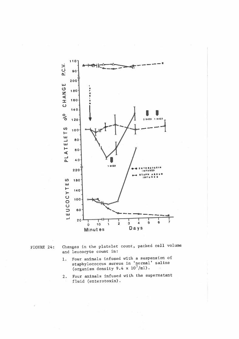
110
ior[
l¡¡oz-C)
o/o
3t,
UJ
l¡Jt-
À
v,l¡¡t-()o()fl¡¡
go
200
180
160
140
120
100
80
60
¡l 0
220
180
140
100
60
20
,rl'I
!ui
å
TI:::î
It OICD
--
Da E¡tEloro¡lLITfU¡ED
¡¡ ltAtH Allu¡t¡FU3ED
-- -¡_ __ _={
o 10
Minut es3456Days
712
FIGURE 24: Changes ln the plaÈelet count, packed cell volume
and leucocyte count ín:1. Four animals infused wÍth a suspenslon of
staphylococcus aureus fn tnormalr saline(oräanrstn densitY 9.4 x 10⁷/m1)
2, Four animals lnfused with the supernaËanÈfluíd (enterotoxin).

154
..,animals. There \^Ias no evidenöe-of fibrin deposition in
any of these.organs.
CON CLUS TON S
This series of experiments confirms the rapid fall in.
the number of circulating ptatelets within 10 minutes of
the intravenous infusion of bacteria noted by previous
authors. It is also possible to demonstrate that the
degree of fall in the platelet count following the infusion
of non-pathogenic organisms .(SarcÍna lutea) is proportional
to the number of bacteria injected; the more organisms
injected the greater is the fall in the platelet count.
This response is very similar to that reported following the
intravenous infusion of colloid.al carbon (Cohen et. alii,
196s).
Unlike the response to co11oida1 carbon where reports
indicate that the platelet count rises within a period of
several hours of the infusion, the platelet count did not
rise until at least 24 hours after the infusion of the
bacteria. The thesis présented by Taniguchi that the plate-
Iets rapidly return to the circulation in such circumstances
does not appear to be substantíated. On the contrary' the
present findings suggest that the platelets are destroyed.

The
day
15s
appearance of a compensatory thrombocytosis seen from
4 adds weight to this suggestion.
Not unexpectedly, changes in the platelet count were
more profound when pathogenic organisms are injected.
There is a major dífference between the two organisms used.
Staph. aureus does not appear to elaborate a toxin capable
of inducing thrombocytopenia. under the-condit,ions of the
experiment,; whereas E. coli endotoxin has an effect as
profound as the organisms themselves. The thrombocytopenia
in Gram-negative infections thereforë appears to be caused
by a combination of the effects of bacteraemia and endo-
toxaemia. This could be an explanation'as'to why thrombo-
cyt,openia is seen more commonly in Gram-negative bacteraemia
than in Gram-positive bacteraemic ínfections.
The response t.o the E. coli endotoxin in these experi-
ments is more profound and persistent than that reported by
other investigators; this is almost certainly due to the
quantity infused. A more rapid recovery in the platelet
count in response to endotoxin is demonstrated in later
experiments.
Although the haemostatic system in general was not

156
examined, the presence of fibrin in the capiltarics of the
E. coli treated animals. does suggest that intravascularclotting was a factor in those which died, whereas no such
evidence was apparent in the staph. aureus treated animals.
The consumption of cr.ottihg Factors iä Response to rnfusionsof SmaL J- Amounts of BacteriaT End.otoxÍn
Factors VII, IX and X persist in the serum of blood
clotted in glass tubesi platelets, fibrinogen, factors II(prothrombin), v and vrrr are consumed" Extensive ctottingin vivo also causes thrombocytopenia and red.uced 1evels of,
factors II, V and VIIT, and in patients with massive intra-vascular clotting there rnay be a reduction of all the coag-
ulation factors (Rapaport et. aIii, Lg64) .
To examlne more accurately and sensitively the effect.of an infusion of dilute tissue thromboplastin on the coag-
ulation system of the rabbit,, Rapaport et alii inhibítedthe synthesis of factors II, VTI, TX and X in the liver by
administering large doses of sodíum warfarin intravenously.
Four hours after the initial dose the animals were infused
with either saline or dilute tíssue thromboplastin. In
both groups of animals there was a fal1 in the leve1s of
the clotting factors but in those infused wíth thromboplastin

]-57
they were able to clearly demonstrate that consumption and
clearance of these factors was increased during episodes of
clotting (napaport et alií, 1966) .
Using this experimental model, small doses of endo-
toxin capable of causing a transient thromloocytopenia but
no significant alteration in the Ievels of factors IT and X
in the intact animal, \^rere infused instead of dilute thrombo-
plastin.
MATERTALS AND METHODS
Bacterial endotoxin: Thi-s was prepared from a culture
of E. coli in broth in the manner previously described. It
was stored at -zOoC and thawed just prior to infusion on
each occasion.
citrated diTutingi fLuid: This contained one part
citrate anticoagulant (a solution of 0.06 M sodium cit.rate
and 0.04 M citric acid) to five parts isotonic saline
solution.
Methods of assav of f actots IÍ ahd X: Factor II \^las
measured by a modification of rnethod described by Owren
(L949) using normal serum and Si-mplastin A (Warner-Chilcott
Laboratories, Morris P1ains, N.J., U.S.A"). The latter

158
reagent supplies factors V, fibrinogen, tissue factor and
calcium.
Factor X was measured by a one-stage method where
dilutions of the test plasma \^rere mixed wíth Seitz filtered
bovine plasma and Russellviper venom' (Stypven, Burroughs
V'Iellcome Co. London) diluted in 0.02? inosithin. The
test was performed in the same manner as the one-stage
prothrombin time with an incubation time from the addition
of the Stypven to the addition of the calcium chloride of
3 minutes (using 0.1 mI of each reagent) .
The clotting times hlere converted to per cent activity
from dilution curves prepared from plasma samples taken at
zero time in each animal. Al-terations ín the levels of
the clotting factors therefore represent the per cent change
in activity from the baseline value (100å) at zero time in
each animal.
Procêclure: The ani-mals were handled in batches of
t.hree; each was gíven 50 mg sodi-um warfarin (Pndo Labor-
atories Inc. U.S.A. ) intravenousl-y. Three infusions of
warfarin at intervals of six hours \^Iere required to induce
a steady faII in factors; Itr and X; Four hours after the
first intravenous injection of warfarin blood (0.5 mI) was

159
collected into a plastic syringe through a 2I SWG needle
from an ear vein. The'bl-ood was then immediately
expelled on to a sheet of 'Parafilm' (American Can Co.
Wisconsin, U.S.A.) and dilutions made by pipettíng 0.05
ml of blood into 0.45 ml citrated dilut.ing fluid. This
procedure had always to be completed within 30 seconds.
One animal \^/as then infused with 0 .2 ml normal saline,
and two animals with 0.2 ml bacterial endotoxin. Further
samples \^rere collected at 5,30 and 60 minutes and at 4,
6 , 10, 18 and 24 hours after the infusion of saline or
endotoxin.
The samples were frozen immediately after collection
and stored at -zOoC for 24 to 48 hours before being thawed
prior to assay of factors II and X.
EXPERTMENT TT (A)
Eight. animals \^rere inf used with 0.2 ml bacterial endo-
toxin at a concentration adjusted to cause a significant
faII in the platelet count. Apart from a transient
pyrexia no apparent. systemd-c manifestations were noted in
any of the rabbits.

160
Resul.Ès.' The changes in platelet count are shown in
Figure 27A; The falI in the ptatelet count is transient,
a response which is in cLose agreement with that observed by
other investigators.
EXPERTMENT TT (B)
Serial estimations of factors II and X were made in
six animals not treated with sodium warfarin; two received
an infusion of 0:2 ml normal,saline arld four 0;2 rnl endo-
toxin. The mean per cent change in the levels of the two
clotting factors from a baseline value of 1003 in the saline
and endot,oxin treated animals are shown'in Figure 25.
Resu-Zts.'. There does appear to be somê fall in factor
II immediately after the infusi.on of the endotoxín but this
is found not to be signifícant on statistical- grounds. The
mean value of both factors in the endotoxin treated animals
does, howeverl remain consistently lowerthan those of the
saline treated animals throughout the period of the exper-
ment. A gradual rise in the levels of the clotting factors,
in particular of factor rI in the salj-ne treated anímals,
occurs during the course of the experiment. This represents,
almost certainly, a response to blood-1oss.

=xooo2l¡¡
I
-_SALIN E IN F USE D
.-ENDOTOXININFUSED
75xr-t 50É.ouÉ
50xrxÉ.ot-UÉ
75
50
FIGI]RE 25:
048 t2 16 20
TIME (HoURs)
24 28 12
Changes ín the levels of factors II and X in:1. T\.¡o anímals ínfused with 0.2 ml tnotmalt
salíne.2. Four animals infused with 0.2 nl endotoxin.

161
A rise in the level of fibrinogen in both groups
increasing from a mean of 348 mg/100 ml príor to the
infusion of endotoxin to 603 mg/ï00.ml at 24 hours also
occurs.
EXPERTMENT TT (C)
Nine animals were given three intravenous doses of 50
mg sodium warfarin at inÈervals of six hours. Three
animals \^Iere infused with O :2 mI normal' saline and six with
0.2 mI endotoxin fourhours.after the 'initial dose of
sodium warfarin. The,mean val-ues Of the serial estlmations
of factors'II and X are shown in Figure 26.
Resutts.. Tn the endotoxin treatêd animaTs the mean
rate of falI in factor II is signifícantly greater (58å
t 8.3 p < 0.001) in the first four hours fotlowing infusion
t.han in the saline treated animals. Duríng the remaind-ng
28 hours the rat,e of fatf in the level'of 'this factor in
the endotoxin and saline treated animals is simllar-
A different pattern in the rate of faIl is seen ín the
levels of factor X; In the'fi-rst six hours the rate of
fa11 of factor X is the same after the infusion of endotoxin
or saline. There is, howeverr'âfi increase in the rate of
decline in the leve1 of this factor in the endotoxin

NtrlÉ.ot-Uf,
\oo\txlÉ.ot-UtL<
roo
75
50
25
roo
15
50
25
o 4 68 12 rö 20 24
TIME (HouRs)
28 32 36
FIGIIRE 26: Changes in Ëhe levels of factors II and X in:
1. Three anímals administered 50 mg sodíumwarfarin intravenously aÈ intervals of sixhours.
2. Síx animals lnfused additlonally htith 0.2 rnlendotoxín four hours after the initial doseof sodium warfarin.
gI=e,úe.
ì
11ls2Hö=õÉôÉ,2<r¡t i}
.- ENDOTO X lNINFUSED
o- SA L lN E INF USED

l-62
treated animals eight t.o sixteen hours later (30å t 8.2
p < 0.001) .
CON CLUS TON S
There is a steady exponential deoline in the levels of
factor II, half life 42 hours, and fact.or X; half life 18
hours,'when synthesis"of:these cl-otting factors by the
liver is inhibited by the,administratlon.of sodium waffarín.
The rate of decline of factor II is significantly increased
in the first four hours after the infusion of endotoxin.
This is very similar to findi-ngs in warfarin treated rabbits
following the infusion of tissue thromboplastin observed by
Rapaport.
It is known that facton IT is consumed in the process
of coagulationi the findd-ng that there is an increased rate
of consumption of this factor does, therefore, suggest thatclotting does occur after the l-nfusion of endotoxin. In
the intact, animal this increased rate of consumptÍon is,however, insufficient to cause a significant fa1I in the
leveI of factor II where,production of clotting factors by
the liver, possibly at an increased rate, compensates forits loss.

163
Factor X is not consumed. in the clotting process; as
expectedr.ño increase in the'rate of decline of this factor
ís noted in the init.ial'hours after the i-nfusion of endo-
toxin. The removal of activated'factor X by the reticulo-
endothelÍa1 system could expJ-ai.n why thete is a morè rapid
fa11 of this'factor eight to,si.xteen hours later in the
endotoxin treated animals "
It is concluded that'Ín the rabbit smal1 intravenous
doses of bacterial endotoxin'which arê capable of causing
a transient fa1I in the platel-et count will also induce an
episode of intravascular clotting.
The Response in PLatelet Coùnt in Rabbits Txeated withHepatin PrÍor to the Infusion of Endotoxin and organjsms
As consumptÍon of clotting factors does occur after
the intravenous infusion.of: evên, srnall quantities of bact-
erial endotoxin, rabbits were anticoagulated with heparin
príor to the infusion of endotoxd-n in order to assess
whether or not inhibitj-on of alotting would prevent the
faIl in the platelet'count.
EXPERTMENT TTI (A)
Six animals were given 1 ml of heparin in a concen-

165
tration of Ir000 units per mL (Weddel Pharmaceutical Ltd.
London) intravenously; This was sufficient to render the
circulating blood incoagualable.: FÍve animals were then
infused with 0.2 mI endotoxin.
ResuL ts .- The changes in the platelêt count are shown
in Figure 278'. There is no difference in the degree of
fall in the platelet counts in the heparinised animals
compared to that occurring i-n the'non-anticoagulated
animals (cf experiment rI (A) , Figure 27A) .
EXPERTMENT TTT (B)
A similar study was made,with respect to the changes in
the platelet count after the infusion of a suspension of
mj-cro-organisms (Sarcina. tutea, organism density 8.9 x tOgl
ml) into the circulation. The changes in platelet numbers
are shown in Figure 28. The response in the heparinized
animals is no different to that seen in the untreated
animals (cf Fi.gures 19 and 20\.
CON CLUSlON S
Despite inhibition of ctr-otting with large doses of
heparin the changes in plat.elet numbers following the
intravenous infusion of endotoxin and of bacteria are not
significantly dÌfferent to those seen in the int4ct, animal.

90
100
80
70
60
50
100
90
80
70
U)
t¡J
l¡Jt-
o-
l¡loz-(J
90
(nÞ¡¡¡
lr¡l-
o-
FIGURX 274 (ABOVE):
FrcuRE 278 (BELO!ü):
.-a EllD0IoxlraItlFusEo
HEPARIN I.< EltD0loxlL
I NFU6EO
G-{ HEPA¡I N
lNFUSlOllAIONE
4812 162024HOU RS
60
50=ÊÀ¡¡¡I
Þoô2U
010
Mins
Changes ín the plateleÈ counÈ in eíghtanímals infused wíth 0.2 ml endotoxin'
Changes in the plaÈelet count in fiveanimals lnfused wíth 1r000 units heparinfollowed by 0.2 ml endotoxin, and one
animal lnfused with 1,000 units heparínalone.

atcnt-UJ
JuJt-
JÈ
160
140
120
100
80
60
40
20
L.tr' ----
I ---_____
.< HEPARIN +ORGANISMS
Þ-{ HEPARINONLY
U'
-2zr'Éc)
o r--T010
Mins't2 ,
7I
6I
534DAYS
FIGURE 28: Changes in Ëhe platelet count in:
Three animals ínfused with 1'000 units heparin.I
2 Slx anímals lnfused with 1r000 unlts heparínfollowed by a suspension of Sarcína lutea Lnsaline (organísm density 8.9 x 10⁸/m1).

166
It does appear that the,faLl- i-n'platelet count induced by
these agents is independent of coagulation-
SUMMARY
Changes in the platelet'count in response to the
intravenous infusion of bacteria and- endotoxin \^Iere studied
in the rabbit.
The observation of-previous'workers who reported a
rapid fall in the numbêr of circulating Platélets followíng
the intravenous infusion of bacteria is confirmed. It is'
further demonstrated that the degree of thrombocytopenia
induced is proportional to the number of organisms infused
when a non-pathogenic strain'is used. The greater the
number of organisms infused, the more profound is the
thrombocytopenia.
The rapid return of the,ptatelet count to nearly normal
levels within a few hours,with the injeCtion of small doses
of endotoxin reported by other investigators is also
confirmed. That this also occurs with bacteríal suspensions
is not substantiated. The,platelêt count in the latter
group remained low for 24 hours before rising to levels in
excess of the baseiine value four to seven days later.

L67
This suggests that there is,destruction of'platelets follow-
ing the infusion of.bacteria.
The changes in the platelet'count are more profound
if the pathogenic organisms E. coli and-Staph" aureus are
used. Thë animals also showed severe constitutional
symptoms and several died. Fibrin demonstrated in the
capillari.es of the lungs and kidnêys of"those animals
which'received the product.s of-Gram-negative bacteria does
suggest that' intravascul-arclottíng had occurred. These
features are not seen in the sections of those animals
which received the products'of the Gram-positive organism.
Staph. aureus also does not.appear to produce, under the
experimental conditions described; a toxin capable of
inducing changes in the platelet count whereas E. coli
endotoxin produces changes as profound as the bacteria
itself.
Vühen some f acets of the coagutr ation system I^tere
studied, it was not possibl-e to demonstrate a significant
change in the levels of.the'cl-otting factors Ir and X after
the infusion of small doses of bacterial endotoxin in the
intact. animal. Vühen synthesi-s of these factors b1r the
liver is inhibit-ed by sodium warfarin, a highllz s:,gnificant

168
increase-in the'rate'of consurnption of factor II occurs
within a few hours of the infusíon'of endotoxin. Factor
II is consumed in the process of clotting;' 'this finding
does, therefore, suggest that, endotoxin' causes an -episode
of intravascularclotting. A corresponding increase in
the rate of fall of factor.X occurs several hours later.
The clearance'of activated,factor Xt which is not consumed'
in the cloÈting process'by the reticulo-êndothélial system,
is the most probable explanation for t,his phenomenon.
Consumption of platelet,s during intravascular clotting
is one mechanism by which endotoxin and bacteria may cause
a fall in the platelet eount.but this is not, however, the
complete explanation, because thrombocytopenia cannot be
prevented by the inhibition of clottíng with heparin.

A¡VI,'JWN S

169
Tn 1968, when this sÈudy of thrombocytopenia in
infectious illnesses was begun, the literature and text-
books of l4edicine and Haematology gaVê little emphasis to
this association; indeed, there appeared to be a general
lack of a\^rareness that thrombocytopenia was a significantmanifestation of sepsis. This prompted several authors
to suggest that thrombocytopenia in bacteraemic illnesses
may be a more frequent occurrence than was then generally
supposed.
That this general una\^rareness of the association of
thrombocytopenia in infectious illnesses should have been
so does not appear to have been entirely warranted since
there is ample literature, especially from France and
Germany, available from Èhe early decades of this century
to indicate that thrombocytopenia is a significant mani-
festation in severe sepsis. Most of the changes inplatelet count observed during the'course of illnesses due
to infections reported in more recent literature were well
documented forty to fifty years ago.
In addition to documenting these changes in platelet
numbers, a number of investigators prior to 1930 did turn

170
their attention to the possibility that platelets may also
in some way be involved in the body's defence against
infections. It was appreciated'that'when organisms entered
the blood stream platelets rapidly clumped about the bact-
eria and disappeared from the circulation. Aggregates of
platelets and bacteria then could be found in the capi11-
aries of many organs, in particular the lungs, liver and
spleen. Only recently has there been a resurgence of
interest in this behaviour of platelets in relation to
foreign material entering the circulation. There is no\^I
evidence to suggest that platelets are involved in the
removal of small particles and bacterial endotoxin.
Haemorrhagic manifestations and the occasional refer-
ence to the finding that the blood of a patient with sepsis
did not clot are also well documented by many of the earlier
observers¡ The concept that other facets of the coagul-
ation system may also be involved were however not expressed
until the late 1950's. This is not surprising as a fuller
understanding of the coagulation system and tests of its
integrity were not devised until the 1940's. More recently
the concept that consumption of coagulation factors, including
platelets, in a prccess termed disseminated intravascular

171
coagulation, has been promulgated as a mechanism for the
lhrombocytopenia, haemorrhagic and other manifestatíons
sometimes seen in infectious diseases. Beginning in the
late 1950's and continuing up to the present time there
has been a considerable and increasing'flow of reports
concerned with the abnormalíties.of haeinostasis in bacter-
ial, protozoal, rickettsial and viral illnesses" Many of
the findings reported in this thesis reflect and confirm
much that has already appeared in this recent and current
literaÈure.
The frequency with which infections in general are
associated with thrombocytopenia is stilI not known. To
ans\^rer this question, a limited prospective study of all
patients admítted to a medical and surgical unit and a
larger survey of all cases of thrombocytopenia detected in
the laboratory by routine haematological examínation was
undertaken. From these surveys it was possible to
estimate that the incídence of thrombocytopenia in
infections in a hospital population to be probably in the
order of five to ten per cent.
In a more detailed study of patients with tþrombocyto-

172
penia and infections it became apparent that a Iow platelet
count ís frequently a transient, phenomenon, wi'th the plate-
let count rapidly rising with resolution of thê illness to
levels in excess of normal in the convalescent period.
Bacteraemia was demonstrated in about half the patients "
It is highly probable that íf an intensive search by means
of repeated blood cultures \^Iere made in alt patients with
bact.erial infections and thrombocytopenia that most would be
found to have a bacteraemia. Gfam*negative bacteria were
much more frequently isolated from these patients with
thrombocytopenia than were Gram-positive bacteria"
Thrombocytopenia also proved to be a useful laboratory
sign suggesting bacteraemd-a, while a persistent or falling
platelet count was highly suggesiive of continuing bacter-
aemia or endoxaemia and usually indicated the presence of the
complication of disseminated intravascular coagulation"
Patients with b'iliary tract infection and jaundice appeared
to be particularly susceptible t.o this complication. The
association of liver disease often tnade it difficult to decide
whether the changes in haemostasls were predominantly due to
the liver disease or to disseminated intravascular coagul-
ation as both produce similar patÈerns.

]-73
In three patients there was unequivocal evidence of
disseminated intravascular coagulation. Two appeared to
have a satisfactory response to the use of heparin, the
third was given an antifibrinotytic agent (Epsilon amino
caproic acid) and at autopsy there was evídence ôf massive
intravascular thrombus formation. This latter case does
add confirmation to the view expressed by most authors that
the fibrinolysis seen in this syndrome is secondary to the
clotting process and represents a compensatory response;
In the remaining patients treated with heparin there was an
improvement in the laboratory coagulation tests but a fatal
outcome to the illness was not averbed in most cases.
It was not practicable to investigate in more detail
the changes induced by septicaemia in man in relation to the
platelets, so further studies of the platelet response to
artificially induced bacteraemia were made in rabbits.
The rapid falI in platelet count withín five minutes of
an intravenous infusion of bacteria noted by prevíous
investi-gators is confirmed. It was also possible to show
that the degree of fall in the platelet count is related to
the number of organisms injected when a non-pathogenic
bacteria, Sarcina lutea, \^ras used. The more organisms

L74
injected the greater was the fall in the platelet count"
The persistence of a low platelet count in these animals for
more than twentyfour hours does suggest that the platelets
\^rere destroyed. The rapid ri-se to levels in excess of the
baseline values seven days later with the highest counts
being inversely proportional to the preceding fall in platelet
count tends to confirm this suspicion. " A similar sequence of
events ín platelet count \4rere seeR in a few uncomplicated
cases of infections in man.
The fall in platelet count was more pronounced when the
pathogenic organism Escherichia coli and Staphylococcus aureus
hrere injected intravenously. Several"of these animals died.
Those infused with the Gram-negative organisms showed evi-
dence of fibrin in the small vessels of the lungs and
kidney, whereas this was not demons.trated ín those animals
infused with the Gram-positive organisms.
Escherichia coli elaborates an endotoxin which is itself
capable of inducing as profor¡:d afall in the platelet count as
the bacteria themselvesi this does not occur with Staphylo-
coccus aureus under the conditions used for these experiments.
This finding again reflects and is in keeping with the
observation that human infections in which Gram-negative

\75
organisms were isolated h/ere more commonly associated with
profound fatls in platelets than in the cases where Gram-
positive organisms \^rere isolated.
A series of experiments \,vere then devised to assess
whether the infusion of smaI1 doses of E" coli endotoxin
capable of causing a transient thrombocytopenia lasting only
a few hours is also associated with activation of the coag-
ulation system. fn the intact animal no significant change
in the levels of the coagulation factors II and X could be
shown following the intravenous injection of small quantities
of endotoxin. If the production of these factors by the
liver is inhibited with targe doses of sodium warfarin, it
\^ras possible to demonstrate that there is an increase in the
rate of fall in leve1 of factor II immediately after the
injection of endotoxin compared to animals which \^Iere treated
with warfarin alone. This strongly suggests that there is
consumption of factor II in response to the ínfusion of endo-
toxin" An increase in the rate of faII in the level of
factor X was not apparent unÈi1 twelve to eighteen hours
after the infusion of endotoxin. This factor, unlike factor
II, is not consumed in the clotting process and this late
change almost cerLarnly reflects removal of activated factor X

l.76
by the reticuloendothelial system.
Although these findings indicate that there is activation
of the coagulation system in response to very sma1l doses of
endotoxin, the fall in platelet count could not be prevented
by prior treatment of the animals wiLh large doses of heparin
sufficient to render the blood incoagulable. The faII in
platelet count was identical in both the heparinized and un-
heparinized animals. A similar finding was also noted with
the intravenous injection of Sarcina lutea. This does
confirm the in vitro findings of other investigators that
platelet adhesion and aggregation can occur independently of
the coagulation system as a whole. Although ít was not
demonstrated, this interaction between platelets, bacteria
and endotoxin most probably depends on the release clf ADP
consequent to the "p1ate1et release" reaction.
Tt would be of interest to devise experiment.s whereby
the role of platelets in clearing endotoxin and bacteria from
the circulation could be more clearly defined. Attempts
were made to render animals thrombocytopenic and neutropeníc
with cytotoxic drugs and radioactive phosphorus-32 with the
intention of comparing the clearance of bactería from the
circulation of rabbits which had received a platetret' trans-

L77
fusion and those which had not. The results obtained have
not been reported in this thesis since the degree of thrombo-
cytopenia and neutropenia induced in individual animals was
extremely variable. Once the animals had been reduced to
this state their physical condition then did not allow for
the manipulation required for such experiments"
There are many cellular and humoral factors involved in
the body's reaction to invasion by micro-organisms. At the
present time it. appears difficult to devise a means by which
the role of the platelet i-n, this scheme could be more
adequately investigated. It is probable that the role of
the platelet is not great since patients with congenital
amegakaryocytic thrombocytopenia (two such cases \^Iere
encountered in the survey period) do not appear to be more
susceptible to infections that the general population.
In conclusion, it is now firmly established that thrombo-
cytopenia is a significant manifestation of sepsis. Indeed
the more recent Èextbooks of Medicine and Haematology have
given increased prominence to this finding. There are
without doubt a number of factors and mechanisms involved in
causing the fall in platelet count. Consumption of platelets.:- ^Æ .i; ^^^-.:--!^-¡ .:*!-^-.^^^,,1^- 'l-+.ia* ì^ +1r^J-t¡ o. vJ-\Jr-gÞÞ (Jr trJ-ÞÞct[[IlIcl,Lc(l IIr LJ-ctvctÞut-tf cLr vvav u¿4 Llv¡¡ rÞ u¡¡ç

l-78
mechanism that has received most attention in the past fiveyears as this complication also appears to be important inthe genesis of other features such as acute renal failureseen in association with septicaemic shock" Tt has been
possible to demonstrate that even very smal1 doses of endo-
toxin will activate and cause consumption of clottingfactors. It is therefore very probable that there is an
increase in turnover of clotting factors in all infectiousillnesses, but only in a few will these changes be so marked
as to cause signs and abnormalities in the laboratory tests
of coagulation.
Other mechanisms are also involved. It has been
demonstrated that the changes in the platelet count inresponse to endotoxin and bacteria cannot be infl-uenced by
the inhibition of the clotting system with heparin. This
strongly suggests that the platelet interaction with these
agents can be independent of the coagulation system as a
whole.
It is also appreciated that many other factors not
studied in this thesis could be involved in the genesis ofthe thrombocytopenia" There appears every indicationthat further research in this field will continue at an

179
increasing rate as it has done in the many other fields of
the behaviour of the complex interactions in which the
platelet is invol-ved.

REFERENCES

ABILDGAARD, C.F; , CORRIGAN; J"J;, SEELER, R"A" , SÏMONE,J. V; 6¡ SCIIULMAN, I . Meningococcaemia associated withinÈravascular coagulation. Pedíatrics, 40: 78-83'1967 .
ACKROYDT J.F. Three cases of thrombocytopenic purpuraoccurring "aft.er Rubellai'with a rreview'of purpuraassociated with' infections. Quart. J. Mêd. 1B: 299-318, L949.
ADELSON, 8., RHEINGOLD, J.J. & CROSBY, Vf .H" The plateletas a sponge: A review. Blood, 17z 767-774t 1961.
AFANASSIEVü, M. Ueber den dritten Formbestandtheil desBlutes im normalen und pathoTogischen Zustande und.llber die Beziehung desselben zur Regeneratíon desBlutes: Dtsch. Arch. Clin: l4ed: 35: 2L7-253, 1884.
AMANO, T.i KATO, K. & SHIMIZU, R. Studies on the roleof iilakin. Med. J. Osaka Univ. 3 ( 2- 3) : 293- 311' 1952 .
r80
K. & SHIMA, M. Studies on the role ofMed. J. Osaka Univ. g(4): 695-7L0, 1953a"
AyIANO, r., KATO,plakin II.
AMANO, r., SHr¡44,plakin ITI "1953b.
M"&Med"
KATO, K.J. Osaka
St.udies on the role ofUniv. 4(2'3): 277'284,
AMANO, I., KATO, K., MORISHIMA, I. & OKADA, K. Studies onthe role of plakin IV" Med. J. Osaka Univ. 4(2-3) ,285-289, 1953c.
AMANO, T., NISHIMOTO, M., KÍN.TO, J", TNOVE, K., TNAI , S.,SEKI , Y. , & KASHIBA, S. Influence of immune bacter-iolysis upon bactería1 respiration. Med" J. OsakaUniv. 5: 145*i65, 1954.

18r
ANGLE, R.M. & ALT, H.L. Thromboclztopenic purpura complic-ating infectious mononucleosis: Report of a case andserial pfatelet counts during the course of infectious' mononucleosis; Blood 5: 449'457; 1950.
AYNAUD, M. L,e globulin des mammiferes; These de Paris,1909.
AYNAUD, M. Action des microbes sur les globulins. 'Comp.Rend. Soc. Bio1. 70: 54-55' 1911.
BANATVALA, J.E;, HORSTMANN, D.M" ' PAYNE, M.C" & GLUCK, L.Rubella syndrome and thrombocytopenic purpura in newborn infants. New Eng. J. Med. 2732 474-478, 1965.
BANNERMAN, R.G. B1osis. Lancet,
ood-çlate counts .in pulmonary tubercul-2z 593-595, L924.
BAYER, W.L. Purpura in congenital and acquired rubella.Comprehensive study of 22 cases. New Eng. J. Med.2732 1362-1366t 1965.
BEHNKE, O., & ZELANDER, T" Filamentous substructure ofmicrotubules of marginal bundle of mammalian bloodplatelets. J. UltrasÈructure Res" 19: Ï47-L65, 1967-
BEHNKE, O. Electron microscopical observations on thesurface coating of human pfatelets. J; UltrastructureRes. 242 5L-69t 1968.
BERG, W., KOR.SAN-BENGSTEN, K., YGGE' J. Determination ofplasminogen in human plasma by the lysis time method.Thrombos. Diathes. Haemorrh. 16: 1-17, 1-966.

L82
BHANCHET; P;; SUWANTK, Ri, HONGHATAT; A: i PLEETIACHINDA,R" & TUCIIINDA, C: Platelet survival and organscannÍng in dengue Thai haemorrhagíc fever" Proc.Intern. Congr. Ilaematol: 12th New York, 1968.
BIGGS, R., DENSON, K.W.Coagulant activity15: 282-296, 1968.
E.,of
RTESENBERG,platelets; '
D. & MCTNTYRE, CBrit: J. Haemat"
BIZZOZEF.OT G. Sul midollo del1a ossa" Naples, 1869.
BIZZOZEPIO, G. Su di un nuovo elemento mortologico delsangue di mammifera della sua importanza vellatrombosi e nella coagulazione. Italia l4ed. 15: 385,1881.
BOCCACIO, G. "Decameron". Translat,ed by J.M" Rigg.Publishers Navarre Society, Ltd. London"
BONCIU, O.T. Contribution a I'etude des variationsnumeriques des plaquettes sanguines dans la scoulatine.Comp. Rend. Soc" Biol. 92: 1453-1455, L925.
BOOYSE, F"M. 6. RAFEISON, M.E. Human platelet contractileproteins: Location, properti.es and function" Ser.Haemat. 4(1) z L52-I74, ]-97l-.
BOUIvIAMEAUX, Y. The coupling of platelets with subendothel-ial fibers. Comp. Rend. Soc. Biol" (Paris) 153: 865-867 , 1959.
BRECHER, G., SCHNETDERMAN, M* & CRONKITE;reproducibility and constancy of theAmer. J. Clin. Path 232 15-26, 1953.
E.P. Theplatelet count.
BRINKHOUS, K.M. BasicplateleË thrombusL967 .
of cell adhesion andFed. Proc. 262 84-87,
mechanismsformation.

183
BROCK, R.C. Observations on the platelet count in tuber-culosis: Tubercle; 15: 241-250 , 1934.
BUCKELL, M. The effect of citrate on euglobulin methods ofestimating fibrinolytic activity; J; CIin. Path. 11:403-405, 1958.
BUCKINGHAM, S. & MAYNERT, -E..W- -The release of S-hydËox1z-tryptamine potassium and amino acids from"platêlets.J. Pharmacol, Exp. Ther. 143: 332-339, 1964"
BULL, C.G. 6' McKEE, ,C"M. The relationshíp of blood plate-lets to the in.vivo agglutination of bacteria andtheír disappearance fióm the blood stream. Amer. J.Hyg. 2z 208-224, 1922.
BULL, 8.S., SCHNEIDERMAN, M.A" & BRECHER, G.counts with the Coulter counter. Amer.Path. 44= 678-688, 1965.
PlateletJ" CIin.
CHARGAFF, 8., BANCROFT, F.,W. & STANLEY-BROWN, M. Studieson the chemistrlz of blood -coagulatíon III. Thechemical constituents of blood.platelets and theirrole in clotting, wíth remarks on the activation ofclotting by tipids. J. Bío1. Chem. 1162 237-25I, 1936.
CLARKE, B.F. & DAVIS, S-H" Severe.thrombocytopenia ininfectious mononucleosis. Amer. J. Med" Sci, 2482705-708, 1964.
CLARKES, N.D"745-759 |
Purpuric chicken pox. Ann. Intern. Med" 542196 1.
CLARK, W.G. & JACOBS¡ 8.. Experi.mental nonthrombocytopenicvascular purpura:. A .review of the Japanese litera-ture with prelíminary confirmatory report. Blood, 5:320-328, 1950.

184
COHEN, P", BRAUNVÍALD, J. & GARDNER, F" Destruction ofcanine and'rabbit platelets following intravenousadministration of .s¿¡5.n particles or endotoxjn"J. Lab. Clin; Med.66:263-27I, 1965"
COHEN, P. & GARDNER, F. Thrombocytopenia as a laboratorlzsign and' complication of Gram'nêgative bacteraemicinfections.' Arch" Intern. Med; 117: 113-L24, L966"
COHEN, P., & WITTELS, B" Energy substrate met.abolism infresh and stored human platelets: J; Clin. Tnvest.492 LI9'I27; 1970.
CONDIE, R.M'. ; HONG; C-Y. & .GOOD..-R.4" Reversal of lesi,onsof the generalized Shwartzman.reaction by treaLment ofrabbits with streptokinase . J " Lab ; Clin " lt4ed. 50 :
803-804, 1957.
COPLEY, A.L. & HOUIIHAN; R..8; The agglutination of plate-lets isolated from human, dog and swine blood; Blood,1: IB2-J-98, 3-947 .
CORRIGAN, J"J., WALKER, L"R. & NORMA MAY. Changes in theblood coagulation .sys,¡em associated withsepticaemia. New Eng. J" Med. 2792 851-855' 1968.
CORRIGAN, J"J. & JORDAN, C.M. Heparin therapy in septic-aemia with disseminated .intravascular coagulation"Effect on mortaliÈy and.correction of hemostatícdefects. New Eng.J.Med. 2832 778-782, 1970"
CORRIGAN, J.J. Effect of ,antieoagulant and non-anticoag-ulant. concentrations.of heparin on the generalizedShwartzman reaction. Thrombos. Diathes. Haemorrh.(Stuttg. ) 24 = 136 , I970 .

185
CRADDOCK, C;G., ADAMS. T¡f .S-i PERRY' S. & LAVüRENCE, J"S"The dynamÍ.cs sf, platelet production "as studied by adepletion technique in normal and irradÍated dogs.J. Lab. Clin. Med" 452 906-9T9, 1955"
DACTE, J.V. & LEWTS, S.M.4th edition. J. & A.
in 'lPractícal Haematology" "Churchill Ltd. London, 1968"
DARLING,' S.T; The blood platelets in..t,ropícal and .otherforms 'of anaemia . ' Trans . Soc; Trop. Med. Hyg. 5 :46-57, 1911.
DAVEY, .M:, G;1ets.
The Survival and -Destruction of Human .Plate-S. Karger, Basel & New York. L966.
DAVEY, M.G. & LUSCHERT :E-F.. Act,ions:-of :thrombin and -othercoagulant and proteolyùic' enzlzmes .on' blood' plat.elets .Nature (London)' 216: 857-858, 1967.
DAVEY, M.G. t r,üsCgnar. E-F'. P-l-atelet proteins. In "Bio-chemistry of blood plateletsi'. Edited by E. Kowalskiand S. Niewiarowski, New York. Academic Press, L967,pp 9-22.
DAVIE, 8.W., HOUGIE, C..&,LUNDBIAD, R.L. l4echanísms of,blood coagulatíon. In "Recent Advances in BloodCoagulatíon". Edited by L. Poller, J. & A" ChurchillLtd. London, 1969. pp 13-28.
DAVïD-FERRIERA, J.F. Surcytaire des plaqueÈte89-103 , 1961.
.st,ructure et le penvoir phago-sanguines. Z. ZelIforschr55 :
1aS
DAVIS, R.B. & tr^fHITE, J..G.. Localízation of S-hydroxy-t,ryptamine in blood .platelets. Autoradiographic andultrastructural changes induced in platelets bychilling. Blood, 30: 625-635t 1967.

lB6
DAY, J.H. & HOLMSEN, H.release reaction"
Concepts of the blood PlateletSer. Haemat. 4 (1) z 3-27 , L97I.
DEFOE, Daniel. Journal of the Plague Year. OxfordUniversity Press I L969.
de GABRIELE, G. & PENINGTON, D.G" Physío1ogy of theregulation of platelet production" Brit. J" Haemat.13: 202-209 | 1967 .
DEGKWïTZ, R"153-189,
Studien tlber Blutplättchen. Fol" Haem. 2521920.
DEKHUYZEN,Anat.
M. C.Anz.
Uber die Thrombocyten (Bï-utp1ättchen) .
19: 529 , 1901.
DELREZ, L. & GOVAERTS, P. L'intervention des globulinsdans 1'eliminatíon des microbes injectes dans lacirculation. Comp. Rend. 81: 53-55r I918.
DENNIS, L.lf., ETCHELBERGER, J.W., TNMAN, M"M" & CONRAD, M"E.Depletion of coagulation factors ín drug resistantptasmodium falciparum malaria" Blood, 29:7I3-72T, 1967-
DENYS, J. Etudes sur la coagulation du sang dans un cas depurpura avec diminution considerable des plaquettes.La Ce1lule, 5: 187-193, 1889.
DES PREZ, R.M., HOROWTTZ, H.r., & HOOK, E"W. Ef fects ofbacterial endotoxin on rabbit platelets" l. Plateletaggregation and release of platelet factors in vitro.J" Exp. l4ed. 114: 857-873, 1961.
DES PREZ, R.M. Effects of bacterial endotoxin on rabbítplatelets. III. Comparison of platelet injuryinduced by thrombin and by endotoxin. J. Exp. Med.L20z 305-313, 1964"
DES PREZ, R. & BRYANT, R"on rabbit platelets.of endotoxin-inducedinjury. J. Exp. Med
187
Effects of bacterial endotoxínIV. The divalent íon requirement
and immunologically-induced plate-L2z 97I-982t 1966.
DEYKIN, D. Thrombogenesis. New Eng. J. Med" 2762 622-628,T967.
DEYKIN, D., & DESSER, R.K. The synthesis of lipids byplatelets. J. Clin" Invest. 47: 1590-1-602, 1968.
human
lasmaz 39-95,L970.
DOERY, J.C.G., HIRSH, J. & COOPER, I. Energy metabolism inhuman platelets: Inter-relationship between glycosísand oxidative metabolism. Bloodt 362 159-168, 1970a.
DOERY, J"C.G., HIRSH, J. & DE GRUCHY, G.C. Plateletglycolytic enzymes: Effect of ce11u1ar disruptionprocedures on activity. Brit. J. Haemat. 19: 145-I57, 1970b.
DONALDSON, V.H.enzymes in
Blood coagulation and relatedinflammation. Ser. Haemat" 3 (1
p)
DONNE, A" De l'originesde formation, et de368, 1842.
des globules du sang,Ieur fin. C.R. Acad.
de leur mocleSci. L4 z 366-
DUCROU, W" & KII4BER, R.J" Stomatocytes, haemolytic anaemiaand abdominal pain in l{editerranean mígrants: Someexamples of a new syndrome. ['led. J. Aust. 2: 1087 ,t969 "
DUDGEON, L.S. 6¡ GOADBYTH.K" The examination of tíssues andsome observations of blood platelets of rabbits at,intervals of five minutes and later after íntravenousinoculations of staphylococcus aureus and Indían ink.J. Hyg. (Camb.) ' 31: 247-256t 1931.

188
DUKE, Id.W. The retation of btood platelets to hemorrhagicdisease. Description of a method for determining thebleed.íng ttme anã coagulation time, and repo-rt ofthree cases of hemorrhagic disease relieved bytransfusion. J. Amer. ¡,ted. Assoc.55:1185-LI92' 1910.
EASTHAM, R.D. Rapid \^/hole-blood platetet counting usíng anelectronic pãrticle counter. J. Clin" Path" 16: t68-L69 , 1963.
EBERTH, J.C. & SCHIMMELBUSCH' C" Experimentelle Untersuch-,rrg"n {lber Thrombose. virchows Arch. 103: 39-87 , 1886.
EGGLETON, M.J. & SHARP, A.A" Platelet counting using thecoulter electronic counter. J. clin. Path. 16: 164-167,r963.
ELLIS, B.C. & STRANSKY, A" A quick and.accurate method forthe determination of fibrinogen in plasma. J. Lab.CIin. Med. 5Bz 477-488, 1961.
ERDOS, E.G. & MMA, I" Effect of endotoxin shock on .theplasma Kaltikrein-kínin system of the rabbit. Fed.Proc . 27 z 92-95 , 196 8.
EVANS, G. & MUSTARD, J.E. Tnhibition of .the platelet-surface reaction in endotoxín shock and the general-ized shwartzman reaction. J" clin, Tnvest" 47: 3Ia,1968 (abstract).
FAMA, P.G., PATON' I¡ü"8. & BOSTOCK'purpura comPlícating mumps.L964.
ThrombocytoPenj-cJ. l4ed. 2z L244,
14, T .Brit.
FARBISZEWSKI, R., NIEWIAROI{SKI, S", WOROWSKI' K. 6¡ LIPINSKI'B. Release of ptatelet factor 4 in vivo during intra-vascular coagulation and in thrombotic states 'Thrombos. oíátfres. Haemorr. (Stuttg.) , I9: 578-583'I96 B.

189
FEARNLEY, G.R. Fibrinolysis. In 'Recent Advances inBlood Coagulation'. Edited by L. Po1ler. Churchill,London, 1969, pp 229.
FERGUSSON ' A 'r'ü
cytopenic& CANTA B. Rubella .as a
purpura. Pediatrics , 25 z
cause of400-408,
thrombo-1960.
FISHER, O. D. & KRASZEI{SKI , T.14. Thrombocytopenic purpurafoltowing measles. Arch. Dis. Childhood 272L44-L46,1952.
FORMAN, E.N., ABILDGA-A,RD, C.F. & BOLGER, J"F. GeneralisedShwartzman reaction. The role of the granulocytes inintravascular coagulation and renal cortícal necrosis.Brit. J. Haemat. l6: 507-515' 1969.
FREfMAN, I. & SUPER, M. Thrombocytopenia andsyphilis in South African Bantu infants.Childhood, 4I: 87-90, L966.
congenitalArch. Dis.
FRENCH, J.E., MACFARLANE, R.G. & SANDERS' A.G. Structureof haemostatic plugs and experimental thrombi in smallarteries. Brit. J. Exper. Path. 452 467-474, L964.
FRENCH, J. E. Bl-ood platelets :
their properties and lífe13: 595-603' 1967.
Morphological studíes oncycle. Brit. J. Haemat.
FRY, G.L. & HOAK, J.C. Improved method for electroniccounting of platelets. J. Lab. CIín. ttled. 742 536-540 | 1969.
GAIRDNER, D. The Schönlein-Henochpurpura). Quart. J. Med. 17:
syndrome ( anaphylactoid95-122, l-948.
GLYNN, M.F., MOVAT, H.2., MURPHY, E.A. & ¡4USTARD, J"P. Studyof platelet adhesíveness and aggregation with latexparticles. J"Lab.CIín.Med. 65: Il9-201' 1965.

190
GOLDENFARB, p.8., ZUCKER, S., CORRTGAN, J,J" & CATHEY, M.H.The coagulation mechanism in acute bacterial infect-ion" BriE" J. HaemaL. 18: 643-652, 1970"
GooD, R.A. & THOIIAS, L. studies on generalized shwartzmanreaction: Production of bilateral cortical necrosisof kídneys by single injection of bacterial toxin inrabbits previously treated with thorotrast or trypanblue. J. Exp. Med. 962 625-64I, 1952"
GooD, R.A. & THOI'IAS, L. studies on the generalized shwart-zman reaction. IV" prevention of local and general-izeð, Shwartzman reactions with heparin. J. eip. Med.972 871-887, 1953.
GOVAERTS, P. La functiorantixenique des plaquettessanguines. Arch. Intern. physiol" 16: L-20, I92I.
GRAM, H.c. The blood platelets in inftuenza with remarkson the technic of counting. Acta l4ed. Scand" 54: l-15,1920.
GRETTE, K. The mechanism of thrombín catalyzed haemostaticreactions in btood praterets, Acta physiol. scand. 56:Suppl. 195, 1962.
HARDAWAY, R.¡4. & JoI{NSo¡ü, D. clottíng mechanism in endo-toxin shock. Arch. Intern. Med. IL2: 775-782t 1963.
HARDAWAY, R.llt. syndromes of disseminated intravascularcoagulation. CharLes C. Thomas, publishers, Spring-field, Illinois, 1966.
HARDAVüAY, R.M. The significance of coagulative and thromb-otic changes after injury. J. Trauma, I0: 354, Lg7O.
HARDISI
Tn
DD
Y, R.14. & TNÇR.A,M, G.I.C. in "Bleeding Dísordersl'vestigation and Management. Btackwell (Oxford) , L965,. 242.

19r
HARDISTY, R"M. Platelet aggregation and platelet factor 3availabílity in bleeding disorders. In ExperimentalBiology and ltÍedicine, vol. 3 " Platelets and Haemo-stasis. Edited by E. Aagen, W. Vüechsler and F"Zilliken, N.Y. Karger t96Br pp 189-l-92"
HASLAM, R.J. Role of ADP in aggregation of human bloodplatelets by thrombin and by fatty acids" Nature(London) 2022 765-768, L964.
HASLAM, R.J. l,Iechanisms of blood platelet aggre"Physiology of Hemostasis and Thrombosis" "S.A. Johnson and W.H. Seegers. Charles C.Publishers, Springfietd, fllinois, 1967 r pp
HAYEM, G., Recherches sur 1'evolution dessang de 1'homme et des vertebrates.Norm. Path. 5: 692-734, 1878a.
gation.Edited
Thomas,88-112.
ïnby
HASLAM, R.J. Biochemical aspects of platelet function. InPlenary Session Papers, edited by E"R. Jaffe, New York.XII Congress International Society of Haematology , J-968,pp 198-2I4.
hematins dans IeArch. Physiol.
H$YEM, G. Sur lamicroscope.
formation de 1aC"R. Acad. Scí.
fibrin du sang, etude au86: 58-61, 1878b.
HAYEM, G. De Ia Crise hematique dans 1esde efrescence brusque" C.R. Acad.1882.
maladiesSci.94z
ar-gues a200-202,
HAYEM, G
c"R
HAYEM, Gl{ed
Sur le mecanísme de lrarret desAcad. Sci. 95: I8-2I, I882.
hemorrhagies.
SurHop
"
un casParís
de diathese hemorrhagies. 8u11. Soc.3s.VIrI, 389-394, 1891.
HAYEM, G. Duformationiques. C
caillot non retractile: sdu serum sang dans quelqu
.R. Acad. Sci. I23: 894-89
ression de Iaetats patholog-1896 "
ppS
ue6 t

L92
HELLEM, A.J. Proc. VIf Congr. Tnt. Soc. Haemat. Rome, 1958(abstract)
"
HELLEM, A.J. & STORMORKEN. H" Platelet adhesion-aggregationreaction and íts clinical significance. Tn 'RecentAdvances in Coagulation'" Edited by L. Poller. J. &
A. Churchill (London) , L969, pp 69-106.
HENDRY, A.T. Purpura and tuberculosis. Tubercle, 36¡ 294-300, 1955.
HERRING, W.8., HERTON, J.C" ¡ T^IALKER, R.I" 6, PALMER, J"G.Distribution and .clearance of circulatíng endotoxin.J. Clin. Invest. 422 79-87t 1963,
HILL, G.J., KNIGHT,vivax malaria.
HIPPOCRATES. Of the El4edicine". Philo98-I42.
V. & JEF!'ERY, G.M. Thrombocytopenia inLancet, 1: 240-24I, !,gAA .
pS
idemics in "g Theory and Practice ofophical Library, New York, L964 r pp
HIRSCH, J.G. Comparative þactericida] activities of bloodserum and plasma serum" J. Exp; Med. LI2zL5-22, 1960"
HrTZrG, W.H., STRAUB, p.Íf ., LO, S"S., & FRfCK, P"G. Cl-inic-a1 experience with anticoagulant therapy in the manage-ment of dísseminated intravascular coagulation inchildren. Proc. Roy. Soc. Med. 61: 1138, 1968.
HJORT, P.F., RAPAPORT, F.r" & JøRGENSEN, L. Purpura fulmin-ans. Report of a case successfqJ.ly treated withheparin and hydrocortisone. Review of 50 cases f rorllthe literature. gcand. J. Haemat. 1: L69-L92, t964.
HJORT, P.F. & RAPAPORI, S.I. The Shwartzman reaction:Pathogenic mechanisms and manifestations" Ann. Rev.Med. 16: 135-168, 1965.

193
HOFFMAN, I/1.8. A simple method for factor V assay. Amer.J. Med. Tech. 33: 28L-284t L967.
HOLMSEN, H. Adenine nucleotide metabolism in platelets andplasma. In "Biochemistrlz of Blood Platelets". Editedby E. Kowalski and S. Niewiarowski. Academic Press,New York , l-967 , pp 81-90.
HOLMSEN, H. & DAY, H.J. Adenine nucleotides and plateletfunction. Ser. Haemat. 4(1) z 28-58, L97L-
HOROWITZ, H.I . , DES PREZ , R.M. & HOOK,' 8 "W. Ef f ects ofbacterial endotoxin on rabbit platelets. II. Enhance-ment of platelet factor 3 activity in vitro and invivo. J. Exp. Med" 116z 619-633, L962-
HOROWITZ, H.I. and FUTI¡4OTO, ¡4.M. Associatj-on of f acÈorsXI and XII with blood platelets. Proc. Soc. Exper.Biol. Med. 1192 487-492, 1965.
HOULIHAN, R.B. & COPLEY, A.L. The adhesíon of rabbit plate-lets to bacteria. J. Bact. 522 439-448, 1946"
HOULfHAN, R.B. Studies of the adhesion of human bloodplatelets and bacteria" B1ood, 1: L42'L54, 1947 '
HOVIG, T. Release of a platelet-aggregating substance(adenosine diphosphate) from rabbit blood plateletsinduced by saline "extract'l of tendons. Thrombos"Diathes. Haemorrh. (Stuttg.) 9z 264-278, 1963a.
HOVIG, T. Aggregation of.rabbit blood platelets producedin vitro by saline "extract" of.tendons. Thrombos"Diathes. Haemorrh. (Stuttg.) 9: 248-263, 1963b.
HOVTG, T. The effect of various enzymes on the ultra-structure, aggregation and clot .retractíon abílity ofrabbit blood ¡.rIatetets. Thrombos. Diathes" Haemorrh.(stuttg.) l-s: 84-113, 1965.

l-94
HOVIG, T., ROWSELL, H.C., DODDS' .'/ü.J. ' JøRGENSEN, L. &
MUSTARD, J.F. Experimental hemostasis in normal dogsand dogs with congeníta1 disorders of blood coagul-ation. Bloodr 30: 636-668' L967.
HOVfc, T. Blood plateletelectron microscopic427 , 1970.
and shape: a screeningScand. J. Haemat" 7: 420-
surfacestudy "
HOWELL, W.H. Observations upon the occurrence,and function of the giant ce1ls of the boneJ. Morph. 4: :-.L7, 1890.
structuremarrow.
& CROSBY, W.H.: Bone marrow panhypoplasía ínexperimentally induced by víral ínfection.IB: 800-801, 1961.
HOWTE, D.L.humansBlood,
INGRAM, G.I.C" The defíbrination syndrome.Advances in Coagulation'. Edited by L.J. & A. Churchill Publishers (London)
'275.
fn ¡RecentPolIer.
L969, pp 263-
ISHIZAKA, T. & ISHTZAKA, K. Biological activities ofaggregated globu1in" V" Agglutination of erythrocytesand platelets. J. fmmunol. 89: 709:7L6, L962"
JERUSHALMY, 2., KOHN, A. 6¿ DE VRIES' A. fnteraction ofmyxoviruses with human platelets in vitro" Proc. Soc.Exp. Biol. Med. t06z 462-469r 1961.
JACKSON, J.M., KNTGHT, .A"D.E-, -NORMAN, J.G.N" & CRAÏ{EORD,I.A. Mediterranean .stomatoclztosis: A possible redcetl membrane defect. Read at the 15th Annual GeneralMeeting of the Royal College of Pathologists ofAustralia, I970.

195
KARPATKIN, S. Studies on human "plateJ-et glycolysis:effect of glucose, clzanide, insulin, citrate, andagglutination and contractíon .on platelet glycolysis.J. CIin" Invest. 462 409-4.L7, L967.
KARPATKIN, S" Heterogeneity of human platelets. Ser.Haemat. 4: 75-97, I97I.
KAUFMAN,252
R.M. Origin of pulmonary megakaryocytes. Blood,767-775, 1965.
KEDA, K. Blood in smallpox -during recent epidemic. Arch.Intern. Med. 372 660-673, L926'.
KIESSELBACH, T.H. & WARNER..R",H. Fíbrin stabílizing factor:thrombin-1abile ptatelet protein. Amer- J. Physiol.2TIz I472-L476, 1966.
KINSELLA, R.A. & BROWN,J. Amer. I4ed. Ass.
Changes in blood in influenza.to70-r072, L920 "
Thromboclztopenrc purpura as aJ.Amer.Med,Ass. 168 z 22I3-22L5,
G. P..742
KJAERHEIM, A. & HOVIG, T. The ultrastructure of haemostaticblood platelet plugs in rabbit mesentery" Thrombos.Diathes. Haemorrh. (Stuttg.) 7: 1-15, 1962'
KLIPIAN, A. & MgKAY, D.G. The prevention of the generalj-sedShwartzman reaction by fibrinolytíc actívity" Arch.Path. 662 7L5-7I9, 1958.
KOLARS, C.P. & SPINK, W-W.complication of mumps.1958.
tt
KR.A,USS, E. Uber purpura. Inaug. Diss. (Heidelberg) 1883.
KURAMOTO, 4., STEINER, ¡lI. $r BALDINT, M.G. Metabolic basis ofplatelet phagocytoçis. Biochim. Biophys. Acta, 20Lz47r-478 | L97o "

L96
LARSSON, S.O.failure.
On coagulation and fibrinolysis in renalScand. J" Haemat" Suppl. 15: t97I.
LEE, L. Reticuloendothelial -cl,earance of circulatingfibrin in the pathogenesis of the generalizedShwartzman reaction. J"Exp" Med", 115:1065-1082,1962-
LEVADITTI, C. Sur I'etat de la cytase dans le plasma desanimaux normaux .et des organísmes .vaccines contre levibrio cholerique" Ann.Tnst.Pasteur l-5: 894=927,1901"
LEVIN, J. Thnombopoiesis -and thrompgíetin. Tn "Hemo-poietic cellu1ar .proliferation". Edited by F.Stohlman, Grune & Stratton, N.Y. L970, PP 299"
MACKAY, W. The blood platelets:Quart. J. Med. 242 285-382,
Its clinj-cal significance.193 1.
¡'ÍACPHERSON, J. C. & HARDISTY, R. M.in screening test. Thrombos"6: 492-497 , 1961.
A modified thromboplast-Diathes. Haemorrh"
MAJERUS, P.l^I., SMïTH, M.B. & CLAMOIü, G.H. Lipid metabolismin human ptatelets. I. Evidence of a complete fattyacid synthesizlng system. J. Clin. rnvest. 4B: I56-L64, L969.
MARCUS, A.J. 6¿ SPAET, T.H. Platelet phosphatides: theirsepartion, identification and clotting activity. J.Clin. Invest" 37: 1836-1-847 ' 1958.
MARcuS, A.J. & zUcKER, M.B. The phlzsío1ogy of blood plate-lets: Recent biocþemj-cal, morphQlogic and clinícalresearch. Grune & Stratton, New York, 1965.
MARCUS, A"J. Role of lípids in blood coagulation"'Advances in Lipid Research', 4z I-37, 1966-
In

197
MARCUS, A"J., ZUCKER-FRANKLTN, D", SAFf,ER, L.B. & ULLMAN,H.I-,. Studies on human platelet granules and membrano.J. C1in. Invest. 45: 14-28t 1966.
MARCUS, A.J.New Eng.
Platelet.function (first of three parts) .J. Med. 280: I2L3-I220t L969.
MARGARETTEN, Vü. & McKAY, D"G.. . The role ofthe generalized. Shwartzman reaction.L29 z 585-590 , L969 .
the platelet ínJ" Exp. Med.
MARTTN, W. & SHORE, B725-748, 1928.
R. Juvenile gangrene" Ann. Surg. BB:
MATTER, M., HARTMAN, J.R.r. KAUTZ, J., DE MARSH, Q.B. &
FINCH, C.A. A studlz of thronrbopoiesis in inducedacute thrombocytopenia. B1ood, 15: L74-185 ' 1960
"
MAYNERT, E.W. & fSAAC,. L. .Uptake and binding ofby the platelet and its granules. AdvancesPharmacol. 6z 114-L22, 1968.
McGEHEE, W.G., RAPAPORT" S"f.coagulation in fulminantMed. 67 z 250-260, L967 .
serotoninin
& HJORT, .P . F " In Lravas cul armeningococcemia, Ann. Tntern.
McKAY, D.G. 6. SHAPTRO, S.S. Alteratíons in blood'.coagulat-íon system induced ,b1z .bacterial endotoxin. I . in vivo(generalized Shwartzman reaction). J. Exp. Med" 107:353-367 | 1958.
McKAY, D.G. A partial slznthesis of the generalizedShwartzman reaction. Fed.Proc . 22:I373'1379, 1963.
McKAY, D.G.Medical
Di sseminated . intravascularDiv, Harper & Ro\^I, 1965.
coagulation. Hoeber

198
McKAY, D.G. & MARGARETTEN, W", Disseminated intravascularcoagulaÈion in virus diseases. Arch" Intern. Med.L20: I29-L52, 1967.
MOLL',ISON, P " L,Bl-ackwe11
"Blood Transfusion in Clinical Medícine".Scientific Publications , oxford ,L969,p. 16 I "
MORSE, 8"E.,purPuraadults.
ZINKMAN, W.H. & JACKSON, D.P. Thrombocytopenicfollowing-rube11a infection in children andArch" frrtern. Med. 117: 573-579, L966.
MOVAT, H.Z.t MUSTARD, J.F-, TATCHMAN, I$
Platelet aggregation .and release .oand histamine associated .with phagantigen-antibody complexes. Proc¡T2Oz 232-237, 1965. i
.S. & URTUHARA, T"f ADP, serotoninoc]ttosis ofSoc. Exp. Biol.
MOVAT, H"Z. , WETSER, Vü.J. ,Platelet phagocytosis27 z 531-543, 1965 "
GLYNN, M.F. & MUSTARD, J"F.and aggregation. J" CeIl Bio1.
MURER, 8.H., HELLEM, J.H. & ROSENBERG,metabolism and platelet function.Lab. fnvest" 19: 280-282, 1967.
M"C. EnergyScand" J. Clin.
MUSTARD, J.F. Haemostasis .and .thrombosis.Haematology r 5: 91-106 , 1968.
Semínars in
Platelet phagocytosís. Ser.M.A"1968.
MUSTARD, J"F. & PACKHA¡{,Haemat. 1: 168-184,
MYLLYLA, G., VAHERT, 4., VESTKARI, T; & PENTTINEN, K" Inter-action between human blood platelet,s, viruses and anti-bodies. IV. Post-rubel1a thrombocytopeni. purpura andplatelet aggregation by rubella antigen-antibody inter-action. CIin. Exp. Tmmunol. 4z 323-332, 1969"
NACHMAN, R" & FERRIS, B. Studies on human platelet proteaseactivity" J" Clin. Invest. 47: 2530'254A, 1968.

L99
NAKAO, K. & ANGRIST, A.A" Membrane surface specializationof the blood pl"atelet and megakarlzocyte. Nature(London) , 2L7: 960-961, 1968.
NICOLAU, J. Le syndrome purpuríque dans les varioleshemorrhagiques. J; Physíol"Path.Gen " 262256-269,1928.
O'BRIEN, J.R. Platelet stickiness. Ann. Rev. Med. L7= 275-290, 1966.
OSKI, F.4", & NAIMAN, J.L. Effect of live measles vaccíneon platelet count. New Eng. J. l4ed" 275: 352-356,1966.
OSLER, W. An account ofliquor sanguinis.391-398, L874.
certain organisms occurring in theProc. Roy" Soc" (London), 222
OWREN, P.A. Quantitativeprothrombin. Scand.
ene-stage method forJ.CIin.Lab.Invest.l:
assay of _81-9 3 ,19 49 . '
PACKHAM, M.4., NTSHTZAWAT E-8" & MUSTARD' J.Eof pIatelêts to tíssue injury. Biochem"Supp. pp 171-184, 1.968.
ResponsePharmacol.
PACKHAN, M.A.Seminars
& MUSTARD ¡ J 'F . .P lateletin Haematology' 8: 30-64,
react,ions.L97L.
PEMBREY, R.G., HANDLEY" D"A" .& KIMBER,of haemostasis in the acutely illAustralian C1inics, 4: 223' L969.
R.J "patient.
ProblemsSouth
PENNY, R., ROZENBERG, M.C-platelet pool. Blood
B.G. The splenicL966.
& FrRKfN,27: 1-16 '

200
PEPPER, O.H"P" The hematologlz of subacute streptococcusviridans endocarditis. J. Amer. Med. Ass. 89: L377-1380 , 1927.
POPESCO-COMBIESCO, C. Recherches .sur 1é role des plaquettessanguines dans 1'.immunite -la fonction antixenique desplaquettes. Arch. Rouen de Path. Exp. Micro. 1: 453-484, 1928. l
POPESCO-COMBIESCO, C. ibíd 3: 343-364, 1930"
PORTER, P.J., SPIEVACK, "4.R. & KASS, E.H" Endotoxin likeactivity in .serum .of patíents wíth severe localízedinfectíons. New Éng. J. Med. 27Lt 445-447, L964.
RAHAL, J.J., MacMAHON, H.E. & WEINSTEINTL. Thrombocyto-penia and symmetrical peripheral gangrene associatedwith staphylococcal and streptococcal bacteraemía.Arch. Intern. Med. 692 35-43, 1968.
RANVIER, M. Du mode de f,ormation de la fibrine dans lesang extrait des vaisseaux. C"R. Soc. Biol. 5: 46-41873.
RAPAPORT, S.I., TATTER, D"., COEUR-BARRON, N" & HJORT, p.F.Pseudomonas septicaemia wíth intravascular clottíngleading to the generalized Shwartzman reaction. NewEng. J. Med" 27I: 80-84, 1964"
RAPAPORT, S. I. , HJORT, -P. F. , PATCH, M" J. A JEREMTC, M.Consumptíon of, .serum .f,actors .and prothrombin duringintravascular clotting in rabbits. Scand. J. Haemat.3: 59-75 , L966.
RAYBAUD, A. & SCARPELLINI," t. .Sur .Ie determinisure desphenomenes hemorrhagiques au cour de certaines infect-íons aigues compatrtement des plaquettes. C.R" Soc.Bio1. 1-J-2: 376-378, 1933.
9

20I
REAM, V.J., DEYKTN, D., GUREWTCK, V" & I^IESSLER, S. ThCaggregation of human platelets by bacterial endo-toxin. J. Lab. C1in" Med. 66: 245-252, 1965"
REIMANN, H.A. The blood plateletsinfectíons. J. Exp. Med. 40:
J-n pneumococcus553-565, L924 "
REIMANN, H"A. ,in typhus
LU, G.Y.C"- &.YANG, C.S" Blood plateletsfever. Arch. Path. 7: 640-645, 1926"
REIMANN, H.A. Thrombocytopenia in acute infections.Amer. l4ed. Ass. 206: 649 | 1968"
J
ROBB-SMITH, A.H.T. Why the platelets vrere discovered.Brit. J. Haemat. 13: 618-637, L967.
ROBSON, H.N. & DUTHTE, J.J.R.adrenocortical activity.1950.
Capillary resistance andBrit" t4ed" J. 2: 97L-977 ,
RODRIGUEZ-ERD¡4ANN, F. Studies .on the pathogenesis of t,hegeneralized Shwartzman,reaction" I" Alterations inthe coagulation system during the generalizedShwartzman reaction of the pregnant ralobiL" Thrombos"Diathes . Haemorrh. (Stuttg. ) 12 z 452-461 , i-96 4a.
RODRTGUEZTERDIvIANT F. Studies on the -pathogenesis of thegeneralized. Shwartzman reaction. II. RoIe played byexpegjimentally índuced .fibrinotysis. Thromb" Diath.Haemorr. (stuttg. ) ]-2z 462-470, ]-964b"
RODRIGUEZ-ERDMÄNN, F" Bleedingvascular blood coagulation.1370-I378, 1965.
due to increased intra-New Eng. J. Med. 273:
ROSKAM, J. La fonctíon antixenique 4"= globulins. Comp.Red. 85: 269-27!r I92J-.
202
ROSKAM, J" Contributíon a 1'etude de Ia physiologienormale et pathologique du -globulin (plaquette deBízzozero). Arch. Int' Physiol" 2Ctz 24L-330, 192'2"
ROSNER, F. & RITZ, N.D. The defibrination syndrome. Arch'rnt. Med. 117: 17-24t 1966.
RUBIO , 1. ,J. D.Amer.
RTLEY, H.D., NÏDA, J-R., BROOKSALER, F" & ÑELSON'Thrombocytopenia,in Rocky Mountain spotted fever"J. Dis. Child. 116: B8-96, 1968"
RUBENBERG, M.L", BAKER¡ L-R.I", McBF.IDE, J.4., SEVÏTT'& BRAIN, M.C. Tntravascular coagulation in a caseclostridíum perfringens.septicaemia.. Treatment.blzexchange transfusion and heparin. erit" Med. J. 4
27r-27 4, L967 .
L.H.of
SALUIDIO,E. & CROSBY, W.H. Thrombocytopeniavenous injection of India ink. J. Lab.7rL-116 , 1960.
SCHAFFNER, h7", McLEOD, -4.C"Rocky Mountain spotted857-765, 1965.
after intra-Clin. Med.56
& KOENTG, M"G. ThrombocYtoPenicfever. Arch" Intern" Med. 116:
SCHULZ, H. Uber die phagozy-tose von kolloidalem silizuim-dioxyd durch thrombozlzten mit Bermerkungen zvrsubmíkroskopischen strukhur der thrombozyten membrane"Folia Haemat. (Frankfurt), 5: 195-205, 1961"
SCHUMACHER, H.R. & BARCLAY, S.J. Haemorrhagicinfectious mononucleosis. Amer" J. Med"L75-182 | 1962,
phenomena inSci.243z
SHAPÍRO, The prevention ofreaction with warfarin.1958.
the generalized ShwartzmanJ. Exp" Med. 107: 377'38I,

203
SHTMAMOTO, T" , YAMAZAKT, H. , OHNO, K., UCHÏDA, H. ,KOIIISHI, T; & IWAHARAT S. Effect of bacterial endo-toxin'on platelets .and -release of serotonín I:Bacterial endotoxin .on .numbers of circulating plate-lets. Proc. Japan. Acad. 342 444 r 1958"
SÉAET, T.H. & CTNTRON T.J. Studíes .on.platelet factor-3avaiJ-abíTity" Brit. J. Haemat. 1l z 269'275, 1965.
STAHL, R. ü¡.t die Blutplatt,chen bei infektions undBlutkránkheiten .insbesonderè llber die unreífenpathologischem pIättchenf,ormen (Thromboblasten) .
Zeitsch. CIin. Med. 962 I82-2L7¡ L923.
srARGL, T.E. BOEHRNTG, H.J., AIVIEMTYA, H.', WTLSEN, C.B. ,DrxoN, F. J. , GTLES. .G" R. , STMPSON, K " M" & HALGRTMSON,C.G. Clotting changes during'rapid renal homographrejection. New Eng. J. Med. 283: 383-389' L970"
STEHBENS, W.E. & ELOREY, H.V'T. The behaviour of, intra-venously injected particles observed in chambers inrabbit ears. Quart. J. Exp. Physiol. 452 252-264 |1960.
STEINER, M., AND KURAMOtrOr A- Energlz metabolism ofaggregating platetets. Ser. Haemat. 4 (1) : 98-J"24 , 1971.
STETSON, C.A. Studies of, the.mechanísm of the Shwartzmanphenomenon. J. Exp. Med. 93: 489-504, 1951.
SVEJGAARD,1ets.
A.A
Tso-antigenic systems of, human blood.plate-survey. Ser. Haemat " Lï/3, L969
"
A.R. Ef,fect upon platelets and bloodinjecting foreign .particles into theJ. Physiol. (r,ond.) 622 L29, L926.
TAIT, J. , ELDRIDGE,coagulation ofblood stream.

204
TANTGUCI{T, T., JOOGETSU, M",blood platelets against8: 55-64 | 1930.
T. A role ofJap. J. Exp" Mecl.
& KASAHARAinfection.
THOI{AS, L. & GOOD, R.A" BíIateral cortical necrosis ofkidneys in cortisone-treated rabbits followinginjection of bacterial toxins (18574). Proc. Soc.Exper. Biol. Med. 762 604-608, 195I.
TOCANTIT{S, L"M. Mammalian blood platelets in health anddisease. Medicine, 17: 155-258 , 1938.
VAN AKEN, W.G., GOOTE, Th.I4., VREEKEN, J" Plateletaggregation: an intermediary mechanism in carbonclearance. Scand. J. Haemat. 5: 333-338, 1968.
vAN HORN, D.L.cytopenicL966.
& JOHNSON,bleeding. '
S.A.Amer.
The mechanism of thrombo-J. Clin. Path; 462 204-2L3,
VAN HORN, D.L. & JOHNSON, S.A. The escape of carbon fromintact capiltaries in experímental thrombocytopenía.J. Lab. C1in. Med. 7l-: 301-3I1, I968"
VAUGIIAN, J. AnaemiaBrit. Med. J. I
associated with trauma and sepsis.35-39, 194 B
"
VON BEHRENS, W., STER.LING, R.K" & LANDER, H- I'{acrothrombo-cytes and thrombocytopenia in healthy Greek and lLaliansubjects. Proc. Haemat. Soc. Aust. 8th Annual Scien-tífic Meeting, 31 , 1970.
WAALKES, T.P. & COBURN, H. The role of platelets and therelease of serotonj.n and histamine during anaphylaxisin the rabbit. J. Allergy, 30: 394-407, L959.
V'IARSHAW, 4.L., LASTER, L.glucose oxidation in452 L923-I934, 1966.
& SHULMAN, N"R.human platelets.
Stimulation ofJ. Clin. Invest.

WARSHAVû, 4"L., LASTER, L.by human platelets.
205
& SHULMAN,.N.R. Protein synthesisJ" Biol" Chem. 242: 2094-2097,L967 "
I
WARDLE, E.N. & WRIGHT' N".4,- Endotoxín and -acute .renalfailure'associated with obstructive jaundice. Brit.Med. J. 4: 472'474, 1970.
WEBB, G.8., , GTLBERTT. G.B.tuberculosis. Arch"
&.HAVENS, L-C" Blood Platelets inInt. Med. L4: 743-756' 1914.
WEIL-¡4ALHERBE, H" & BONE, A.D. Blood.platelets as carriersof adrenalin and noradrenalin. Nature (London) L74z557-558, r954.
WELCH, R.G.Arch.
Thrombocyt,openic -purpura and chicken pox.Dis. Childhood, 31: 3B-41' 1956.
WELCH, W.H. The structure .of .white thrombi. Trans. Path.Soc. Philadelphia, 13 : 28I-302, I8I7.
WINKELSTEIN, A. Fulminant.meníngococcemia and disseminatedintravascular,coagulation. Arch. fntern. Med . 124z55-59 | 1969.
WINTROBE, M.M. 'lC1inicaJ. HematologY"'. Lea & Febiger,Philadelphia, L967 | p 302.
V'IRIGHT, J"H" The hístogenesis.of blood platelets.Morph . 2Lz 263-278, 1910
"
J
VIRIGHT, J.H. & KINNICUTT, R- A new.method.of counting bloodplatelets for clinical.purposes and some resultsobtained with it. J. Amer. Med. Ass" 56: L457-L459,19 11.

I
\
t,
\ 206
IVURZEL, M., MARCUS' A,J- & .ZWEIEACH, B.W" Subcellulardistribution of serotonin 'in rabbit platelets.Proc. Soc.'Exper. Biol. Med" 1182 468-472, 1965.
ycGE, J. Changes in .blood coagulation .and f ibrinollzsisduring the post-operative period. Amer" J. Surg.119: 225, 1970.
ZAHN, F"W. Untersuchungen llber 'Thrombose, Bildung derThrombe. Virchows Arch" 622 BL'L24, 1875.
ZIEVE, P.D. GAMBLE, J"L.6' JACKSON, D.F. Effects ofthrombin on the potassium and ATP content of plate-lets. J. CIin. Invest. 432 2063-2069t 1964.
ZUCKER-FRå,NKLIN, D. Microfibrils .of .blood ptheir relatíonship to microtubules .and'proÈein. J. Clin. Invest. 48: L65-L75,
Iatelets:contractile
1969.
Top Related