


Languages
Pages
Legal

7
IntroductionLymphangiomas are benign tumorus of lymphaticvessels, which are localised in the head and neckarea in about 75% of cases [1]. They manifest fre-quently at birth or before two years of age [2].Lymphangiomas are also known to be associatedwith Turner syndrome, Noonan syndrome, tri-somies, cardiac anomalies, and fetal alcohol syn-drome [3].
Lymphangiomas usually involve the head, neck,and oral region [3]. When a lymphangioma occurs inthe oral cavity, there is a marked predilection for theanterior two-thirds of the tongue [3], which oftenresults in macroglossia [4]. Occurrence in other areassuch as cheeks, lips, floor of the mouth, palate andgingiva has also been reported [4-6].
Although lymphangiomas are benign lesions,the involvement of vital structures or aesthetic andfunctional requirements may necessitate the treat-ment of these pathologies. [2]. In the past, variousmethods have been reported for the treatment oflymphangiomas. Procedures such as surgical exci-
sion, radiation therapy, cryotherapy, electrocautery,sclerotherapy, steroid administration, embolisation,ligation, and laser surgery have been proposed totreat lymphangiomas [7-9].
Complete surgical excision remains the mostaccepted treatment option when it is possible [10-12]. Fortunately, most adult lymphangiomas areencapsulated or partially circumscribed and thussurgical removal is facilitated [1]. For recurrent,residual, unresectable or surgically challengingtumours, pre-operative intralesional injections ofsclerosing agents such as 25% dextrose, hypertonicsaline, bleomycin, aethoxysklerol, or OK-432(picibanil) are recommended [11]. However, thechances of recurrence following the surgery may behigh (10% to 38%) and apparently are a result ofinadequate tumour removal [13-16]. Furthermore,lymphangiomas are thought to be very suitable fortreatment by cryosurgery because of their highfluid content and poor blood supply [17].
The following case report is of a patient with apreviously surgically ablated, recurrent lymphan-
The Treatment of Recurrent Lymphangioma in the Oral BuccalMucosa by Cryosurgery: A Case Report
Necdet Dogan1, Can Engin Durmaz2, Metin Sencimen1, Ozlem Ucok3, Kemal MuratOkcu2, Omer Gunhan4, Osman Kose5, Aydin Gulses6
1 Ph.D., D.D.S. Associate Professor, Gülhane Military Medical Academy Department of Oral and MaxillofacialSurgery, Ankara, Turkey. 2 Ph.D., D.D.S. Gülhane Military Medical Academy Department of Oral and MaxillofacialSurgery, Ankara, Turkey. 3 Associate Professor, Gülhane Military Medical Academy Department of Oral Diagnosis andRadiology, Ankara, Turkey. 4 Ph.D., D.D.S. Professor, Gülhane Military Medical Academy Department of Pathology,Ankara, Turkey. 5 Ph.D., M.D. Gülhane Military Medical Academy Department of Dermatology, Ankara, Turkey.6 D.D.S. Gülhane Military Medical Academy Department of Oral and Maxillofacial Surgery, Ankara, Turkey.
AbstractAims: To present the case of a recurrent lymphangioma in the oral buccal mucosa and briefly discuss the treatmentoptions of the condition. Method: The lesion was surgically ablated; nevertheless, it recurred two months following sur-gery. The recurrent lymphangioma was then treated very successfully with cryosurgery. Result: The patient has nowremained asymptomatic for more than 12 months and experienced no recurrence following the cryosurgery. Conclusion:Cryosurgery is a safe and simple option in the treatment of lymphangiomas.
Key Words: Lymphangioma, Cryosurgery, Case Report
Corresponding author: Aydin Gulses, GATA Dentistry Science Centre, Department of Oral and Maxillofacial Surgery,Etlik, Ankara, Turkey; e-mail: [email protected]
8
OHDMBSC - Vol. IX - No. 1 - March, 2010
gioma in the buccal mucosa, which was successful-ly treated by cryosurgery.
Case ReportA 35-year-old healthy male presented to the
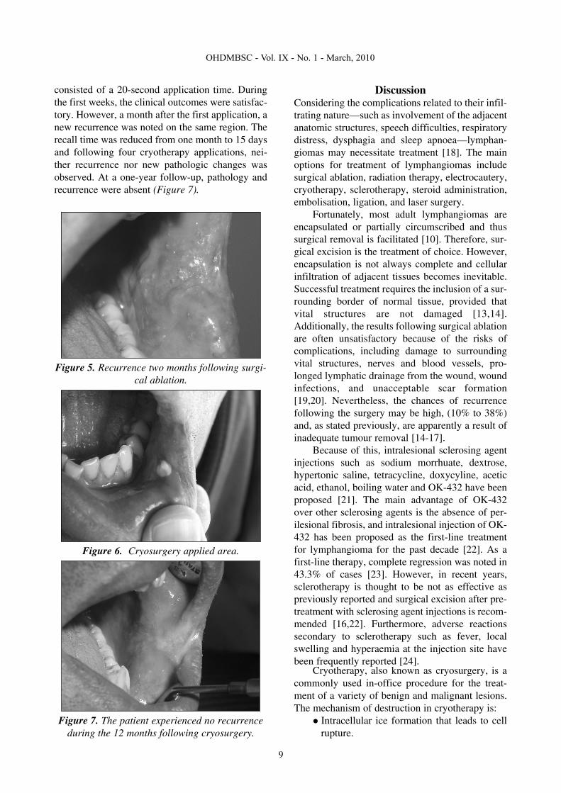
Department of Oral and Maxillofacial Surgery ofGülhane Military Medical Academy, Etlik, Turkey,with a painless swelling of 18 months’ durationinside his left cheek. He said he thought that thelesion had grown in size over the previous twomonths because he was biting it when he ate. Anintraoral clinical examination revealed the presenceof a bluish-purple coloured lesion, 2.5-3.0 cm indiameter, which was connected to the lower part ofthe left inside cheek (Figure 1). When the lesionwas palpated, it was found that its colour wasunchanged and no ischaemic area appeared.Considering the fibrotic appearance, 18 months’duration, and traumatic aetiology, the provisionaldiagnosis was that the lesion was benign and, pos-sibly, a traumatic granuloma.
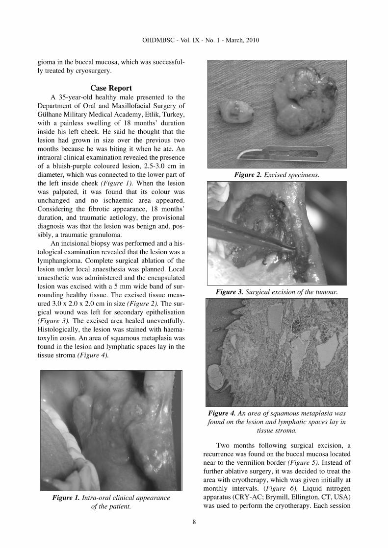
An incisional biopsy was performed and a his-tological examination revealed that the lesion was alymphangioma. Complete surgical ablation of thelesion under local anaesthesia was planned. Localanaesthetic was administered and the encapsulatedlesion was excised with a 5 mm wide band of sur-rounding healthy tissue. The excised tissue meas-ured 3.0 x 2.0 x 2.0 cm in size (Figure 2). The sur-gical wound was left for secondary epithelisation(Figure 3). The excised area healed uneventfully.Histologically, the lesion was stained with haema-toxylin eosin. An area of squamous metaplasia wasfound in the lesion and lymphatic spaces lay in thetissue stroma (Figure 4).
Figure 1. Intra-oral clinical appearance of the patient.
Figure 2. Excised specimens.
Figure 3. Surgical excision of the tumour.
Figure 4. An area of squamous metaplasia wasfound on the lesion and lymphatic spaces lay in
tissue stroma.
Two months following surgical excision, arecurrence was found on the buccal mucosa locatednear to the vermilion border (Figure 5). Instead offurther ablative surgery, it was decided to treat thearea with cryotherapy, which was given initially atmonthly intervals. (Figure 6). Liquid nitrogenapparatus (CRY-AC; Brymill, Ellington, CT, USA)was used to perform the cryotherapy. Each session
9
OHDMBSC - Vol. IX - No. 1 - March, 2010
consisted of a 20-second application time. Duringthe first weeks, the clinical outcomes were satisfac-tory. However, a month after the first application, anew recurrence was noted on the same region. Therecall time was reduced from one month to 15 daysand following four cryotherapy applications, nei-ther recurrence nor new pathologic changes wasobserved. At a one-year follow-up, pathology andrecurrence were absent (Figure 7).
Figure 5. Recurrence two months following surgi-cal ablation.
Figure 6. Cryosurgery applied area.
Figure 7. The patient experienced no recurrenceduring the 12 months following cryosurgery.
DiscussionConsidering the complications related to their infil-trating nature—such as involvement of the adjacentanatomic structures, speech difficulties, respiratorydistress, dysphagia and sleep apnoea—lymphan-giomas may necessitate treatment [18]. The mainoptions for treatment of lymphangiomas includesurgical ablation, radiation therapy, electrocautery,cryotherapy, sclerotherapy, steroid administration,embolisation, ligation, and laser surgery.
Fortunately, most adult lymphangiomas areencapsulated or partially circumscribed and thussurgical removal is facilitated [10]. Therefore, sur-gical excision is the treatment of choice. However,encapsulation is not always complete and cellularinfiltration of adjacent tissues becomes inevitable.Successful treatment requires the inclusion of a sur-rounding border of normal tissue, provided thatvital structures are not damaged [13,14].Additionally, the results following surgical ablationare often unsatisfactory because of the risks ofcomplications, including damage to surroundingvital structures, nerves and blood vessels, pro-longed lymphatic drainage from the wound, woundinfections, and unacceptable scar formation[19,20]. Nevertheless, the chances of recurrencefollowing the surgery may be high, (10% to 38%)and, as stated previously, are apparently a result ofinadequate tumour removal [14-17].
Because of this, intralesional sclerosing agentinjections such as sodium morrhuate, dextrose,hypertonic saline, tetracycline, doxycyline, aceticacid, ethanol, boiling water and OK-432 have beenproposed [21]. The main advantage of OK-432over other sclerosing agents is the absence of per-ilesional fibrosis, and intralesional injection of OK-432 has been proposed as the first-line treatmentfor lymphangioma for the past decade [22]. As afirst-line therapy, complete regression was noted in43.3% of cases [23]. However, in recent years,sclerotherapy is thought to be not as effective aspreviously reported and surgical excision after pre-treatment with sclerosing agent injections is recom-mended [16,22]. Furthermore, adverse reactionssecondary to sclerotherapy such as fever, localswelling and hyperaemia at the injection site havebeen frequently reported [24].
Cryotherapy, also known as cryosurgery, is acommonly used in-office procedure for the treat-ment of a variety of benign and malignant lesions.The mechanism of destruction in cryotherapy is:
Intracellular ice formation that leads to cellrupture.

An increase in solute concentration withinthe damaged tissue.Inflammation in the damaged tissue.Vascular stasis in the area treated.
Following the application, treated areas re-epithelialise. Adverse effects of cryotherapy areusually minor and short-lived. Lymphangiomas arethought to be very suitable for treatment bycryosurgery because of their high fluid content andpoor blood supply [17]. The treatment takes muchless time compared to surgical excision [25].Additionally, its effectiveness in eliminating pain isextremely important for palliative treatment [17].
ConclusionsIn conclusion, total surgical excision is generallyrecommended for the treatment of lymphangiomas[26-28]. Therefore, in the current case, in the firstinstance, complete surgical ablation of the lesionwas performed. Following a recurrence, the patientdid not undergo further surgery. The less invasiveoption of cryosurgery was employed to achieve asuccessful outcome.
This case highlights the advantages ofcryosurgery in that it takes much less time and ismuch more effective in eliminating pain, compared tosurgical excision. In the opinion of the authors, it is asafe and simple option in the treatment of lymphan-giomas.
References1. Mandel L. Parotid area lymphangioma in an adult: case
report. Journal of Oral and Maxillofacial Surgery 2004;62(10): 1320-1323.
2. Brennan TD, Miller AS, Chen SY. Lymphangiomas ofthe oral cavity: a clinicopathologic, immunohistochemical, andelectron-microscopic study. Journal of Oral and MaxillofacialSurgery 1997; 55: 932-935.
3. Neville BW, Damm DD, Allen CM, Bouquot JE. Softtissue tumors. In: Oral & Maxillofacial Pathology. 2nd ed.Philadelphia, PA: WB Saunders; 2002: pp. 475-477.
4. Brennan TD, Miller AS, Chen SY. Lymphangiomas ofthe oral cavity. A clinicopathologic, immunohistochemical,and electron-microscopic study. Journal of Oral andMaxillofacial Surgery 1997; 55: 932-935.
5. Morley SE, Ramesar KC, Macleod DA. Cystic hygro-ma in an adult. A case report. Journal of the Royal College ofSurgeons of Edinburgh 1999; 44: 57-58.
6. Shafer WG, Hine MK, Levy BM. Developmental dis-turbances of the tongue. In: A Textbook of Oral Pathology. 4thed. Philadelphia, PA: WB Saunders; 1983: pp. 159-60.
7. Suen JY, Waner M. Treatment of oral cavity vascularmalformations using the neodymium:YAG laser. Archives ofOtolaryngology—Head and Neck Surgery 1989; 115: 1329-1333.
8. Balakrishnan A, Bailey CM. Lymphangioma of thetongue. A review of pathogenesis, treatment and the use of sur-face laser photocoagulation. Journal of Laryngology andOtology 1991; 105: 924-929.
9. Hellman JR, Myer CM, Prenger EC. Therapeutic alter-natives in the treatment of life-threatening vasoformativetumors. American Journal of Otolaryngology 1992; 13: 48-53.
10. Kennedy TL. Cystic hygroma-lymphangioma. A rareand still unclear entity. Laryngoscope 1989; 99(Suppl 49): 1-10.
11. Mikhail M, Kennedy R, Cramer B, Smith T.Sclerosing of recurrent lymphangioma using OK-432. J PediatrSurg. 1996; 31:1463-4.
12. Brown RL, Azizkhan RG. Pediatric head and necklesions. Pediatric Clinics of North America 1998; 45: 889-905.
13. Fung K, Poenaru D, Sobeleski DA, Kamal IM. Impactof magnetic resonance imaging on the surgical management ofcystic hygromas. Journal of Pediatric Surgery 1998; 33: 839-841.
14. Schefter RP, Olsen KD, Gaffey TA. Cervical lym-phangioma in the adult. Otolaryngology—Head and NeckSurgery 1998; 93: 65-69.
15. Fraser SE, Campbell B, Kajdacsy-Balla A. Pathologicquiz case 2. Cystic lymphangioma. Archives ofOtolaryngology—Head and Neck Surgery 1996; 122: 893, 895.
16. Ricciardelli EJ, Richardson MA. Cervicofacial cystichygroma. Patterns of recurrence and management of the diffi-cult case. Archives of Otolaryngology—Head and NeckSurgery 1991; 117: 546-553
17. Bekke JP, Baart JA. Six years’ experience withcryosurgery in the oral cavity. International Journal of OralSurgery 1979; 8: 251-270.
18. Dinerman WS, Myers EN. Lymphangiomatousmacroglossia. Laryngoscope 1976; 86: 291-296.
19. Kennedy TL, Whitaker M, Pellitteri P, Wood WE.Cystic hygroma/lymphangioma: a rational approach to man-agement. Laryngoscope 2001; 111: 1929-1937.
20. Emery PJ, Bailey CM, Evans JN. Cystic hygroma ofthe head and neck. A review of 37 cases. Journal ofLaryngology and Otology 1984; 98(6): 613-619.
21. Molitch HI, Unger EC, Witte CL, vanSonnenberg E.Percutaneous sclerotherapy of lymphangioma. Radiology1995; 194(2): 343-347.
22. Okazaki T, Iwatani S, Yanai T, Kobayshi H, Kato Y,Marusasa T, et al. Treatment of lymphangioma in children: ourexperience of 128 cases. Journal of Pediatric Surgery 2007;42(2): 386-389.
23. Acevedo JL, Shah RK, Brietzke SE. Nonsurgical ther-apies for lymphangiomas: a systematic review.Otolaryngology—Head and Neck Surgery 2008; 138: 418-424.
24. Okada A, Kubota A, Fukuzawa M, Imura K, KamataS. Injection of bleomycin as a primary therapy of cystic lym-phangioma. Journal of Pediatric Surgery 1992; 27: 440-443.
25. Dawber R, Colver G, Jackson A. CutaneousCryosurgery. Principles and Clinical Practice. 2nd ed.London: Martin Dunitz; 1997; pp. 4-30.
26. Regezi JA, Sciubba J. Oral Pathology: Clinical-Pathologic Correlations. 2nd ed. Philadelphia, PA: WBSaunders; 1993: pp. 214-216.
27. Sato M, Tanaka N, Sato T, Amagasa T. Oral and max-illofacial tumours in children: A review. British Journal ofOral Maxillofacial Surgery 1997; 35: 92-95.
28. Park YW, Kim SM, Min BG, Park IW, Lee SK.Lymphangioma involving the mandible. Immunohistochemicalexpressions for the lymphatic proliferation. Journal of OralPathology and Medicine 2002; 31(5): 280-283.
OHDMBSC - Vol. IX - No. 1 - March, 2010
10
Top Related