


Languages
Pages
Legal


PowerPoint® Lecture Slide Presentation
by Patty Bostwick-Taylor,
Florence-Darlington Technical College
Copyright © 2009 Pearson Education, Inc., publishing as Benjamin Cummings
PART A5
The Skeletal
System
Copyright © 2009 Pearson Education, Inc., publishing as Benjamin Cummings
The Skeletal System
Parts of the skeletal system
Bones (skeleton)
Joints
Cartilages
Ligaments
Two subdivisions of the skeleton
Axial skeleton
Appendicular skeleton
Copyright © 2009 Pearson Education, Inc., publishing as Benjamin Cummings
Functions of Bones
Support the body
Protect soft organs
Allow movement due to attached skeletal muscles
Store minerals and fats in Yellow Marrow
Blood cell formation in Red Marrow
Copyright © 2009 Pearson Education, Inc., publishing as Benjamin Cummings
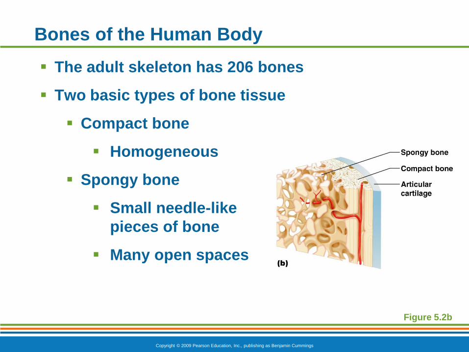
Bones of the Human Body
The adult skeleton has 206 bones
Two basic types of bone tissue
Compact bone
Homogeneous
Spongy bone
Small needle-like
pieces of bone
Many open spaces
Figure 5.2b
Copyright © 2009 Pearson Education, Inc., publishing as Benjamin Cummings
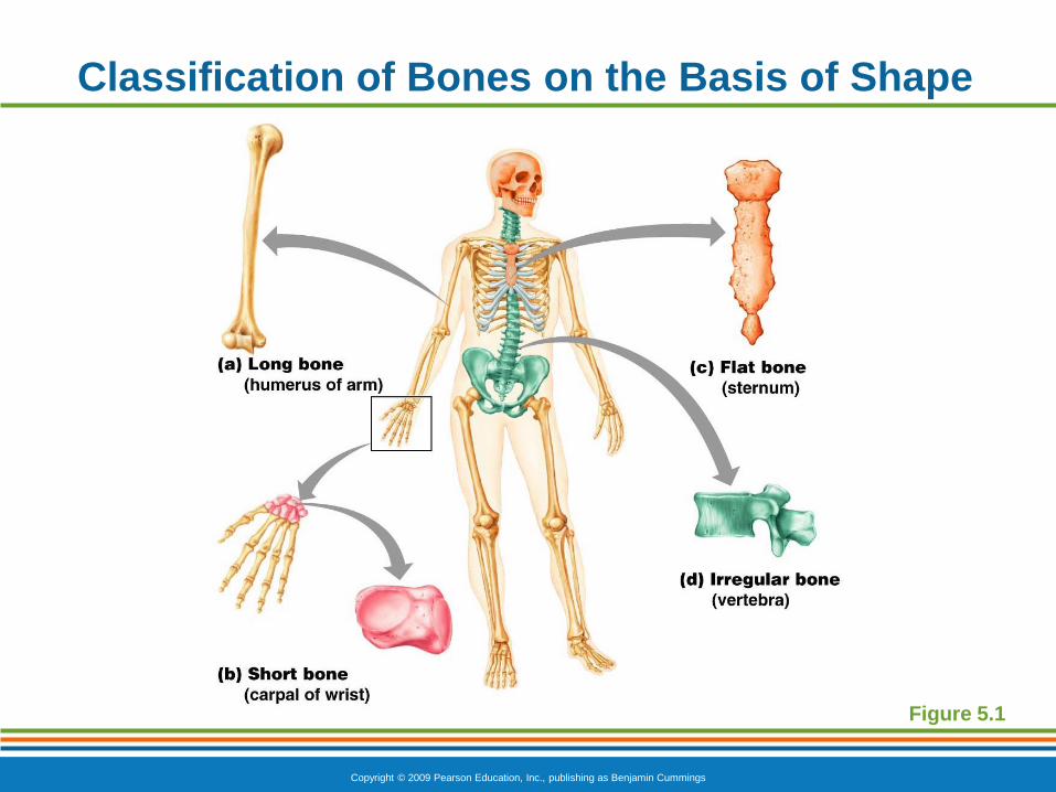
Classification of Bones on the Basis of Shape
Figure 5.1
Copyright © 2009 Pearson Education, Inc., publishing as Benjamin Cummings
Classification of Bones
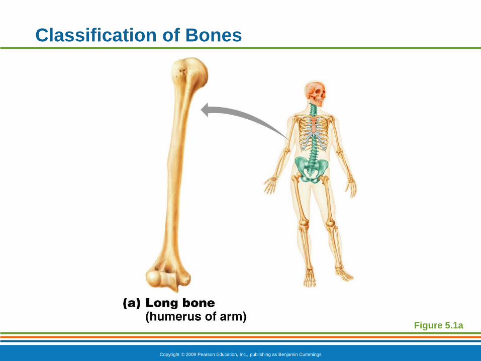
Long bones
Typically longer than they are wide
Have a shaft with heads at both ends
Contain mostly compact bone
Example:
Femur
Humerus
Copyright © 2009 Pearson Education, Inc., publishing as Benjamin Cummings
Classification of Bones
Figure 5.1a
Copyright © 2009 Pearson Education, Inc., publishing as Benjamin Cummings
Classification of Bones
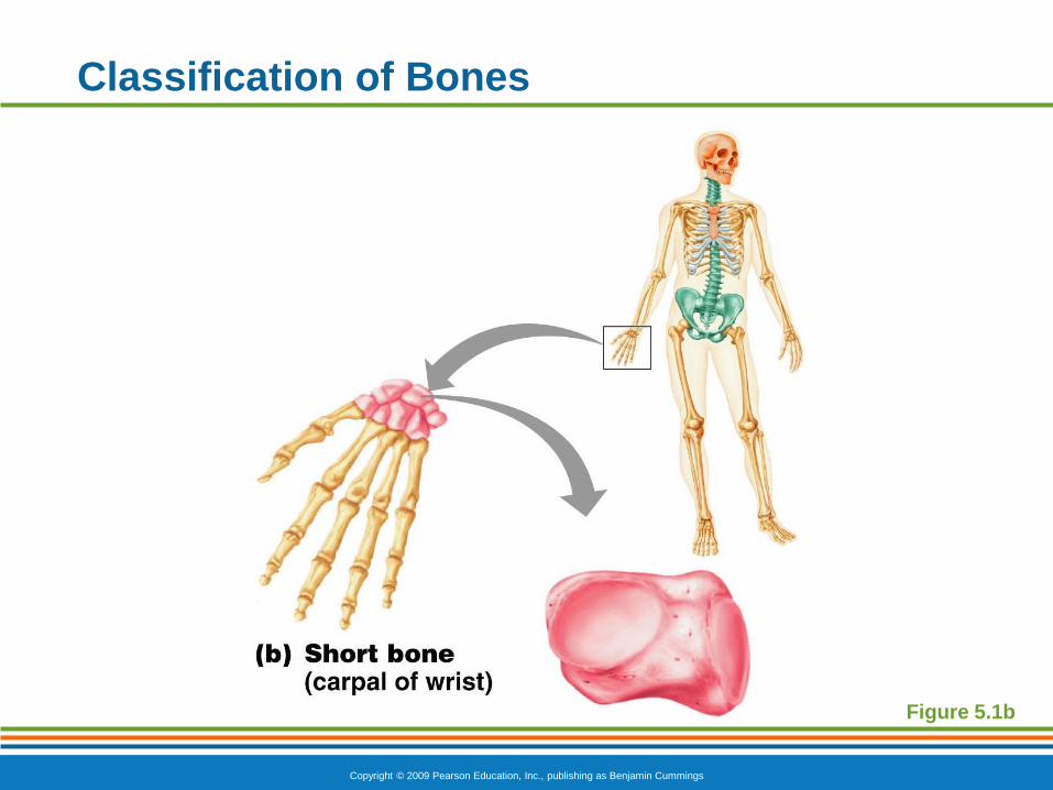
Short bones
Generally cube-shape
Contain mostly spongy bone
Example:
Carpals
Tarsals
Copyright © 2009 Pearson Education, Inc., publishing as Benjamin Cummings
Classification of Bones
Figure 5.1b
Copyright © 2009 Pearson Education, Inc., publishing as Benjamin Cummings
Classification of Bones
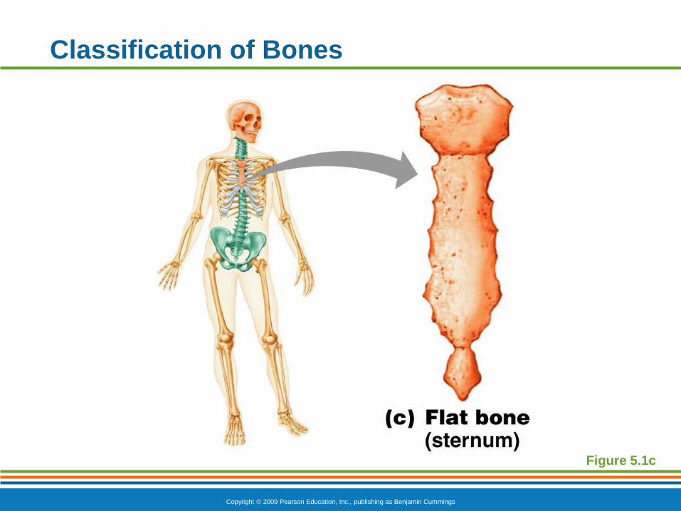
Flat bones
Thin, flattened, and usually curved
Two thin layers of compact bone surround a
layer of spongy bone
Example:
Skull
Ribs
Sternum
Copyright © 2009 Pearson Education, Inc., publishing as Benjamin Cummings
Classification of Bones
Figure 5.1c
Copyright © 2009 Pearson Education, Inc., publishing as Benjamin Cummings
Classification of Bones
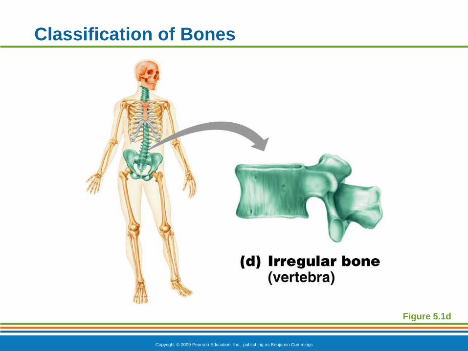
Irregular bones
Irregular shape
Do not fit into other bone classification
categories
Example:
Vertebrae
Hip bones
Copyright © 2009 Pearson Education, Inc., publishing as Benjamin Cummings
Classification of Bones
Figure 5.1d
Copyright © 2009 Pearson Education, Inc., publishing as Benjamin Cummings
Copyright © 2009 Pearson Education, Inc., publishing as Benjamin Cummings
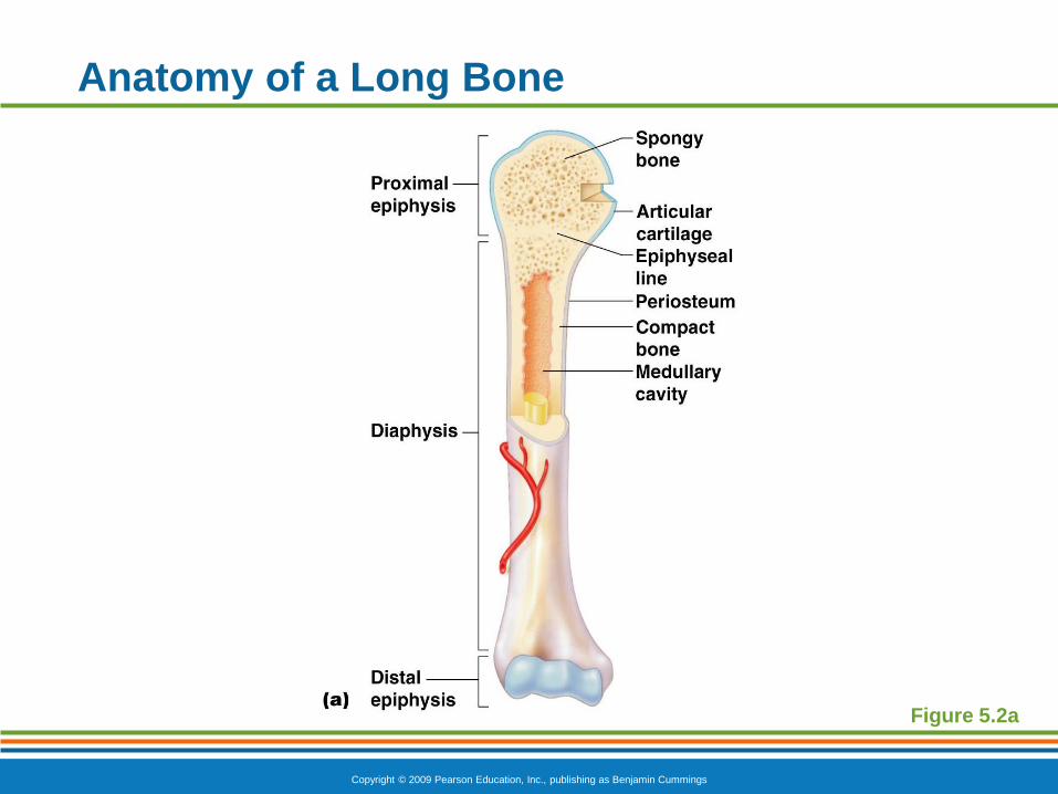
Anatomy of a Long Bone
Diaphysis
Shaft
Composed of compact bone
Epiphysis
Ends of the bone
Composed mostly of spongy bone

Copyright © 2009 Pearson Education, Inc., publishing as Benjamin Cummings
Anatomy of a Long Bone
Figure 5.2a
Copyright © 2009 Pearson Education, Inc., publishing as Benjamin Cummings
Anatomy of a Long Bone
Periosteum
Outside covering of the diaphysis
Fibrous connective tissue membrane
Sharpey’s fibers
Secure periosteum to underlying bone
Arteries
Supply bone cells with nutrients
Copyright © 2009 Pearson Education, Inc., publishing as Benjamin Cummings
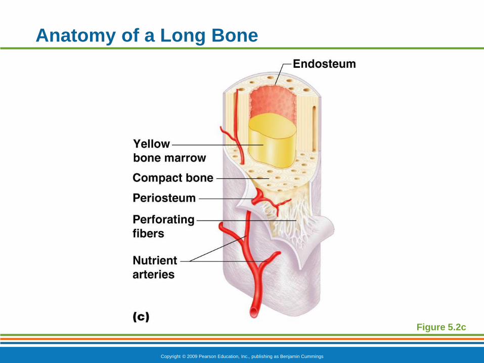
Anatomy of a Long Bone
Figure 5.2c
Copyright © 2009 Pearson Education, Inc., publishing as Benjamin Cummings
Anatomy of a Long Bone
Articular cartilage
Covers the external surface of the epiphyses
Made of hyaline cartilage
Decreases friction at joint surfaces
Copyright © 2009 Pearson Education, Inc., publishing as Benjamin Cummings
Anatomy of a Long Bone
Epiphyseal plate
Flat plate of hyaline cartilage seen in young,
growing bone
Epiphyseal line
Remnant of the epiphyseal plate
Seen in adult bones
Copyright © 2009 Pearson Education, Inc., publishing as Benjamin Cummings
Anatomy of a Long Bone
Figure 5.2a
Copyright © 2009 Pearson Education, Inc., publishing as Benjamin Cummings
Anatomy of a Long Bone
Medullary cavity
Cavity inside of the shaft
Contains yellow marrow (mostly fat) in adults
Contains red marrow (for blood cell formation)
in infants

Copyright © 2009 Pearson Education, Inc., publishing as Benjamin Cummings
Anatomy of a Long Bone
Figure 5.2a
Copyright © 2009 Pearson Education, Inc., publishing as Benjamin Cummings
7.2 Continued: Types of Bone Tissue
Compact bone tissue – continuous extracellular matrix with no spaces. Forms external layer of all bones.
Forms diaphysis of long bones
Spongy bone tissue – consists of numerous branching boney plates.
Makes up inside of most short, flat, or irregular bones.
Also in the epiphysis of long bones.
Contains red bone marrow.
Copyright © 2009 Pearson Education, Inc., publishing as Benjamin Cummings
7.2 Continued: Microscopic Structure
Bone tissue is about 25% water, 25% collagen fibers, & 50% calcium phosphate (inorganic salts)
Calcification: as calcium phosphate is deposited it become hardened / calcified
There are 3 kinds of bone cells
Osteoblasts: bone building cells; synthesize and secrete collagen fibers; build matrix around themselves
Osteocytes: mature bone cells; maintain daily metabolism of bone
Osteoclasts: Huge cells; in endosteum, release enzymes to breakdown matrix >>> part of normal growth & maintenance
Copyright © 2009 Pearson Education, Inc., publishing as Benjamin Cummings
Microscopic Anatomy of Bone
Osteon (Haversian system)
A unit of bone containing central canal and
matrix rings
Central (Haversian) canal
Opening in the center of an osteon
Carries blood vessels and nerves
Perforating (Volkman’s) canal
Canal perpendicular to the central canal
Carries blood vessels and nerves

Copyright © 2009 Pearson Education, Inc., publishing as Benjamin Cummings
Microscopic Anatomy of Bone
Figure 5.3a
Copyright © 2009 Pearson Education, Inc., publishing as Benjamin Cummings
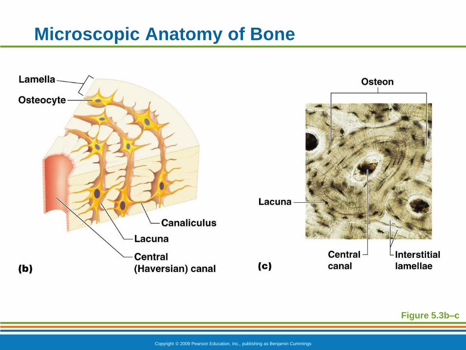
Microscopic Anatomy of Bone
Lacunae
Cavities containing bone cells (osteocytes)
Arranged in concentric rings
Lamellae
Rings around the central canal
Sites of lacunae
Copyright © 2009 Pearson Education, Inc., publishing as Benjamin Cummings
Microscopic Anatomy of Bone
Figure 5.3b–c
Copyright © 2009 Pearson Education, Inc., publishing as Benjamin Cummings
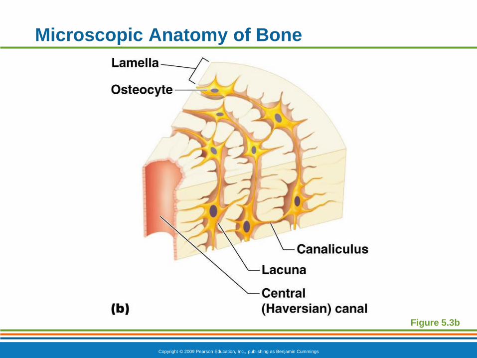
Microscopic Anatomy of Bone
Canaliculi
Tiny canals
Radiate from the central canal to lacunae
Form a transport system connecting all bone
cells to a nutrient supply
Copyright © 2009 Pearson Education, Inc., publishing as Benjamin Cummings
Microscopic Anatomy of Bone
Figure 5.3b
Copyright © 2009 Pearson Education, Inc., publishing as Benjamin Cummings
7.3 Bone Development and Growth Ossification – The process by which bone
forms. The embryo is composed of loose and cartilage connective tissue, which are shaped like bones and are the sites where ossification takes place.
Intramembranous ossification – formation of bone directly on or in loose connective tissue membranes
Flat bones of the skull and mandible
Fetal soft spot
Endochondral ossification – formation of bone within cartilage.
Most bones of the body are formed this way
Steps are on page 134
Copyright © 2009 Pearson Education, Inc., publishing as Benjamin Cummings
Formation of the Human Skeleton
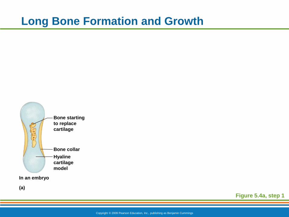
In embryos, the skeleton is primarily hyaline
cartilage
During development, much of this cartilage is
replaced by bone
Cartilage remains in isolated areas
Bridge of the nose
Parts of ribs
Joints
Copyright © 2009 Pearson Education, Inc., publishing as Benjamin Cummings
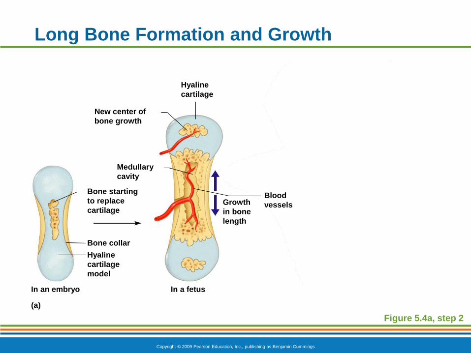
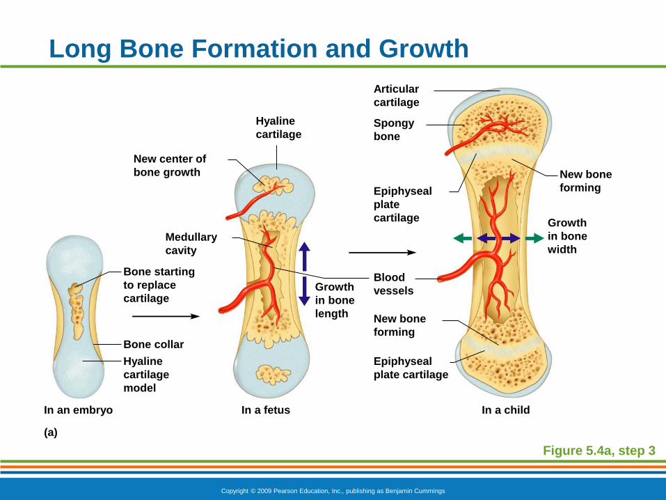
Bone Growth (Ossification)
Epiphyseal plates allow for lengthwise growth of
long bones during childhood
New cartilage is continuously formed
Older cartilage becomes ossified
Cartilage is broken down
Enclosed cartilage is digested away,
opening up a medullary cavity
Bone replaces cartilage through the action
of osteoblasts
Copyright © 2009 Pearson Education, Inc., publishing as Benjamin Cummings
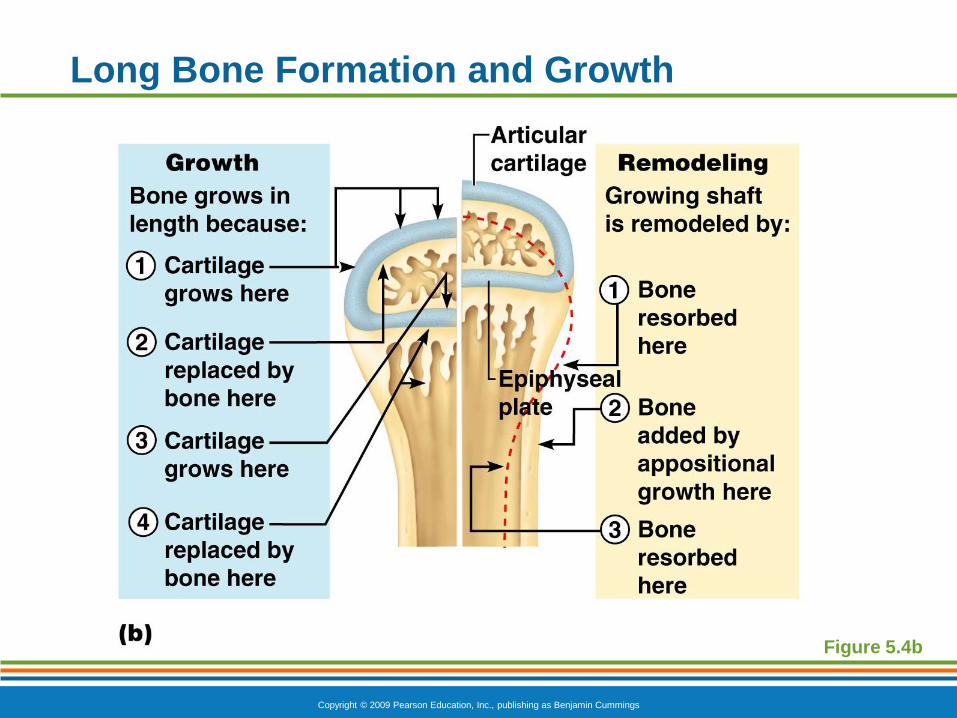
Bone Growth (Ossification)
Bones are remodeled and lengthened until growth
stops
Bones are remodeled in response to two
factors
Blood calcium levels
Pull of gravity and muscles on the
skeleton
Bones grow in width (called appositional
growth)
Copyright © 2009 Pearson Education, Inc., publishing as Benjamin Cummings
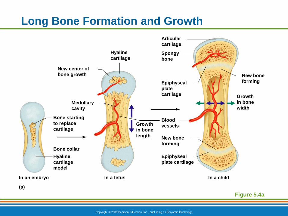
Long Bone Formation and Growth
Figure 5.4a
Bone starting
to replace
cartilage
Epiphyseal
plate
cartilage
Articular
cartilage
Spongy
bone
In a childIn a fetusIn an embryo
New bone
forming
Growth
in bone
width
Growth
in bone
length
Epiphyseal
plate cartilage
New bone
forming
Blood
vessels
Hyaline
cartilage
New center of
bone growth
Medullary
cavity
Bone collar
Hyaline
cartilage
model
(a)
Copyright © 2009 Pearson Education, Inc., publishing as Benjamin Cummings
Long Bone Formation and Growth
Figure 5.4a, step 1
Bone starting
to replace
cartilage
In an embryo
Bone collar
Hyaline
cartilage
model
(a)
Copyright © 2009 Pearson Education, Inc., publishing as Benjamin Cummings
Long Bone Formation and Growth
Figure 5.4a, step 2
Bone starting
to replace
cartilage
In a fetusIn an embryo
Growth
in bone
length
Blood
vessels
Hyaline
cartilage
New center of
bone growth
Medullary
cavity
Bone collar
Hyaline
cartilage
model
(a)
Copyright © 2009 Pearson Education, Inc., publishing as Benjamin Cummings
Long Bone Formation and Growth
Figure 5.4a, step 3
Bone starting
to replace
cartilage
Epiphyseal
plate
cartilage
Articular
cartilage
Spongy
bone
In a childIn a fetusIn an embryo
New bone
forming
Growth
in bone
width
Growth
in bone
length
Epiphyseal
plate cartilage
New bone
forming
Blood
vessels
Hyaline
cartilage
New center of
bone growth
Medullary
cavity
Bone collar
Hyaline
cartilage
model
(a)
Copyright © 2009 Pearson Education, Inc., publishing as Benjamin Cummings
Long Bone Formation and Growth
Figure 5.4b
Copyright © 2009 Pearson Education, Inc., publishing as Benjamin Cummings
7.3 Continued: Homeostasis of Bone
Bone Growth and Maintenance
Bone continually renews itself
Old, worn, & injured bone is continually destroyed and new bone tissue is formed in its place – Remodeling
Osteoclasts resorb bone matrix (resorption)
Osteoblasts replace bone matrix (deposition)
As a result, total mass of bone tissue usually stays the same
Copyright © 2009 Pearson Education, Inc., publishing as Benjamin Cummings
Remember: Calcium can’t be absorbed unless there is enough vitamin D.
Bone stores 99% of total amount of calcium
When bone is remodeled, calcium becomes available to other tissues
Balance must be maintained between blood & bone
Parathyroid hormone (PTH) – responsible for regulating calcium exchange between bone and blood (during resorption)
Low blood calcium levels = bone releases Calcium
Calcitonin – responsible for regulating calcium exchange between blood and bone (during deposition)
High blood calcium levels = bone absorbs Calcium
Calcium Homeostasis
Copyright © 2009 Pearson Education, Inc., publishing as Benjamin Cummings
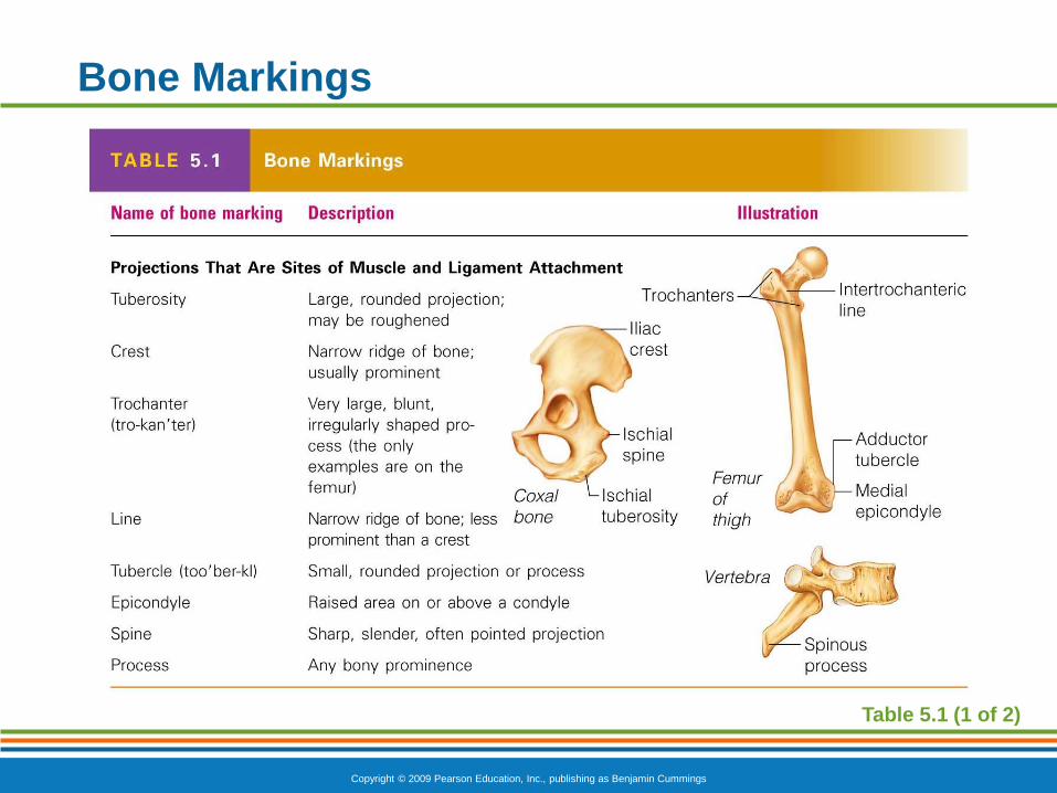
Bone Markings
Surface features of bones
Sites of attachments for muscles, tendons,
and ligaments
Passages for nerves and blood vessels
Categories of bone markings
Projections or processes—grow out from the
bone surface
Depressions or cavities—indentations
Copyright © 2009 Pearson Education, Inc., publishing as Benjamin Cummings
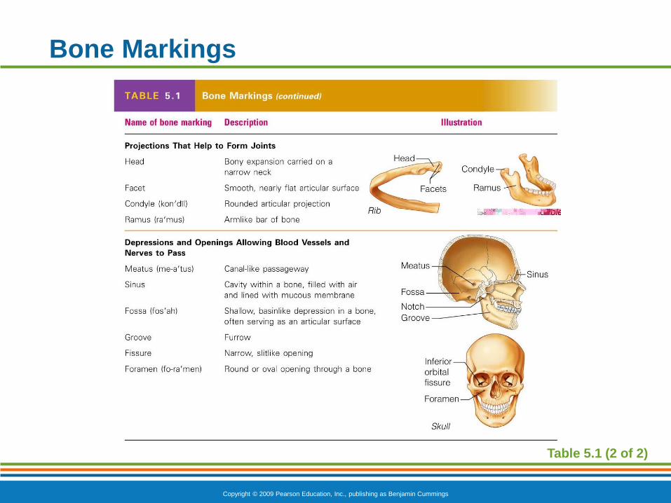
Bone Surface Markings
The surfaces of bones have various structural features adapted to specific functions.
Foramen: opening through with blood vessels, nerves, and ligaments pass
Fossa: a shallow depression in or on a bone
Process: part of a bone that forms a joint or where tendons, ligaments, and connective tissues attach
Copyright © 2009 Pearson Education, Inc., publishing as Benjamin Cummings
Bone Markings
Table 5.1 (1 of 2)
Copyright © 2009 Pearson Education, Inc., publishing as Benjamin Cummings
Bone Markings
Table 5.1 (2 of 2)
PowerPoint® Lecture Slide Presentation
by Patty Bostwick-Taylor,
Florence-Darlington Technical College
Copyright © 2009 Pearson Education, Inc., publishing as Benjamin Cummings
PART A5
The Skeletal
System
Copyright © 2009 Pearson Education, Inc., publishing as Benjamin Cummings
Bone Fractures
Fracture—break in a bone
The main categories of bone fractures are: complete, incomplete, compound and simple.
Complete: bone snaps into two or more parts
Incomplete: bone cracks but does not break all the way through
Compound (open fracture): bone breaks through the skin; it may then recede back into the wound and not be visible through the skin
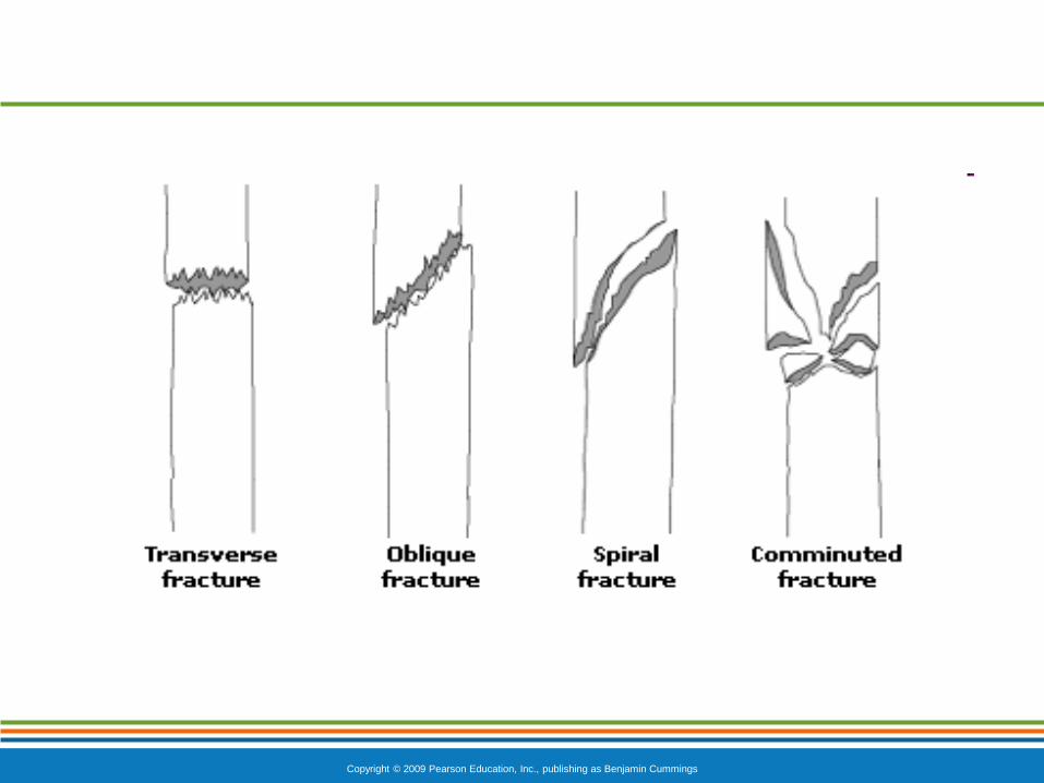
Simple (closed fracture): bone breaks but there is no open wound in the skin. There are many kinds; some are:
Transverse, Oblique, Spiral, Comminuted, Impact, Pathologic, and Stress
Copyright © 2009 Pearson Education, Inc., publishing as Benjamin Cummings
Copyright © 2009 Pearson Education, Inc., publishing as Benjamin Cummings
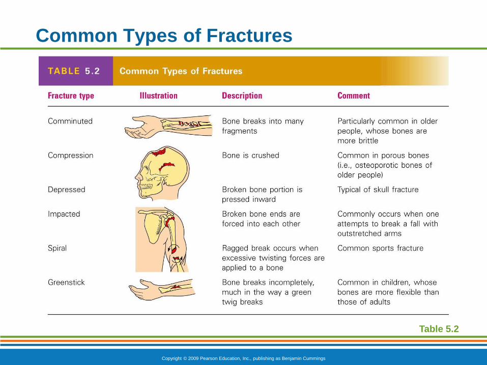
Common Types of Fractures
Table 5.2
Copyright © 2009 Pearson Education, Inc., publishing as Benjamin Cummings
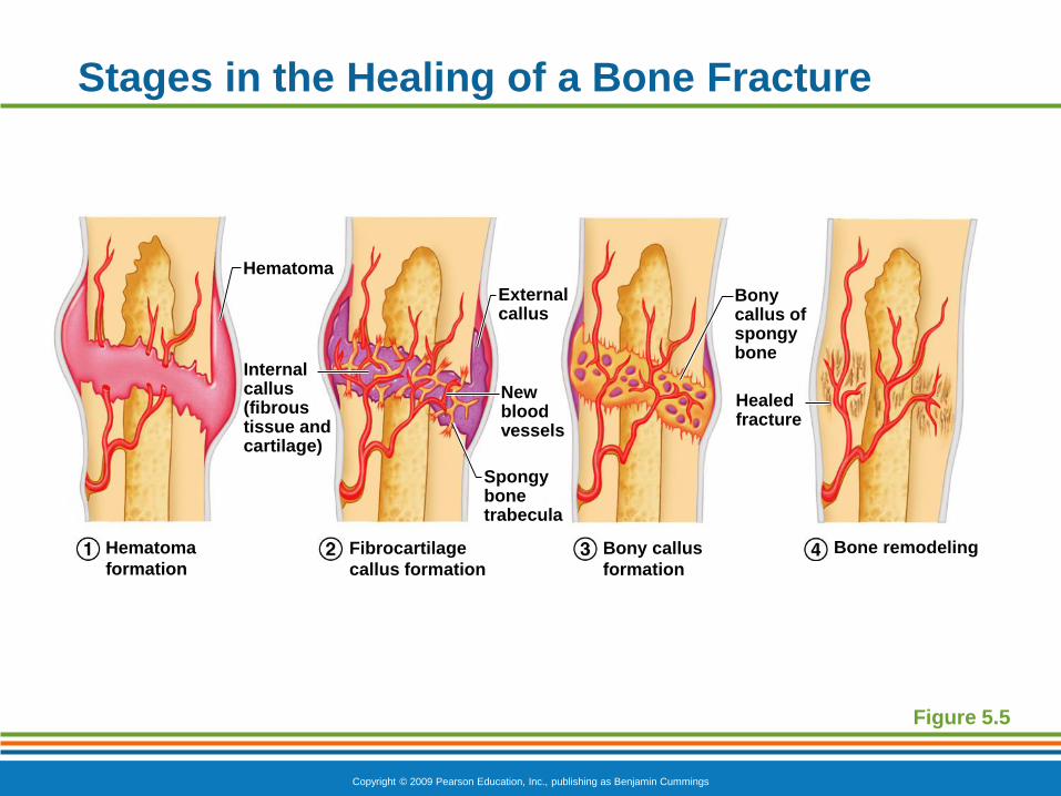
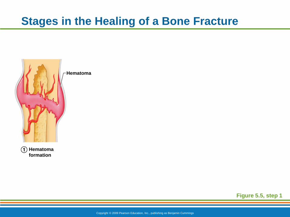
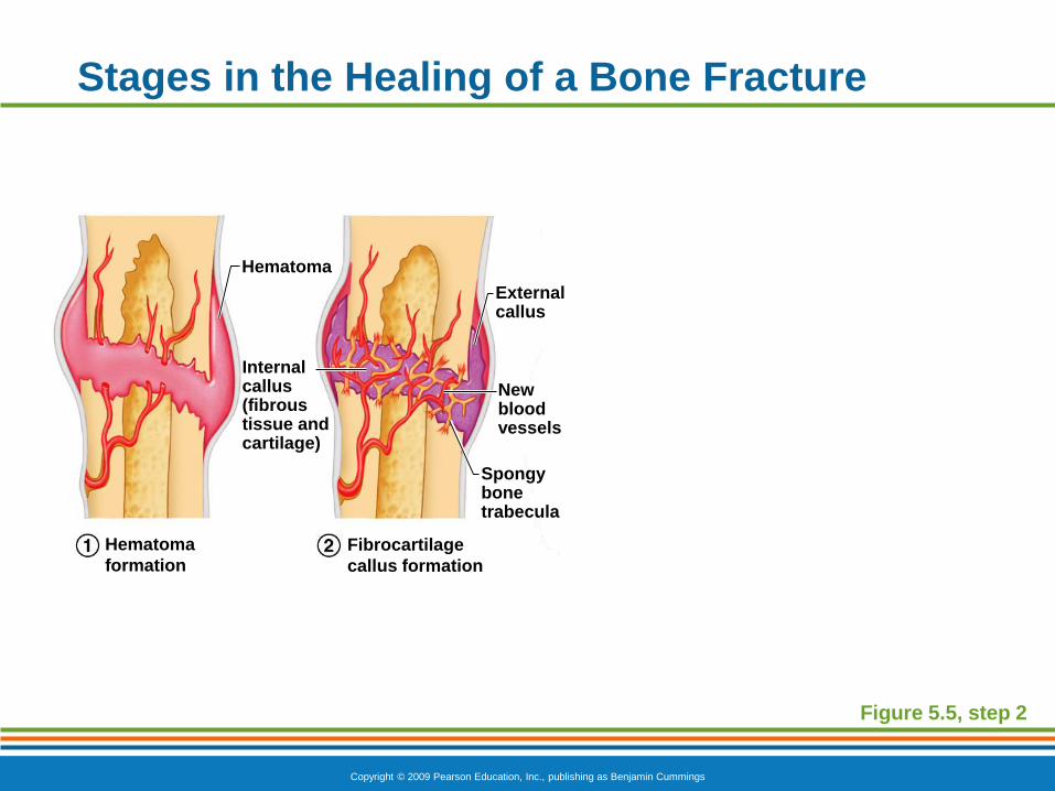
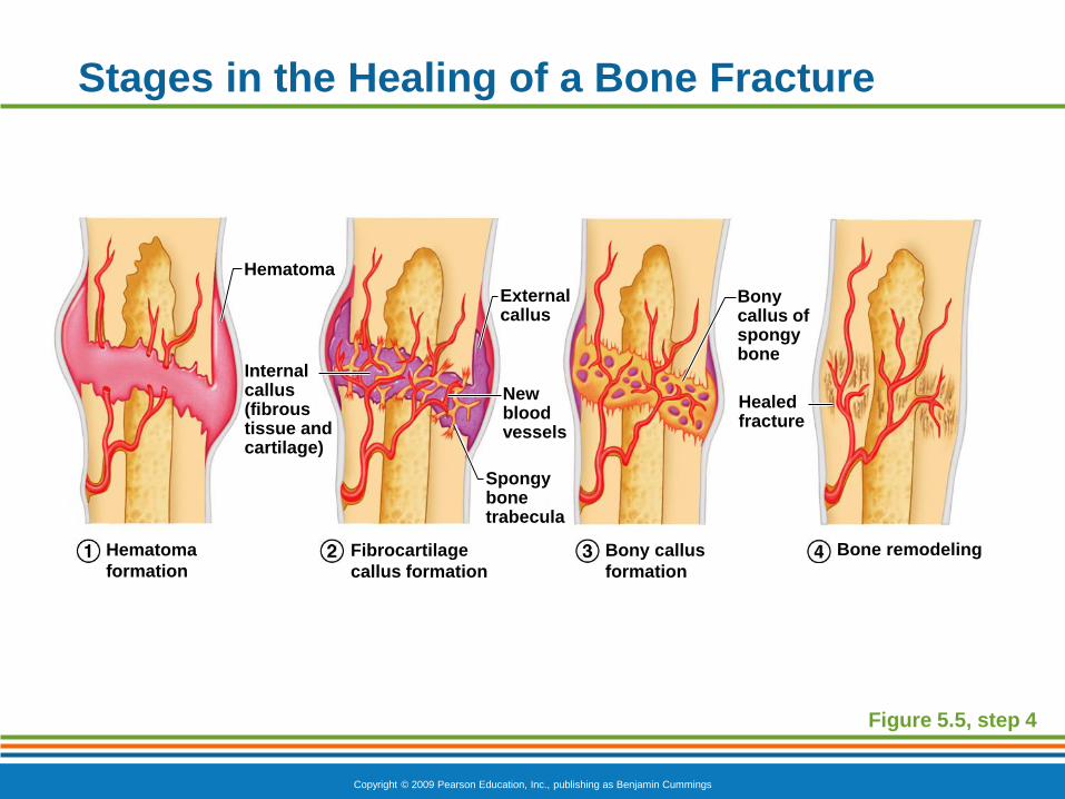
Repair of Bone Fractures
Hematoma (blood-filled swelling) is formed
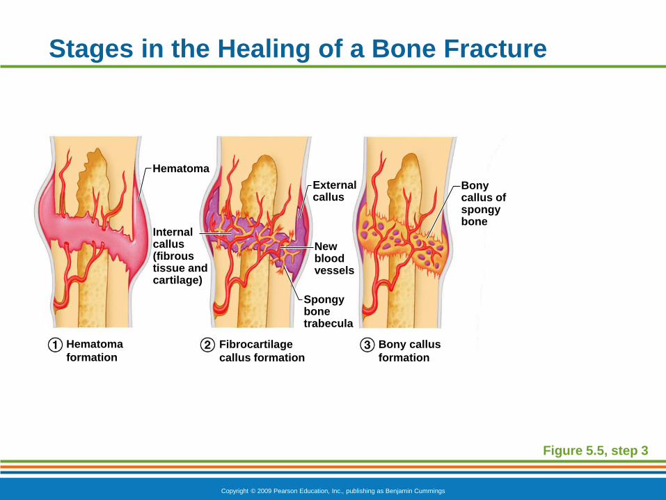
Break is splinted by fibrocartilage to form a callus
Fibrocartilage callus is replaced by a bony callus
Bony callus is remodeled to form a permanent
patch
Copyright © 2009 Pearson Education, Inc., publishing as Benjamin Cummings
Stages in the Healing of a Bone Fracture
Figure 5.5
Hematoma
Externalcallus
Bonycallus ofspongybone
Healedfracture
Newbloodvessels
Internalcallus(fibroustissue andcartilage)
Spongybonetrabecula
Hematoma
formation
Fibrocartilage
callus formation
Bony callus
formation
Bone remodeling
Copyright © 2009 Pearson Education, Inc., publishing as Benjamin Cummings
Stages in the Healing of a Bone Fracture
Figure 5.5, step 1
Hematoma
Hematoma
formation
Copyright © 2009 Pearson Education, Inc., publishing as Benjamin Cummings
Stages in the Healing of a Bone Fracture
Figure 5.5, step 2
Hematoma
Externalcallus
Newbloodvessels
Internalcallus(fibroustissue andcartilage)
Spongybonetrabecula
Hematoma
formation
Fibrocartilage
callus formation
Copyright © 2009 Pearson Education, Inc., publishing as Benjamin Cummings
Stages in the Healing of a Bone Fracture
Figure 5.5, step 3
Hematoma
Externalcallus
Bonycallus ofspongybone
Newbloodvessels
Internalcallus(fibroustissue andcartilage)
Spongybonetrabecula
Hematoma
formation
Fibrocartilage
callus formation
Bony callus
formation
Copyright © 2009 Pearson Education, Inc., publishing as Benjamin Cummings
Stages in the Healing of a Bone Fracture
Figure 5.5, step 4
Hematoma
Externalcallus
Bonycallus ofspongybone
Healedfracture
Newbloodvessels
Internalcallus(fibroustissue andcartilage)
Spongybonetrabecula
Hematoma
formation
Fibrocartilage
callus formation
Bony callus
formation
Bone remodeling
Copyright © 2009 Pearson Education, Inc., publishing as Benjamin Cummings
Divisions of the Skeleton
The adult human skeleton has 206 bones
Axial skeleton – contains 80 bones.
Consists of cranial bones, hyoid, vertebral column, and thorax (ribs, sternum).
Appendicular skeleton – Contains 126 bones.
Consists of clavicle, scapula, upper limbs, pelvic girdle, and lower limbs.
Copyright © 2009 Pearson Education, Inc., publishing as Benjamin Cummings
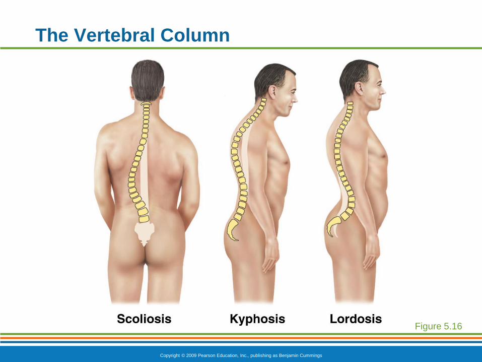
Homeostatic Imbalances
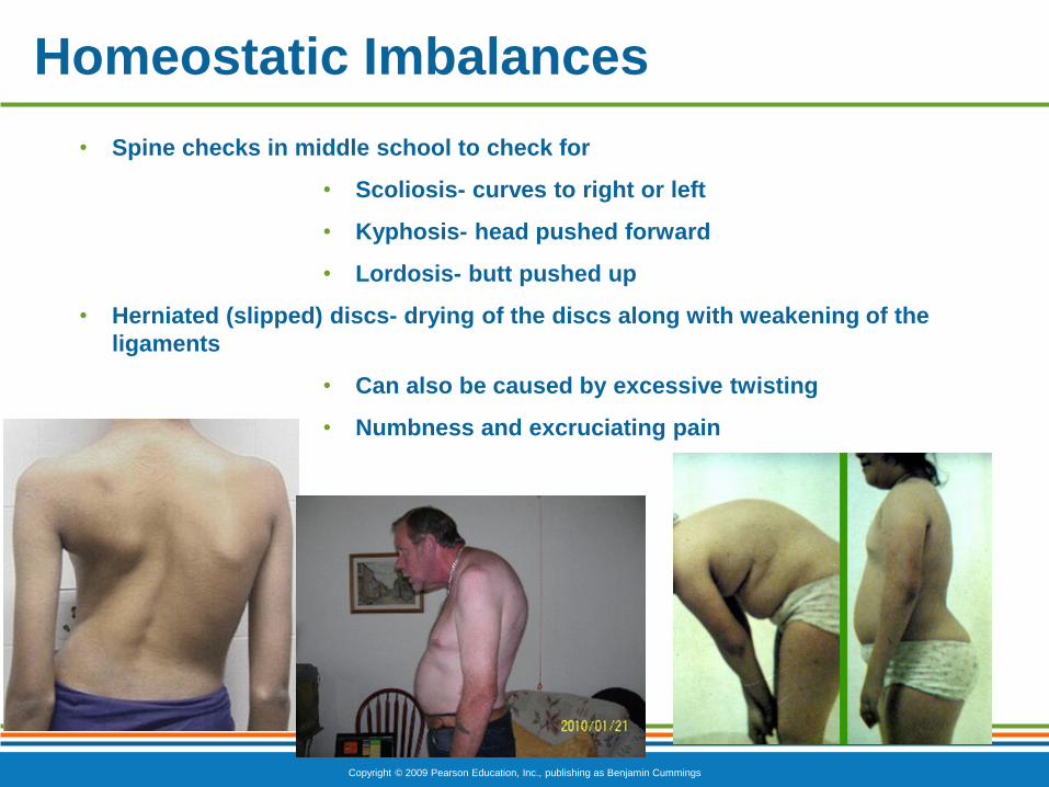
• Spine checks in middle school to check for
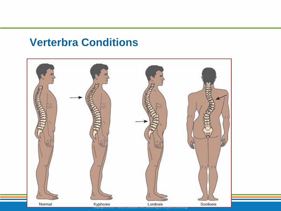
• Scoliosis- curves to right or left
• Kyphosis- head pushed forward
• Lordosis- butt pushed up
• Herniated (slipped) discs- drying of the discs along with weakening of the
ligaments
• Can also be caused by excessive twisting
• Numbness and excruciating pain
Copyright © 2009 Pearson Education, Inc., publishing as Benjamin Cummings
Verterbra Conditions
Copyright © 2009 Pearson Education, Inc., publishing as Benjamin Cummings
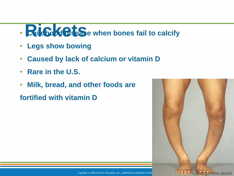
Rickets• Childhood disease when bones fail to calcify
• Legs show bowing
• Caused by lack of calcium or vitamin D
• Rare in the U.S.
• Milk, bread, and other foods are
fortified with vitamin D
Copyright © 2009 Pearson Education, Inc., publishing as Benjamin Cummings
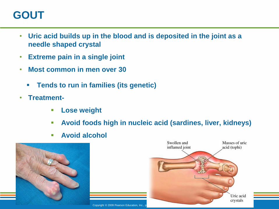
GOUT
• Uric acid builds up in the blood and is deposited in the joint as a
needle shaped crystal
• Extreme pain in a single joint
• Most common in men over 30
Tends to run in families (its genetic)
• Treatment-
Lose weight
Avoid foods high in nucleic acid (sardines, liver, kidneys)
Avoid alcohol
Copyright © 2009 Pearson Education, Inc., publishing as Benjamin Cummings
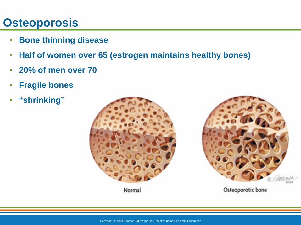
Osteoporosis
• Bone thinning disease
• Half of women over 65 (estrogen maintains healthy bones)
• 20% of men over 70
• Fragile bones
• “shrinking”
Copyright © 2009 Pearson Education, Inc., publishing as Benjamin Cummings
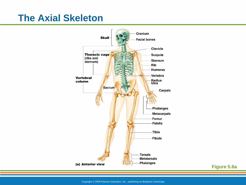
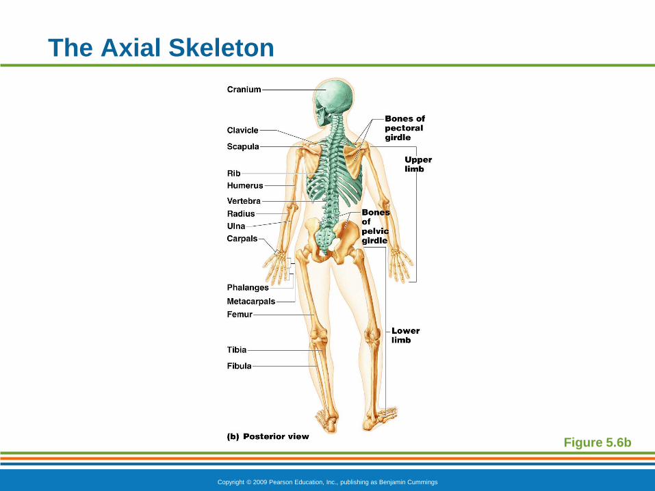
The Axial Skeleton
Forms the longitudinal axis of the body
Divided into three parts
Skull
Vertebral column
Bony thorax
Copyright © 2009 Pearson Education, Inc., publishing as Benjamin Cummings
The Axial Skeleton
Figure 5.6a
Copyright © 2009 Pearson Education, Inc., publishing as Benjamin Cummings
The Axial Skeleton
Figure 5.6b
Copyright © 2009 Pearson Education, Inc., publishing as Benjamin Cummings
The Skull
Two sets of bones
Cranium
Facial bones
Bones are joined by sutures
Only the mandible is attached by a freely movable
joint
Copyright © 2009 Pearson Education, Inc., publishing as Benjamin Cummings
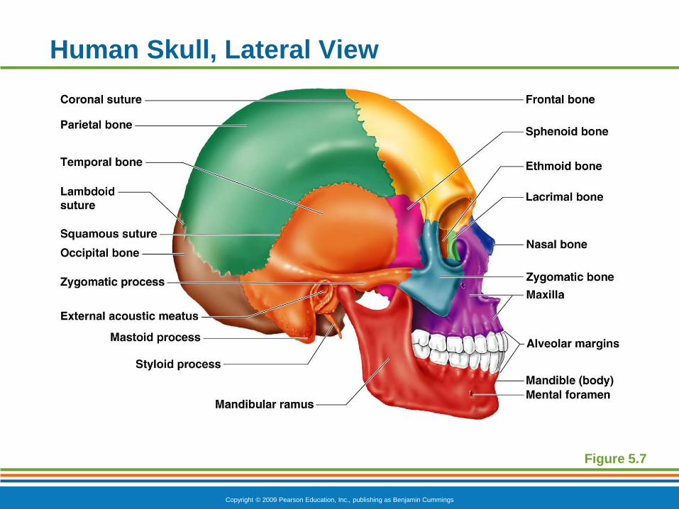
Human Skull, Lateral View
Figure 5.7
Copyright © 2009 Pearson Education, Inc., publishing as Benjamin Cummings
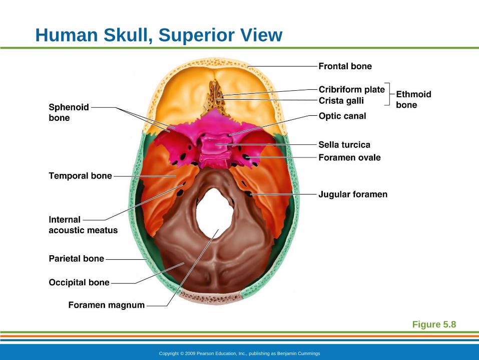
Human Skull, Superior View
Figure 5.8
Copyright © 2009 Pearson Education, Inc., publishing as Benjamin Cummings
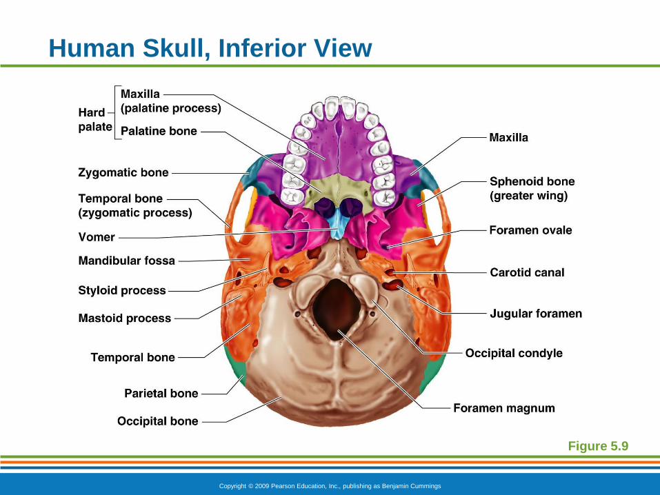
Human Skull, Inferior View
Figure 5.9
Copyright © 2009 Pearson Education, Inc., publishing as Benjamin Cummings
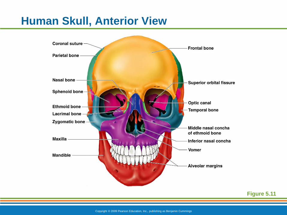
Human Skull, Anterior View
Figure 5.11
Copyright © 2009 Pearson Education, Inc., publishing as Benjamin Cummings
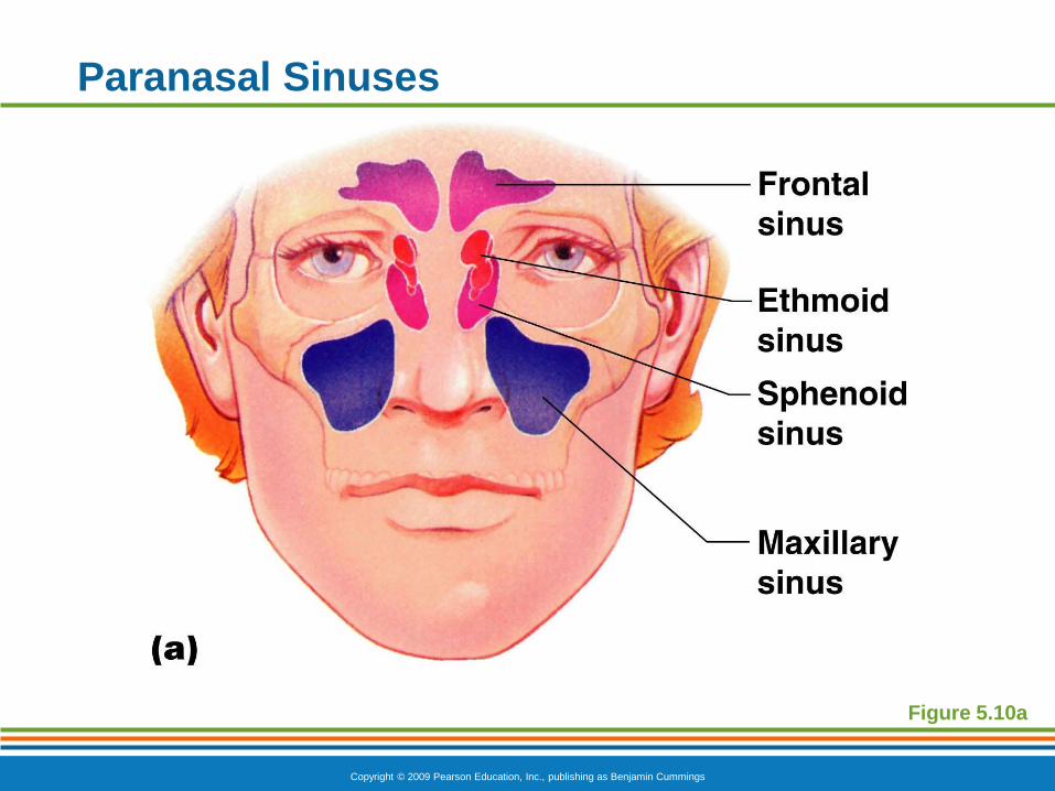
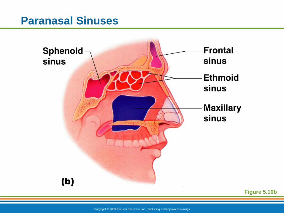
Paranasal Sinuses
Hollow portions of bones surrounding the nasal
cavity
Functions of paranasal sinuses
Lighten the skull
Give resonance and amplification to voice
Copyright © 2009 Pearson Education, Inc., publishing as Benjamin Cummings
Paranasal Sinuses
Figure 5.10a
Copyright © 2009 Pearson Education, Inc., publishing as Benjamin Cummings
Paranasal Sinuses
Figure 5.10b
Copyright © 2009 Pearson Education, Inc., publishing as Benjamin Cummings
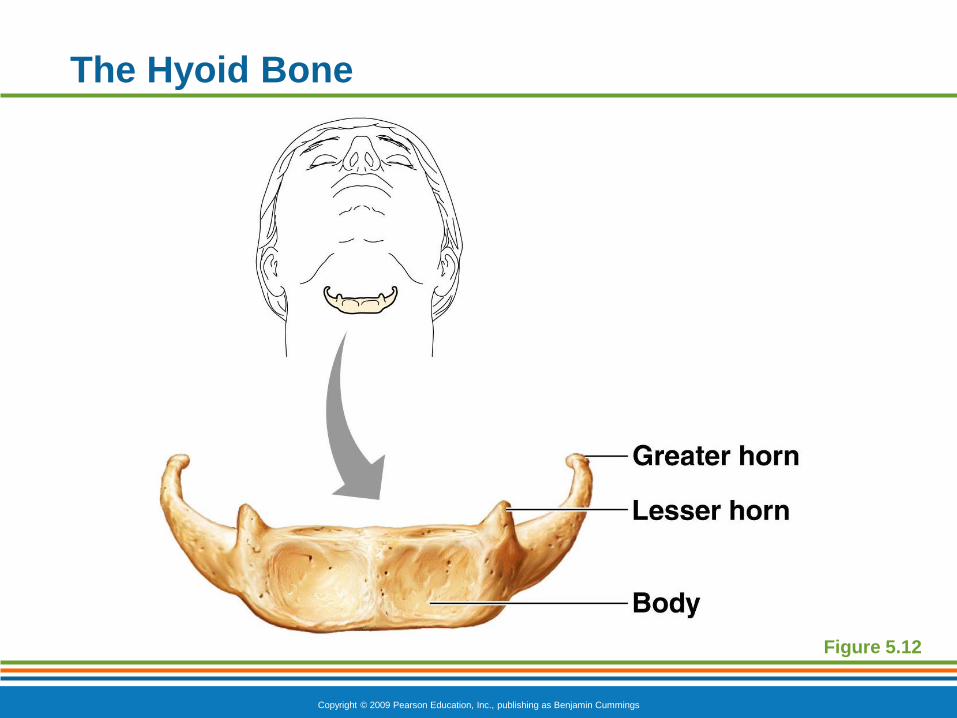
The Hyoid Bone
The only bone that does not articulate with
another bone
Serves as a moveable base for the tongue
Aids in swallowing and speech
Copyright © 2009 Pearson Education, Inc., publishing as Benjamin Cummings
The Hyoid Bone
Figure 5.12

PowerPoint® Lecture Slide Presentation
by Patty Bostwick-Taylor,
Florence-Darlington Technical College
Copyright © 2009 Pearson Education, Inc., publishing as Benjamin Cummings
PART A5
The Skeletal
System
Copyright © 2009 Pearson Education, Inc., publishing as Benjamin Cummings
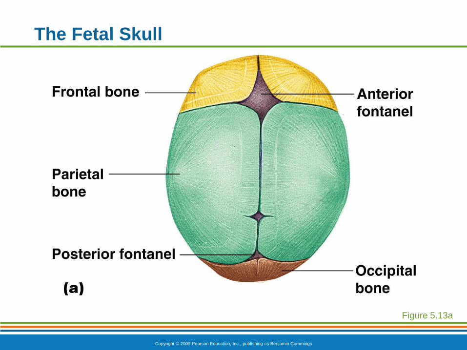
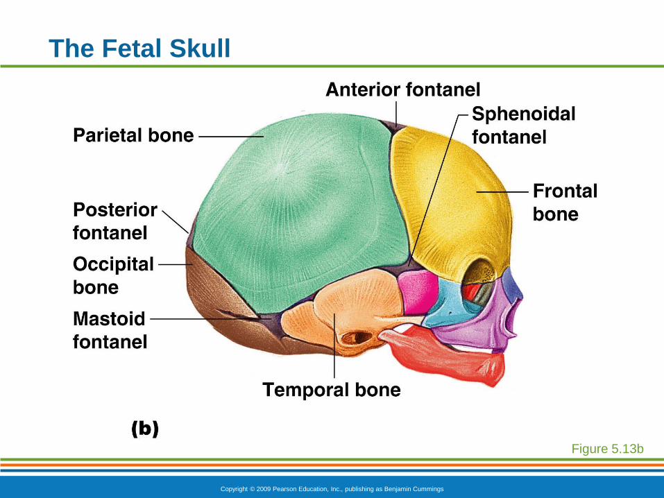
The Fetal Skull
The fetal skull is large compared to the infant’s
total body length
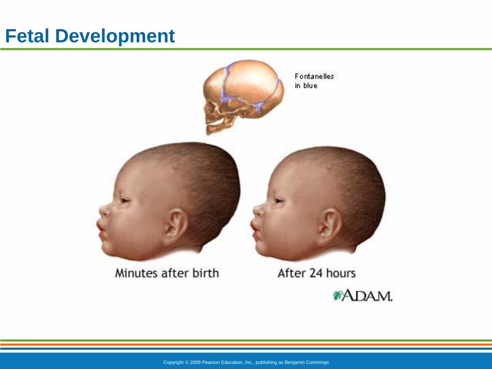
Fontanels—fibrous membranes connecting the
cranial bones
Allow the brain to grow
Convert to bone within 24 months after birth
Copyright © 2009 Pearson Education, Inc., publishing as Benjamin Cummings
The Fetal Skull
Figure 5.13a
Copyright © 2009 Pearson Education, Inc., publishing as Benjamin Cummings
The Fetal Skull
Figure 5.13b
Copyright © 2009 Pearson Education, Inc., publishing as Benjamin Cummings
Fetal Development
Copyright © 2009 Pearson Education, Inc., publishing as Benjamin Cummings
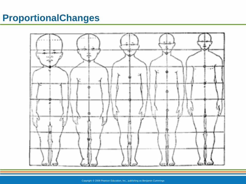
ProportionalChanges
Copyright © 2009 Pearson Education, Inc., publishing as Benjamin Cummings
The Vertebral Column
Each vertebrae is given a name according to its
location
There are 24 single vertebral bones separated
by intervertebral discs
Seven cervical vertebrae are in the neck
Twelve thoracic vertebrae are in the chest
region
Five lumbar vertebrae are associated with
the lower back
Copyright © 2009 Pearson Education, Inc., publishing as Benjamin Cummings
The Vertebral Column
Nine vertebrae fuse to form two composite bones
Sacrum
Coccyx
Copyright © 2009 Pearson Education, Inc., publishing as Benjamin Cummings
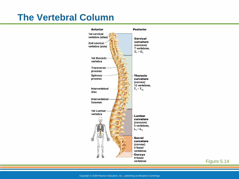
The Vertebral Column
Figure 5.14
Copyright © 2009 Pearson Education, Inc., publishing as Benjamin Cummings
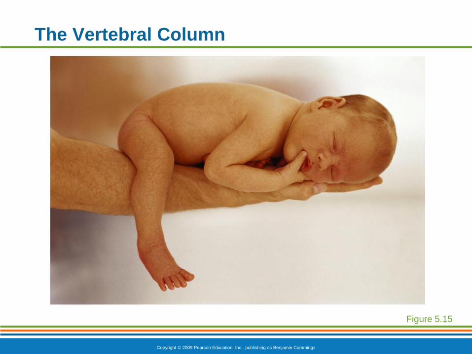
The Vertebral Column
The spine has a normal curvature
Primary curvatures are the spinal curvatures
of the thoracic and sacral regions
Present from birth
Secondary curvatures are the spinal
curvatures of the cervical and lumbar regions
Develop after birth
Copyright © 2009 Pearson Education, Inc., publishing as Benjamin Cummings
The Vertebral Column
Figure 5.15
Copyright © 2009 Pearson Education, Inc., publishing as Benjamin Cummings
The Vertebral Column
Figure 5.16
Copyright © 2009 Pearson Education, Inc., publishing as Benjamin Cummings
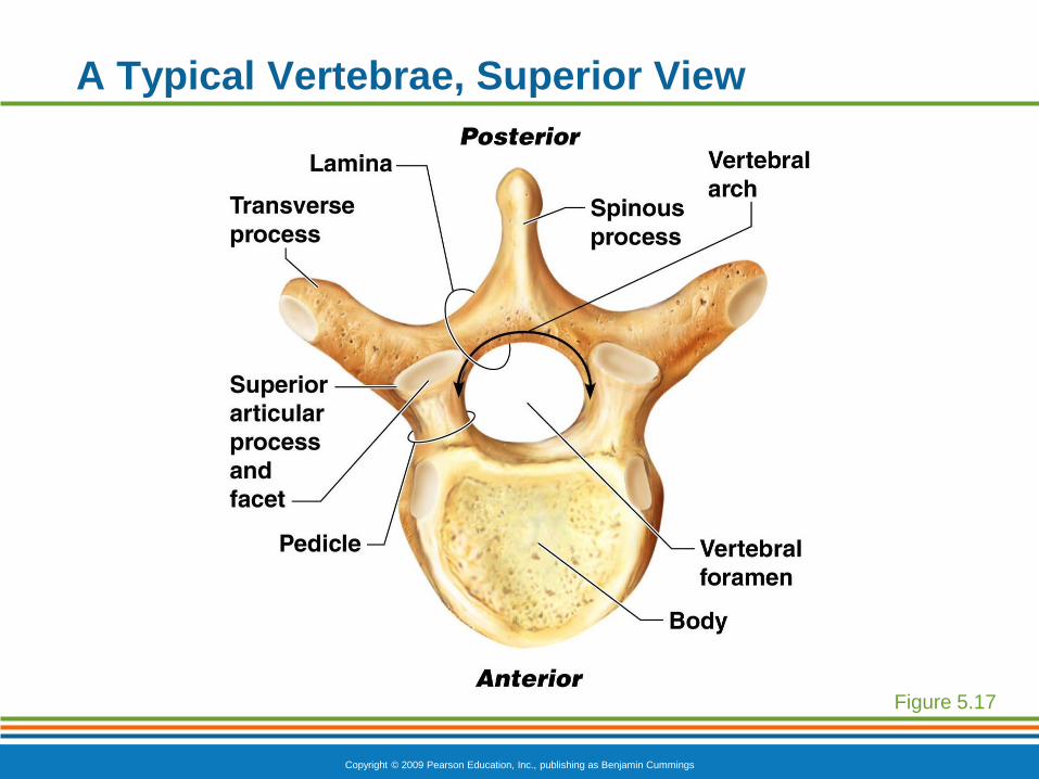
A Typical Vertebrae, Superior View
Figure 5.17
Copyright © 2009 Pearson Education, Inc., publishing as Benjamin Cummings
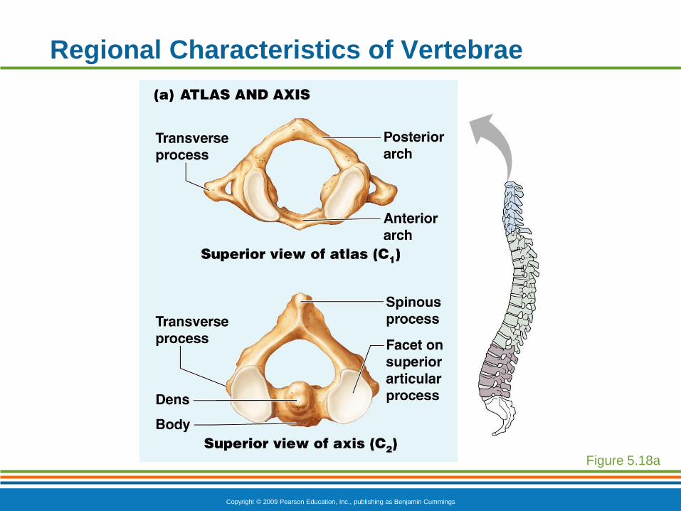
Regional Characteristics of Vertebrae
Figure 5.18a
Copyright © 2009 Pearson Education, Inc., publishing as Benjamin Cummings
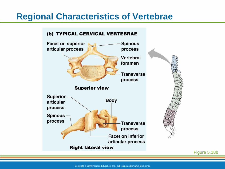
Regional Characteristics of Vertebrae
Figure 5.18b
Copyright © 2009 Pearson Education, Inc., publishing as Benjamin Cummings
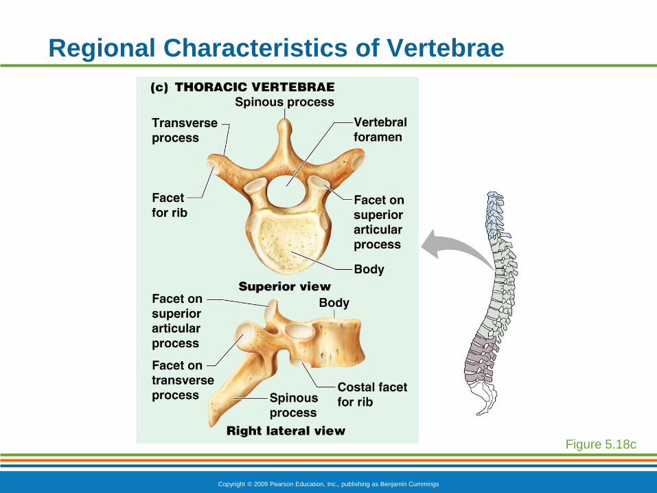
Regional Characteristics of Vertebrae
Figure 5.18c
Copyright © 2009 Pearson Education, Inc., publishing as Benjamin Cummings
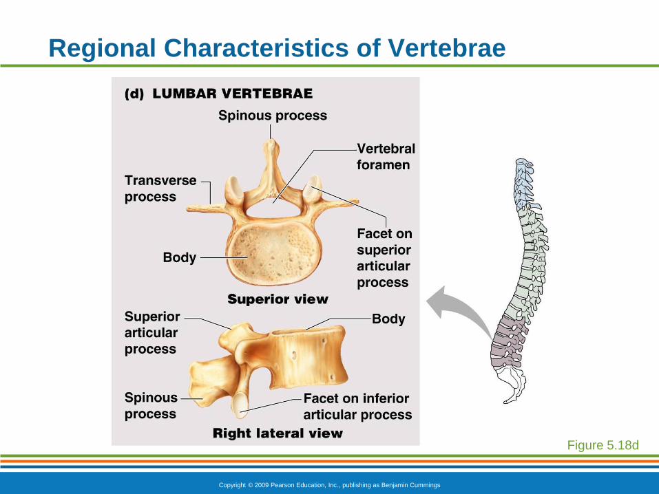
Regional Characteristics of Vertebrae
Figure 5.18d
Copyright © 2009 Pearson Education, Inc., publishing as Benjamin Cummings
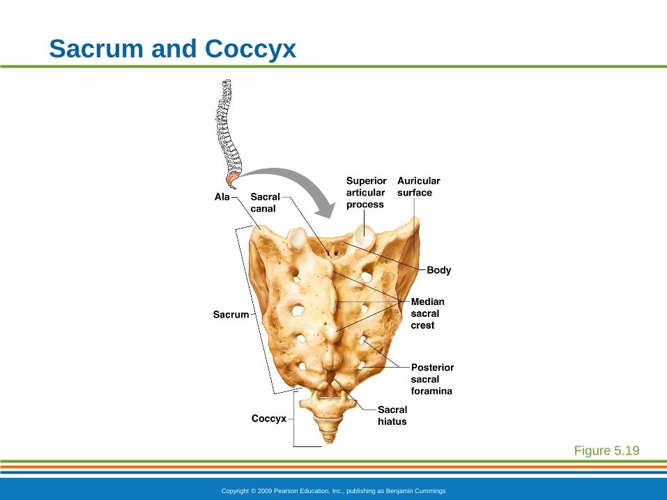
Sacrum and Coccyx
Sacrum
Formed by the fusion of five vertebrae
Coccyx
Formed from the fusion of three to five
vertebrae
“Tailbone,” or remnant of a tail that other
vertebrates have
Copyright © 2009 Pearson Education, Inc., publishing as Benjamin Cummings
Sacrum and Coccyx
Figure 5.19
Copyright © 2009 Pearson Education, Inc., publishing as Benjamin Cummings
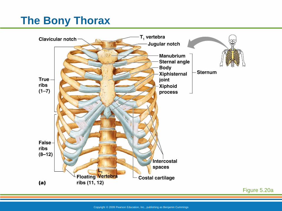
The Bony Thorax
Forms a cage to protect major organs
Consists of three parts
Sternum
Ribs
True ribs (pairs 1–7)
False ribs (pairs 8–12)
Floating ribs (pairs 11–12)
Thoracic vertebrae
Copyright © 2009 Pearson Education, Inc., publishing as Benjamin Cummings
The Bony Thorax
Figure 5.20a

PowerPoint® Lecture Slide Presentation
by Patty Bostwick-Taylor,
Florence-Darlington Technical College
Copyright © 2009 Pearson Education, Inc., publishing as Benjamin Cummings
PART A5
The Skeletal
System
Copyright © 2009 Pearson Education, Inc., publishing as Benjamin Cummings
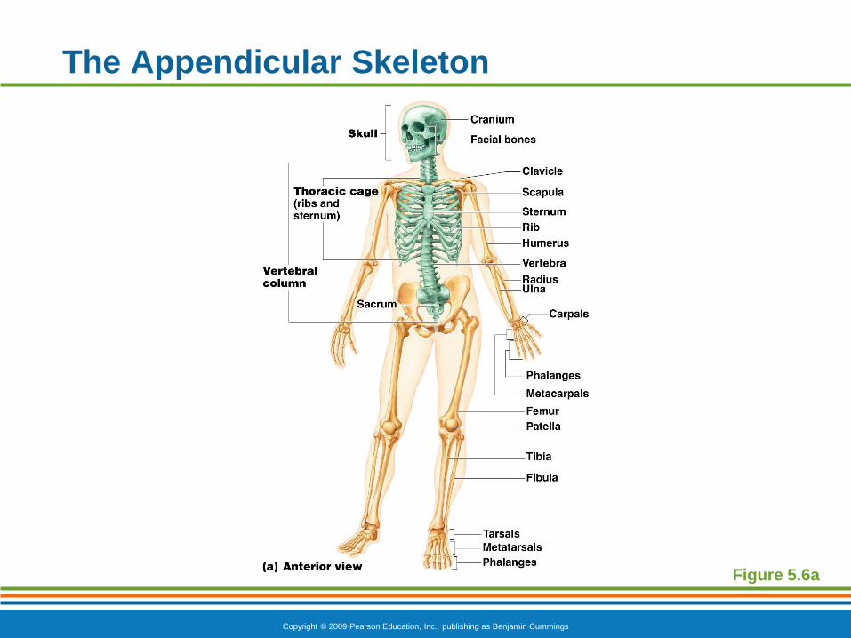
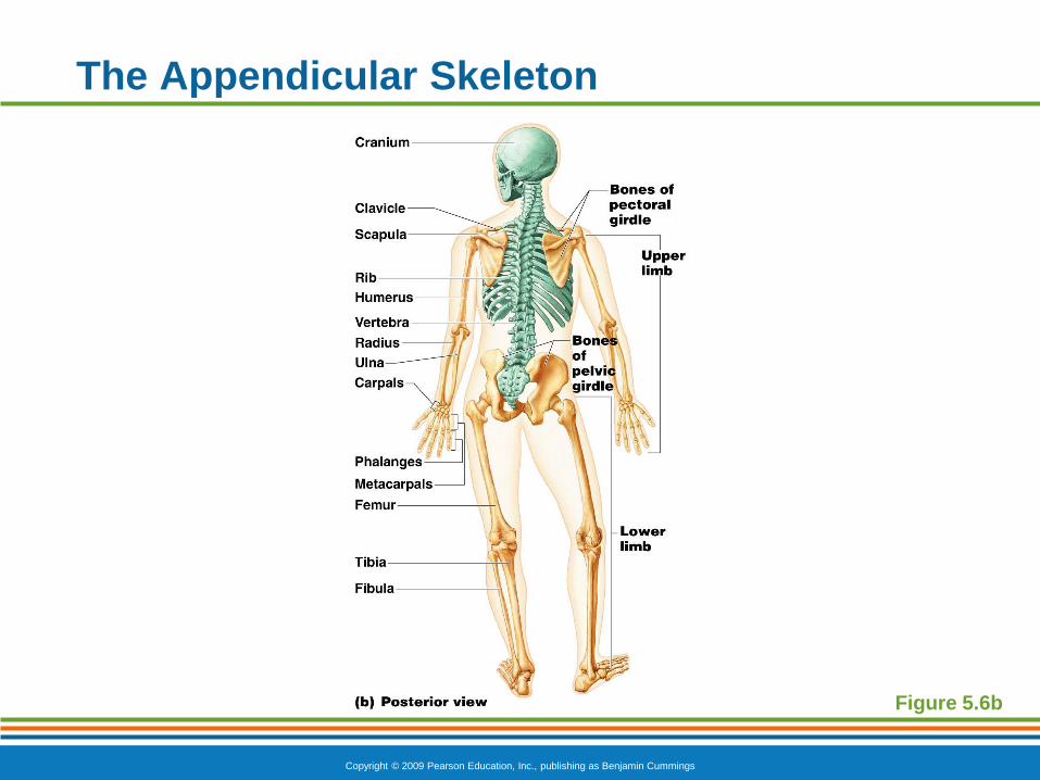
The Appendicular Skeleton
Composed of 126 bones
Limbs (appendages)
Pectoral girdle
Pelvic girdle
Copyright © 2009 Pearson Education, Inc., publishing as Benjamin Cummings
The Appendicular Skeleton
Figure 5.6a
Copyright © 2009 Pearson Education, Inc., publishing as Benjamin Cummings
The Appendicular Skeleton
Figure 5.6b
Copyright © 2009 Pearson Education, Inc., publishing as Benjamin Cummings
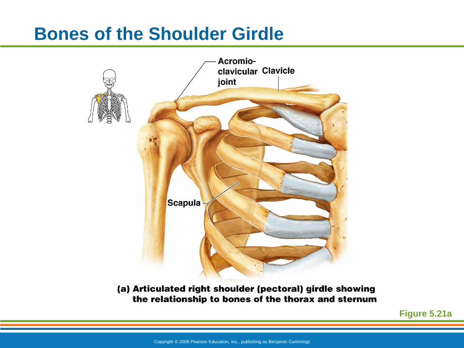
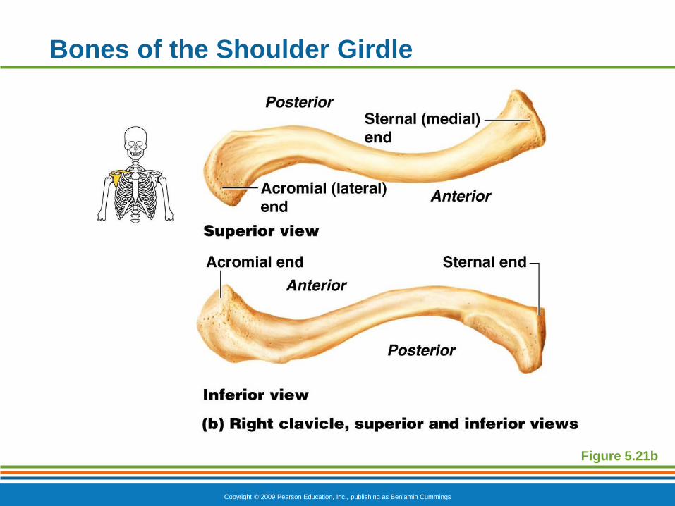
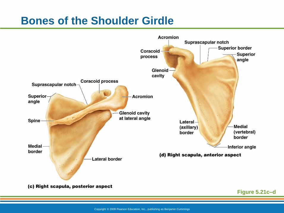
The Pectoral (Shoulder) Girdle
Composed of two bones
Clavicle—collarbone
Scapula—shoulder blade
These bones allow the upper limb to have
exceptionally free movement
Copyright © 2009 Pearson Education, Inc., publishing as Benjamin Cummings
Bones of the Shoulder Girdle
Figure 5.21a
Copyright © 2009 Pearson Education, Inc., publishing as Benjamin Cummings
Bones of the Shoulder Girdle
Figure 5.21b
Copyright © 2009 Pearson Education, Inc., publishing as Benjamin Cummings
Bones of the Shoulder Girdle
Figure 5.21c–d
Copyright © 2009 Pearson Education, Inc., publishing as Benjamin Cummings
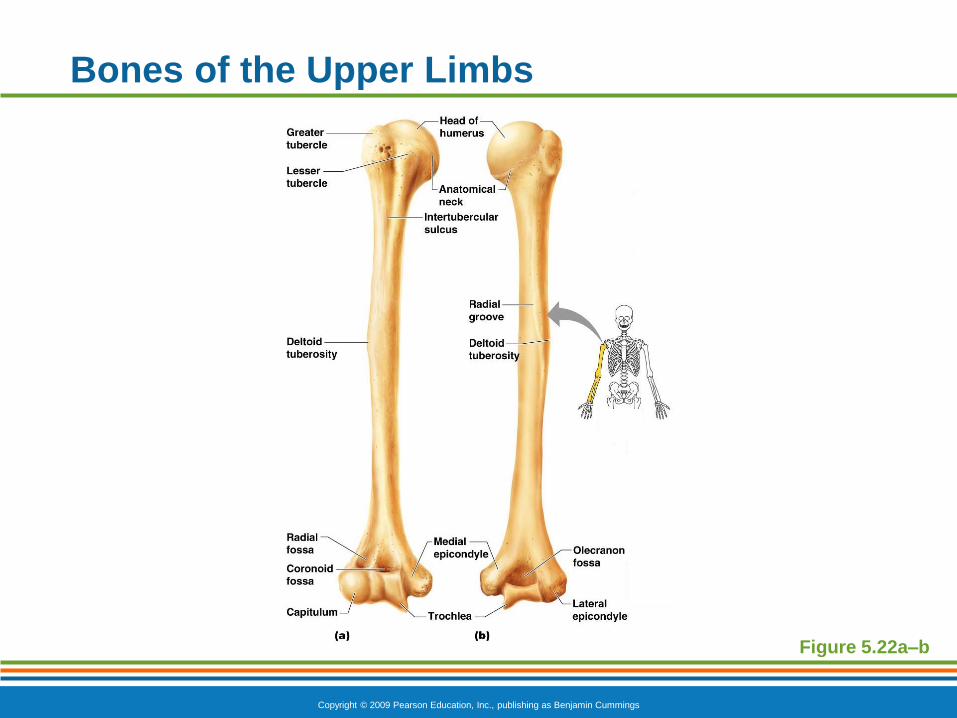
Bones of the Upper Limbs
Humerus
Forms the arm
Single bone
Copyright © 2009 Pearson Education, Inc., publishing as Benjamin Cummings
Bones of the Upper Limbs
Figure 5.22a–b
Copyright © 2009 Pearson Education, Inc., publishing as Benjamin Cummings
Bones of the Upper Limbs
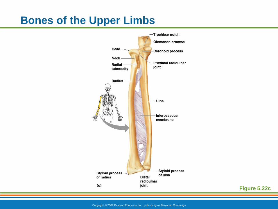
The forearm has two bones
Ulna
Medial bone in anatomical position
Radius
Lateral bone in anatomical position
Copyright © 2009 Pearson Education, Inc., publishing as Benjamin Cummings
Bones of the Upper Limbs
Figure 5.22c
Copyright © 2009 Pearson Education, Inc., publishing as Benjamin Cummings
Bones of the Upper Limbs
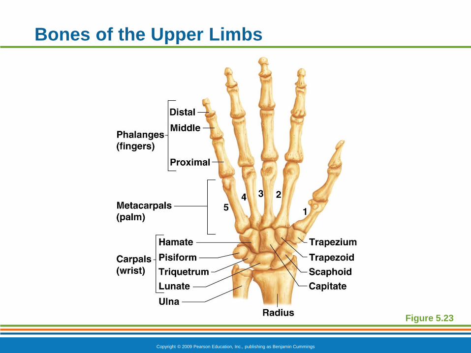
The hand
Carpals—wrist
Metacarpals—palm
Phalanges—fingers
Copyright © 2009 Pearson Education, Inc., publishing as Benjamin Cummings
Bones of the Upper Limbs
Figure 5.23

PowerPoint® Lecture Slide Presentation
by Patty Bostwick-Taylor,
Florence-Darlington Technical College
Copyright © 2009 Pearson Education, Inc., publishing as Benjamin Cummings
PART A5
The Skeletal
System
Copyright © 2009 Pearson Education, Inc., publishing as Benjamin Cummings
Bones of the Pelvic Girdle
Formed by two coxal (ossa coxae) bones
Composed of three pairs of fused bones
Ilium
Ischium
Pubis
Copyright © 2009 Pearson Education, Inc., publishing as Benjamin Cummings
Bones of the Pelvic Girdle
The total weight of the upper body rests on the
pelvis
It protects several organs
Reproductive organs
Urinary bladder
Part of the large intestine
Copyright © 2009 Pearson Education, Inc., publishing as Benjamin Cummings
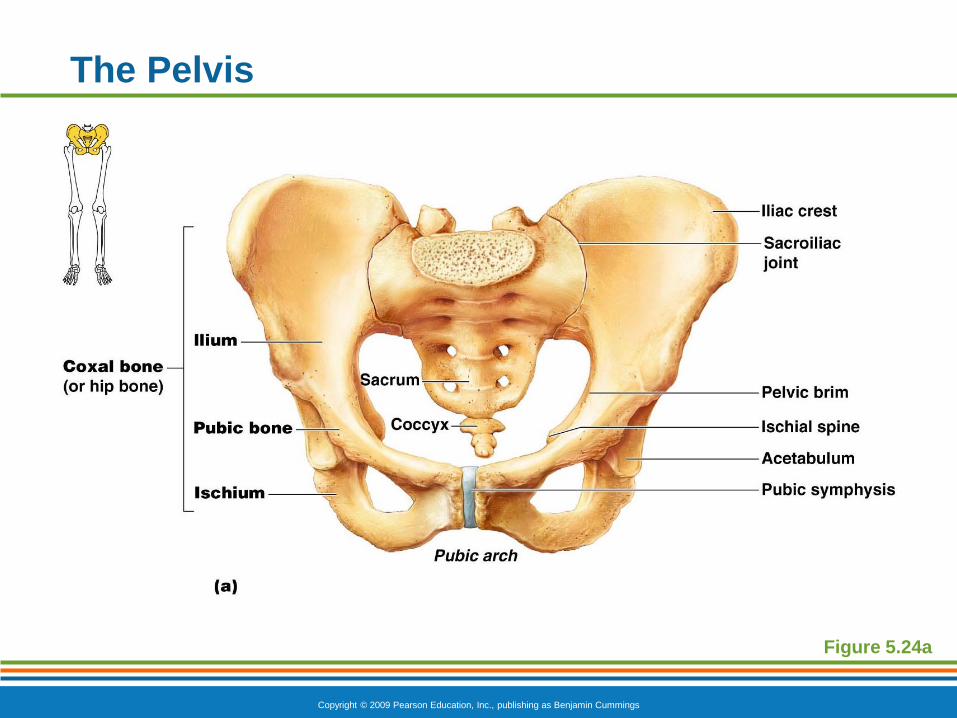
The Pelvis
Figure 5.24a
Copyright © 2009 Pearson Education, Inc., publishing as Benjamin Cummings
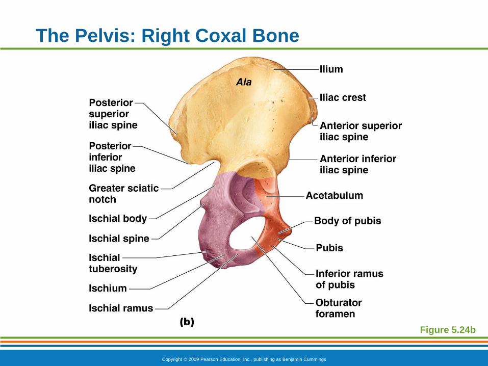
The Pelvis: Right Coxal Bone
Figure 5.24b
Copyright © 2009 Pearson Education, Inc., publishing as Benjamin Cummings
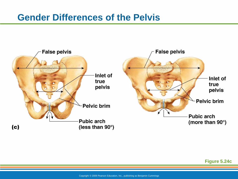
Gender Differences of the Pelvis
The female inlet is larger and more circular
The female pelvis as a whole is shallower, and the
bones are lighter and thinner
The female ilia flare more laterally
The female sacrum is shorter and less curved
The female ischial spines are shorter and farther
apart; thus the outlet is larger
The female pubic arch is more rounded because
the angle of the pubic arch is greater
Copyright © 2009 Pearson Education, Inc., publishing as Benjamin Cummings
Gender Differences of the Pelvis
Figure 5.24c

Copyright © 2009 Pearson Education, Inc., publishing as Benjamin Cummings
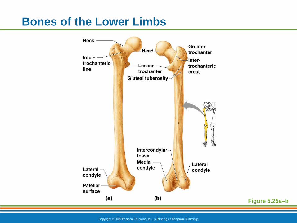
Bones of the Lower Limbs
The thigh has one bone
Femur
The heaviest, strongest bone in the body
Copyright © 2009 Pearson Education, Inc., publishing as Benjamin Cummings
Bones of the Lower Limbs
Figure 5.25a–b
Copyright © 2009 Pearson Education, Inc., publishing as Benjamin Cummings
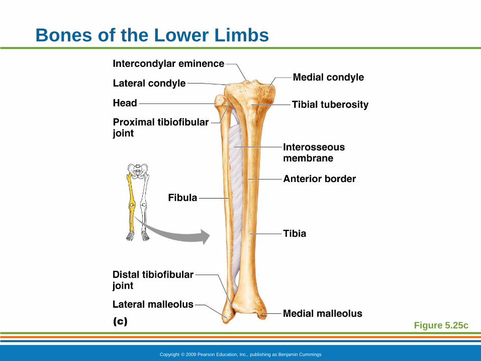
Bones of the Lower Limbs
The lower leg has two bones
Tibia
Shinbone
Larger and medially oriented
Fibula
Thin and sticklike
Copyright © 2009 Pearson Education, Inc., publishing as Benjamin Cummings
Bones of the Lower Limbs
Figure 5.25c
Copyright © 2009 Pearson Education, Inc., publishing as Benjamin Cummings
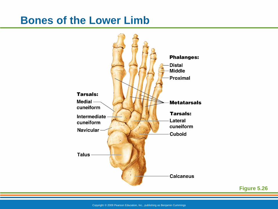
Bones of the Lower Limbs
The foot
Tarsals
Two largest tarsals
Calcaneus (heelbone)
Talus
Metatarsals—sole
Phalanges—toes
Copyright © 2009 Pearson Education, Inc., publishing as Benjamin Cummings
Bones of the Lower Limb
Figure 5.26
Copyright © 2009 Pearson Education, Inc., publishing as Benjamin Cummings
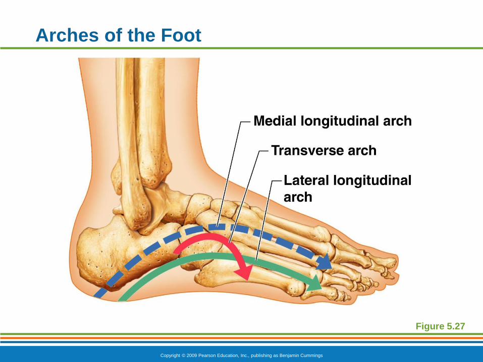
Arches of the Foot
Bones of the foot are arranged to form three
strong arches
Two longitudinal
One transverse
Copyright © 2009 Pearson Education, Inc., publishing as Benjamin Cummings
Arches of the Foot
Figure 5.27
Top Related