


Languages
Pages
Legal


Systemic Inflammatory Response to Smoking in ChronicObstructive Pulmonary Disease: Evidence of a GenderEffectRosa Faner1,2,3., Nuria Gonzalez1., Tamara Cruz1,2, Susana Graciela Kalko4, Alvar Agustı1,2,3,5,6*
1 Fundacio Privada Clınic per a la Recerca Biomedica, Barcelona, Spain, 2 Institut d’investigacions Biomediques August Pi i Sunyer (IDIBAPS), Barcelona, Spain, 3 CIBER
Enfermedades Respiratorias (CIBERES), Barcelona, Spain, 4 Bioinformatics Core Facility, IDIBAPS, Barcelona, Spain, 5 Thorax Institute, Hospital Clinic, University of Barcelona,
Barcelona, Spain, 6 Fundacio de Investigacio Sanitaria Illes Balears (FISIB), Mallorca, Spain
Abstract
Background: Tobacco smoking is the main risk factor of chronic obstructive pulmonary disease (COPD) but not all smokersdevelop the disease. An abnormal pulmonary and systemic inflammatory response to smoking is thought to play a majorpathogenic role in COPD, but this has never been tested directly.
Methods: We studied the systemic biomarker and leukocyte transcriptomic response (Affymetrix microarrays) to smokingexposure in 10 smokers with COPD and 10 smokers with normal spirometry. We also studied 10 healthy never smokers (notexposed to smoking) as controls. Because some aspects of COPD may differ in males and females, and the inflammatoryresponse to other stressors (infection) might be different in man and women, we stratified participant recruitment by sex.Differentially expressed genes were validated by q-PCR. Ontology enrichment was evaluated and interaction networksinferred.
Results: Principal component analysis identified sex differences in the leukocyte transcriptomic response to acute smoking.In both genders, we identified genes that were differentially expressed in response to smoking exclusively in COPD patients(COPD related signature) or smokers with normal spirometry (Smoking related signature), their ontologies and interactionnetworks.
Conclusions: The use of an experimental intervention (smoking exposure) to investigate the transcriptomic response ofperipheral leukocytes in COPD is a step beyond the standard case-control transcriptomic profiling carried out so far, and hasfacilitated the identification of novel COPD and Smoking expression related signatures which differ in males and females.
Citation: Faner R, Gonzalez N, Cruz T, Kalko SG, Agustı A (2014) Systemic Inflammatory Response to Smoking in Chronic Obstructive Pulmonary Disease: Evidenceof a Gender Effect. PLoS ONE 9(5): e97491. doi:10.1371/journal.pone.0097491
Editor: Mehrdad Arjomandi, University of California San Francisco, United States of America
Received January 27, 2014; Accepted April 18, 2014; Published May 15, 2014
Copyright: � 2014 Faner et al. This is an open-access article distributed under the terms of the Creative Commons Attribution License, which permitsunrestricted use, distribution, and reproduction in any medium, provided the original author and source are credited.
Funding: Supported, in part, by Mutua Madrilena PI041966/2012, Fundacio Catalana de Pneumologia (Fucap)-Beca Esteve 2011, Instituto de Salud Carlos III-Fondo de Investigaciones Sanitarias (FIS) 10/00523, SEPAR 133/2011. The funders had no role in study design, data collection and analysis, decision to publish, orpreparation of the manuscript.
Competing Interests: The authors have declared that no competing interests exist.
* E-mail: [email protected]
. These authors contributed equally to this work.
Introduction
Tobacco smoking is the major risk factor for Chronic
Obstructive Pulmonary Disease (COPD) [1]. Yet, only a
proportion of smokers, so called ‘‘susceptible smokers’’, develop
the disease [2]. The genetic and epigenetic background of each
smoker is likely to regulate the type and intensity of his/her
inflammatory response to smoking [1,3–5]. In ‘‘susceptible
smokers’’, this response is thought to be ‘‘enhanced’’, both in the
lungs [6] and in the systemic circulation [7], and is believed to
drive disease progression [1,6]. However, despite the wide
acceptance of this notion [1], no previous study has actually
studied the ‘‘response’’ to smoking (i.e., the specific inflammatory
changes that occur before and after smoking) in susceptible
smokers (i.e., patients with COPD) and resistant smokers (i.e.,
smokers with normal spirometry). Rather, available evidence
compares a number of inflammatory markers in these two groups
of smokers ‘‘after’’ many years of smoking exposure [6].
To address this gap in knowledge, we compared a number of
systemic inflammatory biomarkers and the transcriptome of
circulating leukocytes, before and after smoking in susceptible
(COPD patients) and resistant smokers. We hypothesized that
smoking exposure will induce a different inflammatory signature,
at the cellular, protein and/or transcriptome levels, in these two
groups of smokers. Importantly, because several previous reports
suggest that there may be significant gender differences in the
natural history of COPD [8–10] and some experimental
observations show that the leukocyte transcriptional response to
other acute stressors (infection) is different in males and females
[11], we recruited participants stratified by sex.
PLOS ONE | www.plosone.org 1 May 2014 | Volume 9 | Issue 5 | e97491
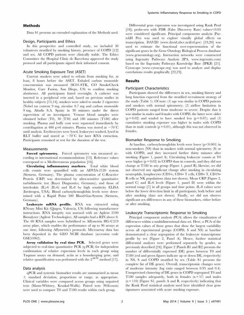
Methods
Data S1 presents an extended explanation of the Methods used.
Design, Participants and EthicsIn this prospective and controlled study, we included 30
volunteers stratified by smoking history, presence of COPD [12]
and sex. All COPD patients were clinically stable. The Ethics
Committee the Hospital Clinic de Barcelona approved the study
protocol and all participants signed their informed consent.
Acute Smoking Exposure Test (ASET)Current smokers were asked to refrain from smoking for, at
least, 8 hours before the ASET. Exhaled carbon monoxide
concentration was measured (SC01-STK, CO SmokeCheck
Monitor, Care Fusion, San Diego, US) to confirm smoking
abstinence. All participants fasted overnight. A catheter was
inserted in a peripheral vein and, based on previous studies in
healthy subjects [13,14], smokers were asked to smoke 2 cigarettes
(Nobel tar content 9 mg, nicotine 0.7 mg and carbon monoxide
9 mg, Altadis S.A. Spain) in 30 minutes under the direct
supervision of an investigator. Venous blood samples were
obtained before (T0), 30 (T30) and 180 minutes (T180) after
smoking. Plasma and buffy coat were separated immediately by
centrifugation at 400 g66 minutes. Plasma was stored at 270uCuntil analysis. Erythrocytes were lysed, leukocytes washed, lysed in
RLT buffer and stored at 270uC for later RNA extraction.
Participants remained at rest for the duration of the test.
MeasurementsForced spirometry. Forced spirometry was measured ac-
cording to international recommendations [15]. Reference values
correspond to a Mediterranean population [16].
Circulating inflammatory markers. Total white blood
cells counts were quantified with an ADVIA-2120 system
(Siemens, Germany). The plasma concentration of C-Reactive
Protein (CRP) was determined by ultra-sensitive quantitative
turbidimetric test (Bayer Diagnostics, Germany), and those of
interleukin (IL)-6 (IL-6) and IL-8 by high sensitivity ELISA
(Invitrogen, USA). Blood carboxyhemoglobin levels were deter-
mined with a Rapid Point 500 Blood-Gas-System (Siemens,
Germany).
Leukocyte mRNA profile. RNA was extracted using
RNeasy Mini Kit (Qiagen, Valencia, US) following manufacturer
instructions. RNA integrity was assessed with an Agilent 2100
Bionalyzer (Agilent Technologies). All samples had a RIN above 8.
The 60 RNA samples were hybridized to Affymetrix HG-U219
array plate, which enables the performance of up to 96 arrays at
one time, following Affymetrix’s protocols. Microarray data has
been deposited in the GEO NCBI database (accession code
GSE55962).
Array validation by real time PCR. Selected genes were
subjected to real-time quantitative PCR (q-PCR) for independent
confirmation of relative expression levels in each group using
Taqman assays on demand, actin as a housekeeping gene, and
relative quantification was performed with the 2(DCP) method [17].
Data analysisqPCR and systemic biomarker results are summarized as mean
6 standard deviation, proportions or range, as appropriate.
Clinical variables were compared across groups using unpaired
tests (Mann-Whitney, Kruskal-Wallis). Paired tests (Wilcoxon)
were used to compare T0 and T180 results within each group.
Differential gene expression was investigated using Rank Prod
[18]; probe-sets with FDR (False Discovery Rate) values,0.05
were considered significant. Principal components analysis (Par-
tekH Pro) was used to explore visually global effects on
transcription. DAVID (www.david.abcc.ncifcrf.gov) [19,20] was
used to estimate the functional over-representation of the
significant genes in the Gene Ontology Biological Process database
(www.geneontology.org). Interaction networks were constructed
using Ingenuity Pathways Analysis (IPA, www.ingenuity.com)
based on the Ingenuity Pathways Knowledge Base (IPKB) [21].
Cytoscape (www.cytoscape.org) was used to analyze and display
correlations results graphically [22,23].
Results
Participant CharacteristicsParticipants showed the differences in sex, smoking history and
lung function expected from the stratified recruitment strategy of
the study (Table 1). Of note: (1) age was similar in COPD patients
and smokers with normal spirometry; (2) airflow limitation in
COPD patients ranged from moderate to severe. Despite that it
was similar in males and females with COPD, the latter were older
(p = 0.02) and tended to have smoked less (p = 0.07); and (3)
cumulative smoking exposure was higher in males with COPD
than in male controls (p = 0.01), although this was not observed in
females.
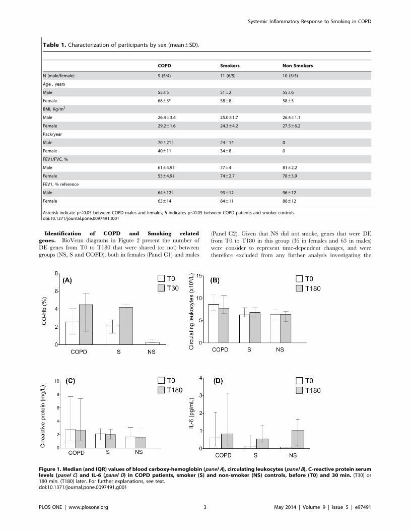
Biomarker Response to SmokingAt baseline, carboxyhemoglobin levels were lower (p,0.001) in
non-smokers (NS) than in smokers with normal spirometry (S) or
with COPD, and they increased further thirty minutes after
smoking (Figure 1, panel A). Circulating leukocyte counts at T0
were higher (p = 0.02) in COPD than in controls, and they did not
change at T180 in any group (Figure 1, panel B); likewise, we did
not observed any significant change after smoking in circulating
neutrophils, lymphocytes (CD3+), CD4+ T cells, CD8+ T, CD19+B cells or NK populations (data not shown). Mean CRP (Figure 2,
panel C) and IL-6 levels (Figure 1, panel D) were within the
normal range [7] in all groups and time points. IL-8 values were
below the lower detection limit in all participants, both before and
after smoking (data not shown). Finally, we did not observe
significant sex differences in any of these biomarkers, either before
or after smoking.
Leukocyte Transcriptomic Response to SmokingPrincipal component analysis (PCA) allows the visualization of
differences within a multidimensional data set. Here, a PCA of the
expression values of those genes that show the largest variability
across all experimental groups (COPD, S and NS) at baseline
demonstrated a clear segregation of the leukocyte transcriptome
profile by sex (Figure 2, Panel A). Hence, further statistical
differential analyses were performed separately by gender, as
previously described [24]. Figure 2 (Panels B1 and B2) presents the
number of differentially expressed (DE) genes between T0 and
T180 (red and green figures indicate up or down DE, respectively)
in NS, S and COPD stratified by sex (Table S1 presents the
complete list of DE genes). Overall, transcriptomic changes were
of moderate intensity (log ratio ranged between 0.95 and 0.4).
Unsupervised clustering of DE genes in COPD segregated T0 and
T180 samples adequately, both in females (n = 57) and males
(n = 110) (Figure S1, panels A and B, respectively) indicating that
the Rank Prod statistical analysis used here identified clear gene
signatures associated with acute smoking exposure.
Systemic Inflammatory Response to Smoking in COPD
PLOS ONE | www.plosone.org 2 May 2014 | Volume 9 | Issue 5 | e97491
Identification of COPD and Smoking related
genes. BioVenn diagrams in Figure 2 present the number of
DE genes from T0 to T180 that were shared (or not) between
groups (NS, S and COPD), both in females (Panel C1) and males
(Panel C2). Given that NS did not smoke, genes that were DE
from T0 to T180 in this group (36 in females and 63 in males)
were consider to represent time-dependent changes, and were
therefore excluded from any further analysis investigating the
Table 1. Characterization of participants by sex (mean6SD).
COPD Smokers Non Smokers
N (male/female) 9 (5/4) 11 (6/5) 10 (5/5)
Age , years
Male 5565 5162 5566
Female 6863* 5868 5865
BMI, Kg/m2
Male 26.463.4 25.061.7 26.461.1
Female 29.261.6 24.364.2 27.566.2
Pack/year
Male 706211 24614 0
Female 40611 3468 0
FEV1/FVC, %
Male 6164.91 7764 8162.2
Female 5364.91 7462.7 7863.9
FEV1, % reference
Male 646121 93612 96612
Female 63614 84611 88612
Asterisk indicate p,0.05 between COPD males and females, 1 indicates p,0.05 between COPD patients and smoker controls.doi:10.1371/journal.pone.0097491.t001
Figure 1. Median (and IQR) values of blood carboxy-hemoglobin (panel A), circulating leukocytes (panel B), C-reactive protein serumlevels (panel C) and IL-6 (panel D) in COPD patients, smoker (S) and non-smoker (NS) controls, before (T0) and 30 min. (T30) or180 min. (T180) later. For further explanations, see text.doi:10.1371/journal.pone.0097491.g001
Systemic Inflammatory Response to Smoking in COPD
PLOS ONE | www.plosone.org 3 May 2014 | Volume 9 | Issue 5 | e97491

response to smoking in S or COPD. Likewise, we operationally
considered that those genes that were DE in response to smoking
only in COPD patients represent COPD related genes, whereas
those genes DE in response to smoking only in smokers with
normal spirometry represent Smoking related genes. Accordingly, we
identified 29 COPD related genes in females (78 in males), and 61
Smoking genes in females (16 in males) (Figure 2, Panels C1 and
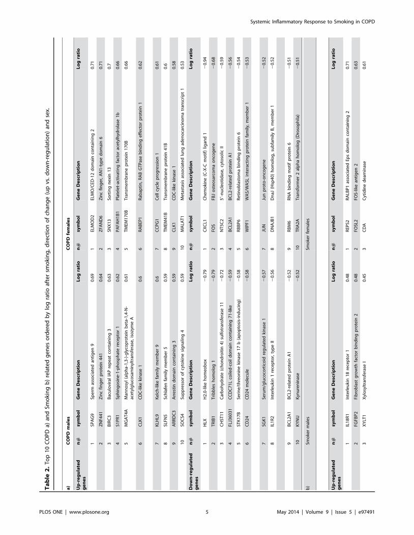
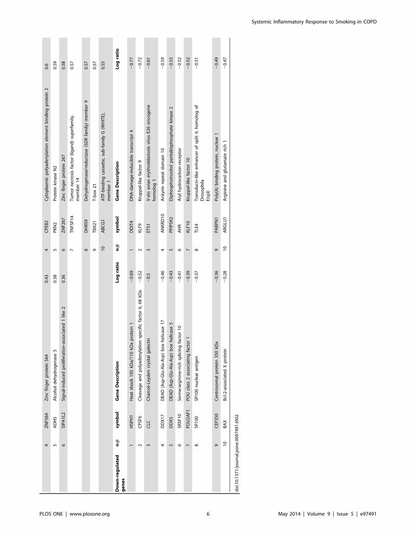
C2, respectively). Table 2 lists the top 10 susceptibility and
resistance genes ordered by log ratio after smoking, direction of
change and sex.
Additionally, we investigated if baseline differences in gene
expression between groups (S vs. COPD) and/or sex (male vs.
female) influence the transcriptional response to smoking. Results
are explained in detail in the data S1 (and Tables S2 and S3) but,
by and large, DE at baseline was only marginally related to the
transcriptomic response to smoking.
COPD and Smoking related genes shared by males and
females. We identified five COPD related genes shared by
males and females: (1) WIPF1 (mean log ratio = 20.479), an
important regulator of the immunological synapsis formation and
T cell activation[25]; (2) BCL2A1 (mean log ratio = 20.538), that
regulates p53tumor suppression induced apoptosis [26]; (3) SGK1
(mean log ratio = 20.512), whose up regulation is involved in
hypertension, obesity, diabetes, thrombosis, stroke, fibrosing
disease, infertility and tumor growth, and its inhibition mediates
skeletal muscle homeostasis and function [27]; (4) ZNF397 (mean
log ratio = 0.551), thought to have a role in centromere formation
and gene transcription [28]; and, finally, (5) CLK1 (mean log
ratio = 0.589), an alternative splicing factor induced by hypoxia
[29].
Similarly, we identified two Smoking related genes shared by
males and females: POU2AF1 (log ratio = 20.40), a nuclear factor
responsible of immunoglobulin transcription regulation [30], and
XYLT1 (log ratio = 0.45), the initial enzyme responsible of GAG
biosynthesis that has been associated with tissue remodeling [31].
Gene Ontologies Associated with COPD and Smoking
related genes. We used DAVID [19,20] to identify enriched
gene ontologies (% FDR,25) associated with the COPD
susceptibility and resistance genes identified above. In females,
ontologies enriched in COPD related genes (n = 29) included genes
Figure 2. Panel A: Principal component analysis (PCA) of those genes with the largest variability of expression values across the experimental groups(COPD patients, smokers (S) and non-smokers (NS)) at baseline (T0). Because the sex effect observed, further analysis was stratified by sex (right andleft columns.) Panels B1 (females) and B2 (males) present the number of differentially expressed (DE) genes from T0 to T180 in the three groups ofparticipants studied. Red and green figures indicate up and down-regulation, respectively. Panels C1 (females) and C2 (males) show a BioVenndiagram (www.cmbi.ru.nl/cdd/biovenn) with the number of DE genes shared between COPD, S and NS controls. For further explanations, see text.doi:10.1371/journal.pone.0097491.g002
Systemic Inflammatory Response to Smoking in COPD
PLOS ONE | www.plosone.org 4 May 2014 | Volume 9 | Issue 5 | e97491
Ta
ble
2.
To
p1
0C
OP
Da)
and
Smo
kin
gb
)re
late
dg
en
es
ord
ere
db
ylo
gra
tio
afte
rsm
oki
ng
,d
ire
ctio
no
fch
ang
e(u
pvs
.d
ow
n-r
eg
ula
tio
n)
and
sex.
a)
CO
PD
ma
les
CO
PD
fem
ale
s
Up
-re
gu
late
dg
en
es
n#
sym
bo
lG
en
eD
esc
rip
tio
nL
og
rati
on
#sy
mb
ol
Ge
ne
De
scri
pti
on
Lo
gra
tio
1SP
AG
9Sp
erm
asso
ciat
ed
anti
ge
n9
0.6
91
ELM
OD
2EL
MO
/CED
-12
do
mai
nco
nta
inin
g2
0.7
1
2Z
NF4
41
Zin
cfi
ng
er
pro
tein
44
10
.64
2Z
FAN
D6
Zin
cfi
ng
er,
AN
1-t
ype
do
mai
n6
0.7
1
3B
IRC
3B
acu
lovi
ral
IAP
rep
eat
con
tain
ing
30
.63
3SN
X1
3So
rtin
gn
exi
n1
30
.7
4S1
PR
1Sp
hin
go
sin
e-1
-ph
osp
hat
ere
cep
tor
10
.62
4P
AFA
H1
B1
Pla
tele
t-ac
tiva
tin
gfa
cto
rac
ety
lhyd
rola
se1
b0
.66
5M
GA
T4
AM
ann
osy
l(a
lph
a-1
,3-)
-gly
cop
rote
inb
eta
-1,4
-N-
ace
tylg
luco
sam
inyl
tran
sfe
rase
,is
ozy
me
A0
.61
5T
MEM
17
0B
Tra
nsm
em
bra
ne
pro
tein
17
0B
0.6
6
6C
LK1
CD
C-l
ike
kin
ase
10
.66
RA
BEP
1R
abap
tin
,R
AB
GT
Pas
eb
ind
ing
eff
ect
or
pro
tein
10
.62
7K
LHL9
Ke
lch
-lik
efa
mily
me
mb
er
90
.67
CC
PG
1C
ell
cycl
ep
rog
ress
ion
10
.61
8SL
FN5
Sch
lafe
nfa
mily
me
mb
er
50
.59
8T
MEM
41
BT
ran
sme
mb
ran
ep
rote
in4
1B
0.6
9A
RR
DC
3A
rre
stin
do
mai
nco
nta
inin
g3
0.5
99
CLK
1C
DC
-lik
eki
nas
e1
0.5
8
10
SOC
S4Su
pp
ress
or
of
cyto
kin
esi
gn
alin
g4
0.5
91
0M
ALA
T1
Me
tast
asis
asso
ciat
ed
lun
gad
en
oca
rcin
om
atr
ansc
rip
t1
0.5
3
Do
wn
-re
gu
late
dg
en
es
n#
sym
bo
lG
en
eD
esc
rip
tio
nL
og
rati
on
#sy
mb
ol
Ge
ne
De
scri
pti
on
Lo
gra
tio
1H
LXH
2.0
-lik
eh
om
eo
bo
x2
0.7
91
CX
CL1
Ch
em
oki
ne
(C-X
-Cm
oti
f)lig
and
12
0.9
4
2T
RIB
1T
rib
ble
sh
om
olo
g1
20
.79
2FO
SFB
Jo
ste
osa
rco
ma
on
cog
en
e2
0.6
8
3C
HST
11
Car
bo
hyd
rate
(ch
on
dro
itin
4)
sulf
otr
ansf
era
se1
12
0.7
23
NT
5C
25
9-n
ucl
eo
tid
ase
,cy
toso
licII
20
.59
4FL
J36
03
1C
CD
C7
1L
coile
d-c
oil
do
mai
nco
nta
inin
g7
1-l
ike
20
.59
4B
CL2
A1
BC
L2-r
ela
ted
pro
tein
A1
20
.56
5ST
K1
7B
Seri
ne
/th
reo
nin
eki
nas
e1
7b
(ap
op
tosi
s-in
du
cin
g)
20
.58
5R
BB
P6
Re
tin
ob
last
om
ab
ind
ing
pro
tein
62
0.5
4
6C
D2
4C
D2
4m
ole
cule
20
.58
6W
IPF1
WA
S/W
ASL
inte
ract
ing
pro
tein
fam
ily,
me
mb
er
12
0.5
3
7SG
K1
Seru
m/g
luco
cort
ico
idre
gu
late
dki
nas
e1
20
.57
7JU
NJu
np
roto
-on
cog
en
e2
0.5
2
8IL
1R
2In
terl
eu
kin
1re
cep
tor,
typ
eII
20
.56
8D
NA
JB1
Dn
aJ(H
sp4
0)
ho
mo
log
,su
bfa
mily
B,
me
mb
er
12
0.5
2
9B
CL2
A1
BC
L2-r
ela
ted
pro
tein
A1
20
.52
9R
BM
6R
NA
bin
din
gm
oti
fp
rote
in6
20
.51
10
KY
NU
Kyn
ure
nin
ase
20
.52
10
TR
A2
AT
ran
sfo
rme
r2
alp
ha
ho
mo
log
(Dro
sop
hila
)2
0.5
1
b)
Smo
ker
mal
es
Smo
ker
fem
ale
s
Up
-re
gu
late
dg
en
es
n#
sym
bo
lG
en
eD
esc
rip
tio
nL
og
rati
on
#sy
mb
ol
Ge
ne
De
scri
pti
on
Lo
gra
tio
1IL
18
R1
Inte
rle
uki
n1
8re
cep
tor
10
.48
1R
EPS2
RA
LBP
1as
soci
ate
dEp
sd
om
ain
con
tain
ing
20
.71
2FG
FBP
2Fi
bro
bla
stg
row
thfa
cto
rb
ind
ing
pro
tein
20
.48
2FO
SL2
FOS-
like
anti
ge
n2
0.6
3
3X
YLT
1X
ylo
sylt
ran
sfe
rase
I0
.45
3C
DA
Cyt
idin
ed
eam
inas
e0
.61
Systemic Inflammatory Response to Smoking in COPD
PLOS ONE | www.plosone.org 5 May 2014 | Volume 9 | Issue 5 | e97491
4Z
NF5
64
Zin
cfi
ng
er
pro
tein
56
40
.43
4C
PEB
2C
yto
pla
smic
po
lyad
en
ylat
ion
ele
me
nt
bin
din
gp
rote
in2
0.6
5A
DH
5A
lco
ho
ld
eh
ydro
ge
nas
e5
0.3
85
PK
N2
Pro
tein
kin
ase
N2
0.5
9
6SI
PA
1L2
Sig
nal
-in
du
ced
pro
life
rati
on
-ass
oci
ate
d1
like
20
.36
6Z
NF2
67
Zin
cfi
ng
er
pro
tein
26
70
.58
7T
NFS
F14
Tu
mo
rn
ecr
osi
sfa
cto
r(l
igan
d)
sup
erf
amily
,m
em
be
r1
40
.57
8D
HR
S9D
eh
ydro
ge
nas
e/r
ed
uct
ase
(SD
Rfa
mily
)m
em
be
r9
0.5
7
9T
BX
21
T-b
ox
21
0.5
7
10
AB
CG
1A
TP
-bin
din
gca
sse
tte
,su
b-f
amily
G(W
HIT
E),
me
mb
er
10
.55
Do
wn
-re
gu
late
dg
en
es
n#
sym
bo
lG
en
eD
esc
rip
tio
nL
og
rati
on
#sy
mb
ol
Ge
ne
De
scri
pti
on
Lo
gra
tio
1H
SPH
1H
eat
sho
ck1
05
kDa/
11
0kD
ap
rote
in1
20
.69
1D
DIT
4D
NA
-dam
age
-in
du
cib
letr
ansc
rip
t4
20
.77
2C
PSF
6C
leav
age
and
po
lyad
en
ylat
ion
spe
cifi
cfa
cto
r6
,6
8kD
a2
0.5
22
KLF
9K
rup
pe
l-lik
efa
cto
r9
20
.72
3C
LCC
har
cot-
Leyd
en
crys
tal
gal
ect
in2
0.5
3ET
S1V
-ets
avia
ne
ryth
rob
last
osi
svi
rus
E26
on
cog
en
eh
om
olo
g1
20
.61
4D
DX
17
DEA
D(A
sp-G
lu-A
la-A
sp)
bo
xh
elic
ase
17
20
.46
4A
NK
RD
10
An
kyri
nre
pe
atd
om
ain
10
20
.59
5D
DX
5D
EAD
(Asp
-Glu
-Ala
-Asp
)b
ox
he
licas
e5
20
.43
5P
PIP
5K
2D
iph
osp
ho
ino
sito
lp
en
taki
sph
osp
hat
eki
nas
e2
20
.53
6SR
SF1
0Se
rin
e/a
rgin
ine
-ric
hsp
licin
gfa
cto
r1
02
0.4
16
AH
RA
ryl
hyd
roca
rbo
nre
cep
tor
20
.52
7P
OU
2A
F1P
OU
clas
s2
asso
ciat
ing
fact
or
12
0.3
97
KLF
10
Kru
pp
el-
like
fact
or
10
20
.52
8SP
10
0SP
10
0n
ucl
ear
anti
ge
n2
0.3
78
TLE
4T
ran
sdu
cin
-lik
ee
nh
ance
ro
fsp
lit4
,h
om
olo
go
fD
roso
ph
ilaE(
spl)
20
.51
9C
EP3
50
Ce
ntr
oso
mal
pro
tein
35
0kD
a2
0.3
69
PA
BP
N1
Po
ly(A
)b
ind
ing
pro
tein
,n
ucl
ear
12
0.4
9
10
BA
XB
cl-2
-ass
oci
ate
dX
pro
tein
20
.28
10
AR
GLU
1A
rgin
ine
and
glu
tam
ate
rich
12
0.4
7
do
i:10
.13
71
/jo
urn
al.p
on
e.0
09
74
91
.t0
02
Systemic Inflammatory Response to Smoking in COPD
PLOS ONE | www.plosone.org 6 May 2014 | Volume 9 | Issue 5 | e97491

Table 4. Gene ontology enrichment analysis, by sex. Females ontologies/genes associated with Smoking.
Term Genes % FDR p value
GO:0006955 immune response CYBB, POU2AF1, IL18RAP, CST7, ETS1, BCL2, LILRB4, TGFBR3, TNFSF14, CLEC4D 0.7 4.50E-04
GO:0043065 positive regulation of apoptosis ADRB2, ETS1, MMP9, KLF10, BCL2, TNFSF14, FADD, RRM2B 0.9 5.70E-04
GO:0043068 positive regulation of programed cell death ADRB2, ETS1, MMP9, KLF10, BCL2, TNFSF14, FADD, RRM2B 0.9 6.00E-04
GO:0010942 positive regulation of cell death ADRB2, ETS1, MMP9, KLF10, BCL2, TNFSF14, FADD, RRM2B 0.9 6.10E-04
GO:0051270 regulation of cell motion ETS1, IL6ST, MMP9, BCL2, TGFBR3 6 4.00E-03
GO:0051272 positive regulation of cell motion ETS1, IL6ST, MMP9, BCL2 7 4.00E-03
GO:0042127 regulation of cell proliferation ADRB2, FOSL2, ETS1, IL-6ST, KLF10, BCL2, CHST11, TGFBR3, ING1 7 5.00E-03
GO:0042981 regulation of apoptosis ADRB2, ETS1, MMP9, KLF10, BCL2, CHST11, TNFSF14, FADD, RRM2B 8 5.00E-03
GO:0043067 regulation of programmed cell death ADRB2, ETS1, MMP9, KLF10, BCL2, CHST11, TNFSF14, FADD, RRM2B 8 6.00E-03
GO:0010941 regulation of cell death ADRB2, ETS1, MMP9, KLF10, BCL2, CHST11, TNFSF14, FADD, RRM2B 9 6.00E-03
GO:0045926 negative regulation of growth ADRB2, BCL2, CDA, ING1 9 6.00E-03
GO:0005976 polysaccharide metabolic process XYLT1, IL6ST, CHST11, MGAM 10 6.00E-03
GO:0048643 positive regulation of skeletal muscle tissuedevelopment
ADRB2, BCL2 15 0.01
GO:0045844 positive regulation of striated muscledevelopment
ADRB2, BCL2 19 0.01
GO:0048636 positive regulation of muscle development ADRB2, BCL2 19 0.01
GO:0051094 positive regulation of developmental process ADRB2, ETS1, IL-6ST, KLF10, BCL2 21 0.01
Bold italic text indicates up regulated genes and normal text indicates down regulated genes in response to smoking.doi:10.1371/journal.pone.0097491.t004
Table 5. Gene ontology enrichment analysis, by sex. Males ontologies/genes associated with COPD.
Term Genes % FDR p value
GO:0006955 immune response GPR183, IL1R2, SH2D1A, KYNU, CLEC4E, THEMIS, SNCA, EOMES, TGFBR3, CLEC4D, CD24 1 9.00E-04
GO:0040012 regulation of locomotion SPAG9, S1PR1, SNCA, TGFBR3, CBLL1, TRIB1 3 1.71E-03
GO:0030334 regulation of cell migration SPAG9, S1PR1, TGTBR3, CBLL1, TRIB1 10 7.06E-03
GO:0051270 regulation of cell motion SPAG9, S1PR1, TGTBR3, CBLL1, TRIB1 16 1.00E-02
GO:0030098 lymphocyte differentiation GRP183, THEMIS, EOMES, CD24 16 1.00E-02
GO:0001824 blastocyst development HOPX, EOMES, TGFBR3 23 2.00E-02
Bold italic text indicates up regulated genes and normal text indicates down regulated genes in response to smoking. Analysis could not be done for ontologies/genesassociated with COPD resistance in males due to the small number of DE genes (n = 16) identified in smokers with normal spirometry.doi:10.1371/journal.pone.0097491.t005
Table 3. Gene ontology enrichment analysis, by sex. Females ontologies/genes associated with COPD.
Term Genes % FDR p value
GO:0009991 response to extracellular stimulus FOS, MLL5, JUN, KLF4 7 5.20E-03
GO:0045893 positive regulation of transcription, DNA-dependent FOS, MLL5, JUN, RBM4, KLF4 9 6.74E-03
GO:0051254 positive regulation of RNA metabolic process FOS, MLL5, JUN, RBM4, KLF4 9 6.94E-03
GO:0060395 SMAD protein signal transduction FOS, JUN 12 0.01
GO:0045941 positive regulation of transcription FOS, MLL5, JUN, RBM4, KLF4 15 0.01
GO:0010628 positive regulation of gene expression FOS, MLL5, JUN, RBM4, KLF4 17 0.01
GO:0007611 learning or memory FOS, JUN, PAFAH1B1 18 0.01
GO:0045935 positive regulation of nucleobase, nucleoside, nucleotide and nucleic acid metabolic process FOS, MLL5, JUN, RBM4, KLF4 21 0.02
GO:0051173 positive regulation of nitrogen compound metabolic process FOS, MLL5, JUN, RBM4, KLF4 23 0.02
GO:0010557 positive regulation of macromolecule biosynthetic process FOS, MLL5, JUN, RBM4, KLF4 24 0.02
Bold italic text indicates up regulated genes and normal text indicates down regulated genes in response to smoking.doi:10.1371/journal.pone.0097491.t003
Systemic Inflammatory Response to Smoking in COPD
PLOS ONE | www.plosone.org 7 May 2014 | Volume 9 | Issue 5 | e97491
that were mostly down regulated (Table 3). Of note, MLL5, JUN
and FOS, were involved in most of the ontologies. It is known that
FOS and JUN dimerize to form the transcription factor complex
AP-1 a key player in the regulation of several biological processes
[32]. In keeping with this we observed that enriched ontologies
included regulation of transcription control, metabolism of nucleic
acids and biosynthetic processes (Table 3) as well as ontologies
related with the immune system, such as SMAD signaling and
response to extracellular stimulus. On the other hand, ontologies
enriched in Smoking related genes in females (n = 61) included both up
and down-regulated genes, and were related with the immune
system, cell cycle, cellular growth, apoptosis and cell locomotion
(Table 4). The former included genes involved in T cell activation
(ETS1 and TNFSF14) and antigen presentation control (CYBB
and CST7).
On the other hand, in males COPD related genes (n = 78) were
related to immune system, cellular locomotion and cellular
activation ontologies (Table 5). Interestingly, some of these
ontologies were similar to those identified in females as Smoking
related ontologies but with an opposite DE sign (up vs. down-
regulation). For instance, as shown in Table 5, genes involved in
cell migration were mainly activated in response to smoking in
males with COPD but mostly repressed in female S with normal
lung function. The ontology enrichment analysis of Smoking related
genes in males failed to obtain significant terms because of the small
number of genes identified (n = 16) (Figure 2, panel C2).
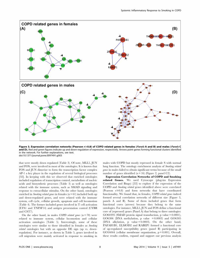
Expression Correlation Networks of COPD and Smoking
related Genes. We used Cytoscape (plug-ins Expression
Correlation and Bingo) [22] to explore if the expression of the
COPD and Smoking related genes identified above were correlated
(Pearson r$0.8) and form networks that have coordinated
functionality. We found that, in females, COPD related genes indeed
formed several correlation networks of different size (Figure 3,
panels A and B). Some of them included genes that form
functional cores (arrows) because they belong to the same
ontologies. For instance, MLL5, JUN and FOS define a functional
core of (repressed) genes (Panel A) that belong to three ontologies:
GO60395 (SMAD protein signal transduction, p value = 0.0001),
GO6306 (DNA methylation, p value = 0.0003) and GO6305
(DNA alkylation, p value = 0.0003). On the other hand,
PAFAH1B1, ELMOD2 and RABEP1 formed a functional core
of up-regulated susceptibility genes (panel B) participating in
GO16044 (cellular membrane organization, p = 0.001). Overall,
these results confirm, expand and support our previous analysis
Figure 3. Expression correlation networks (Pearson r$0.8) of COPD related genes in females (Panels A and B) and males (Panels Cand D). Red and green figures indicate up and down-regulation of expression, respectively. Arrows point genes forming functional clusters identifiedin the network. For further explanations, see text.doi:10.1371/journal.pone.0097491.g003
Systemic Inflammatory Response to Smoking in COPD
PLOS ONE | www.plosone.org 8 May 2014 | Volume 9 | Issue 5 | e97491

using DAVID. Figure 4 depicts the correlation networks of Smoking
related genes in females. Two down-regulated networks were
identified containing ontologies related to GO44237 cellular
metabolic process (p = 0.015) and GO42221 response to chemical
stimulus (p = 0.0005) among others. A large positive correlation
network was identified, containing several ontologies as GO7275
multicellular organismal development (p = 0.0039) and GO50794
regulation of cellular process (p = 0.006).
In males, COPD related genes form (Figure 3): (1) a functional core
of repressed genes (arrows, Panel C) that belong to ‘‘Response to
chemical stimulus’’ (GO42221, p = 0.001); and, (2) a cluster
(arrows) of up-regulated genes (Panel D) involved in GO2376
(Immune system process, p = 0.007) and GO30097 (Hemopoiesis,
p = 0.0002). The network analysis of Smoking in males was not
possible because of the low number of genes identified (n = 16).
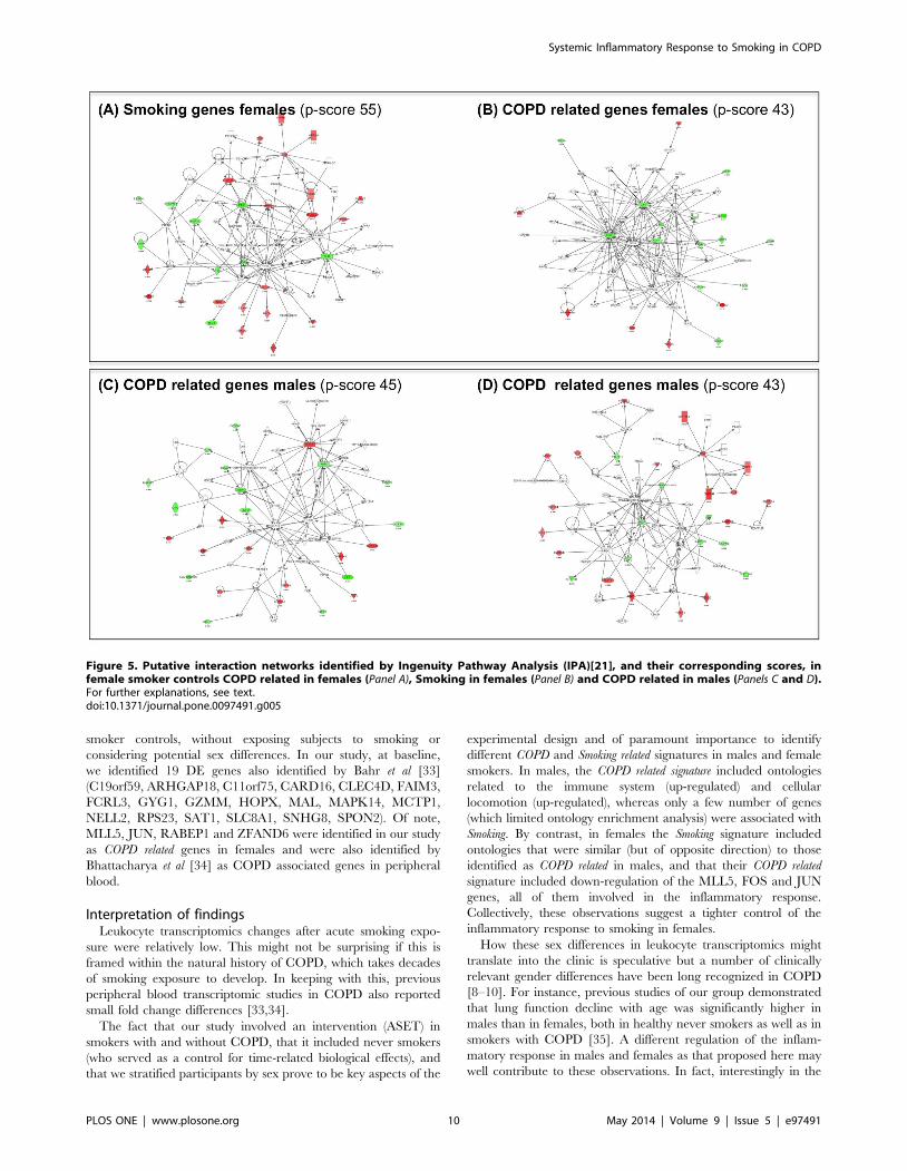
Interaction Network Analysis of COPD and Smoking
related genes. To infer interaction networks of COPD and
Smoking related genes we used Ingenuity Pathways Analysis (IPA), a
database of known molecule interactions [21]. In females, from the
61 Smoking genes (Figure 2, panel C1) IPA identified one significant
network (IPA p-score 55) (Figure 5, panel A) whose main
functional terms included organismal injury and abnormalities,
cell death and survival and cell morphology. We selected the ETS
gene, a central hub in this network, for independent validation by
real time PCR (qPCR); the later confirmed the observed
microarrays transcriptional change (Figure S2). Likewise, from
the 29 COPD related genes identified in females (Figure 2, panel
C1), IPA identified a network (p-score 43) enriched in cell death
and survival, lipid metabolism and small molecule biochemistry
functions (Figure 5, panel B). In this network, we selected FOS and
CXCL1 for independent qPCR validation; results (Figure S2)
confirmed changes in both CXCL1 (p = 0.01) and FOS (p = 0.07).
The low number of Smoking genes (n = 16) identified in males did
not allow obtaining significant networks in the IPA analysis. By
contrast, from the 78 COPD related genes identified in males
(Figure 2, panel C2), IPA identified two large networks. The first
one (IPA p-score 45) included infectious disease, cellular function
and maintenance, hematological system development and function
as main functional terms (Figure 5, panel C). The second one (p-
score 43) included DNA replication, recombination, cell cycle and
cell morphology terms (Figure 5, panel D). From these two
networks, we selected HLX, TRIB1 and BIRC3, known to be
involved in cell activation, cell migration and apoptosis respec-
tively, for qPCR validation (Figure S2).
Discussion
Despite that an abnormal inflammatory response to smoking is
believed to play a major pathogenic role in COPD [1], to our
knowledge this is the first study to directly compare the systemic
biomarker and transcriptomic leukocyte response to acute smoking
exposure in susceptible (i.e. COPD patients) and resistant smokers
(i.e., those with normal spirometry). By doing so, our results
provide several novel and potentially relevant observations. First,
we identified a differential transcriptomic pattern, both at baseline
and in response to smoking, in males and females, whose
implications go beyond COPD, as discussed below. Second, we
identified a number of COPD and Smoking related genes (signatures),
both in males and females, and we identified their enriched
ontologies and gene interaction networks. Yet, due to the relatively
small number of subjects studied, reproducibility in an indepen-
dent and larger set of COPD patients and smokers is needed.
Previous studiesSeveral previous studies have investigated the biological
response to acute smoking exposure in healthy subjects [13,14].
In fact, we designed the dose and timing of our ASET (see
Methods) mostly based on these previous experiences. Yet, to our
knowledge, no previous study has directly contrasted the
transcriptomic response of circulating leukocytes to ASET in
smokers with or without COPD. Bahr et al [33] compared the
basal leukocyte transcriptomic pattern in COPD patients and non-
Figure 4. Expression correlation networks (Pearson r$0.8) of Smoking genes in females. Arrows indicate genes identified by geneontology analysis in the network. Red and green indicate up and down-regulation, respectively. For further explanations, see text.doi:10.1371/journal.pone.0097491.g004
Systemic Inflammatory Response to Smoking in COPD
PLOS ONE | www.plosone.org 9 May 2014 | Volume 9 | Issue 5 | e97491
smoker controls, without exposing subjects to smoking or
considering potential sex differences. In our study, at baseline,
we identified 19 DE genes also identified by Bahr et al [33]
(C19orf59, ARHGAP18, C11orf75, CARD16, CLEC4D, FAIM3,
FCRL3, GYG1, GZMM, HOPX, MAL, MAPK14, MCTP1,
NELL2, RPS23, SAT1, SLC8A1, SNHG8, SPON2). Of note,
MLL5, JUN, RABEP1 and ZFAND6 were identified in our study
as COPD related genes in females and were also identified by
Bhattacharya et al [34] as COPD associated genes in peripheral
blood.
Interpretation of findingsLeukocyte transcriptomics changes after acute smoking expo-
sure were relatively low. This might not be surprising if this is
framed within the natural history of COPD, which takes decades
of smoking exposure to develop. In keeping with this, previous
peripheral blood transcriptomic studies in COPD also reported
small fold change differences [33,34].
The fact that our study involved an intervention (ASET) in
smokers with and without COPD, that it included never smokers
(who served as a control for time-related biological effects), and
that we stratified participants by sex prove to be key aspects of the
experimental design and of paramount importance to identify
different COPD and Smoking related signatures in males and female
smokers. In males, the COPD related signature included ontologies
related to the immune system (up-regulated) and cellular
locomotion (up-regulated), whereas only a few number of genes
(which limited ontology enrichment analysis) were associated with
Smoking. By contrast, in females the Smoking signature included
ontologies that were similar (but of opposite direction) to those
identified as COPD related in males, and that their COPD related
signature included down-regulation of the MLL5, FOS and JUN
genes, all of them involved in the inflammatory response.
Collectively, these observations suggest a tighter control of the
inflammatory response to smoking in females.
How these sex differences in leukocyte transcriptomics might
translate into the clinic is speculative but a number of clinically
relevant gender differences have been long recognized in COPD
[8–10]. For instance, previous studies of our group demonstrated
that lung function decline with age was significantly higher in
males than in females, both in healthy never smokers as well as in
smokers with COPD [35]. A different regulation of the inflam-
matory response in males and females as that proposed here may
well contribute to these observations. In fact, interestingly in the
Figure 5. Putative interaction networks identified by Ingenuity Pathway Analysis (IPA)[21], and their corresponding scores, infemale smoker controls COPD related in females (Panel A), Smoking in females (Panel B) and COPD related in males (Panels C and D).For further explanations, see text.doi:10.1371/journal.pone.0097491.g005
Systemic Inflammatory Response to Smoking in COPD
PLOS ONE | www.plosone.org 10 May 2014 | Volume 9 | Issue 5 | e97491

context of this discussion, systemic levels of inflammatory
biomarkers appear reduced in females (vs. males) with COPD
[36].
The relevance of these observations for other common smoking
related diseases, such as cardiovascular disease [37–39] or lung
cancer [40–42], deserves further investigation. In fact, recent
publications support similar sex differences in other immune
related diseases, such as multiple sclerosis [43], and it is well
established that autoimmune diseases are particularly prevalent in
females [44]. All in all, therefore, these observations call for a
careful exploration and re-analysis of available COPD genetic
data by gender.
Finally, we used several network analysis techniques to infer
potential interactions among the identified COPD and Smoking
related genes and thus to get further insight into the pathobiology of
the disease. The expression correlation networks in conjunction
with gene ontology identified functional cores of differentially
expressed genes. The possible interaction networks between
differentially expressed genes identify by IPA (on the basis of
already know molecule interactions) showed good IPA p-scores
and their main gene hub changes were validated by qPCR. In
females, resistance genes networks included genes related to Gpcr
signaling (SIPR5, GPR56), inflammation and Th1 responses
(ETS-1, MMP9, TBX21), apoptosis (BCL2) and sensing of planar
aromatic hydrocarbons that are present in tobacco (AHR),
whereas the main hubs in their susceptibly gene networks include
the repression of JUN and FOS, both involved also in the
regulation of immune responses through the formation of the AP
transcription factor. Overall, therefore, these observations suggest
again a differential regulation of immune processes in females with
or without COPD. On the other hand, in males, we identified two
COPD related gene networks that involved, respectively, the
response to planar aromatic hydrocarbons (hub gene AHR) and
the immune response (hub gene RORA) on the one hand, and
Gpcr signaling, ubiquitinization (TRIB1, RAD23B, USP-15) and
apoptosis-autophagy (BIRC3, TRIM13, TRIM23) on the other,
which were validated by qPCR. Interestingly, other studies have
reported alterations in some of these processes in COPD [45–47].
Strengths and LimitationsOur study has strengths and limitations. The fact that, despite
the widespread assumption that COPD is characterized by an
abnormal response to smoking, this is the first study to investigate
this possibility specifically at the transcriptional level is a clear
strength. Likewise, the use of an active experimental intervention
(ASET) is a step beyond the standard transcriptomic case-control
studies [48] and has in fact facilitated the identification of novel
COPD and Smoking related genes in response to acute tobacco
exposure. Finally, the use of network analysis has allowed the
description of clear gender differences in the systemic inflamma-
tory response to smoking in COPD patients and Smokers, an
observation that may have implications beyond COPD in other
smoking-related diseases. Among its limitations, we acknowledge
that the transcriptomic changes after ASET were measured only in
circulating leukocytes, so it is possible that changes in the
pulmonary parenchyma may differ in type and/or intensity;
needless to say that ethical and logistic difficulties involved in
obtaining lung tissue samples before and after smoking are
notorious. Finally, we included in the study 30 volunteers but,
when stratified by presence of disease, smoking status and sex,
each subgroup includes 5 individuals only. Due to the relatively
small number of subjects studied, results need replication in an
independent and larger cohort of COPD patients.
ConclusionsThis study shows that the transcriptomic response to smoke in
circulating leukocytes is different in smokers with and without
COPD, and also in males and females.
Supporting Information
Figure S1 Unsupervised clustering of DE genes inCOPD segregated T0 and T180 samples adequately,both in females (n = 57) and males (n = 110) (panels A andB, respectively).
(TIFF)
Figure S2 qPCR validation of array results, RQ =relative quantification of target gene mRNA to ActinmRNA calculated using the comparative CT method.Central hub genes in IPA networks were selected for the
validation; a) COPD males (HLX, TRIB1 and BIRC3), b)
Healthy smoker females (ETS 1) and c) COPD females (CXCL1
and FOS1).
(TIFF)
Table S1 Complete list of differentially expressed genes(DE) in COPD patients, smokers (S) and non-smokers(NS) stratified by sex. Gene ID, affymetrix probe ID, log ratio
and FDR.
(DOCX)
Table S2 Top 10 differentially expressed genes atbaseline between COPD patients and Smokers, strati-fied by sex. Gene ID, affymetrix probe ID, log ratio and FDR.
(DOCX)
Table S3 Top 10 differentially expressed genes atbaseline due to gender differences in COPD patientsand in healthy smokers. Gene ID, affymetrix probe ID, log
ratio and FDR.
(DOCX)
Data S1 Detailed methods and additional results.
(DOCX)
Acknowledgments
Authors thank all volunteers participating in the study for their willingness
to contribute to medical research, Ms. Gemma Sunyer for her excellent
technical support during the study, Drs. Sellares, Soler and Ballester, from
the Respiratory Department of our hospital, for their help recruiting
COPD patients, and the different institutions that financed the study. This
work was developed at the Centre de Recerca Biomedica Cellex,
Barcelona, Spain. Affymetrix arrays were performed in the Genomics
platform of the IDIBAPS.
Author Contributions
Conceived and designed the experiments: AA RF. Performed the
experiments: NG RF TC. Analyzed the data: SGK RF NG AA. Wrote
the paper: RF NG SGK AA.
References
1. Vestbo J, Hurd SS, Agusti AG, Jones PW, Vogelmeier C, et al. (2013) Global
Strategy for the Diagnosis, Management and Prevention of Chronic Obstructive
Pulmonary Disease, GOLD Executive Summary. Am J Respir Crit Care Med
187: 347–365. rccm.201204-0596PP [pii];10.1164/rccm.201204-0596PP [doi].
Systemic Inflammatory Response to Smoking in COPD
PLOS ONE | www.plosone.org 11 May 2014 | Volume 9 | Issue 5 | e97491

2. Fletcher C, Peto R (1977) The natural history of chronic airflow obstruction. Br
Med J 1: 1645–1648.
3. Lomas DA, Silverman EK (2001) The genetics of chronic obstructive pulmonarydisease. Respir Res 2: 20–26.
4. Silverman EK, Speizer FE, Weiss ST, Chapman HA Jr, Schuette A, et al. (2000)Familial aggregation of severe, early-onset COPD: candidate gene approaches.
Chest 117: 273S–274S.
5. DeMeo DL, Carey VJ, Chapman HA, Reilly JJ, Ginns LC, et al. (2004) Familialaggregation of FEF25-75 and FEF25-75/FVC in families with severe, early
onset COPD. Thorax 59: 396–400.
6. Hogg JC, Chu F, Utokaparch S, Woods R, Elliott WM, et al. (2004) The Natureof Small-Airway Obstruction in Chronic Obstructive Pulmonary Disease.
N Engl J Med 350: 2645–2653.
7. Agusti A, Edwards LD, Rennard SI, MacNee W, Tal-Singer R, et al. (2012)
Persistent Systemic Inflammation is Associated with Poor Clinical Outcomes in
COPD: A Novel Phenotype. PLoS ONE 7: e37483.
8. Aryal S, Diaz-Guzman E, Mannino DM (2013) COPD and gender differences:
an update. Transl Res 162: 208–218. S1931-5244(13)00138-2 [pii];10.1016/j.trsl.2013.04.003 [doi].
9. de Torres JP, Casanova C, Hernandez C, Abreu J, guirre-Jaime A, et al. (2005)
Gender and COPD in patients attending a pulmonary clinic. Chest 128: 2012–2016.
10. Dransfield MT, Washko GR, Foreman MG, San Jose ER, Reilly J, et al. (2007)
Gender differences in the severity of CT emphysema in COPD. Chest.
11. McDunn JE, Husain KD, Polpitiya AD, Burykin A, Ruan J, et al. (2008)
Plasticity of the systemic inflammatory response to acute infection during criticalillness: development of the riboleukogram. PLoS ONE 3: e1564. 10.1371/
journal.pone.0001564 [doi].
12. Rabe KF, Hurd S, Anzueto A, Barnes PJ, Buist SA, et al. (2007) Global strategyfor the diagnosis, management, and prevention of chronic obstructive
pulmonary disease: GOLD executive summary. Am J Respir Crit Care Med176: 532–555.
13. van der Vaart H, Postma DS, Timens W, Hylkema MN, Willemse BW, et al.
(2005) Acute effects of cigarette smoking on inflammation in healthy intermittentsmokers. Respir Res 6: 22.
14. van der Vaart H, Postma DS, Timens W, ten Hacken NH (2004) Acute effects of
cigarette smoke on inflammation and oxidative stress: a review. Thorax 59: 713–721.
15. American Thoracic Society Official Statement (1995) Standardization ofSpirometry. 1994 Update. Am J Respir Crit Care Med 152: 1107–1136.
16. Roca J, Sanchis J, Agustı-Vidal A, Segarra J, Navajas D, et al. (1986)
Spirometric reference values for a mediterranean population. Bull EurPhysiopathol Respir 22: 217–224.
17. Livak KJ, Schmittgen TD (2001) Analysis of relative gene expression data using
real-time quantitative PCR and the 2(-Delta Delta C(T)) Method. Methods 25:402–408. 10.1006/meth.2001.1262 [doi];S1046-2023(01)91262-9 [pii].
18. Breitling R, Armengaud P, Amtmann A, Herzyk P (2004) Rank products: a
simple, yet powerful, new method to detect differentially regulated genesin replicated microarray experiments. FEBS Lett 573: 83–92. 10.1016/
j.febslet.2004.07.055 [doi];S0014579304009354 [pii].
19. Huang dW, Sherman BT, Lempicki RA (2009) Systematic and integrative
analysis of large gene lists using DAVID bioinformatics resources. Nat Protoc 4:
44–57. nprot.2008.211 [pii];10.1038/nprot.2008.211 [doi].
20. Dennis G Jr, Sherman BT, Hosack DA, Yang J, Gao W, et al. (2003) DAVID:
Database for Annotation, Visualization, and Integrated Discovery. Genome Biol4: 3.
21. Jimenez-Marin A, Collado-Romero M, Ramirez-Boo M, Arce C, Garrido JJ
(2009) Biological pathway analysis by ArrayUnlock and Ingenuity PathwayAnalysis. BMC Proc 3 Suppl 4: S6. 1753-6561-3-S4-S6 [pii];10.1186/1753-
6561-3-S4-S6 [doi].
22. Smoot M, Ono K, Ideker T, Maere S (2011) PiNGO: a Cytoscape plugin to findcandidate genes in biological networks. Bioinformatics 27: 1030–1031. btr045
[pii];10.1093/bioinformatics/btr045 [doi].
23. Smoot ME, Ono K, Ruscheinski J, Wang PL, Ideker T (2011) Cytoscape 2.8:
new features for data integration and network visualization. Bioinformatics 27:
431–432. btq675 [pii];10.1093/bioinformatics/btq675 [doi].
24. Menon R, Di DM, Cordiglieri C, Musio S, La ML, et al. (2012) Gender-based
blood transcriptomes and interactomes in multiple sclerosis: involvement of SP1dependent gene transcription. J Autoimmun 38: J144–J155. S0896-
8411(11)00117-X [pii];10.1016/j.jaut.2011.11.004 [doi].
25. Anton IM, de la Fuente MA, Sims TN, Freeman S, Ramesh N, et al. (2002) WIPdeficiency reveals a differential role for WIP and the actin cytoskeleton in T and
B cell activation. Immunity 16: 193–204. S1074761302002686 [pii].
26. Vogler M (2012) BCL2A1: the underdog in the BCL2 family. Cell Death Differ
19: 67–74. cdd2011158 [pii];10.1038/cdd.2011.158 [doi].27. Lang F, Voelkl J (2013) Therapeutic potential of serum and glucocorticoid
inducible kinase inhibition. Expert Opin Investig Drugs 22: 701–714. 10.1517/
13543784.2013.778971 [doi].28. Bailey SL, Chang SC, Griffiths B, Graham AN, Saffery R, et al. (2008) ZNF397,
a new class of interphase to early prophase-specific, SCAN-zinc-finger,mammalian centromere protein. Chromosoma 117: 367–380. 10.1007/
s00412-008-0155-7 [doi].
29. Eisenreich A, Zakrzewicz A, Huber K, Thierbach H, Pepke W, et al. (2013)Regulation of pro-angiogenic tissue factor expression in hypoxia-induced human
lung cancer cells. Oncol Rep 30: 462–470. 10.3892/or.2013.2413 [doi].30. Luo Y, Roeder RG (1995) Cloning, functional characterization, and mechanism
of action of the B-cell-specific transcriptional coactivator OCA-B. Mol Cell Biol15: 4115–4124.
31. Gotting C, Kuhn J, Zahn R, Brinkmann T, Kleesiek K (2000) Molecular cloning
and expression of human UDP-d-Xylose:proteoglycan core protein beta-d-xylosyltransferase and its first isoform XT-II. J Mol Biol 304: 517–528. 10.1006/
jmbi.2000.4261 [doi];S0022-2836(00)94261-X [pii].32. Schonthaler HB, Guinea-Viniegra J, Wagner EF (2011) Targeting inflammation
by modulating the Jun/AP-1 pathway. Ann Rheum Dis 70 Suppl 1: i109–i112.
70/Suppl_1/i109 [pii];10.1136/ard.2010.140533 [doi].33. Bahr TM, Hughes GJ, Armstrong M, Reisdorph R, Coldren CD, et al. (2013)
Peripheral Blood Mononuclear Cell Gene Expression in Chronic ObstructivePulmonary Disease. Am J Respir Cell Mol Biol.
34. Bhattacharya S, Tyagi S, Srisuma S, DeMeo DL, Shapiro SD, et al. (2011)Peripheral blood gene expression profiles in COPD subjects. J Clin Bioinforma
1: 12. 2043-9113-1-12 [pii];10.1186/2043-9113-1-12 [doi].
35. Kohansal R, Martinez-Camblor P, Agusti A, Buist AS, Mannino DM, et al.(2009) The Natural History of Chronic Airflow Obstruction Revisited: An
Analysis of the Framingham Offspring Cohort. Am J Respir Crit Care Med 180:3–10. 200901-0047OC [pii];10.1164/rccm.200901-0047OC [doi].
36. de Torres JP, Casanova C, Pinto-Plata V, Varo N, Restituto P, et al. (2011)
Gender Differences in Plasma Biomarker Levels in a Cohort of COPD Patients:A Pilot Study. PLoS ONE 6: e16021. 10.1371/journal.pone.0016021 [doi].
37. Roeters van Lennep JE, Westerveld HT, Erkelens DW, van der Wall EE (2002)Risk factors for coronary heart disease: implications of gender. Cardiovasc Res
53: 538–549.38. Erbel R, Mohlenkamp S, Lehmann N, Schmermund A, Moebus S, et al. (2008)
Sex related cardiovascular risk stratification based on quantification of
atherosclerosis and inflammation. Atherosclerosis 197: 662–672. S0021-9150(07)00132-3 [pii];10.1016/j.atherosclerosis.2007.02.031 [doi].
39. Zhu J, Shearer GM, Norman JE, Pinto LA, Marincola FM, et al. (2000) Hostresponse to cytomegalovirus infection as a determinant of susceptibility to
coronary artery disease: sex-based differences in inflammation and type of
immune response. Circulation 102: 2491–2496.40. Gawron A, Hou L, Ning H, Berry JD, Lloyd-Jones DM (2012) Lifetime risk for
cancer death by sex and smoking status: the lifetime risk pooling project. CancerCauses Control 23: 1729–1737. 10.1007/s10552-012-9959-0 [doi].
41. Ryu JS, Jeon SH, Kim JS, Lee JH, Kim SH, et al. (2011) Gender differences insusceptibility to smoking among patients with lung cancer. Korean J Intern Med
26: 427–431. 10.3904/kjim.2011.26.4.427 [doi].
42. Park SK, Cho LY, Yang JJ, Park B, Chang SH, et al. (2010) Lung cancer riskand cigarette smoking, lung tuberculosis according to histologic type and gender
in a population based case-control study. Lung Cancer 68: 20–26. S0169-5002(09)00318-3 [pii];10.1016/j.lungcan.2009.05.017 [doi].
43. Menon R, Di DM, Cordiglieri C, Musio S, La ML, et al. (2012) Gender-based
blood transcriptomes and interactomes in multiple sclerosis: involvement of SP1dependent gene transcription. J Autoimmun 38: J144–J155. S0896-
8411(11)00117-X [pii];10.1016/j.jaut.2011.11.004 [doi].44. Pennell LM, Galligan CL, Fish EN (2012) Sex affects immunity. J Autoimmun
38: J282–J291. S0896-8411(11)00126-0 [pii];10.1016/j.jaut.2011.11.013 [doi].
45. Zeng H, Kong X, Peng H, Chen Y, Cai S, et al. (2012) Apoptosis and Bcl-2family proteins, taken to chronic obstructive pulmonary disease. Eur Rev Med
Pharmacol Sci 16: 711–727.46. Ryter SW, Lee SJ, Choi AM (2010) Autophagy in cigarette smoke-induced
chronic obstructive pulmonary disease. Expert Rev Respir Med 4: 573–584.10.1586/ers.10.61 [doi].
47. Barrero CA, Perez-Leal O, Aksoy M, Moncada C, Ji R, et al. (2013) Histone 3.3
participates in a self-sustaining cascade of apoptosis that contributes to theprogression of chronic obstructive pulmonary disease. Am J Respir Crit Care
Med 188: 673–683. 10.1164/rccm.201302-0342OC [doi].48. Faner R, Tal-Singer R, Riley JH, Celli B, Vestbo J+, et al. (2013) Lessons from
ECLIPSE: a review of COPD biomarkers. Thorax. In press.
Systemic Inflammatory Response to Smoking in COPD
PLOS ONE | www.plosone.org 12 May 2014 | Volume 9 | Issue 5 | e97491
Top Related