


Languages
Pages
Legal

Synthesis of porphyrin- graphene oxide nanocomposite for an optical
chemical sensor application
Rahmatollah Rahimi*, Rouholah Zare – Dorabei*, Asgar Koohi, Solmaz Zargari
Department of Chemistry, Iran University of Science and Technology, Tehran 16846-13114,
Iran
Abstract
A specific and sensitive optical chemical sensor (optode) was fabricated for the determination
of mercury ions. The optode was prepared by graphene oxide–porphyrin composite. The
graphene oxide–porphyrin composite was synthesized and characterized by field emission
scanning electron microscopy (FE-SEM), UV-Vis and Fourier transform infrared (FT-IR)
spectroscopic techniques. The various experimental parameters such as pH of aqueous
solution of Hg (II) and content of the THPP as well as the grapheme oxide have been
optimized. This optode exhibited a linear range of 6.0×10⁻⁵ to 6.0×10⁻⁹ mol.L-1 Hg (II) with a
detection limit of 3.2×10⁻⁹ mol.L−1 and a response time of ∼210 s.
Keywords:
Optical sensor, Graphene oxide nanosheets, Porphyrin, Mercury ions, Spectrophotometry
UV-Vis.
*Corresponding authors
E-mail address: [email protected]

1. Introduction
Heavy metal pollution due to the indiscriminate disposal of wastewater is a worldwide
environment concern. Wastewaters from many industries such as metallurgical, mining,
chemical manufacturing, and battery manufacturing industries contain many kinds of toxic
heavy metal ions. Mercury is a highly toxic heavy metal that causes human health problems
and environmental contamination, and is also more toxic even from lead and arsenic. This
metal represents a major toxicity to microorganisms and environment even in low
concentrations. Mercury triggers several serious disorders for humans including allergic
reactions and brain and neurological damage. If ingested by a pregnant woman, it can cause
developmental delays in children. Therefore, the detection of very little amounts of mercury
is extraordinarily important [1-4].
Design of control systems such as optical chemical sensors for determination of mercury in
very low concentrations in water samples and rivers can be very useful and effective in the
control and remove pollutants from the environment [1, 5]. In recent years, extensive research
is allocated into optical chemical sensors. Optical chemical sensors used in different contexts
such as environmental, clinical and industrial analysis. The use of optical chemical sensors,
nutrients is very important particularly for the measurement of trace amounts of heavy metal
ions in environmental samples. The application of optical chemical sensors is highly regarded
especially for the determination of trace amounts of heavy metal ions in environmental
samples, a variety of water and food [6-7].
Porphyrins have been studied as probe molecules in different materials and their application
has been examined as detector for chemical sensors. Porphyrins are sensitive to metal ions.
Nitrogen atoms in the structure of porphyrin ring is located inward, and with their only
electron pairs can act as a strong ligand, and they keep the metal ions in the center and the
formation a complexes [8-14]. Choosing a proper substrate and detector stabilizer, are a very

important issue as another part of the membrane components in optical sensors. A proper
grounds should be suitable functional groups to bind and detector stabilize on the plastic or
glass fulcrum. Graphene Oxide with carboxyl functional groups, hydroxyl and epoxy on itself
carbon surface, has been studied as a substrate and important factor to bind and stabilize
detector on the Fulcrum [15-17]. Therefore, in this work, a specific and sensitive optical
chemical sensor based graphene oxide–porphyrin composite was fabricated for the
determination of mercury ions.
2. Experimental
2.1 . Chemicals
All reagents were prepared from analytical reagent grade chemicals supplied from Merck
(Darmstadt, Germany), except HgCl2 which was obtained from Aldrich. The 10-3 M mercury
standard stock solution was prepared by dissolving 0.0811 g of HgCl2 in a 250 mL
volumetric flask and diluting to the mark with distilled water. Lower concentrations were
prepared by serial dilution of the stock solution with phosphate buffer, pH 7.5 Universal
buffer solutions (0.1 M) were prepared from phosphoric acid solutions and sodium hydroxide
solutions and adjusting pH to the desired value (0.08 M). The final pH was adjusted with the
addition of a sodium hydroxide.
2.2 . Synthesis of GO-THPP
2.2.1. Synthesis of THPP
Tetrakis (4-hydroxyphenyl) porphyrin (THPP) was prepared using a modification of A. D.
Adler-f. R. Longo method [18]. Briefly in a typical reaction, 80 mL of propionic acid was
loaded in a 250 mL round-bottomed flask and refluxed for 0.5 h. Then 1.146 g 4-
Hydroxybenzaldehyde dissolved in 10 mL of propionic acid are added. After about 15
minutes the amount of 6.0 g of fresh distilled pyrrole was slowly added into the solution.
After 45 min the mixture was placed at room temperature to precipitate formed. The dark
purple product was filtered and washed thoroughly with ethanol until the filtrate became
clear.
2.2.2. Synthesis of graphene oxide nanosheets (GO)
Graphene oxide was prepared using a modification of Hummers and Offeman’s method [19,
20]. Briefly in a typical reaction, 1 g graphite, 1 g NaNO3, and 46 mL H2SO4 were stirred
together in an ice bath. KMnO4 (8 g) was slowly added while stirring, and the rate of addition
was controlled to prevent the mixture temperature from exceeding 20 °C. The mixture was
then transferred to a 35 °C water bath and stirred for about 1 h, forming a thick paste.
Subsequently, 80 mL de-ionized water was added gradually and the temperature was raised to
90 °C. The mixture was further treated with 10 mL 30% H2O2 solution. The solution was
then filtered and washed with de-ionized water until the pH was 6 and dried at 65 °C under
vacuum.
2.2.3. Synthesis of GO–COCl
Briefly in a typical reaction, 0.5 g GO–COOH was suspended in 30 mL SOCl2 and 5 mL of
DMF. The mixture was refluxed for 24 h at 70°C under nitrogen atmosphere. The resultant
solution was filtered and washed with anhydrous tetrahydrofuran (THF) and dried under
vacuum.
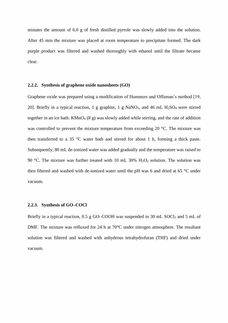
2.2.4. Synthesis of covalently attached porphyrin graphene oxide hybrids (GO-THPP)
Briefly in a typical reaction, a mixture of 30 mg GO–COCl and 60 mg THPP were taken in a
100 mL round bottom flask and 3 mL of triethylamine and 15 mL of DMF were added and
heated to 80 ° C for 72 h under a nitrogen atmosphere. After the reaction, the solution was
cooled to room temperature, and then poured into 250 mL diethyl ether to precipitate the
product. The precipitate was collected by centrifuging at 8000 rpm for 30 min. The
supernatant which contained dissolved THPP was discarded and the precipitate was washed
thoroughly. After adding another 100 mL of diethyl ether, the mixture was sonicated for 5
min and then centrifuged at 8000 rpm for 30 min to collect the GO-THPP. Finally, the
precipitate was washed with CHCl3 five times following the above procedure.
Fig. 1. The schematic illustration of GO-THPP
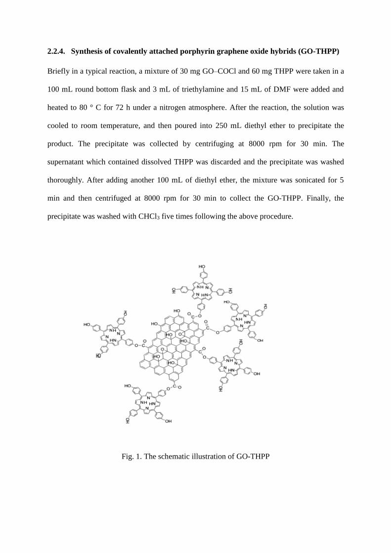
2.3. Structural and spectroscopic characterization of GO-THPP
The FTIR spectra were recorded using KBr plates in the range 500–4000 cm using a Nicolet
6700 FT-IR spectrometer. Fig. 2 shows the typical FTIR spectrum obtained for our graphite
oxide material. The most characteristic features are the broad, intense band at 3430 cm-1 )O–
H stretching vibrations) and the bands at 1725 cm-1 (C=O stretching vibrations from carbonyl
and carboxylic groups), 1639 cm-1 (skeletal vibrations from unoxidized graphitic domains),
1380 cm-1 (C–OH stretching vibrations), and 1027 cm-1 (C–O stretching vibrations). The
appearance of the peaks at around 2900 cm-1 is ascribed to the aromatic stretching vibrations
of C–H bonds of GO. After covalent functionalization with porphyrins, a new peak appears at
~ 1582 cm-1 corresponding to the C=C vibrations of porphyrins and the peak of the C–O
stretching vibration shifts to 1108 cm-1 ,and broadens. The peak present at ~1707 cm-1 is
assigned to the bending vibration of the C=N of the porphyrin ring. The disappearance of the
peak at 1380 cm-1 clearly indicates that in graphene oxide–porphyrin composites, the
porphyrin molecules are covalently bonded to the graphene oxide via carboxylic acid linkage.
Fig. 2. FT-IR spectrum of graphene oxide and porphyrin–GO composites
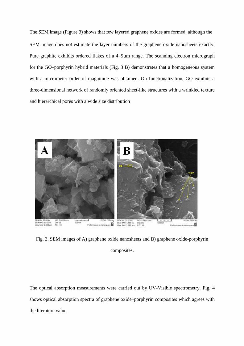
The SEM image (Figure 3) shows that few layered graphene oxides are formed, although the
SEM image does not estimate the layer numbers of the graphene oxide nanosheets exactly.
Pure graphite exhibits ordered flakes of a 4–5µm range. The scanning electron micrograph
for the GO–porphyrin hybrid materials (Fig. 3 B) demonstrates that a homogeneous system
with a micrometer order of magnitude was obtained. On functionalization, GO exhibits a
three-dimensional network of randomly oriented sheet-like structures with a wrinkled texture
and hierarchical pores with a wide size distribution
Fig. 3. SEM images of A) graphene oxide nanosheets and B) graphene oxide-porphyrin
composites.
The optical absorption measurements were carried out by UV-Visible spectrometry. Fig. 4
shows optical absorption spectra of graphene oxide–porphyrin composites which agrees with
the literature value.

Fig. 4. UV/Vis spectrum of graphene oxide–porphyrin composites in DMF.
2.4. Membrane preparation
A mixture of 0.6 mg graphene oxide–porphyrin composites, was dissolved in 1.0mL of DMF.
The membrane solution was homogenized with a magnetic stirrer for 15 min. A Plexiglas
slides with 9 mm × 40 mm dimensions were cut to fit into standard spectrophotometer cells.
The slides were cleaned with ethanol, then water and finally dried in an oven at 70 °C. The
membranes were cost by pipetting 10 μL of the membrane solution onto a Plexiglas slide and
spread it by a spin coater was deposit uniform thin films to flat substrates.
2.5. Measurement procedure
The membrane was conditioned by inserting it into a cell including 3 mL of phosphate buffer
(pH 7.5). After 5 min the membrane absorbance was measured at 426 nm. Then, the cell was
filled with the Hg (II) standard solution and after 210 s its absorbance was measured in the
same wavelength

3. Results and discussion
3.2. Principle of the operation
Tetrakis (4-hydroxyphenyl) porphyrin (THPP) contains a nitrogen donor atom which could
form internal bonds with soft metal ions such as mercury (II). THPP has high affinity to make
a complex with mercury (II) ions. Mercury (II) ions form a complex with the ligand when the
organic membrane contacts with the aqueous solution; therefore, the following ion-exchange
reaction takes place:
H2 (L)(m) + Hg2+(aq) ↔ (L)Hg(m) + 2H+
(aq) (1)
In this equation, ‘L’ is the Ligand. It can be seen that by the addition of mercury ions into the
aqueous solution, the chromoionophore in the organic membrane is more deprotonated. Fig. 5
shows the absorption spectra of the optode with different concentrations of Hg (II) in the
range 1.0 × 10⁻¹⁴ to 1.0 × 10⁻³ mol L−1 .
Fig. 5. Absorption spectra of the optical sensor in phosphate buffer solution (pH
7.5)containing different concentrations of Hg(II) as: 1) Blank solution (buffer); 2) 1.0 × 10⁻¹⁴;
3) 1.0 × 10⁻¹³; 4) 1.0 × 10⁻¹²; 5) 1.0 × 10⁻¹¹; 6) 1.0 × 10⁻¹⁰; 7) 1.0 × 10⁻⁹; 8) 1.0 × 10⁻⁸; 9) 1.0 ×
10-7; 10) 1.0 × 10⁻⁶; 11) 1.0 × 10⁻⁵; 12) 1.0 × 10⁻⁴; 13) 1.0 × 10⁻³ mol.L-1 Hg(II).
4. Conclusion
In this work a new composites was synthesized based on the tetrakis (4-hydroxyphenyl)
porphyrin (THPP) stabilized with graphene oxide as compound for the designs optical
chemical sensor. The optode described in this work is easily prepared and provides a simple
and inexpensive means for the determination of ultra-trace amounts of Hg (II) ions. Based on
the results, it is clear that the determination of Hg (II) ions with optode prepared from
graphene oxide–porphyrin composites were a selective and sensitive methods. The amount
of mercury was measured in different water samples by the optode and also by ICP
method. The results confirm that there was not any significant difference between the
results.
References
[1] A. R. Firooz, M. Movahedi A. A. Ensafi, "Selective and sensitive optical chemical sensor
for the determination of Hg(II) ions based on tetrathia-12-crown-4 and chromoionophore I",
Sensors and Actuators B, 171-172, 492-498, (2012).
[2] Y. Yang, J. Jiang, G. Shen, R.Yu, "An optical sensor for mercury ion based on the
fluorescence quenching of tetra (p-dimethylaminophenyl) porphyrin", Analytical Chemical
Acta, 636, 83–88, (2009).
[3] K. Alizadeh ,R. Parooi ,P. Hashemi ,B. Rezaei, M. R. Ganjali, "A new Schiff’s base
ligand immobilized agarose membrane optical sensor for selective monitoring of mercury
ion", Journal of Hazardous Materials, 186, 1794–1800, (2011).
[4] A. Oehmen, D. Vergel, J. Fradinho, M. A. M. Reis, "Mercury removal from water
streams through the ion exchange membrane bioreactor concept", Journal of Hazardous
Materials, 264, 65-70, (2014).
[5] A. R. Firooz, A. A. Ensafi, K. Karimi, R. Khalifeh, " Specific sensing of mercury(II) ions
by an optical sensor based on a recently synthesized ionophore", Sensors and Actuators B,
185, 84-90, (2013).
[6] J. Janata, A. Bezegh, "Chemical Sensors", Anal. Chem, 60, 62R-74R, (1988).
[7] P. Gründler, "Chemical Sensors", Springer-Verlag Berlin Heidelberg, (2007).
[8] R. Yang, K. Li, K. Wang, F. Liu, N. Li, F. Zhao, "Cyclodextrin-porphyrin supramolecular
sensitizer for mercury(II) ion", Analytical Chemical Acta, 469, 285-293, (2002).
[9] W. H. Chana, R. H. Yang, K. M. Wang, "Development of a mercury ion-selective optical
sensor based on fluorescence quenching of 5, 10, 15, 20-tetraphenylporphyrin", Analytical
Chemical Acta, 444, 261-269, (2001).
[10] P. Kumar, Y.B. Shim, "A novel Mg (II)-selective sensor based on 5, 10, 15, 20-tetrakis
(2-furyl)-21, 23-dithiaporphyrin as an electroactive material", Journal of Electroanalytical
Chemistry, 661, 25-30, (2011).
[11] I. Leray, M. C. Vernieres, R. L. Saibi, C. B. Charreton, J. Faure, "Porphyrins as probe
molecules in the detection of gaseous pollutants I: Diffusion of pyddine in polystyrene films
containing zinc- tetraphenyl porphyrin", Sensors and Actuators B, 37, 67-74, (1996).
[12] H. K. Lee, K. Song, H. R. Seo, Y. K. Choi, S. Jeon, "Lead(II) selective electrodes based
on tetrakis(2-hydroxy-1-naphthyl)porphyrins: the effect of atropisomers", Sensors and
Actuators B, 99, 323-329, (2004).
[13] K. Kilian, K. Pyrzynska, "Spectrophotometric study of Cd(II), Pb(II), Hg(II) and Zn(II)
complexes with 5,10,15,20-tetrakis(4-carboxylphenyl) porphyrin", Talanta, 60, 669-678,
(2003).
[14] M. Biesaga, K. Pyrzynska, M. Trojanowicz, "Porphyrins in analytical chemistry. A
review", Talanta, 51, 209-224, (2000).

[15] G. Zhao, J. Li, X. Ren, C. Chen, X. Wang, "Few-Layered Graphene Oxide Nanosheets
As Superior Sorbents for Heavy Metal Ion Pollution Management", Environ. Sci. Technol,
45, 10454-10462, (2011).
[16] Q. Xiang, J. Yu, M. Jaroniec, "Graphene-based semiconductor photocatalysts", Chem.
Soc. Rev, 41, 782-796, (2012).
[17] V. Georgakilas, M. Otyepka, A. B. Bourlinos, V. Chandra, N. Kim, K. C. Kemp, P.
Hobza, R. Zboril, K. S. Kim, " Functionalization of Graphene: Covalent and Non-Covalent
Approaches, Derivatives and Applications", Chem. Rev, (2012).
[18] S. Shanmugathasan, C. Edwards, R. W. Boyle, "Advances in Modern Synthetic
Porphyrin Chemistry", Tetrahedron, 56, 1025-1046, (2000).
[19] M. B. M. Krishna, N. Venkatramaiah, R. Venkatesan, D. N. Rao, "Synthesis and
structural, spectroscopic and nonlinear optical measurements of graphene oxide and its
composites with metal and metal free porphyrins", J. Mater. Chem, 22, 3059-3068, (2012).
[20] W. S. Hummers, R.E. Offeman, "Synthesis and characterization of graphene oxide", J.
Am. Chem. Soc, 80, 1339-1339, (1958).
Top Related