


Languages
Pages
Legal


S1
Supporting information
Synthesis and antiplasmodial activity of new indolone N-oxide derivatives
Françoise Nepveu1,2 *
, Sothea Kim1,2
, Jeremie Boyer1,2
, Olivier Chatriant3, Hany Ibrahim
1,2,
Karine Reybier1,2
, Marie-Carmen Monje1,2
, Severine Chevalley1,2
, Pierre Perio1,2
, Barbora H.
Lajoie1,2,
, Jalloul Bouajila1,2
, Eric Deharo1,2
, Michel Sauvain1,2
, Rachida Tahar4, Leonardo
Basco4, Antonella Pantaleo
5, Francesco Turini
5, Paolo Arese
5, Alexis Valentin
1,2, Eloise
Thompson, Livia Vivas6, Serge Petit
3, Jean-Pierre Nallet
3.
1 Université de Toulouse, UPS; UMR 152 (Laboratoire de pharmacochimie des substances naturelles et
pharmacophores redox), F-31062 Toulouse cedex 9, France 2
IRD, UMR 152, F-31062 Toulouse cedex 9, France 3 IDEALP-PHARMA, Bâtiment CEI, 66 Bd Niels Bohr, BP 2132, 69603 Villeurbanne cedex, France
4 Organisation de Coordination pour la lutte contre les Endémies en Afrique Centrale (OCEAC), BP 288,
Yaoundé, Cameroun 5 University of Turin Medical School, Via Santena 6 bis, 10126 Torino, Italy
6 London School of Hygiene and Tropical Medicine (LSHTM), Keppel Street, London C1E 7HT United
Kingdom.
Table of contents
1. Hematin binding assays
2. Metabolism of some selected compounds by liver microsomes
3. Lactate dehydrogenase (LDH) assay
4. Elemental analysis
5. Selected 1H- and
13C-NMR spectra
6. Selected IR spectra
7. References cited in Supporting Information
* Corresponding author:
Françoise Nepveu Tel: +33-5-62-25-68-69; Fax: +33-5-62-25-98-02; e-mail: [email protected]

S2
1. Hematin binding assays
Degradation of hemoglobin by malaria parasites releases ferriprotoporphyrin IX (FP)
that is detoxified by crystallization into hemozoin (HZ). Antimalarial drugs such as
chloroquine and chloroquine-like derivatives bind to FP and impair this process. To approach
the mechanisms of action of the new series, we examine the capacity of indolone-N-oxides to
interfere with hematin dimerization, to bind to iron or to form heme-adducts. First, the drug
concentration required to inhibit β-hematin formation by 50% (IC50) was determined for
selected analogues from the new series of indolone-N-oxide using the UV-visible
spectrophotometric assay of Deharo et al1. The results are given in Table 1 for five selected
compounds and are the results of two different experiments performed in triplicate.
Table 1. Comparison of IC50 required to inhibit P.f parasite growth (FCB1) to IC50
required to inhibit β-hematin formation for seven selected compound compared to
chloroquine.
Compound IC50/P.f. FcB1 (nM) IC50/FBIT (nM)
1 66 ± 28 238
49 3 940 ± 200 2 840
52 899 ± 88 inactif
51 3 560 ± 400 39
15 156 ± 57 inactif
CQ 151 ± 6 28
β-hematin formation inhibition UV-visible assay:
The analysis were done using 96-well plates non sterile with a flat bottom (Greiner
Bio-one, Poitiers, France). The well plates were charged by the addition of the following
solutions (N.B. the ordre of addition is important): i) 50 μL hemin chloride (0.5 mg/mL)
freshly prepared into DMSO, ii) 100 μL sodium acetate buffer (0.5 M, pH = 4.4), iii) 50 μL
tested compound/ DMSO (50 μL DMSO for the blank). The final pH must be adjusted
between 5 and 5.2. After incubation at 37 °C for 18-24 h, the well plates were centrifuged at
3000 rpm for 8 minutes. The supernatant was rejected and the residue was re-suspended using
200 μL de DMSO, to eliminate the non-reacted hematine. The well plate was centrifuged
again and the supernatant was rejected. The residue, ß-hematine, was dissolved into 200 μL
de NaOH 0.1 M and the absorbance was measured at 405 nm using microplate reader
(Metertech S960, Avantech, Illkirch, France). The results were expressed as a percentage
inhibition of hemin biocrystallization calculated by the following equation:
%inhibiton = 100 x (Ablank – Atest)/Ablank.
The experiments were done in triplicate (n = 3).
2. Metabolism of some selected compounds by liver microsomes
Liver microsomes pooled from male mouse (CD-1) 20 mg protein/mL and NADPH
were obtained from Biopredic International (Rennes, France). Microsomes (20 mg protein/mL
in 250 mM sucrose, stored at - 80 °C) were quickly thawed at 37 °C using a water bath and
kept on ice until use. NADPH (10 mM in phosphate buffer) was prepared in the previous
prepared buffer solution.
Due to the poor aqueous solubility, the cosolvent method2 was used as follows. DMSO
stock solutions of test compounds were prepared at 0.5 mM concentration. Diluted solutions
of test compounds were prepared by adding 50 µL of each DMSO stock solution to 200 µL of
acetonitrile to make 0.1 mM solutions in 20% DMSO/ 80% acetonitrile. Mouse liver
microsomal solution was prepared by adding 1.582 mL of concentrated mouse liver
S3
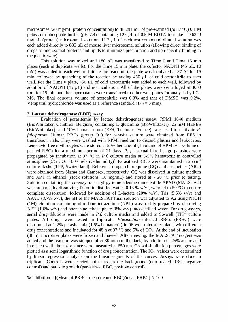
microsomes (20 mg/mL protein concentration) to 48.291 mL of pre-warmed (to 37 °C) 0.1 M
potassium phosphate buffer (pH 7.4) containing 127 µL of 0.5 M EDTA to make a 0.6329
mg/mL (protein) microsomal solution. 11.2 µL of each test compound diluted solution was
each added directly to 885 µL of mouse liver microsomal solution (allowing direct binding of
drugs to microsomal proteins and lipids to minimize precipitation and non-specific binding to
the plastic ware).
This solution was mixed and 180 µL was transferred to Time 0 and Time 15 min
plates (each in duplicate wells). For the Time 15 min plate, the cofactor NADPH (45 µL, 10
mM) was added to each well to initiate the reaction; the plate was incubated at 37 °C for 15
min, followed by quenching of the reaction by adding 450 µL of cold acetonitrile to each
well. For the Time 0 plate, 450 µL of cold acetonitrile was added to each well, followed by
addition of NADPH (45 µL) and no incubation. All of the plates were centrifuged at 3000
rpm for 15 min and the supernatants were transferred to other well plates for analysis by LC–
MS. The final aqueous volume of acetonitrile was 0.8% and that of DMSO was 0.2%.
Verapamil hydrochloride was used as a reference standard (T1/2 = 6 min).
3. Lactate dehydrogenase (LDH) assay
Evaluation of parasitemia by lactate dehydrogenase assay: RPMI 1640 medium
(BioWhittaker, Cambrex, Belgium) containing L-glutamine (BioWhittaker), 25 mM HEPES
(BioWhittaker), and 10% human serum (EFS, Toulouse, France), was used to cultivate P.
falciparum. Human RBCs (group O±) for parasite culture were obtained from EFS in
transfusion vials. They were washed with RPMI medium to discard plasma and leukocytes.
Leucocyte-free erythrocytes were stored at 50% hematocrit (1 volume of RPMI + 1 volume of
packed RBC) for a maximum period of 21 days. P. f. asexual blood stage parasites were
propagated by incubation at 37 °C in P.f. culture media at 3-5% hematocrit in controlled
atmosphere (5% CO2, 100% relative humidity)3. Parasitized RBCs were maintained in 25 cm
2
culture flasks (TPP, Switzerland). Reference drugs, chloroquine (CQ) and artemether (ART)
were obtained from Sigma and Cambrex, respectively. CQ was dissolved in culture medium
and ART in ethanol (stock solutions: 10 mg/mL) and stored at - 20 °C prior to testing.
Solution containing the co-enzymz acetyl pyridine adenine dinucleotide APAD (MALSTAT)
was prepared by dissolving Triton in distilled water (0.13 % w/v), warmed to 50 °C to ensure
complete dissolution, followed by addition of L-lactate (20% w/v), Tris (5.5% w/v) and
APAD (3.7% w/v), the pH of the MALSTAT final solution was adjusted to 9.2 using NaOH
(1M). Solution containing nitro blue tetrazolium (NBT) was freshly prepared by dissolving
NBT (1.6% w/v) and phenazine ethosulphate (8% w/v) into distilled water. For drug assays,
serial drug dilutions were made in P.f. culture media and added to 96-well (TPP) culture
plates. All drugs were tested in triplicate. Plasmodium-infected RBCs (PRBC) were
distributed at 1-2% parasitaemia (1.5% hematocrit) in 96-well microtiter plates with different
drug concentrations and incubated for 48 h at 37 °C and 5% of CO2. At the end of incubation
(48 h), microtiter plates were frozen and thawed. After thawing, the MALSTAT reagent was
added and the reaction was stopped after 30 min (in the dark) by addition of 25% acetic acid
into each well, the absorbance were measured at 650 nm. Growth-inhibition percentages were
plotted as a semi logarithmic function of drug concentration. The IC50 values were determined
by linear regression analysis on the linear segments of the curves. Assays were done in
triplicate. Controls were carried out to assess the background (non-treated RBC, negative
control) and parasite growth (parasitized RBC, positive control).
% inhibition = [(Mean of PRBC- mean treated RBC)/mean PRBC] X 100

S4
4. Elemental analysis
Compound C H N
Cal. Found Cal. Found Cal. Found
1 59.72 59.16 2.67 2.13 4.64 4.48
3 71.14 71.05 4.38 4.32 5.53 5.55
5 75.24 75.18 4.32 4.11 4.62 4.64
7 70.19 70.13 3.65 3.51 3.90 3.83
26 68.91 68.65 5.44 5.12 9.45 9.31
49 70.92 70.33 6.45 6.24 6.89 6.80
50 69.83 69.71 5.86 5.78 7.40 7.25

S5
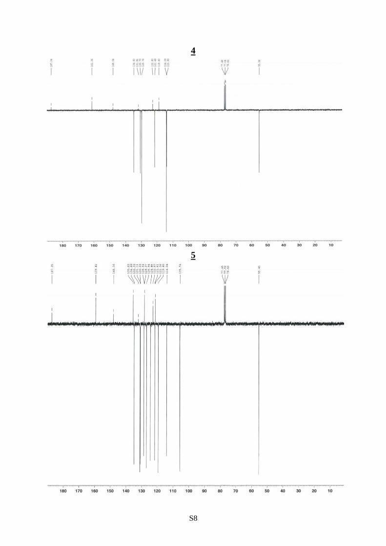
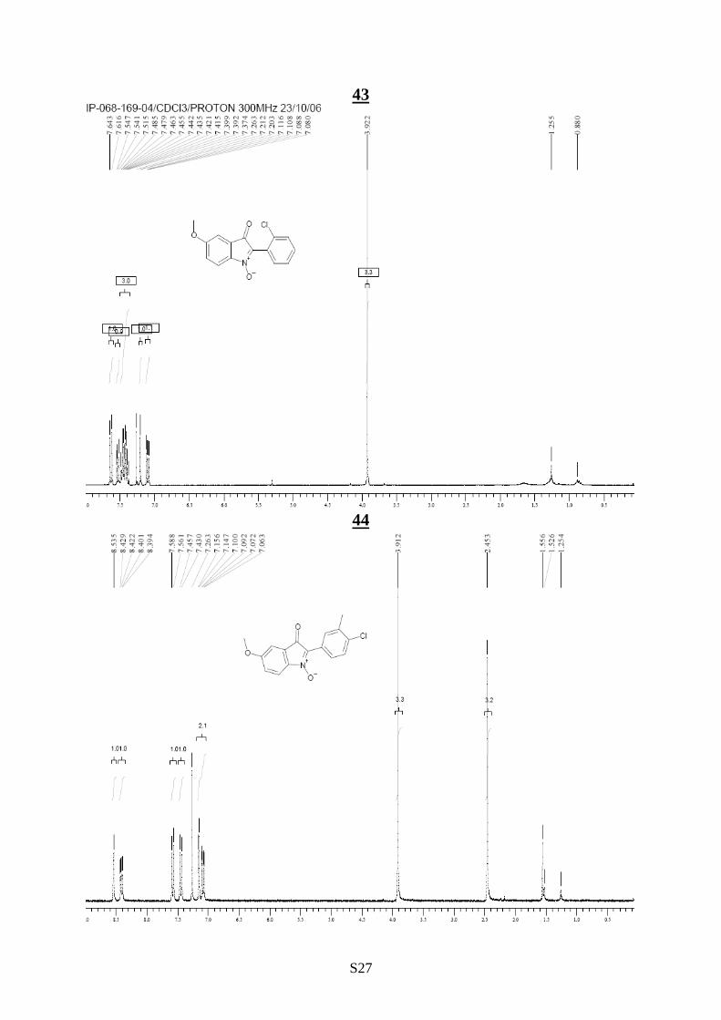
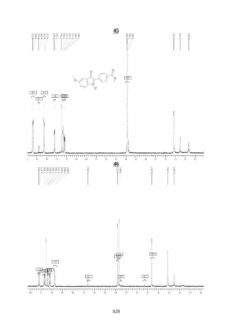
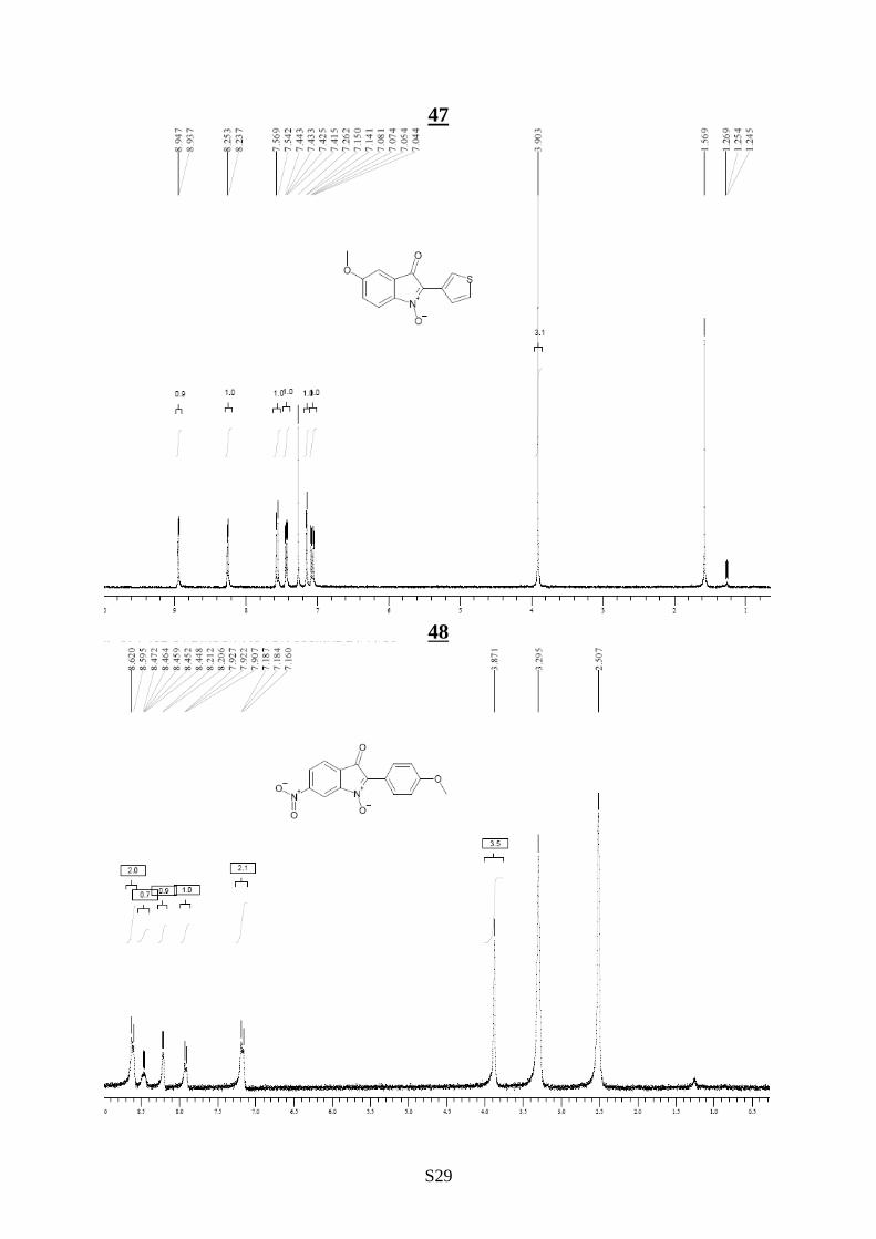
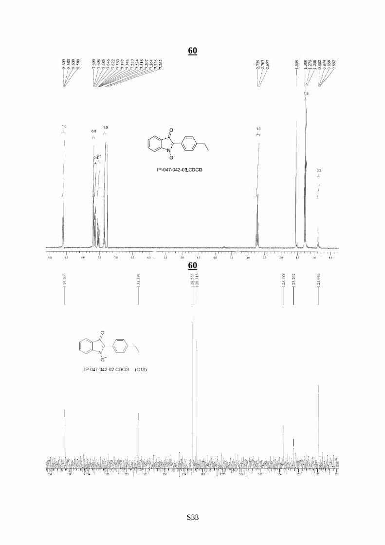
5. Selected 1H and
13C NMR
1H NMR spectrum of 1
13
C NMR spectrum of 1
S6
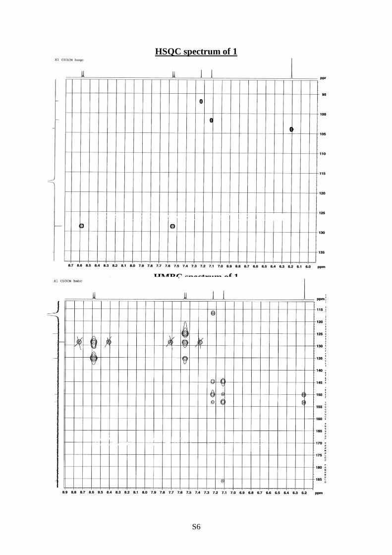
HSQC spectrum of 1
HMBC spectrum of 1

S7
2
4
S8
4
5

S9
6
7

S10
8
9

S11
10
11

S12
12
13

S13
14
16

S14
17
18

S15
20
21

S16
22
23
S17
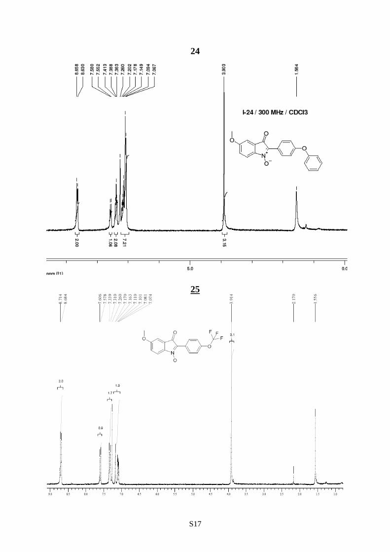
24
25

S18
13CNMR zgpg30 spectrum of 25
26

S19
27
28

S20
29
30

S21
31
32

S22
33
34
S23
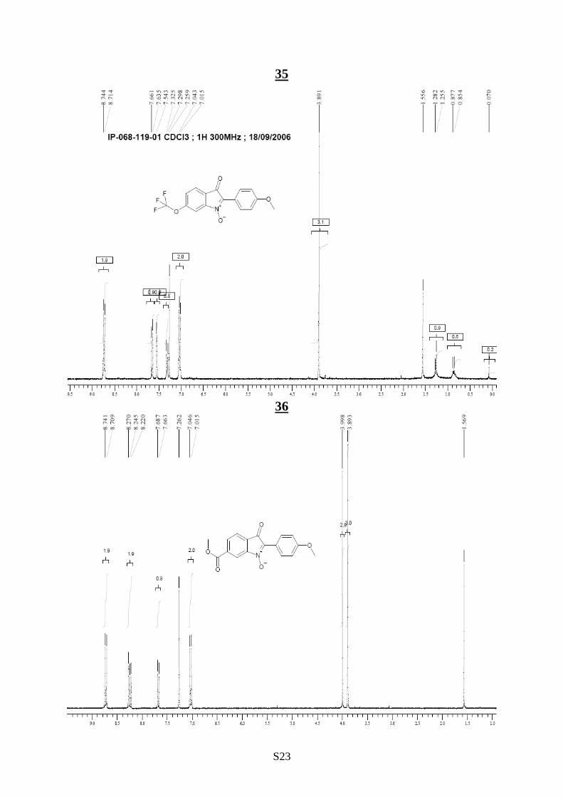
35
36

S24
37
38

S25
39
40
S26
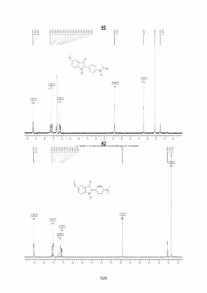
41
42
S27
43
44
S28
45
46
S29
47
48

S30
49
49

S31
50
50

S32
53
59
S33
60
60

S34
61

S35
62
63

S36
64
65

S37
66

S38
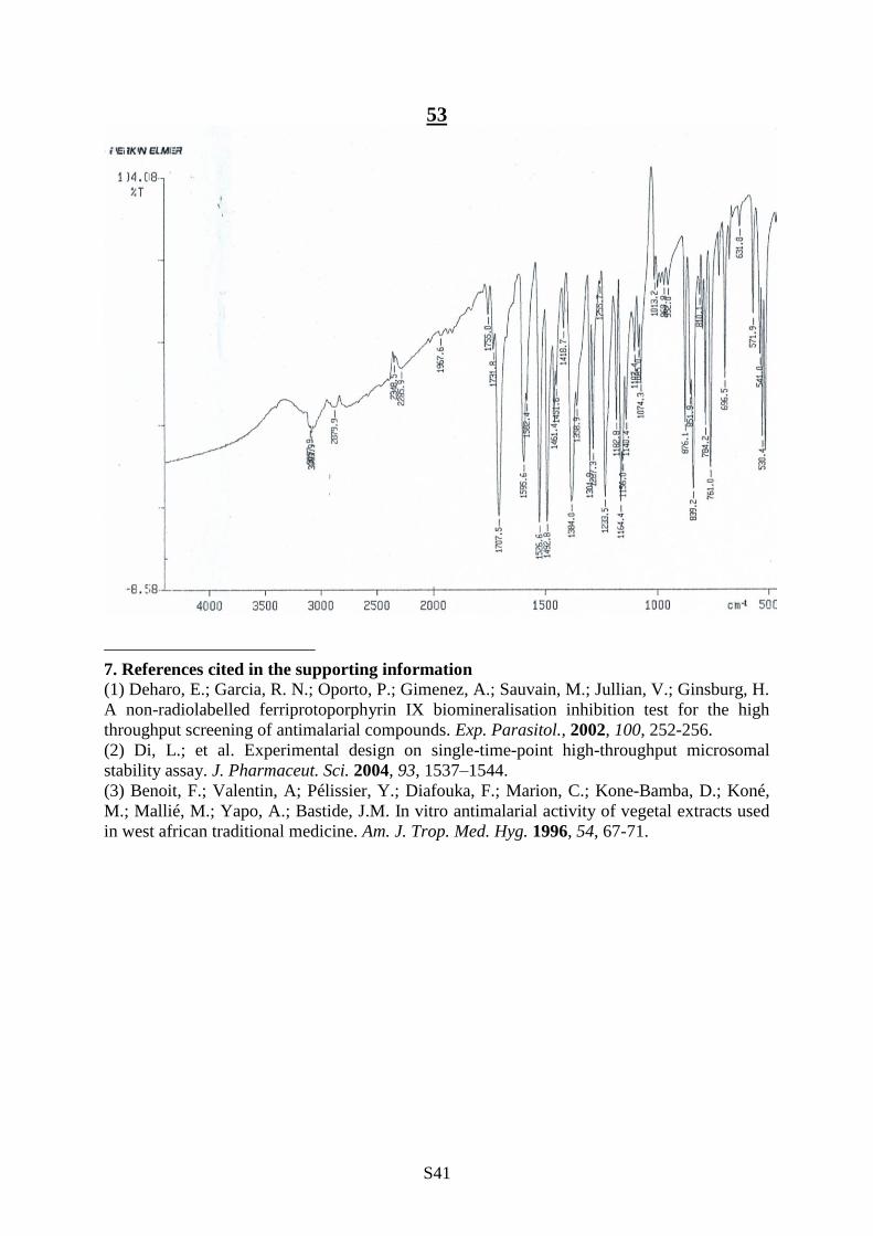
6. Selected IR Spectra
1
3

S39
27
49

S40
50
52
S41
53
7. References cited in the supporting information
(1) Deharo, E.; Garcia, R. N.; Oporto, P.; Gimenez, A.; Sauvain, M.; Jullian, V.; Ginsburg, H.
A non-radiolabelled ferriprotoporphyrin IX biomineralisation inhibition test for the high
throughput screening of antimalarial compounds. Exp. Parasitol., 2002, 100, 252-256.
(2) Di, L.; et al. Experimental design on single-time-point high-throughput microsomal
stability assay. J. Pharmaceut. Sci. 2004, 93, 1537–1544.
(3) Benoit, F.; Valentin, A; Pélissier, Y.; Diafouka, F.; Marion, C.; Kone-Bamba, D.; Koné,
M.; Mallié, M.; Yapo, A.; Bastide, J.M. In vitro antimalarial activity of vegetal extracts used
in west african traditional medicine. Am. J. Trop. Med. Hyg. 1996, 54, 67-71.
Top Related