


Languages
Pages
Legal

Supporting InformationJogler et al. 10.1073/pnas.1012694108SI Materials and Methods.Electron Microscopy. For chemical fixation, cells were incubated in2.5% glutardialdehyde containing fixative buffer (75 mM sodiumcacodylate, 2 mM MgCl2, pH 7.0), for 1 h at room temperature.Afterward, samples were rinsed several times in fixative bufferand postfixed at room temperature for 1 h with 1% osmiumtetroxide in fixative buffer. After two washing steps in water, thecells were stained en bloc for 30 min with 1% uranyl acetate in20% acetone. Dehydration was performed with a graded acetoneseries. Samples were then infiltrated and embedded in Spurr’slow-viscosity resin.For high-pressure freezing, aluminum platlets were filled with
concentrated cell suspensions and the cells immobilized by high-pressure freezing (Leica; HPM100). Freeze substitution wasperformed in acetone with 2% osmium tetroxide and 0.2% uranylacetate, including 5% water. After embedding the samples inEpon, ultrathin sections were cut with a diamond knife andmounted onto uncoated copper grids. The sections were post-stained with aqueous lead citrate (100 mM, pH 13.0).Transmission electron micrographs were taken with an EM 912
electron microscope (Zeiss) equipped with an integratedOMEGA energy filter operated at 80 kV in the zero loss mode.The FIB serial sectioning was performed by a Zeiss-Aurigaworkstation. The focused ion beam consisted of Ga+ ions ac-celerated by a voltage of 30 kV. In the cut-and-view mode,sections ranging in thickness between 5 nm and 10 nm (de-pendent on the magnification) were produced with the FIB andFESEM images, which were recorded at 1.5 kV using the in-lensenergy selective backscattered (EsB) detector. Specimens weretilted to an angle of 54°; images were tilt corrected for un-distorted surface view.For SEM, drops of the sample were placed onto a glass slide,
covered with a coverslip, and rapidly frozen with liquid nitrogen.The coverslip was removed with a razor blade and the glass slidewas immediately fixed with 2.5% glutaraldehyde in 75 mMcacodylate buffer (pH 7.0), postfixed with 1% osmium tetroxide infixative buffer, dehydrated in a graded series of acetone solutions,and critical-point dried after transfer to liquid CO2. Specimenswere mounted on stubs, coated with 3 nm platinum usinga magnetron sputter coater, and examined with a Zeiss Aurigascanning electron microscope operated at 1–2 kV. For cryo-scanning electron microscopy high-pressure frozen samples werefractured with a Leica EM MED020, sublimated for 1–2 min a
−95 °C and coated with 3 nm of tungsten, transferred to thescanning electron microscope and examined at 1 kV.
Single Cell Sorting and Whole Genome Amplification (WGA). Singlecell sorting was achieved via an Eppendorf TransferMan NK2micromanipulator and the Eppendorf CellTram Oil manual hy-draulic pressure-control system mounted to an Olympus BX61microscope equipped with a 40× LD objective and a double slideholder. Samples were kept on microscopic slides attached toa custom-build plastic frame, while an Advalytix AmpliGridAG480F was placed next to it in the double slide holder (Fig.S6C). Five to 15 individual Mbav cells were transferred via mi-cromanipulation from the 5-μL sample droplet (containing var-iable amounts of MTB) into two different washing droplets(5 μL) and finally into 0.75 μL sample buffer (Illustra GenomiPhiV2 DNA amplification kit; GE Healthcare) covered with 5 μL ofsealing solution (Advalytix). Together, this washing procedureresulted in a 1018-fold dilution of sample liquid, which is likely tohave outdiluted any contaminating DNA to extinction (Fig. S10).Loaded AmpliGrid was transferred into a customized Ampli-Speed slide cycler (Advalytix) calibrated to operate at 4 °C.Samples were heated to 95 °C for 3 min and incubated at 4 °Cfor 10 min. A total of 0.75 μL of reaction buffer containing en-zyme mix (Illustra GenomiPhi V2 DNA amplification kit; GEHealthcare) was added by pipetting on top of the sealing solution(Advalytix) and reactions were incubated for 4 h at 30 °C. Thephi29 enzyme was inactivated by incubating the sample at 65 °Cfor 10 min. After amplification, seven independent reactions—based on a total number of 158 individually sorted Mbav cells—were pooled to overcome phi29 bias and a 16S rRNA gene librarywas constructed as described previously (1). Sequence analysis of25 clones revealed identical Mbav sequences (cutoff 99%).
Screening of Fosmid Libraries. Six fosmid libraries that were con-structed before were screened via endsequencing as previouslydescribed (2, 3). Based on sequence analysis of individually sortedcells after WGA, the primers CJ272 (cactacgccacccttgaagt) andCJ273 (tgaggtaatcggcatcaaca) targeting a 130-bp region ofmamE, as well as the primers CJ280 (ttgatattacatgacatctg) andCJ281 (cgaggcaacggagaagatac) targeting a 555-bp region ofmamP were deduced. The PCR-based fosmid library screeningwas performed as previously described (3).
1. Lin W, Pan Y (2009) Uncultivated magnetotactic cocci from yuandadu park in beijing,china. Appl Environ Microbiol 75:4046–4052.
2. Jogler C, et al. (2009) Toward cloning of the magnetotactic metagenome:Identification of magnetosome island gene clusters in uncultivated magnetotacticbacteria from different aquatic sediments. Appl Environ Microbiol 75:3972–3979.
3. Jogler C, et al. (2010) Cultivation-independent characterization of ‘CandidatusMagnetobacterium bavaricum’ via ultrastructural, geochemical, ecological andmetagenomic methods. Environ Microbiol 12:2466–2478.
4. Preusting H, Kingma J, Huisman G, Steinbüchel A, Witholt B (1993) Formation ofpolyester blends by a recombinant strain of Pseudomonas oleovorans: Different poly(3-hydroxyalkanoates) are stored in separate granules. J Environ Polym Degrad 1:11–21.
Jogler et al. www.pnas.org/cgi/content/short/1012694108 1 of 13
OL2
OL1
CMPG
OM
Ribosomes
S
PHB
MM
PPS
50 nm
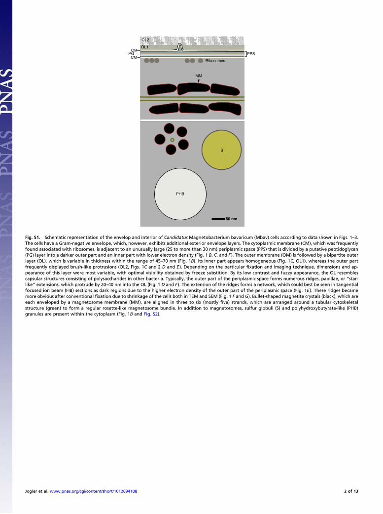
Fig. S1. Schematic representation of the envelop and interior of Candidatus Magnetobacterium bavaricum (Mbav) cells according to data shown in Figs. 1–3.The cells have a Gram-negative envelope, which, however, exhibits additional exterior envelope layers. The cytoplasmic membrane (CM), which was frequentlyfound associated with ribosomes, is adjacent to an unusually large (25 to more than 30 nm) periplasmic space (PPS) that is divided by a putative peptidoglycan(PG) layer into a darker outer part and an inner part with lower electron density (Fig. 1 B, C, and F). The outer membrane (OM) is followed by a bipartite outerlayer (OL), which is variable in thickness within the range of 45–70 nm (Fig. 1B). Its inner part appears homogeneous (Fig. 1C, OL1), whereas the outer partfrequently displayed brush-like protrusions (OL2, Figs. 1C and 2 D and E). Depending on the particular fixation and imaging technique, dimensions and ap-pearance of this layer were most variable, with optimal visibility obtained by freeze substition. By its low contrast and fuzzy appearance, the OL resemblescapsular structures consisting of polysaccharides in other bacteria. Typically, the outer part of the periplasmic space forms numerous ridges, papillae, or “star-like” extensions, which protrude by 20–40 nm into the OL (Fig. 1 D and F). The extension of the ridges forms a network, which could best be seen in tangentialfocused ion beam (FIB) sections as dark regions due to the higher electron density of the outer part of the periplasmic space (Fig. 1E). These ridges becamemore obvious after conventional fixation due to shrinkage of the cells both in TEM and SEM (Fig. 1 F and G). Bullet-shaped magnetite crystals (black), which areeach enveloped by a magnetosome membrane (MM), are aligned in three to six (mostly five) strands, which are arranged around a tubular cytoskeletalstructure (green) to form a regular rosette-like magnetosome bundle. In addition to magnetosomes, sulfur globuli (S) and polyhydroxybutyrate-like (PHB)granules are present within the cytoplasm (Fig. 1B and Fig. S2).
Jogler et al. www.pnas.org/cgi/content/short/1012694108 2 of 13
AA BB
100 nm100 nm 100 nm100 nm
SSSS
SS
PHBPHB PHBPHB
500 nm500 nm
CC DD BSESE 1
SE 2
S-Kα
C-Kα
Fe-Lα
O-Kα
mixmix
1 µm1 µm
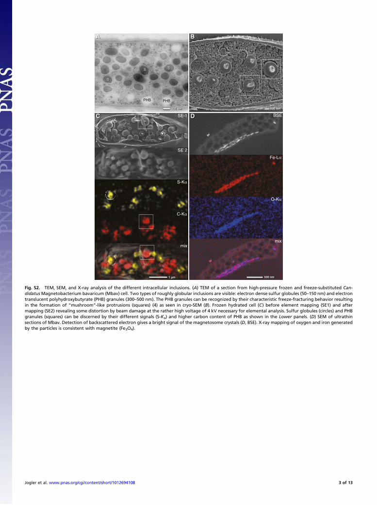
Fig. S2. TEM, SEM, and X-ray analysis of the different intracellular inclusions. (A) TEM of a section from high-pressure frozen and freeze-substituted Can-didatusMagnetobacterium bavaricum (Mbav) cell. Two types of roughly globular inclusions are visible: electron dense sulfur globules (50–150 nm) and electrontranslucent polyhydroxybutyrate (PHB) granules (300–500 nm). The PHB granules can be recognized by their characteristic freeze-fracturing behavior resultingin the formation of “mushroom”-like protrusions (squares) (4) as seen in cryo-SEM (B). Frozen hydrated cell (C) before element mapping (SE1) and aftermapping (SE2) revealing some distortion by beam damage at the rather high voltage of 4 kV necessary for elemental analysis. Sulfur globules (circles) and PHBgranules (squares) can be discerned by their different signals (S-Ka) and higher carbon content of PHB as shown in the Lower panels. (D) SEM of ultrathinsections of Mbav. Detection of backscattered electron gives a bright signal of the magnetosome crystals (D, BSE). X-ray mapping of oxygen and iron generatedby the particles is consistent with magnetite (Fe3O4).
Jogler et al. www.pnas.org/cgi/content/short/1012694108 3 of 13
BB
AA
CC
GG
100 nm100 nm
100 nm100 nm
10 nm10 nm
500 nm500 nm
MMMM
MMMM
FF
DD
EE
500 nm500 nm
100 nm100 nm
100 nm100 nm
****
FlFl
FlFl
50 nm50 nm
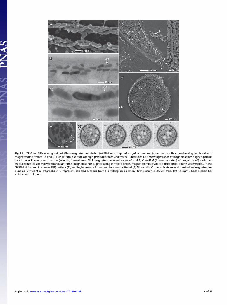
Fig. S3. TEM and SEM micrographs of Mbav magnetosome chains. (A) SEMmicrocraph of a cryofractured cell (after chemical fixation) showing two bundles ofmagnetosome strands. (B and C) TEM ultrathin sections of high-pressure frozen and freeze-substituted cells showing strands of magnetosomes aligned parallelto a tubular filamentous structure (asterisk, framed area; MM, magnetosome membrane). (D and E) Cryo-SEM (frozen hydrated) of tangential (D) and cross-fractured (E) cells of Mbav (rectangular frame, magnetosomes aligned along MF; solid circles, magnetosomes crystals; dotted circle, empty MM vesicles). (F andG) SEM of focused ion beam (FIB) sections (F), and high-pressure frozen and freeze-substituted (G) Mbav cells. Circles indicate several rosette-like magnetosomebundles. Different micrographs in G represent selected sections from FIB-milling series (every 10th section is shown from left to right). Each section hasa thickness of 8 nm.
Jogler et al. www.pnas.org/cgi/content/short/1012694108 4 of 13
sediment sample
fosmid selection
and sequencing
A I E QBQMP
identification of magnetosome
gene sequences mamP / mamE
D
magnetic
enrichment
deducing
primers
B´ C´
whole genomeamplification
construction and end-sequencing of fosmid-libraries
PCR screening of
10,000 fosmids
B” C”
A B
single
cell sorting
C
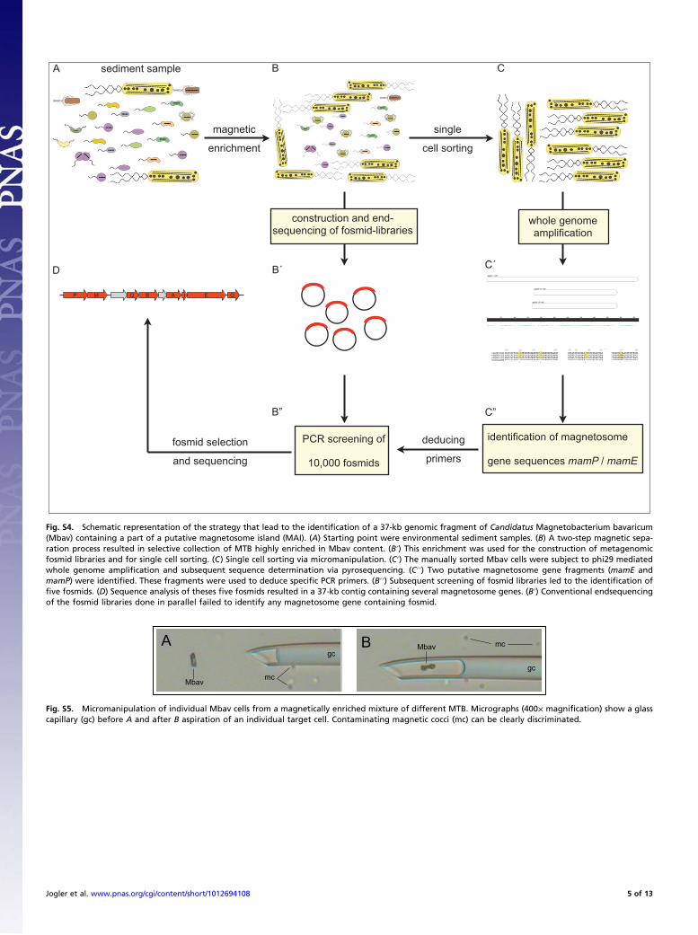
Fig. S4. Schematic representation of the strategy that lead to the identification of a 37-kb genomic fragment of Candidatus Magnetobacterium bavaricum(Mbav) containing a part of a putative magnetosome island (MAI). (A) Starting point were environmental sediment samples. (B) A two-step magnetic sepa-ration process resulted in selective collection of MTB highly enriched in Mbav content. (B′) This enrichment was used for the construction of metagenomicfosmid libraries and for single cell sorting. (C) Single cell sorting via micromanipulation. (C′) The manually sorted Mbav cells were subject to phi29 mediatedwhole genome amplification and subsequent sequence determination via pyrosequencing. (C′′) Two putative magnetosome gene fragments (mamE andmamP) were identified. These fragments were used to deduce specific PCR primers. (B′′) Subsequent screening of fosmid libraries led to the identification offive fosmids. (D) Sequence analysis of theses five fosmids resulted in a 37-kb contig containing several magnetosome genes. (B′) Conventional endsequencingof the fosmid libraries done in parallel failed to identify any magnetosome gene containing fosmid.
A B
Mbav
Mbav
mc
mcgc
gc
Fig. S5. Micromanipulation of individual Mbav cells from a magnetically enriched mixture of different MTB. Micrographs (400× magnification) show a glasscapillary (gc) before A and after B aspiration of an individual target cell. Contaminating magnetic cocci (mc) can be clearly discriminated.
Jogler et al. www.pnas.org/cgi/content/short/1012694108 5 of 13
11 2 NCNC 1 2 NCNC 1 2 NCNCM M
mamMmamM mamPmamP mamEmamE
1000 bp1000 bp
500 bp500 bp
700 bp700 bp
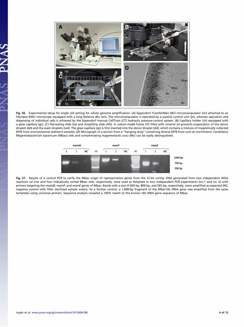
Fig. S7. Results of a control PCR to verify the Mbav origin of representative genes from the 37-kb contig. DNA generated from two independent WGAreactions on one and four individually sorted Mbav cells, respectively, were used as template in two independent PCR experiments (no.1 and no. 2) withprimers targeting the mamM, mamP, and mamE genes of Mbav. Bands with a size of 693 bp, 809 bp, and 565 bp, respectively, were amplified as expected (NC,negative control with filter sterilized sample water). As a further control, a 1,068-bp fragment of the Mbav16S rRNA gene was amplified from the sametemplates using universal primers. Sequence analysis revealed a 100% match to the known 16S rRNA gene sequence of Mbav.
A B
C
jo
CT
mi
chgc
cf
AG
gc
dd wdhs
Mbav
Mc
Mc
Mbav
D
Fig. S6. Experimental setup for single cell sorting for whole genome amplification. (A) Eppendorf TransferMan NK2 micromanipulator (mi) attached to anOlympus BX61 microscope equipped with a long distance 40× lens. The micromanipulator is operated by a joystick control unit (jo), whereas aspiration anddispensing of individual cells is achieved by the Eppendorf manual CellTram (CT) hydraulic pressure-control system. (B) Capillary holder (ch) equipped witha glass capillary (gc). (C) Harvesting slide (hs) and AmpliGrip slide (AG). A custom-made frame (cf) filled with mineral oil prevents evaporation of the donordroplet (dd) and the wash droplets (wd). The glass capillary (gc) is first inserted into the donor droplet (dd), which contains a mixture of magnetically collectedMTB from environmental sediment samples. (D) Micrograph of a section from a “hanging drop” containing diverse MTB from such an enrichment. CandidatusMagnetobacterium bavaricum (Mbav) cells and contaminating magnetotactic cocci (Mc) can be easily distinguished.
Jogler et al. www.pnas.org/cgi/content/short/1012694108 6 of 13
AMB-1
MS-1
MSR-1
Fos002
Fos001
MV-1
MC-1
RS-1
M. bavaricum
100
100
100
86
94
99
0.1
MamAAMB-1
MS-1
MSR-1
Fos002
Fos001
MV-1
MC-1
M. bavaricum
RS-1
100
100
100
99
100
99
0.1
MamE
AMB-1
MS-1
MSR-1
Fos002
Fos001
MV-1
MC-1
M. bavaricum
RS-1
100
100
89
99
89
0.05
MamPAMB-1
MS-1
MSR-1
Fos002
Fos001
MV-1
M. bavaricum-I
RS-1
M. bavaricum-II
MC-1
99
100
85
71
100
53
67
0.05
MamQ
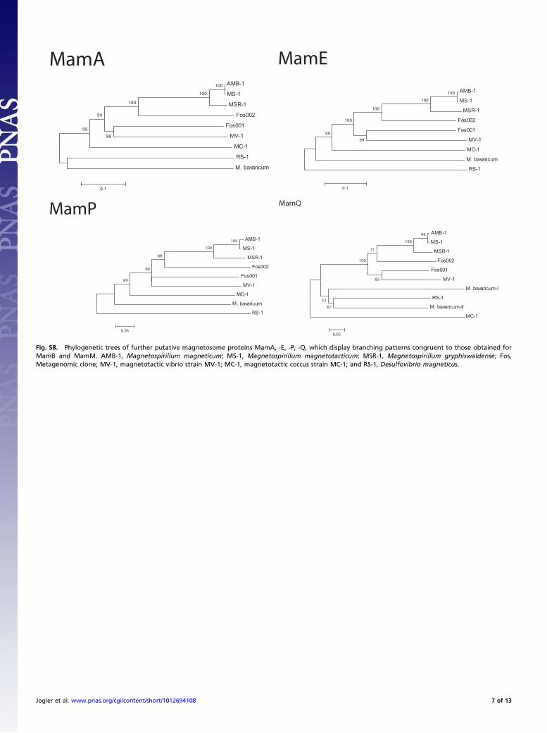
Fig. S8. Phylogenetic trees of further putative magnetosome proteins MamA, -E, -P, -Q, which display branching patterns congruent to those obtained forMamB and MamM. AMB-1, Magnetospirillum magneticum; MS-1, Magnetospirillum magnetotacticum; MSR-1, Magnetospirillum gryphiswaldense; Fos,Metagenomic clone; MV-1, magnetotactic vibrio strain MV-1; MC-1, magnetotactic coccus strain MC-1; and RS-1, Desulfovibrio magneticus.
Jogler et al. www.pnas.org/cgi/content/short/1012694108 7 of 13

Fig. S9. (A) Multiple sequence alignment of MamI homologs from Candidatus Magnetobacterium bavaricum (Mbav), selected cultivated MTB, and a meta-genomic clone (Fos001). Amino acid positions correspond to the MamI protein of M. gryphiswaldense MSR-1. The putative Mbav MamI protein is shorter thanall other MamI proteins. (B) Phylogenetic tree calculated from the alignment shown in A. MamI homologs from Desulfovibriomagneticus RS-1 and Mbav clustertogether, but are separated from other MTB. Bootstrap values and the bootstrap consensus tree are shown.
1 2 3 M
1.0 kb1.5 kb
Fig. S10. Results of a nontemplate control experiment for whole genome amplification (WGA).The control experiment was performed as the previouslydescribed sorting experiment and the preparation of WGA DNA for pyrosequencing (Fig. S5 and Materials and Methods), except one modification: About 15Candidatus Magnetobacterium bavaricum (Mbav) cells were transferred from a 5-μL sample droplet into a 5-μL sorting droplet, which consists of 5 μL of filter-sterilized (0.45 μm pore size) sample water (identical to the 5-μL sample droplet, but without cells). As previously described, sorted cells were subsequentlytransferred into a 5-μL H2O droplet for further washing before transfer onto the Ampligrid was performed. After processing 82 cells in total, the capillary wasused to harvest nontemplate control samples from sorting- and washing droplet. Together with the sorted cells, these samples were subjected to WGA andsubsequent PCR experiments to check for extracellular DNA contaminations. PCR amplification of the 16S rRNA gene from the MTB sample (line 1), the filter-sterilized sample water (line 2), and the H2O washing droplet (line 3) was performed as previously described (2), (lane M, Fermentas 1-kb DNA ladder). Onlylane 1 shows the expected band corresponding to the full-length 16S rRNA gene, whereas lanes 2 and 3 do not show any band. Therefore, it can be confidentlyconcluded from the nontemplate WGA control that the washing procedure was sufficiently stringent to remove any extracellular DNA contaminations.
Jogler et al. www.pnas.org/cgi/content/short/1012694108 8 of 13
MamB: M. magneticum AMB-1
MamB: M. magnetotacticum MS-1
MamB: M. gryphiswaldense MSR-1
MamB: Fos002
MamB: Fos001
MamB: magnetic vibrio MV-1
MamB: magnetic coccus MC-1
MamB: D. magneticus RS-1
MamB: “Candidatus M. bavaricum”
MamM: D. magneticus RS-1
MamM: “Candidatus M. bavaricum”
MamM: magnetic coccus MC-1
MamM: magnetic vibrio MV-1
MamM: Fos001
MamM: Fos002
MamM: M. gryphiswaldense MSR-1
MamM: M. magneticum AMB-1
MamM: M. magnetotacticum MS-1
FieF: C. metallidurans CH34
FieF: E. coli K-12
FieF: M. gryphiswaldense
FieF: M. magnetotacticum
FieF: M. magneticum
0.05
MamB
MamM
FieF
100
100
100
100
100100
100
100100
100
100
65
69
99
44
76
79
38
9348
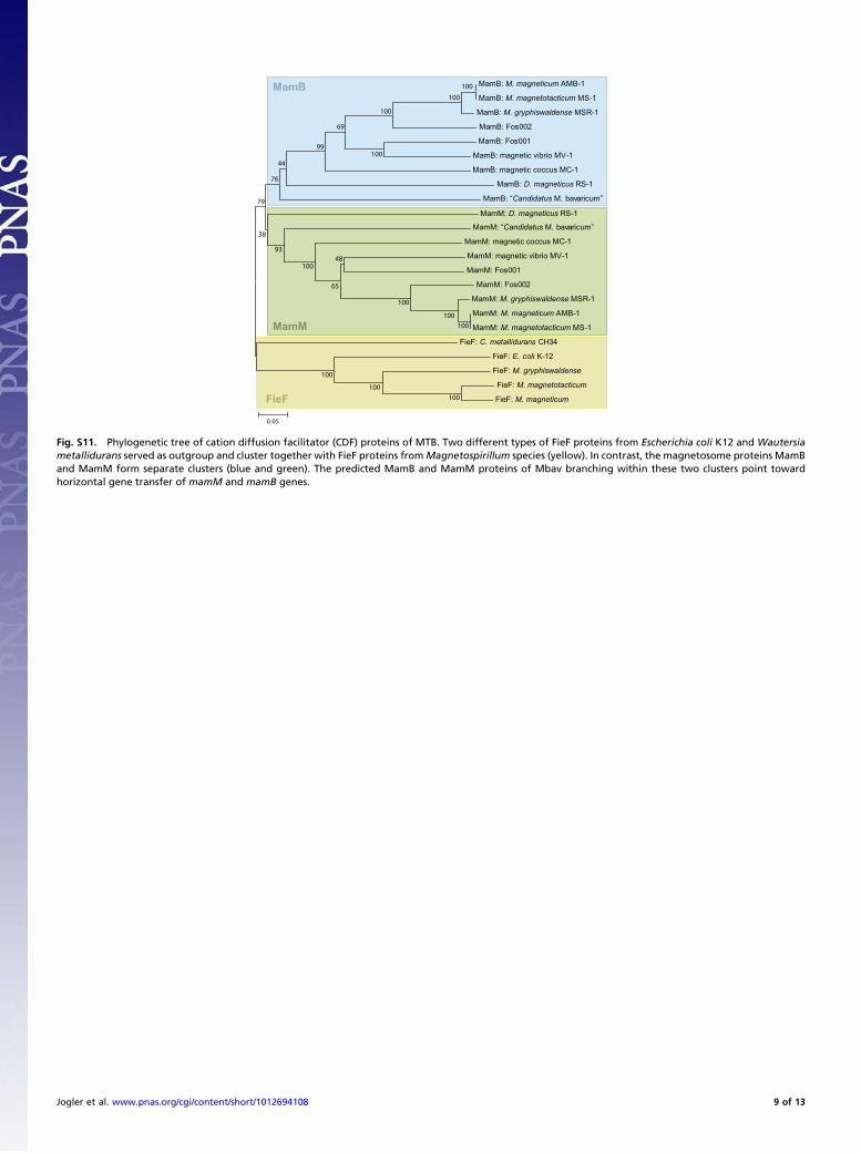
Fig. S11. Phylogenetic tree of cation diffusion facilitator (CDF) proteins of MTB. Two different types of FieF proteins from Escherichia coli K12 and Wautersiametallidurans served as outgroup and cluster together with FieF proteins fromMagnetospirillum species (yellow). In contrast, the magnetosome proteins MamBand MamM form separate clusters (blue and green). The predicted MamB and MamM proteins of Mbav branching within these two clusters point towardhorizontal gene transfer of mamM and mamB genes.
Jogler et al. www.pnas.org/cgi/content/short/1012694108 9 of 13
Table S1. Blast analysis of proteins encoded by predicted genes against the NCBI database
Locus tag Annotation Best hit Identity, % e-value aa
emg00001 Hypothetical protein Gloeobacter violaceus 38 1–32 516emg00002 XRE family transcriptional regulator Yersinia pseudotuberculosis 33 7–17 206emg00003 Aspartate 1-decarboxylase Thermodesulfovibrio yellowstonii 52 4–32 117emg00004 4-hydroxy-3-methylbut-2-en-1-yl diphosphate synthase Thermodesulfovibrio yellowstonii 60 3–120 348emg00005 Hypothetical protein Thermodesulfovibrio yellowstonii 45 3–72 319emg00006 Hypothetical protein Thermodesulfovibrio yellowstonii 61 1–51 180emg00007 Transcriptional regulator, MerR family Sphaerobacter thermophilus 33 4–16 128emg00008 IS1 transposase B Escherichia coli 100 1–94 167emg00009 Hypothetical protein Syntrophomonas wolfei 30 4–57 791emg00010 Transposase Plasmid R100 100 0.0 402emg00011 Hypothetical protein Syntrophobacter fumaroxidans 41 1–67 1,038emg00012 Hypothetical protein Chloroflexus aggregans 32 5–64 1,135emg00013 Polysaccharide biosynthesis protein Methanococcus aeolicus 23 2–20 489emg00014 Hypothetical protein Sulfurihydrogenibium azorense 51 0.032 63emg00015 Conserved hypothetical protein Aspergillus flavus 47 5–04 229emg00016 Hypothetical protein Desulfovibrio magneticus 39 1–15 237emg00017 Pyruvate phosphate dikinase Anaplasma marginale 23 0.003 196emg00018 Hypothetical protein Desulfovibrio magneticus 39 1–73 543emg00019 Putative membrane protein Carboxydothermus hydrogenoformans 43 5–46 358emg00020 Hypothetical protein Bacteroides coprocola 35 0.16 383emg00021 NA NA NA N.A. 69emg00022 Condensin subunit Smc Methanohalophilus mahii 27 1.9 252emg00023 NA NA NA N.A. 46emg00024 Similar to GA14224-PA Tribolium castaneum 22 0.14 146emg00025 LemA protein (mamQ-I) Mitsuokella multacida 36 5–25 186emg00026 Magnetosome protein MamE Uncultured bacterium 31 2–57 603emg00027 Magnetosome protein MamI Magnetic vibrio MV-1 29 0.78 62emg00028 TPR Domain containing protein (mamA) Tetrahymena thermophila 32 8–23 216emg00029 Hypothetical membrane protein Desulfovibrio magneticus 28 1–07 127emg00030 Magnetosome protein MamB Uncultured bacterium 32 3–41 297emg00031 LemA protein (mamQ-II) Campylobacter showae 32 2–17 182emg00032 Hypothetical protein Desulfitobacterium hafniense 27 7–08 227emg00033 Magnetosome protein MamM Magnetospirillum gryphiswaldense 35 2–44 307emg00034 Magnetosome protein MamP Magnetic vibrio MV-1 37 2–23 375
Results of BlastP analysis of proteins encoded by predicted genes of the 37,160-bp genomic fragment from Candidatus Magnetobacterium bavaricumagainst the NCBI database. Best BlastP hits are shown. Genes of a magnetosome cluster are in boldface type. The genes egm00025 and egm00031 encodeproteins that were found to contain a LemA motive and were annotated as mamQ-like. Gene egm00028 encodes a protein containing a TPR domain and wasannotated as mamA-like (see text for details). All other genes were annotated according their best BlastP hits. NCBI, National Center for BiotechnologyInformation.
Table S2. BlastP similarities of putative magnetosome proteinsfrom Candidatus Magnetobacterium bavaricum with theirhomologs from all MTB sequenced thus far
Protein MSR-1, % AMB-1, % MS-1, % MV-1, % MC-1, % RS-1, %
MamP 38 38 39 37 43 40MamM 35 35 36 32 36 27MamQ-II 24 21 21 23 22 26MamB 31 30 30 30 32 34MamA 24 23 23 26 NA 25MamI 25 23 23 29 35 35MamE 40 26 26 36 30 30MamQ-I 28 27 28 26 NA 36
Local identity values are shown. See Figs. 4 and 5 and Figs. S7 and S8 foranalysis involving ClustalW alignments of entire protein sequences. MTB,magnetotactic bacteria.
Jogler et al. www.pnas.org/cgi/content/short/1012694108 10 of 13
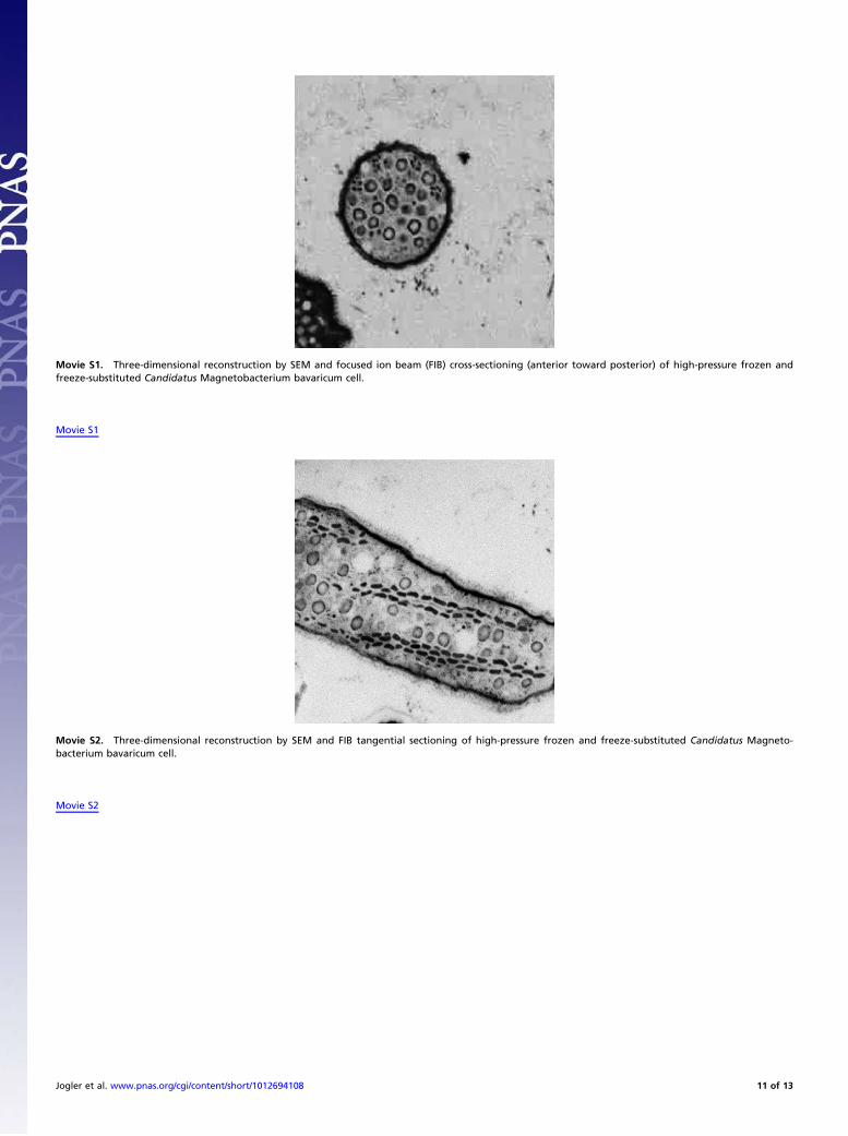
Movie S1. Three-dimensional reconstruction by SEM and focused ion beam (FIB) cross-sectioning (anterior toward posterior) of high-pressure frozen andfreeze-substituted Candidatus Magnetobacterium bavaricum cell.
Movie S1
Movie S2. Three-dimensional reconstruction by SEM and FIB tangential sectioning of high-pressure frozen and freeze-substituted Candidatus Magneto-bacterium bavaricum cell.
Movie S2
Jogler et al. www.pnas.org/cgi/content/short/1012694108 11 of 13
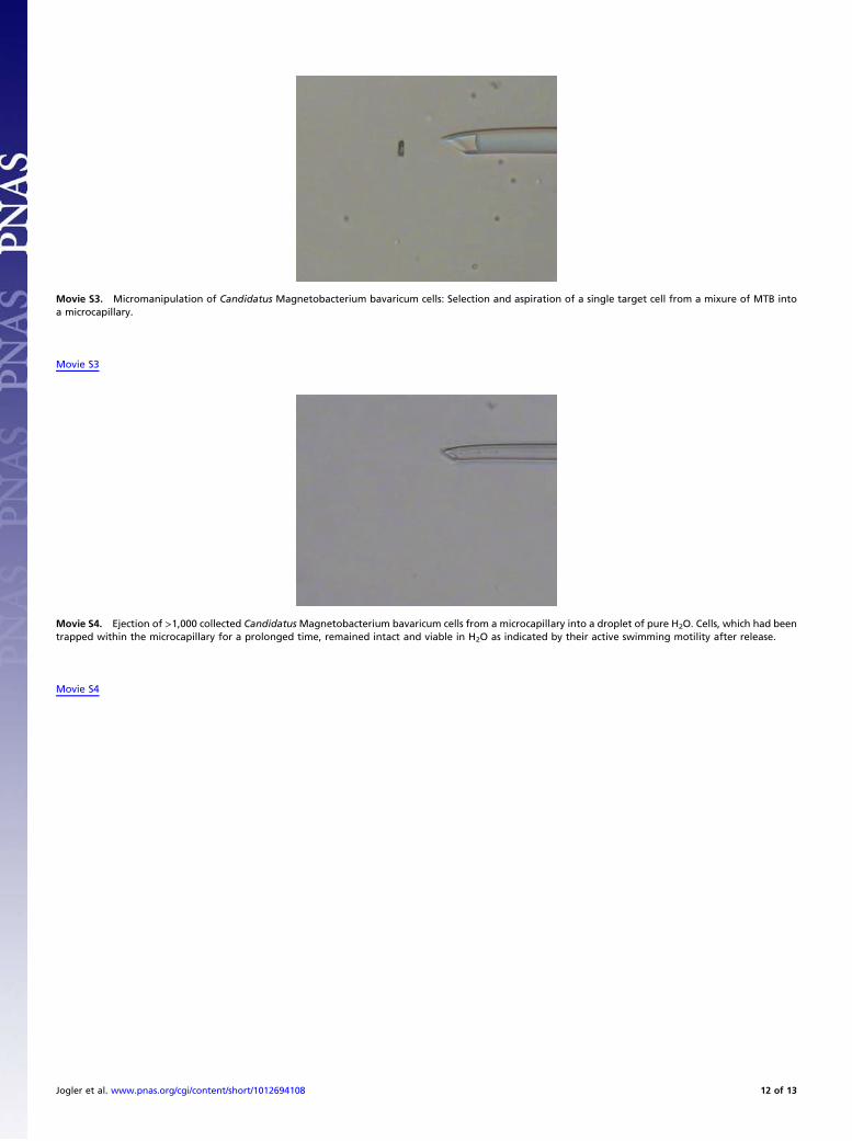
Movie S3. Micromanipulation of Candidatus Magnetobacterium bavaricum cells: Selection and aspiration of a single target cell from a mixure of MTB intoa microcapillary.
Movie S3
Movie S4. Ejection of >1,000 collected CandidatusMagnetobacterium bavaricum cells from a microcapillary into a droplet of pure H2O. Cells, which had beentrapped within the microcapillary for a prolonged time, remained intact and viable in H2O as indicated by their active swimming motility after release.
Movie S4
Jogler et al. www.pnas.org/cgi/content/short/1012694108 12 of 13
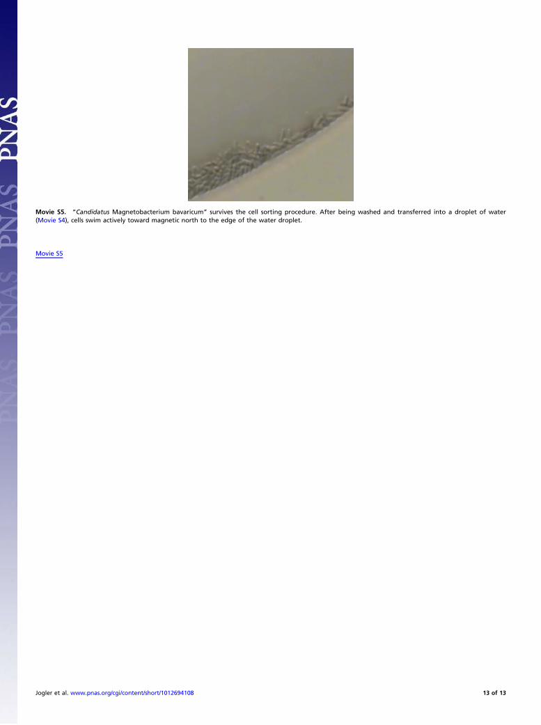
Movie S5. “Candidatus Magnetobacterium bavaricum” survives the cell sorting procedure. After being washed and transferred into a droplet of water(Movie S4), cells swim actively toward magnetic north to the edge of the water droplet.
Movie S5
Jogler et al. www.pnas.org/cgi/content/short/1012694108 13 of 13
Top Related