


Languages
Pages
Legal

SingleSingle-lung Ventilation for -lung Ventilation for PulmoPulmonarynary LLobe Resection obe Resection inin
a Nea Newbowbornrn
SingleSingle lung Ventilation for- lung Ventilation for-PulmoPulmonarynary LL obe Resection obe Resectioninin
a Nea Newbowbornrn
Tariq AlzahraniTariq Alzahrani
DemonstratorDemonstrator
College of MedicineCollege of Medicine
King Saud UniversityKing Saud University
Introduction • For lung isolation - double-lumen tubes - univent tube - single-lumen endobronchial tubes - endobronchial blockers• Lung isolation is generally limited
to children older than 1yr.
History • 34 wk gestation.• 3 kg .• Congenital emphysema of the left
upper lobe.• Infective pulmonary complication .• Intermittent ventilatory support
with 3.5mm(ID) nasotracheal tube
• Increasing volume of the left upper pulmonary lobe caused progressive mediastinal shift & the need for urgent surgery.
• At the time of surgery , the infant was spontaneously breathing with a natural airway & was 40 days old .
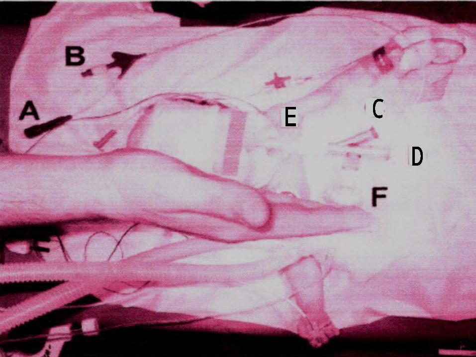
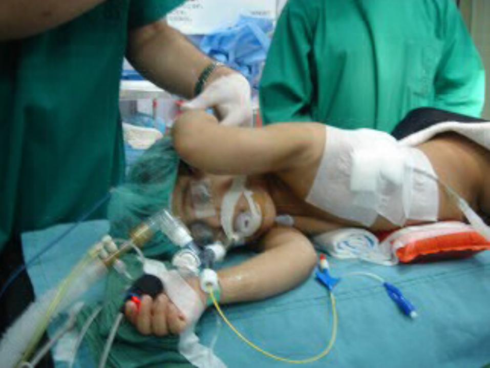
Intra Operative • G.A (thiopental ,
sufentanil ,rocuronium , & sevoflurane )• Monitored (pulse oximetry , ECG,
temperature probe , capnography & Lt radial artery invasive B.P)
• A 22G central venous catheter was placed via the Rt internal jugular vein .
• Intubated nasally with an uncuffed 4mm ID.
• FWEB (fiberoptically directed ,wire-guided 5f endobronchial blocker) was coaxially guided into the left main stem bronchus using a 2 mm pediatric fiberscope.
• The left lung collapsed by continuously suctioning the 0.7 mm lumen of the FWEB after removal of the guidewire.
• Right-sided decubitus position & positioning of FWEB was verified fiberoptically .
• 2.5 h , R.R 30-40/min , PAWP was limited to 25cm H2o resulting in a minute volume of 1.4-1.7L , fio2 1, pao2
350mmhg . • Paco2 increased during mechanical
ventilation to maximum of 84 mmhg corresponding to a pH of 7.06 .
• Manually ventilated .• After resection of the left upper
pulmonary lobe , the left lung was cautiously expanded under visual control & the FWEB removed .
• PICU with 4mm ET still in place .• 1 day postoperative , the infant was
nasally extubated & after uneventful recovery discharged to the word .
Discussion • Youngest child for SLV.• A regular cuffed ET that is 0.5-1 mm
ID smaller than indicated for endotracheal intubation can be used to allow the cuff to fit the main stem bronchus .
• 3 mm cuffed tubes have been applied in infants < 12 months old , this technique has certain limitations :
1. The mainstem bronchi are out of reach for conventional ETS if the nasotracheal route is preferred to minimize the risk of dislocation .
2. Significant airway trauma may result from advancing an ET blindly , especially if a stylet is used to enter the left mainstem bronchus .
3. Hypoxemia may result from obstruction of the upper lobe bronchus by the cuff of the ET , typically when the short right mainstem bronchus is intubated .
4. Suction cannot be applied to the operative side to promote lung collapse .
5. O2 & continuous positive airway pressure cannot be administered to the operative lung if the patient experiences O2 desaturation .
• The component of the system (multiport adaptor , moisture exchange filter ) significantly add to dead space ventilation leading to the limits of acceptable respiratory acidosis in this case .
• Others have used continues caudal epidural anesthesia in congenital lobar emphysema in an attempt to avoid positive pressure ventilation .

Conclusion
They have demonstrated the feasibility of SLV in a newborn using a coaxially placed fiberoptically directed endobronchial blocker .
Top Related