


Languages
Pages
Legal

Aus dem Institut für Klinische Chemie und Laboratoriumsmedizin
(Leiter Prof. Dr. med. M. Nauck)
der Universitätsmedizin der Ernst-Moritz-Arndt-Universität Greifswald
Serum Prolaktin-Konzentrationen alsRisikofaktor des Metabolischen
Syndroms oder Typ 2 Diabetes Mellitus?
INAUGURAL � DISSERTATION
zur
Erlangung des akademischen Grades
Doktor der Medizin
(Dr. med.)
der
Universitätsmedizin
der
Ernst-Moritz-Arndt-Universität
Greifswald
2013
vorgelegt von
Lisa Balbach
geb. am 10.06.1988
in Berlin

Dekan:1. Gutachter:2. Gutachter:
Ort, Raum:Tag der Disputation:
Prof. Dr. med. H. WallaschowskiProf. Dr. rer. nat. Max P. Bauer
Prof. Dr. med. M. Blüher
Greifswald, Universitätsklinikum, Raum 0.6503.06.2016
Inhaltsverzeichnis
1 Einleitung 1
2 Methoden 2
2.1 Studienpopulation und -design . . . . . . . . . . . . . . . . . . . . . 2
2.2 Datenerhebung und Laboranalysen . . . . . . . . . . . . . . . . . . 3
2.3 Statistische Methoden . . . . . . . . . . . . . . . . . . . . . . . . . 5
3 Ergebnisse 6
4 Diskussion 11
4.1 Fazit . . . . . . . . . . . . . . . . . . . . . . . . . . . . . . . . . . . 14
5 Zusammenfassung 15
6 Literatur 16
7 Anhang
7.1 Publikation
7.2 Eidesstattliche Erklärung
7.3 Lebenslauf
7.4 Danksagung

Abbildungsverzeichnis
1 Boxplots für den Zusammenhang der baseline Prolaktin-Konzentra-
tionen (25. und 75. Perzentil) und der Anzahl Metabolischer Syn-
drom Komponenten zum 5-Jahres-Follow-up; getrennt nach Män-
nern und Frauen. . . . . . . . . . . . . . . . . . . . . . . . . . . . . 10
Tabellenverzeichnis
1 Baseline Charakteristika der Studienpopulation; strati�ziert nach
dem Geschlecht. . . . . . . . . . . . . . . . . . . . . . . . . . . . . . 7
2 Assoziation zwischen der Serum Prolaktin-Konzentration und dem
Metabolischen Syndrom sowie dem Typ 2 Diabetes Mellitus im
Quer- und Längsschnitt. . . . . . . . . . . . . . . . . . . . . . . . . 9
Abkürzungsverzeichnis
ATC-Code Anatomical Therapeutic Chemical Code
(Anatomisch-therapeutisch-chemisches Klassi�kationssystem)
BMI Body Mass Index
CI Kon�denzintervall
HDL high-density lipoprotein (Lipoprotein hoher Dichte)
HOMA-IR Homeostasis Model Assessment Index
LDL low-density lipoprotein (Lipoprotein niedriger Dichte)
MetS Metablisches Syndrom
PRL Prolaktin
Q Quartil
RR Relatives Risiko
SD Standardabweichung
SHIP Study of Health in Pomerania
T2DM Typ 2 Diabetes Mellitus
Einleitung
1 Einleitung
Prolaktin (PRL), ein Hormon der Hypophyse, ist essenziell für verschiedene phy-
siologische Funktionen im menschlichen Körper.1�3 Es ist nicht nur bedeutsam
für die Initiation und Aufrechterhaltung der Laktation, sondern ist darüber hin-
aus involviert in Reproduktion, Wachstum und Entwicklung, Osmoregulation, Ge-
hirnfunktion, Verhalten und andere metabolische Vorgänge im menschlichen Or-
ganismus.1�3 Der PRL-Rezeptor wird in verschiedenen Geweben und Zellen, wie
lymphoiden Zellen, im Endometrium, in der Prostata und in den Adipozyten ex-
primiert.1�3
Das im Zusammenhang mit der vorliegenden Arbeit bedeutsame Metablisches Syn-
drom (MetS) beschreibt das gehäufte Auftreten von kardiovaskulären Risikofak-
toren, wie Übergewicht, Hypertriglyceridämie, Hypertonus und Insulinresistenz4,5
und ist häu�g mit der Entwicklung eines Typ 2 Diabetes Mellitus (T2DM) assozi-
iert.
Es existieren jedoch nur wenige Beobachtungsstudien hinsichtlich eines potenti-
ellen Zusammenhangs zwischen PRL und einem T2DM beziehungsweise einem
MetS. Dabei lassen experimentelle Studien durchaus einen Ein�uss von PRL auf
den T2DM mit metabolischen E�ekten auf das Fettgewebe,2,3 die Entwicklung
und das Wachstum der β-Zellen des Pankreas,6,7 eine Insulinresistenz3,8 sowie den
Lipidsto�wechsel3,9 vermuten. Die Fähigkeit von PRL, die Insulinfreisetzung zu
stimulieren sowie die Adiponektin- und Interleukin-6-Freisetzung zu unterdrücken,
deutet auf eine potentielle Rolle von PRL hinsichtlich der Manifestation einer In-
sulinresistenz hin.2 Während einige Studien zu dem Ergebnis kamen, dass PRL das
Wachstum bzw. die Entwicklung von pankreatischen β-Zellen fördert und die In-
sulinsekretion unterstützt,6,7 konnten andere Studien keinen Zusammenhang zwi-
schen PRL und metabolischen Störungen feststellen.10
Eine Beobachtungsstudie unter Patienten mit erektiler Dysfunktion zeigte eine
Assoziation zwischen niedrigen PRL-Konzentrationen und ungünstigen kardiovas-
kulären Risikopro�len sowie anderen klinischen Ereignissen.11 Allerdings stand in
den meisten der früheren Studien10,11 die Korrelation zwischen PRL und kardio-
metabolischen Risikofaktoren, einschlieÿlich des MetS oder des T2DM, nicht im
1
Studienpopulation und -design Methoden
Hauptfokus. Zudem wurden die früheren Studien lediglich mit kleinen und stark se-
lektierten Patientenpopulationen durchgeführt, wobei relevante Störfaktoren nicht
ausreichend berücksichtigt wurden.
Aufgrund der schlechten Vergleichbarkeit und der nur beschränkten Aussagekraft
der oben aufgeführten bisherigen Studien ist es deshalb das Ziel der vorliegen-
den Arbeit, auf Basis der populationsbasierten Kohortenstudie Study of Health in
Pomerania (SHIP) mit einem groÿen, repräsentativen Kollektiv von 3.993 Proban-
den, die möglichen Assoziationen von PRL zu einem MetS beziehungsweise einem
T2DM zu untersuchen.
2 Methoden
2.1 Studienpopulation und -design
Die SHIP-Studie ist eine populationsbasierte Kohortenstudie aus Vorpommern,
einer Region im nord-östlichen Teil Deutschlands. Details bezüglich des Studi-
endesigns, der Rekrutierung und Datenerhebung wurden bereits verö�entlicht.12
Dabei erfolgte im Jahr 1996 aus der Gesamtbevölkerung von Vorpommern mit
213.057 Einwohnern eine zweistu�g geschichtete Stichprobe von Erwachsenen im
Alter von 20-79 Jahren. Die Netto-Stichprobe (ohne migrierte oder verstorbene
Personen) umfasste 6.265 teilnahmefähige Probanden. Es wurden nur Personen
mit deutscher Staatsangehörigkeit und Wohnsitz in dem Hauptuntersuchungsge-
biet eingeschlossen. Alle Probanden erhielten ein Maximum von drei schriftlichen
Einladungen. In Fällen von Nichtbeantwortung folgten auf die Briefe wiederholte
Telefonanrufe oder aber Hausbesuche, wenn der Kontakt per Telefon nicht möglich
war.13
Nach Erteilung der schriftlichen Einwilligung wurden zwischen 1997 und 2001 4.308
Teilnehmer (davon 2.192 Frauen) der Basisstudie (SHIP-0) untersucht (Respon-
se 68,8%). Während des Fünf-Jahres-Follow-ups (SHIP-1) wurden zwischen 2002
und 2006 3.300 Teilnehmer (davon 1.711 Frauen) erneut untersucht (Response
83,6%). Die Studie entsprach den Prinzipien der Deklaration von Helsinki; die
Ethikkommission der Universität Greifswald genehmigte das Protokoll. Von den
2
Datenerhebung und Laboranalysen Methoden
4.308 Teilnehmern der Baseline Studie wurden Individuen nach folgenden Kriterien
ausgeschlossen: fehlende PRL-Daten (N = 169), PRL-Konzentration > 100µg/l
(N = 16), Schwangerschaft (N = 8), Erkrankungen der Hypophyse (N = 1) oder
fehlende Daten für Kovarianten (N = 121). Die endgültige Studiengröÿe umfasste
somit 3.993 Personen (davon 2.027 Frauen).
2.2 Datenerhebung und Laboranalysen
Informationen über sozio-demographische Merkmale und den gesundheitsbezoge-
nen Lebensstil einschlieÿlich Rauchgewohnheiten (kategorisiert in Raucher, ehe-
malige-Raucher und Nicht-Raucher), Bildungsniveau (< 10, = 10 oder > 10 Jahre
Schulbildung), Parität (Anzahl leiblicher Kinder), körperliche Aktivität
(> 1 h/Woche körperliches Training im Sommer oder Winter) und Alkoholkon-
sum wurden durch persönliche Interviews erhoben. Der Taillenumfang wurde auf
0,1 cm genau mit einem unelastischen Band in der Mitte zwischen dem unteren
Rippenbogen und dem Beckenkamm in der horizontalen Ebene, in stehender Hal-
tung und mit gleichmäÿiger Gewichtsverteilung auf beide Füÿe gemessen. Kör-
pergröÿe und Gewicht wurden für die Berechnung des Body Mass Index (BMI)
(Körpergewicht [kg]/(Körpergröÿe [m])2) gemessen.
Nach einer Ruhezeit von mindestens fünf Minuten wurde der systolische und dia-
stolische Blutdruck dreimal am rechten Arm des sitzenden Teilnehmers mit einem
oszillometrischen digitalen Blutdruckmessgerät (HEM-705CP, Omron Corporati-
on, Tokyo, Japan) gemessen. Das Intervall zwischen den Messungen betrug drei
Minuten. Der mittlere systolische und diastolische Blutdruck wurde aus der zwei-
ten und dritten Messung berechnet.14
Der menopausale Status (prä- und postmenopausal) wurde nach einer vorher fest-
gelegten De�nition bestimmt. Als prämenopausal wurden Frauen < 40 Jahre oder
zwischen 40 und 60 Jahren und bestehendem Menstruationszyklus eingestuft; als
postmenopausal eingeordnet wurden Frauen ≥ 60 Jahre und alle Frauen zwischen
40 und 60 Jahren, die über keinen Menstruationszyklus berichteten.15
Die Messung der Serum HDL-(High-Density-Lipoprotein) Konzentrationen erfolg-
te zu Studienbeginn photometrisch (Hitachi 704, Roche, Mannheim, Deutsch-
land), wohingegen die high-density lipoprotein (Lipoprotein hoher Dichte) (HDL)-
3
Datenerhebung und Laboranalysen Methoden
Konzentrationen beim Follow-up mit Hilfe einer Lipid-Elektrophorese (HELENA
SAS-3-System, 7 Helena Bio Sciences Europe, Tyne & Wear, UK) ermittelt wur-
den.
Um die Vergleichbarkeit der HDL-Analysen im Längsschnitt zu gewährleisten, der
Ausgangswert der SHIP-0-HDL-Konzentrationen als Referenz verwendet. Mithilfe
einer zuvor verö�entlichten Umrechnungsformel konnten die korrigierten SHIP-1-
HDL-Konzentrationen berechnet werden.16 Dabei konnte festgestellt werden, dass
die durchschnittlichen HDL-Konzentrationen beider Methoden praktisch identisch,
und damit die Unterschiede im HDL nur von geringer Relevanz waren.17 Die
Serum Triglycerid- und Glukosekonzentrationen wurden enzymatisch unter Ver-
wendung von Reagenzien von Roche Diagnostics ermittelt (Hitachi 717, Roche
Diagnostics, Mannheim, Deutschland). Die Bestimmung von glykiertem Hämoglo-
bin (HbA1c) erfolgte durch eine Hochleistungs-Flüssigchromatographie (Bio-Rad,
München, Deutschland).
Im Verlauf der Studie betrug der Variationskoe�zient zwischen den Analysen für
HbA1c 2,76% im niedrigen Pool und 1,38% im hohen Pool. Alle Analysen wurden
nach Empfehlungen der Hersteller von quali�ziertem Fachpersonal durchgeführt
und täglichen internen Qualitätskontrollen unterzogen. Darüber hinaus beteiligte
sich das Labor an den o�ziellen deutschen, vierteljährlichen externen Eignungs-
prüfungsprogrammen.
Der T2DM wurde auf der Basis eigener Angaben, der ärztlichen Diagnose oder
dem Einsatz von Antidiabetika (ATC-Code A10) in den letzten sieben Tagen
oder durch HbA1c Konzentrationen > 6,5% de�niert. Die diagnostischen Kriteri-
en für die Beurteilung der MetS Komponenten wurden gemäÿ der gemeinsamen
wissenschaftlichen Erklärung zum MetS18 de�niert und wie bereits zuvor in der
SHIP-Studie19�22 und anderen groÿen Kohortenstudien23 für die Verwendung der
Blutproben wie folgt modi�ziert:
1. erhöhter Taillenumfang: Männer > 94 cm, Frauen > 80 cm;
2. erhöhter Blutzucker: ≥ 8,0mmol/l oder antidiabetische Behandlung
(ATC-Codes A10A, A10B);
3. vermindertes HDL-Cholesterin: Männer < 1,0mmol/l, Frauen < 1,3mmol/l,
oder lipidsenkende Medikation (ATC-Codes C10AB, A10AD);
4
Statistische Methoden Methoden
4. erhöhte Nicht-Nüchtern-Triglyceride: ≥ 2,3mmol/l oder lipidsenkende Me-
dikation (ATC-Codes C10AB, A10AD);
5. erhöhter Blutdruck: ≥ 130/85mmHg oder selbstberichtete medikamentöse
antihypertensive Therapie.
Von einem MetS bei den Teilnehmern wurde dann ausgegangen, wenn mindestens
drei von diesen fünf Komponenten vorhanden waren.
Die Analyse der PRL-Konzentrationen erfolgte aus eingefrorenen Seren (−80 ◦C)
der Basis-Teilnehmer. Die Blutproben wurden aus der Kubitalvene in Rückenla-
ge zwischen 7.00 und 19.00 Uhr abgenommen. Die Analyse erfolgte unter Ver-
wendung eines chemiluminescentimmunometric Assay mittels eines Immulite 2500
Analysators (Ref. L5KPR, DPC Biermann GmbH, Bad Nauheim, Deutschland).
Ein Aliquot von zwei alternierenden Niveaus von einem dritten kommerziellen
Kontrollmaterial (Bio-Rad Lyphochek Immunoassay Plus-Control, Bio-Rad, Mün-
chen, Deutschland) war in jeder Serie beider einzelnen Bestimmungen enthalten.
Im Verlauf der Studie lag der Variationskoe�zient zwischen den Analysen bei 5,6%
mit einer systematischen Abweichung von −4,3% auf dem 6,3µg/l-Niveau, und
4,3% mit einer systematischen Abweichung von −6,4% auf dem 14,5µg/l-Niveau.
Die analytische Sensitivität betrug 0,5µg/l bei einem e�ektiven Messbereich von
0,5 bis 150µg/l.
2.3 Statistische Methoden
Kategoriale Daten wurden in Prozent und kontinuierliche Daten als Median (p25th,
p75th) ausgedrückt. Geschlechtsbezogene Unterschiede wurden mit dem χ2-Test
(kategoriale Daten) und dem Mann-Whitney-U-Test (kontinuierliche Daten) un-
tersucht. Die stark asymmetrisch verteilten PRL-Werte wurde log-transformiert,
um eine Normalverteilung zu modellieren.
Zur Analyse von Quer- und Längsschnittassoziationen zwischen PRL, MetS und
T2DMwurden alters-adjustierte und multivariable Poisson-Regressionsmodelle mit
robusten Standardfehlern implementiert. Die Ergebnisse wurden präsentiert als
Relatives Risiko (RR), inklusive 95% Kon�denzintervalle (95% CI), pro SD An-
stieg der kontinuierlichen log-PRL-Variablen beziehungsweise für geschlechtsspe-
5
Ergebnisse
zi�sch kategorisierte PRL-Quartile. Die longitudinalen Inzidenz-Analysen wurden
nur bei Personen ohne MetS und T2DM zu SHIP-0 durchgeführt. In Trend-Tests
wurden die PRL-Quartile als ordinale Variablen in den Regressionsmodellen be-
rücksichtigt.
Multivariable Regressionsmodelle wurden für die Kovariablen Alter, BMI, Rau-
chen, körperliche Aktivität, Bildungsniveau und Alkoholkonsum adjustiert.
Alle Analysen wurden geschlechtsgetrennt durchgeführt. Die Validität der Ergeb-
nisse wurde durch verschiedene Sensitivitätsanalysen überprüft. Bei Frauen wur-
den die multivariablen Regressionsmodelle zusätzlich für den menopausalen Sta-
tus und die Parität adjustiert. Sämtliche Regressionsanalysen wurden nach dem
Ausschluss von 184 Individuen, die PRL beein�ussende Medikamente wie Me-
toclopramid (A03FA01, A03FA03, N02CX59), Cimetidin (A02BA01), Reserpin
(C02LA01), Methyldopa (C02AB) und Psychoanaleptika (ATC-Code N06) ein-
nahmen, wiederholt.24 Der mögliche Ein�uss des Non-Response-Bias auf die un-
tersuchten Zusammenhänge wurde durch eine Gewichtung der Teilnahmewahr-
scheinlichkeit abgeschätzt.25
Schlieÿlich wurden die multivariablen Regressionsmodelle bezüglich des Blutent-
nahmezeitpunkts adjustiert, um die potenziellen Auswirkungen des zirkadianen
Rhythmus der PRL-Sekretion auf die Ergebnisse zu bewerten. Das Signi�kanzni-
veau wurde auf p < 0.05 festgelegt. Alle statistischen Analysen wurden mit Hilfe
des Statistiksoftwarepakets Stata 11.0 (Stata Corporation, College Station, TX,
USA) durchgeführt.
3 Ergebnisse
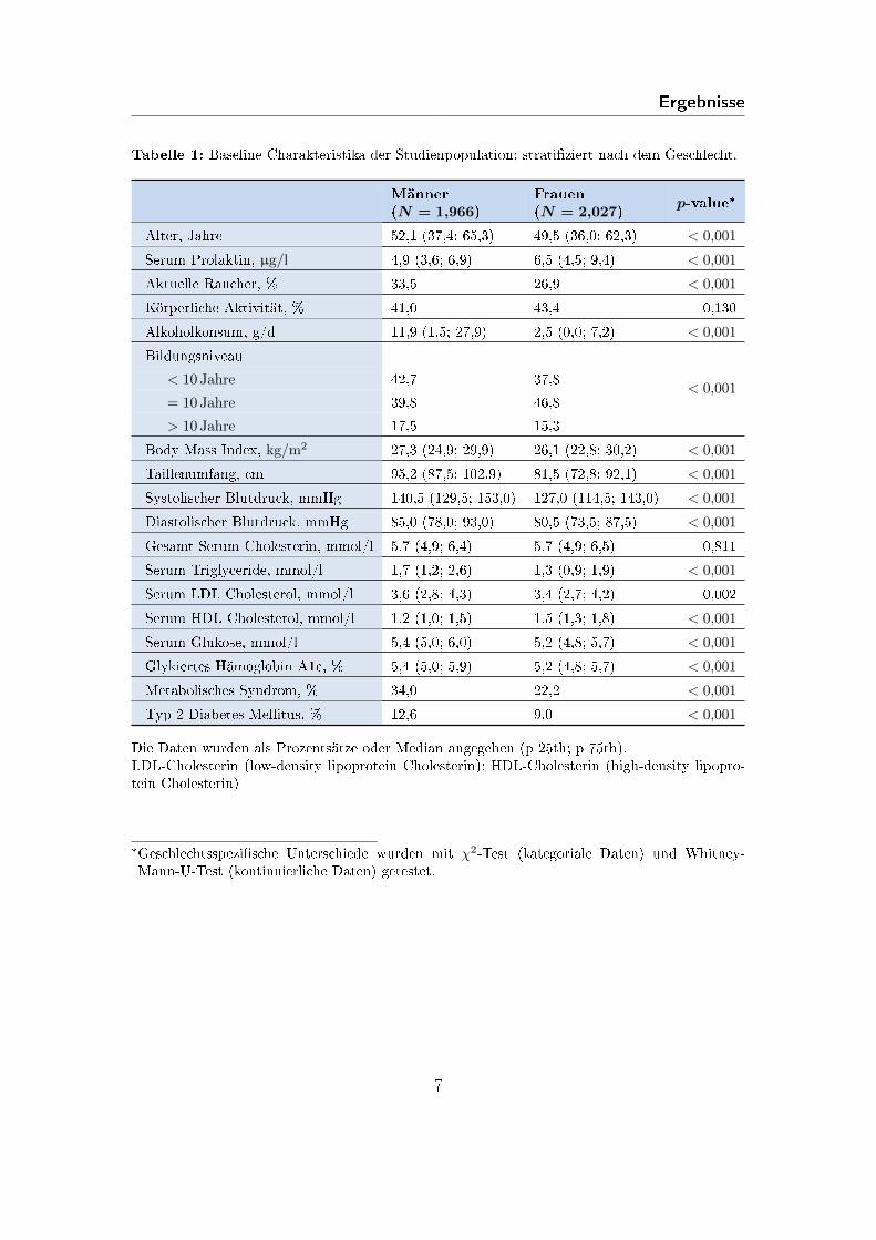
Vergleicht man die Baseline Charakteristika der Studienstichprobe bezogen auf
das Geschlecht, so wiesen männliche Probanden signi�kant niedrigere PRL-Kon-
zentrationen auf und hatten ein höheres Herz-Kreislauf-Risiko (unter Berücksich-
tigung von Rauchgewohnheiten, Alkoholkonsum, BMI, Blutdruck, Blutfettwerten
und Blutzucker) als Frauen (Tabelle 1).
6
Ergebnisse
Tabelle 1: Baseline Charakteristika der Studienpopulation; strati�ziert nach dem Geschlecht.
Männer(N = 1,966)
Frauen(N = 2,027)
p-value*
Alter, Jahre 52,1 (37,4; 65,3) 49,5 (36,0; 62,3) < 0,001
Serum Prolaktin, µg/l 4,9 (3,6; 6,9) 6,5 (4,5; 9,4) < 0,001
Aktuelle Raucher, % 33,5 26,9 < 0,001
Körperliche Aktivität, % 41,0 43,4 0,130
Alkoholkonsum, g/d 11,9 (1,5; 27,9) 2,5 (0,0; 7,2) < 0,001
Bildungsniveau
< 0,001< 10 Jahre 42,7 37,8
= 10 Jahre 39,8 46,8
> 10 Jahre 17,5 15,3
Body Mass Index, kg/m2 27,3 (24,9; 29,9) 26,1 (22,8; 30,2) < 0,001
Taillenumfang, cm 95,2 (87,5; 102,9) 81,5 (72,8; 92,1) < 0,001
Systolischer Blutdruck, mmHg 140,5 (129,5; 153,0) 127,0 (114,5; 143,0) < 0,001
Diastolischer Blutdruck, mmHg 85,0 (78,0; 93,0) 80,5 (73,5; 87,5) < 0,001
Gesamt Serum Cholesterin, mmol/l 5,7 (4,9; 6,4) 5,7 (4,9; 6,5) 0,811
Serum Triglyceride, mmol/l 1,7 (1,2; 2,6) 1,3 (0,9; 1,9) < 0,001
Serum LDL Cholesterol, mmol/l 3,6 (2,8; 4,3) 3,4 (2,7; 4,2) 0,002
Serum HDL Cholesterol, mmol/l 1,2 (1,0; 1,5) 1,5 (1,3; 1,8) < 0,001
Serum Glukose, mmol/l 5,4 (5,0; 6,0) 5,2 (4,8; 5,7) < 0,001
Glykiertes Hämoglobin A1c, % 5,4 (5,0; 5,9) 5,2 (4,8; 5,7) < 0,001
Metabolisches Syndrom, % 34,0 22,2 < 0,001
Typ 2 Diabetes Mellitus, % 12,6 9,0 < 0,001
Die Daten wurden als Prozentsätze oder Median angegeben (p 25th; p 75th).LDL-Cholesterin (low-density lipoprotein Cholesterin); HDL-Cholesterin (high-density lipopro-tein Cholesterin)
*Geschlechtsspezi�sche Unterschiede wurden mit χ2-Test (kategoriale Daten) und Whitney-Mann-U-Test (kontinuierliche Daten) getestet.
7
Ergebnisse
Auch die Prävalenz des MetS (34,0% vs. 22,2%) und des T2DM (12,6% vs. 9,0%)
war bei Männern höher als bei Frauen.
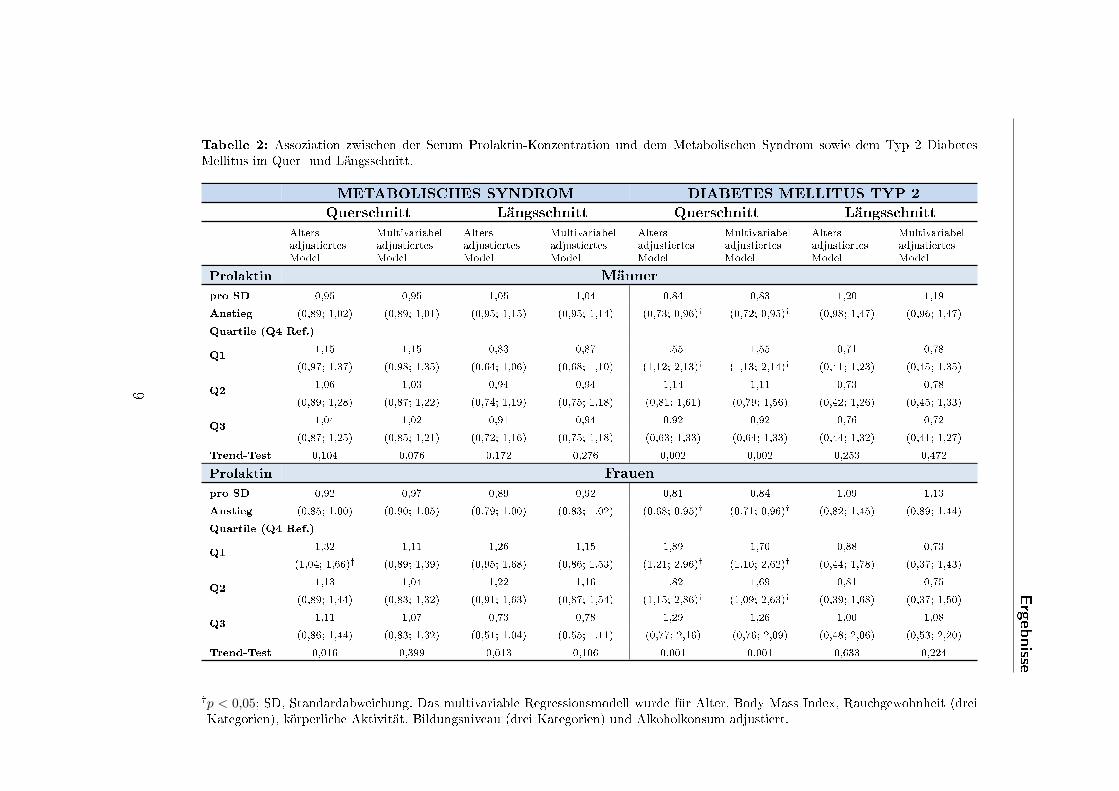
Im Querschnitt zeigte sich im altersadjustierten Regressionsmodell eine inverse
Assoziation zwischen PRL und dem MetS Risiko bei Frauen (Q1 vs. Q4: RR,
1,32, 95% CI, 1,04-1,66), aber nicht bei Männern (Q1 vs. Q4: RR, 1,15, 95%
CI, 0,97-1,37). Diese geschlechtsspezi�schen Zusammenhänge konnten jedoch nach
multivariabler Adjustierung nicht aufrechterhalten werden (Tabelle 2).
Dagegen fand sich in alters- und multivariabel adjustierten Regressionsmodellen
eine konsistente Assoziation zwischen niedrigen PRL-Konzentrationen und einem
prävalenten T2DM (Männer: Q1 vs. Q4: RR, 1,55; 95% CI, 1,13-2,14; Frauen: Q1
vs. Q4: RR, 1,70; 95% CI, 1,10-2,62). Darüber hinaus zeigten die Trend-Analysen
eine progressive inverse Beziehung über alle PRL-Quartile hinweg. Ebenso waren
höhere PRL-Konzentrationen mit einem deutlich niedrigeren T2DM Risiko assozi-
iert (RR pro SD Anstieg des log-PRL: 0,83; 95% CI, 0,72-0,95 bei Männern, und
0,84; 95% CI, 0,71-0,98 bei Frauen).
8
Ergebnisse
Tabelle 2: Assoziation zwischen der Serum Prolaktin-Konzentration und dem Metabolischen Syndrom sowie dem Typ 2 DiabetesMellitus im Quer- und Längsschnitt.
METABOLISCHES SYNDROM DIABETES MELLITUS TYP 2
Querschnitt Längsschnitt Querschnitt Längsschnitt
AltersadjustiertesModel
MultivariabeladjustiertesModel
AltersadjustiertesModel
MultivariabeladjustiertesModel
AltersadjustiertesModel
MultivariabeladjustiertesModel
AltersadjustiertesModel
MultivariabeladjustiertesModel
Prolaktin Männer
pro SD 0,95 0,95 1,05 1,04 0,84 0,83 1,20 1,19
Anstieg (0,89; 1,02) (0,89; 1,01) (0,95; 1,15) (0,95; 1,14) (0,73; 0,96)� (0,72; 0,95)� (0,98; 1,47) (0,96; 1,47)
Quartile (Q4 Ref.)
Q11,15 1,15 0,83 0,87 1,55 1,55 0,71 0,78
(0,97; 1,37) (0,98; 1,35) (0,64; 1,06) (0,68; 1,10) (1,12; 2,13)� (1,13; 2,14)� (0,41; 1,23) (0,45; 1,35)
Q21,06 1,03 0,94 0,94 1,14 1,11 0,73 0,78
(0,89; 1,28) (0,87; 1,22) (0,74; 1,19) (0,75; 1,18) (0,81; 1,61) (0,79; 1,56) (0,42; 1,26) (0,45; 1,33)
Q31,04 1,02 0,91 0,94 0,92 0,92 0,76 0,72
(0,87; 1,25) (0,85; 1,21) (0,72; 1,16) (0,75; 1,18) (0,63; 1,33) (0,64; 1,33) (0,44; 1,32) (0,41; 1,27)
Trend-Test 0,104 0,076 0,172 0,276 0,002 0,002 0,253 0,472
Prolaktin Frauen
pro SD 0,92 0,97 0,89 0,92 0,81 0,84 1,09 1,13
Anstieg (0,85; 1,00) (0,90; 1,05) (0,79; 1,00) (0,83; 1,02) (0,68; 0,95)� (0,71; 0,96)� (0,82; 1,45) (0,89; 1,44)
Quartile (Q4 Ref.)
Q11,32 1,11 1,26 1,15 1,89 1,70 0,88 0,73
(1,04; 1,66)� (0,89; 1,39) (0,95; 1,68) (0,86; 1,53) (1,21; 2,96)� (1,10; 2,62)� (0,44; 1,78) (0,37; 1,43)
Q21,13 1,04 1,22 1,16 1,82 1,69 0,81 0,75
(0,89; 1,44) (0,83; 1,32) (0,91; 1,63) (0,87; 1,54) (1,15; 2,86)� (1,09; 2,63)� (0,39; 1,68) (0,37; 1,50)
Q31,11 1,07 0,73 0,78 1,29 1,26 1,00 1,08
(0,86; 1,44) (0,83; 1,32) (0,51; 1,04) (0,55; 1,11) (0,77; 2,16) (0,76; 2,09) (0,48; 2,06) (0,53; 2,20)
Trend-Test 0,016 0,399 0,013 0,106 0,001 0,001 0,633 0,224
�p < 0,05; SD, Standardabweichung. Das multivariable Regressionsmodell wurde für Alter, Body Mass Index, Rauchgewohnheit (dreiKategorien), körperliche Aktivität, Bildungsniveau (drei Kategorien) und Alkoholkonsum adjustiert.
9
Ergebnisse
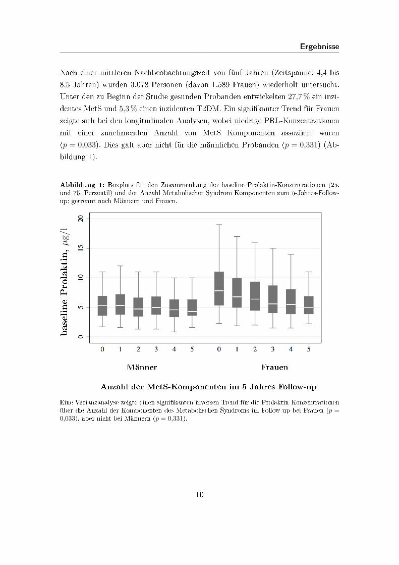
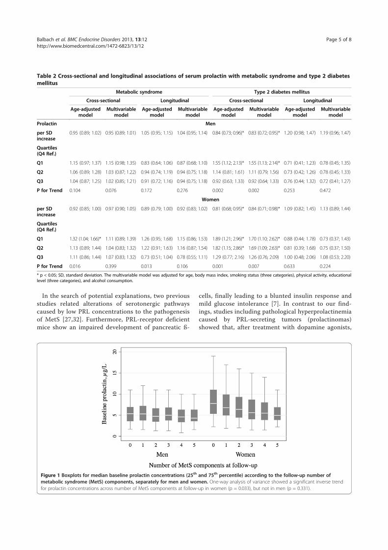
Nach einer mittleren Nachbeobachtungszeit von fünf Jahren (Zeitspanne: 4,4 bis
8,5 Jahren) wurden 3.078 Personen (davon 1.589 Frauen) wiederholt untersucht.
Unter den zu Beginn der Studie gesunden Probanden entwickelten 27,7% ein inzi-
dentes MetS und 5,3% einen inzidenten T2DM. Ein signi�kanter Trend für Frauen
zeigte sich bei den longitudinalen Analysen, wobei niedrige PRL-Konzentrationen
mit einer zunehmenden Anzahl von MetS Komponenten assoziiert waren
(p = 0,033). Dies galt aber nicht für die männlichen Probanden (p = 0,331) (Ab-
bildung 1).
Abbildung 1: Boxplots für den Zusammenhang der baseline Prolaktin-Konzentrationen (25.und 75. Perzentil) und der Anzahl Metabolischer Syndrom Komponenten zum 5-Jahres-Follow-up; getrennt nach Männern und Frauen.
baselinePro
laktin,µg/l
Männer Frauen
Anzahl der MetS-Komponenten im 5 Jahres Follow-up
Eine Varianzanalyse zeigte einen signi�kanten inversen Trend für die Prolaktin-Konzentrationenüber die Anzahl der Komponenten des Metabolischen Syndroms im Follow-up bei Frauen (p =0,033), aber nicht bei Männern (p = 0,331).
10
Diskussion
Multivariable Regressionsanalysen zeigten keinen Zusammenhang zwischen PRL
und der Inzidenz eines MetS beziehungsweise T2DM (Tabelle 2).
Die durchgeführten Sensitivitätsanalysen mit zusätzlicher Berücksichtigung des
menopausalen Status und der Parität, dem Ausschluss von Individuen mit Hin-
weis auf die Einnahme von PRL beein�ussenden Medikamenten, der zusätzlichen
Aufnahme von Ausschlusskriterien sowie der Anpassung des Blutentnahmezeit-
punktes zeigten keinen Ein�uss auf die Ergebnisse.
4 Diskussion
Die vorliegende Arbeit ist die erste umfassende, populationsbasierte Untersuchung
eines möglichen Zusammenhangs zwischen PRL und dem MetS beziehungswei-
se dem T2DM. Auf der Basis einer groÿen Stichprobe von 3.993 Individuen der
allgemeinen Bevölkerung Vorpommerns konnte in Querschnittsanalysen anhand
alters-adjustierter und multivariabler Regressionsmodelle eine inverse Assoziati-
on zwischen niedrigen PRL-Konzentrationen und einem erhöhten Risiko für einen
prävalenten T2DM festgestellt werden.
Die Ergebnisse bezüglich der Beziehung zwischen PRL und T2DM stehen im Ein-
klang mit früheren Beobachtungsstudien.11,26,27 Darunter war eine groÿ angeleg-
te Studie mit 2.351 männlichen Patienten mit erektiler Dysfunktion, in welcher
niedrige PRL-Konzentrationen mit einer steigenden Anzahl von MetS Faktoren
assoziiert waren.26 Vergleichbar mit der vorliegenden Arbeit konnten diese26 und
die darauf folgende Anschlussstudie11 eine inverse Assoziation zwischen PRL und
einem prävalenten T2DM feststellen.
Allerdings basieren beide Studien auf Daten von ausschlieÿlich männlichen Patien-
ten, was die Vergleichbarkeit mit der von uns präsentierten populationsbasierten
Studie limitiert. Des Weiteren konnte eine Querschnittsstudie mit 345 gesunden
Probanden im Alter von 30-55 Jahren eine negative Assoziation zwischen PRL
und einer Insulinresistenz sowie ein doppeltes Risiko für ein prävalentes MetS pro
Standardabweichung (SD) bei niedrigen PRL-Konzentrationen feststellen.27
11
Diskussion
Hinweise aus patientenbasierten Querschnittsstudien mit medikamentennaiven schi-
zophrenen Patienten deuten darauf hin, dass hohe Dopaminspiegel mit daraus
folgenden niedrigen PRL-Konzentrationen28,29 mit einem erhöhten T2DM Risiko,
einschlieÿlich Insulinresistenz und erhöhter Nüchternglukose,30 verbunden sind. So
wird vermutet, dass die typischen Psychopharmaka, durch Erhöhung der PRL-
Konzentrationen mittels Inhibierung der D2 Rezeptoren, möglicherweise vor dem
Entstehen und dem Voranschreiten eines T2DM schützten könnten.31 Da jedoch
diese früheren Studien28�30 ausschlieÿlich Querschnittsstudien waren, sind Ursache
und Wirkung in der Assoziation zwischen PRL und T2DM schwer zu bestimmen.
So lassen die Ergebnisse einer Interventionsstudie beispielsweise vermuten, dass die
Insulinsekretion eine wichtige Determinante für die PRL-Freisetzung darstellt.9
Auf der Suche nach möglichen Erklärungen zeigen zwei frühere Studien, dass Ver-
änderungen im serotonergen System und die dadurch bedingten niedrigen PRL-
Konzentrationen die Pathogenese des MetS beein�ussen.27,32 Darüber hinaus wei-
sen PRL-Rezeptor-de�ziente Mäuse eine Beeinträchtigung der Entwicklung der
pankreatischen β-Zellen auf, die letztlich zu einer abgestumpften Insulinantwort
und milden Glucose-Intoleranz führt.7
Im Gegensatz zu den vorliegenden Ergebnissen konnten Studien, welche Patien-
ten mit pathologischer Hyperprolaktinämie aufgrund PRL sezernierender Tumoren
(Prolaktinom) betrachteten, zeigen, dass nach Behandlung mit Dopaminagonis-
ten, die reduzierten PRL-Konzentrationen zu einer erhöhten Insulinemp�ndlich-
keit führten.33,34 Diese Ergebnisse wurden jedoch lediglich in kleinen Patienten-
studien mit klinisch bestätigten Prolaktinomen erhoben. Somit lagen die Serum
PRL-Konzentrationen oberhalb des Referenzbereiches, wodurch die Übertragbar-
keit auf die allgemeine Bevölkerung begrenzt ist.
Im Gegensatz dazu ändert sich der HOMA-IR als Maÿ für die Insulinresistenz
nach der Behandlung mit Dopaminagonisten nicht,33 und es besteht keine Korre-
lation der PRL-Konzentration mit der HOMA-IR Änderung oder der Abnahme
der Blutglukose.8,34 Interessanterweise zeigte auch eine frühere Querschnittsstu-
die bei adipösen Nicht-Prolaktinom Patienten eine fehlende Korrelation der PRL-
Konzentration mit Insulin, dem HOMA-IR und dem Glukosespiegel.10
12
Diskussion
Bereits verö�entlichte in-vitro Studien legen einen Ein�uss von PRL auf die β-Zell-
sekretion sowohl durch eine erhöhte Glukokinaseaktivität,35 als auch ein verbesser-
tes spezi�sches Überleben der β-Zellen36,37 beziehungsweise durch die Hemmung
der β-Zellapoptose38 nahe. Um allerdings die einzelnen Details bezüglich der Wir-
kungsweise von PRL auf die verschiedenen Gewebe und deren Auswirkungen auf
die kardiometabolischen Risikofaktoren zu verstehen, sind weitere in-vitro Studien
nötig.
Frühere, bereits publizierte Forschungsergebnisse deuten auf eine positive Asso-
ziation zwischen PRL und Entzündungsmarkern39 sowie der Mortalität (kardio-
vaskuläre und Gesamtmortalität)40 als auch auf eine inverse Korrelation zwischen
PRL und dem kardialen Umbau41 hin. Allerdings liefern die empirischen Ergeb-
nisse dieser und anderer Studien keine konsistenten Assoziationen. Infolgedessen
ergibt sich die Notwendigkeit weiterer Studien, um die mögliche Rolle des PRL als
Herz-Kreislauf-Risiko-Marker weiter zu untersuchen.
Für die Interpretation der vorliegenden Ergebnisse sollten folgende Stärken und
Schwächen berücksichtigt werden: Wesentliche Stärken bestehen in der groÿen
Stichprobe, dem longitudinalen Studiendesign und in der breiten Altersspanne.
Darüber hinaus wurden umfangreiche Sensitivitätsanalysen durchgeführt, um die
Gültigkeit der aktuellen Ergebnisse zu bewerten.
Eine wichtige Limitation dagegen stellt die einmalige Serum PRL-Messung von
Nicht-Nüchtern-Blutproben dar, welche nicht der pulsatilen Freisetzung von PRL
angepasst wurde. Jedoch konnte gezeigt werden, dass die pulsatile Sekretion von
PRL vor allem während der Nacht auftritt und in der Zeit von 9.00 bis 17.00 Uhr
relativ konstant bleibt.42 Weitere Einschränkungen ergeben sich aus der kaukasi-
schen Studienpopulation, die die Generalisierbarkeit der vorliegenden Ergebnisse
erschwert. Zudem blieben weitere endokrine Erkrankungen wie zum Beispiel eine
Hyperthyreose,43,44 eine Akromegalie oder das Cushing-Syndrom,45 welche mögli-
cherweise ebenfalls einen T2DM auslösen könnten, unberücksichtigt und führten
nicht zum Ausschluss von der Studienpopulation.
Angesichts der inhärenten Limitationen empirischer Forschung,46 kann die de�ni-
tive Rolle von PRL als Risikofaktor oder Risikomarker nicht allein auf der Grund-
13
Fazit Diskussion
lage epidemiologischer Daten aufgeklärt werden. Assoziationen zwischen PRL und
dem Erkrankungsrisiko, welche in früheren klinischen Studien beobachtet wurden,
konnten in der vorliegenden Studie nicht oder nur teilweise in der allgemeinen
Bevölkerung repliziert werden. Dies deutet darauf hin, dass PRL eher als ein spe-
zi�scher Krankheitsmarker angesehen werden kann und nicht als ein kausaler Ri-
sikofaktor für das MetS und/oder dem T2DM. Aktuell ist somit ihre Bedeutung
in der klinischen Praxis sehr begrenzt.
4.1 Fazit
Zusammenfassend ist die vorliegende Arbeit die erste bevölkerungsbezogene Stu-
die, welche zeigt, dass niedrige PRL-Konzentrationen mit einem höheren Risiko
für einen T2DM bei beiden Geschlechtern assoziiert ist. Angesichts der vielfältigen
Funktionen von PRL, dessen Rezeptoren in mehreren unterschiedlichen Geweben
exprimiert werden, sowie der Beein�ussung von PRL durch die Wechselwirkung
mit anderen Hormonen, Cytokinen als auch psychologischen Faktoren, kann ver-
mutet werden, dass PRL als Marker für die komplexen mehrstu�gen Änderungen
angesehen werden kann, welche an der Entstehung und dem Verlauf des T2DM
beteiligt sind. Aus diesem Grund sind weitere Forschungen notwendig, um die
Rolle von PRL im Zusammenhang mit den komplexen Wechselwirkungen noch
spezi�scher aufklären zu können.
14
Zusammenfassung
5 Zusammenfassung
Das Ziel dieser Arbeit ist es, auf Basis einer groÿen populationsbasierten Kohorte
im Rahmen der SHIP-Studie (Study of Health in Pomerania) die mögliche Asso-
ziation zwischen der Serum PRL-Konzentration mit dem MetS und dem T2DM
aufzuzeigen. Dieser Sachverhalt wurde bereits in früheren ausgewählten Studien
mit kleineren Kohorten untersucht.
In der aktuellen Studie wurden dazu die Daten von 3.993 Individuen (davon 2.027
Frauen) in einem Alter von 20-79 Jahren aus der populationsbasierten SHIP-Studie
verwendet. Die Assoziation zwischen PRL-Konzentrationen und einem MetS so-
wie dem T2DM wurden sowohl im Quer- als auch im Längsschnitt mittels alters-
und multivariabel-adjustierten Poisson-Regressionsmodellen untersucht. PRL wur-
den log-transformiert und als kontinuierliche (per Anstieg der Standardabweichung
(SD)) oder kategoriale (geschlechtsspezi�sches Quartil) Ein�ussvariable, getrennt
nach Männern und Frauen, dargestellt.
Die Querschnittsanalyse zeigte eine inverse Assoziation zwischen niedrigen PRL-
Konzentrationen und einem prävalenten T2DM Risiko sowohl in Männern als auch
in Frauen nach multivariabler Adjustierung (Männer: Q1 vs. Q4: Relatives Risi-
ko (RR), 1,55; 95% Kon�denzintervall (CI), 1,13 - 2,14; Frauen: Q1 vs. Q4: RR,
1,70; 95% CI, 1,10 - 2,62). Gleichermaÿen wurden höhere PRL Konzentrationen
mit einem signi�kant niedrigerem T2DM Risiko assoziiert (RR pro SD Anstieg in
log-transformierten PRL: 0,83, 95% CI, 0,72-0,95 bei Männern und 0,84, 95% CI,
0,71 bis 0,98 bei Frauen).
Die inverse Assoziation zwischen PRL und dem MetS konnte nach der multiva-
riablen Adjustierung nicht beibehalten werden. In der Längsschnittanalyse konnte
die Assoziation zwischen PRL und inzidentem MetS oder T2DM nicht aufrechter-
halten werden.
Zusammenfassend ist die vorliegende Arbeit die erste groÿe populationsbasierte
Studie, welche im Querschnitt eine inverse Assoziation zwischen PRL und präva-
lentem T2DM bei beiden Geschlechtern feststellen kann. Jedoch deutet die fehlende
longitudinale Assoziation darauf hin, dass PRL keine kausale Rolle als Risikofaktor
für einen inzidenten T2DM oder ein MetS darstellt.
15
Literatur
6 Literatur
[1] Cejkova, P.; Fojtikova, M.; Cerna, M.: Immunomodulatory role of
prolactin in diabetes development. Autoimmunity reviews 2009, 9(1):23-
27.
[2] Brandebourg, T.; Hugo, E.; Ben-Jonatahn, N.: Adipocyte prolactin:
regulation of release and putative functions. Diabetes Obes Metab 2007,
9(4):464-476.
[3] Ben-Jonathan, N.; Hugo, ER.; Brandebourg, TD; LaPensee, CR: Fo-
cus on prolactin as a metabolic hormone. Trends in endocrinology and
metabolism: TEM 2006, 17(3):110-116.
[4] Prasad, H.; Ryan, DA.; Celzo, MF.; Stapleton, D.: Metabolic syn-
drome: de�nition and therapeutic implications. Postgraduate medicine
2012, 124(1):21-30.
[5] Rosenzweig, JL.; Ferrannini, E.; Grundy, SM.; Haffner, SM.; Heine,
RJ.; Horton, ES.; Kawamori, R.: Primary prevention of cardiovas-
cular disease and type 2 diabetes in patients at metabolic risk: an
endocrine society clinical practice guideline. The Journal of clinical
endocrinology and metabolism 2008, 93(10):3671-3689.
[6] Brelje, TC.; Stout, LE.; Bhagroo, NV.; Sorenson, RL.: Distinctive
roles for prolactin and growth hormone in the activation of signal
transducer and activator of transcription 5 in pancreatic islets of
langerhans. Endocrinology 2004, 145(9):4162-4175.
[7] Freemark, M.; Avril, I.; Fleenor, D.; Driscoll, P.; Petro, A.; Opara,
E.; Kendall, W.; Oden, J.; Bridges, S.; Binart, N. et al : Targeted de-
letion of the PRL receptor: e�ects on islet development, insulin pro-
duction, and glucose tolerance. Endocrinology 2002, 143(4):1378-1385
[8] Serri, O.; Li, L.; Mamputu, JC.; Beauchamp, MC.; Maingrette, F.;
Renier, G: The in�uences of hyperprolactinemia and obesity on car-
16
Literatur
diovascular risk markers: e�ects of cabergoline therapy. Clin Endo-
crinol (Oxf) 2006, 64(4):366-370.
[9] Mingrone, G.; Manco, M.; Iaconelli, A.; Gniuli, D.; Bracaglia, R.;
Leccesi, L.; Calvani, M.; Nolfe, G.; Basi, S.; Berria, R.: Prolactin
and insulin ultradian secretion and adipose tissue lipoprotein lipase
expression in severely obese women after bariatric surgery. Obesity
(Silver Spring) 2008, 16(8):1831-1837.
[10] Ernst, B.; Thurnheer, M.; Schultes, B.: Basal serum prolactin levels
in obesity � unrelated to parameters of the metabolic syndrome and
unchanged after massive weight loss. Obesity surgery 2ÿÿ9, 19(8):1159-
1162.
[11] Corona, G.; Rastrelli, G.; Boddi, V.;Monami, M.;Melani, C.; Balzi,
D.; Sforza, A.; Forti, G.; Mannucci, E.; Maggi, M.: Prolactin levels
independently predict major cardiovascular events in patients with
erectile dysfunction. International journal of andrology 2011, 34(3):217-
224.
[12] Völzke, H.; Alte, D.; Schmidt, CO.; Radke, D.; Lorbeer, R.; Fried-
rich, N.; Aumann, N.; Lau, K.; Piontek, M.; Born, G. et al : Cohort
pro�le: the study of health in pomerania. International journal of epi-
demiology 2011, 40(2):294-307.
[13] Haring, R.;Alte, D.;Volzke, H.; Sauer, S.;Wallaschofski, H.;Hohn,
U.; Schmidt, CO.: Extended recruitment e�orts minimize attrition
but not necessarily bias. J Clin Epidemiol 2009, 62(3):252-260.
[14] Torkler, S.;Wallaschofski, H.; Baumeister, SE.; Volzke, H.; Dorr,
M.; Felix, S.; Rettig, R.; Nauck, M.; Haring, R.: Inverse associati-
on between total testosterone concentrations, incident hypertension
and blood pressure. Aging Male 2011, 14(3):176-182.
[15] Haring, R.; Hannemann, A.; John, U.; Radke, D.; Nauck, M.; Walla-
schofski, H.; Owen, L.; Adaway, J.; Keevil, BG.; Brabant, G.: Age-
17
Literatur
speci�c reference ranges for serum testosterone and androstenedio-
ne concentrations in women measured by liquid chromatography-
tandem mass spectrometry. The Journal of clinical endocrinology and
metabolism 2012, 97(2):408-415.
[16] Nauck, M.; Winkler, K.; Marz, W.; Wieland, H.: Quantitative de-
termination of high-, low-, and very-low-density lipoproteins and
lipoprotein(a) by agarose gel electrophoresis and enzymatic choles-
terol staining. Clinical chemistry 1995, 41(12 Pt 1):1761-1767.
[17] Haring, R.; Baumeister, SE.; Völzke, H.; Dorr, M.; Felix, SB.; Kro-
emer, HK.; Nauck, M.; Wallaschofski, H.: Prospective Association
of Low Total Testosterone Concentrations with an Adverse Lipid
Pro�le and Increased Incident Dyslipidemia. Eur J Cardiovasc Prev
Rehabil 2011, 18(1):86-96.
[18] Alberti, KG.; Eckel, RH.; Grundy, SM.; Zimmet, PZ.; Cleeman, JI.;
Donato, KA.; Fruchart, JC.; James, WP.; Loria, CM.; Smith, SC., Jr.:
Harmonizing the metabolic syndrome: a joint interim statement
of the International Diabetes Federation Task Force on Epidemio-
logy and Prevention; National Heart, Lung, and Blood Institute;
American Heart Association; World Heart Federation; Internatio-
nal Atherosclerosis Society; and International Association for the
Study of Obesity. Circulation 2009, 120(16):1640-1645.
[19] Hannemann, A.; Meisinger, C.; Bidlingmaier, M.; Doring, A.; Tho-
rand, B.; Heier, M.; Belcredi, P.; Ladwig, KH.; Wallaschofski, H.;
Friedrich, N. et al :Association of plasma aldosterone with the meta-
bolic syndrome in two German populations. European journal of endo-
crinology / European Federation of Endocrine Societies 2011, 164(5):751-758.
[20] Schipf, S.; Alte, D.; Voelzke, H.; Friedrich, N.; Haring, R.; Loh-
mann, T.; Nauck, M.; Felix, SB.; Hoffmann, W.; John, U. et al : Präva-
lenz des Metabolischen Syndroms in Deutschland: Ergebnisse der
18
Literatur
Study of Health in Pomerania (SHIP). Diabetologie & Sto�wechsel 2010,
5 161�168.
[21] Haring, R.;Volzke, H.; Felix, SB.; Schipf, S.;Dorr, M.; Rosskopf, D.;
Nauck, M.; Schofl, C.; Wallaschofski, H.: Prediction of metabolic
syndrome by low serum testosterone levels in men: results from the
study of health in Pomerania. Diabetes 2009, 58(9):2027-2031.
[22] Haring, R.;Wallaschofski, H.; Nauck, M.; Felix, SB.; Schmidt, CO.;
Dorr, M.; Sauer, S.; Wilmking, G.; Volzke, H.: Total and cardio-
vascular disease mortality predicted by metabolic syndrome is in-
ferior relative to its components. Exp Clin Endocrinol Diabetes 2010,
118(10):685-691.
[23] Lidfeldt, J.; Nyberg, P.; Nerbrand, C.; Samsioe, G.; Schersten, B.;
Agardh.; CD.: Socio-demographic and psychosocial factors are as-
sociated with features of the metabolic syndrome. The Women's
Health in the Lund Area (WHILA) study. Diabetes Obes Metab 2003,
5(2):106-112.
[24] Molitch, ME.: Drugs and prolactin. Pituitary 2008, 11(2):209-218.
[25] Afifi, AA.; Kotlerman, JB.; Ettner, SL.; Cowan, M.: Methods for
improving regression analysis for skewed continuous or counted re-
sponses. Annual review of public health 2007, 28:95-111.
[26] Corona, G.;Mannucci, E.; Jannini, EA.; Lotti, F.;Ricca, V.;Monami,
M.; Boddi, V.; Bandini, E.; Balercia, G.; Forti, G et al : Hypoprolacti-
nemia: a new clinical syndrome in patients with sexual dysfunction.
J Sex Med 2009, 6(5):1457-1466.
[27] Muldoon, MF.; Mackey, RH.; Korytkowski, MT.; Flory, JD.; Pol-
lock, BG.; Manuck, SB.: The metabolic syndrome is associated
with reduced central serotonergic responsivity in healthy communi-
ty volunteers. The Journal of clinical endocrinology and metabolism 2006,
91(2):718-721.
19
Literatur
[28] Meaney, AM.; O'Keane V.: Prolactin and schizophrenia: clinical con-
sequences of hyperprolactinaemia. Life sciences 2002, 71(9):979-992.
[29] Rao, ML.; Gross, G.; Strebel, B.; Halaris, A.; Huber, G.; Braunig,
P.; Marler, M.: Circadian rhythm of tryptophan, serotonin, mela-
tonin, and pituitary hormones in schizophrenia. Biol Psychiatry 1994,
35(3):151-163.
[30] Ryan, MC.; Collins, P,; Thakore JH.: Impaired fasting glucose tole-
rance in �rst-episode, drug-naive patients with schizophrenia. The
American journal of psychiatry 2003, 160(2):284-289.
[31] Oberweis, B.; Gragnoli, C.: Potential role of prolactin in
antipsychotic-mediated association of schizophrenia and type 2 dia-
betes. Journal of cellular physiology 2012, 227(8):3001-3006.
[32] Muldoon, MF.; Mackey, RH.; Williams, KV.; Korytkowski, MT.;
Flory, JD.; Manuck, SB.: Low central nervous system serotonergic
responsivity is associated with the metabolic syndrome and physi-
cal inactivity. The Journal of clinical endocrinology and metabolism 2004,
89(1):266-271.
[33] Berinder, K.; Nystrom, T.; Hoybye, C.; Hall, K.; Hulting, AL.: In-
sulin sensitivity and lipid pro�le in prolactinoma patients before
and after normalization of prolactin by dopamine agonist therapy.
Pituitary 2011, 14(3):199-207.
[34] dos Santos Silva, CM.; Barbosa, FR.; Lima, GA.; Warszawski, L.;
Fontes, R.; Domingues, RC.; Gadelha, MR.: BMI and metabolic pro-
�le in patients with prolactinoma before and after treatment with
dopamine agonists. Obesity (Silver Spring) 2011, 19(4):800-805.
[35] Weinhaus, AJ.; Stout, LE.; Bhagroo, NV.; Brelje, TC.; Sorenson,
RL.: Regulation of glucokinase in pancreatic islets by prolactin: a
mechanism for increasing glucose-stimulated insulin secretion du-
ring pregnancy. The Journal of endocrinology 2007, 193(3):367-381.
20
Literatur
[36] Yamamoto, T.; Ricordi, C.; Mita, A.; Miki, A.; Sakuma, Y.; Mola-
no, RD.; Fornoni, A.; Pileggi, A.; Inverardi, L.; Ichii, H.: beta-Cell
speci�c cytoprotection by prolactin on human islets. Transplantation
proceedings 2008, 40(2):382-383.
[37] Yamamoto, T.;Mita, A.;Ricordi, C.;Messinger, S.;Miki, A.; Sakuma,
Y.; Timoneri, F.; Barker, S.; Fornoni, A.; Molano, RD. et al : Prolac-
tin supplementation to culture medium improves beta-cell survival.
Transplantation 2010, 89(11):1328-1335.
[38] Terra, LF.; Garay-Malpartida, MH.; Wailemann, RA.; Sogayar,
MC.; Labriola, L.: Recombinant human prolactin promotes human
beta cell survival via inhibition of extrinsic and intrinsic apoptosis
pathways. Diabetologia 2011, 54(6):1388-1397.
[39] Friedrich, N.; Schneider, HJ.; Spielhagen, C.;Markus, MR.; Haring,
R.; Grabe, HJ.; Buchfelder, M.;Wallaschofski, H.; Nauck, M.: The
association of serum prolactin concentration with in�ammatory bio-
markers - cross-sectional �ndings from the population-based Study
of Health in Pomerania. Clin Endocrinol (Oxf) 2011, 75(4):561-566.
[40] Haring, R.; Friedrich, N.; Volzke, H.; Vasan, RS.; Felix, SB.; Dorr,
M.; Meyer Zu Schwabedissen, HE.; Nauck, M.; Wallaschofski, H.:
Positive association of serum prolactin concentrations with all-cause
and cardiovascular mortality. European heart journal 2012.
[41] Haring, R.; Volzke, H.; Vasan, RS.; Felix, SB.; Nauck, M.; Dorr, M.;
Wallaschofski, H.: Sex-speci�c associations of serum prolactin con-
centrations with cardiac remodeling: longitudinal results from the
Study of Health Pomerania (SHIP). Atherosclerosis 2012, 221(2):570-
576.
[42] Katznelson, L.;Riskind, PN.; Saxe, VC.;Klibanski, A.:Prolactin pul-
satile characteristics in postmenopausal women. The Journal of clinical
endocrinology and metabolism 1998, 83(3):761-764.
21
Literatur
[43] Maratou, E.; Hadjidakis, DJ.; Peppa, M.; Alevizaki, M.; Tsegka, K.;
Lambadiari, V.; Mitrou, P.; Boutati, E.; Kollias, A.; Economopou-
los, T. et al : Studies of insulin resistance in patients with clinical
and subclinical hyperthyroidism. European journal of endocrinology /
European Federation of Endocrine Societies 2010, 163(4):625-630.
[44] Mitrou, P.; Raptis, SA.; Dimitriadis, G.: Insulin action in hyperthy-
roidism: a focus on muscle and adipose tissue. Endocrine reviews 2010,
31(5):663-679.
[45] Resmini, E.; Minuto, F.; Colao, A.; Ferone, D.: Secondary diabe-
tes associated with principal endocrinopathies: the impact of new
treatment modalities. Acta diabetologica 2009, 46(2):85-95.
[46] Rothman, KJ.; Greenland, S.: Causation and causal inference in
epidemiology. American journal of public health 2005, 95 Suppl 1:S144-
150.
22

Publikation Anhang
7 Anhang
7.1 Publikation

RESEARCH ARTICLE Open Access
Serum prolactin concentrations as risk factor ofmetabolic syndrome or type 2 diabetes?Lisa Balbach1†, Henri Wallaschofski1,2†, Henry Völzke2,3, Matthias Nauck1,2, Marcus Dörr2,4 and Robin Haring1,2*
Abstract
Background: To investigate potential associations of serum prolactin concentration (PRL) with metabolic syndrome(MetS) and type 2 diabetes mellitus (T2DM), previously observed in small and selected study samples, in a largepopulation-based cohort.
Methods: Data from 3,993 individuals (2,027 women) aged 20-79 years from the population-based Study of Healthof Pomerania (SHIP) were used to analyse cross-sectional and longitudinal associations of PRL with MetS and T2DMrisk in age- and multivariable-adjusted Poisson regression models. PRL were log-transformed and modelled ascontinuous (per standard deviation (SD) increase) and categorical predictor (sex-specific quartiles) variable,separately for men and woman.
Results: Cross-sectional analyses showed an inverse association between low PRL concentrations and prevalentT2DM risk in men and women after multivariable-adjustment (men: Q1 vs. Q4: relative risk (RR), 1.55; 95%confidence interval (CI), 1.13 – 2.14; women: Q1 vs. Q4: RR, 1.70; 95% CI, 1.10 – 2.62). Likewise, higher PRLconcentrations were associated with significantly lower T2DM risk (RR per SD increase in log-PRL: 0.83; 95% CI,0.72 – 0.95 in men, and 0.84; 95% CI, 0.71 – 0.98 in women, respectively). An inverse association between PRL andMetS risk was not retained after multivariable adjustment. Longitudinal analyses yielded no association of PRL withincident MetS or T2DM.
Conclusion: The present study is the first large population-based study reporting a cross-sectional inverseassociation between PRL and prevalent T2DM in both genders. But the absent longitudinal associations do notsupport a causal role of PRL as a risk factor of incident MetS or T2DM.
BackgroundProlactin (PRL) is a pituitary hormone essential for vari-ous physiological functions in the human body [1-3]. It isnot only important for the initiation and maintenance oflactation, but seems to be also involved in reproduction,growth and development, osmoregulation, immune regu-lation, brain function, behaviour, and metabolism [1-3].These different functions of PRL can only be fulfilled dueto the fact that the PRL receptor is expressed in differenttissues and cells such as lymphoid cells, endometrium,prostate, and adipocytes [1-3].
The metabolic syndrome (MetS), a cluster of car-diometabolic risk factors including obesity, hypertrigly-ceridemia, hypertension, and insulin resistance [4,5], isoften associated with type 2 diabetes mellitus (T2DM)[1]. Although previous investigations about the potentialeffects of PRL in T2DM and its complications are scarce,existing experimental studies suggest an influence ofPRL on T2DM via its metabolic effects on adipose tissue[2,3], development and growth of pancreatic ß-cells[6,7], insulin resistance [3,8], and lipid metabolism [3,9].The ability of PRL to stimulate insulin [6] and suppressadiponectin as well as interleukin-6 release further sug-gests a potential role in the manifestation of insulin re-sistance [2]. But although these studies support the viewthat PRL promotes the growth and survival of pancreaticß-cells and supports insulin secretion [6,7], other studieswere not able to detect any correlation between PRL andmetabolic disturbances [10].
* Correspondence: [email protected]†Equal contributors1Institute of Clinical Chemistry and Laboratory Medicine, University MedicineGreifswald, Ferdinand-Sauerbruch-Straße, Greifswald 17475, Germany2DZHK (German Centre for Cardiovascular Research), partner site Greifswald,Greifswald, GermanyFull list of author information is available at the end of the article
© 2013 Balbach et al.; licensee BioMed Central Ltd. This is an Open Access article distributed under the terms of the CreativeCommons Attribution License (http://creativecommons.org/licenses/by/2.0), which permits unrestricted use, distribution, andreproduction in any medium, provided the original work is properly cited.
Balbach et al. BMC Endocrine Disorders 2013, 13:12http://www.biomedcentral.com/1472-6823/13/12
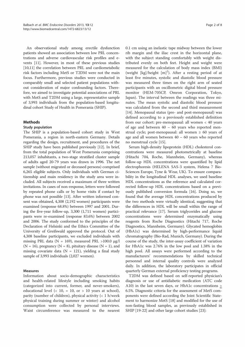
An observational study among erectile dysfunctionpatients showed an association between low PRL concen-trations and adverse cardiovascular risk profiles and e-vents [11]. However, in most of these previous studies[10,11] the correlation between PRL and cardiometabolicrisk factors including MetS or T2DM were not the mainfocus. Furthermore, previous studies were conducted incomparably small and selected patient populations with-out consideration of major confounding factors. There-fore, we aimed to investigate potential associations of PRLwith MetS and T2DM using a large, representative sampleof 3,993 individuals from the population-based longitu-dinal cohort Study of Health in Pomerania (SHIP).
MethodsStudy populationThe SHIP is a population-based cohort study in WestPomerania, a region in north-eastern Germany. Detailsregarding the design, recruitment, and procedures of theSHIP study have been published previously [12]. In brief,from the total population of West Pomerania comprising213,057 inhabitants, a two-stage stratified cluster sampleof adults aged 20-79 years was drawn in 1996. The netsample (without migrated or deceased persons) comprised6,265 eligible subjects. Only individuals with German ci-tizenship and main residency in the study area were in-cluded. All subjects received a maximum of three writteninvitations. In cases of non-response, letters were followedby repeated phone calls or by home visits if contact byphone was not possible [13]. After written informed con-sent was obtained, 4,308 (2,192 women) participants wereexamined (response 68.8%) between 1997 and 2001. Dur-ing the five-year follow-up, 3,300 (1,711 women) partici-pants were re-examined (response 83.6%) between 2002and 2006. The study conformed to the principles of theDeclaration of Helsinki and the Ethics Committee of theUniversity of Greifswald approved the protocol. Out of4,308 baseline participants, we excluded individuals withmissing PRL data (N = 169), measured PRL >100.0 μg/l(N = 16), pregnancy (N = 8), pituitary disease (N = 1), andmissing covariate data (N = 121), yielding a final studysample of 3,993 individuals (2,027 women).
MeasuresInformation about socio-demographic characteristicsand health-related lifestyle including smoking habits(categorized into current, former, and never-smokers),educational level (< 10, = 10, or > 10 years at school),parity (number of children), physical activity (< 1 h/weekphysical training during summer or winter) and alcoholconsumption were collected by personal interviews.Waist circumference was measured to the nearest
0.1 cm using an inelastic tape midway between the lowerrib margin and the iliac crest in the horizontal plane,with the subject standing comfortably with weight dis-tributed evenly on both feet. Height and weight weremeasured for the calculation of body mass index (BMI)(weight [kg]/height [m]2). After a resting period of atleast five minutes, systolic and diastolic blood pressurewas measured three times on the right arm of seatedparticipants with an oscillometric digital blood pressuremonitor (HEM-705CP, Omron Corporation, Tokyo,Japan). The interval between the readings was three mi-nutes. The mean systolic and diastolic blood pressurewas calculated from the second and third measurement[14]. Menopausal status (pre- and post-menopausal) wasdefined according to a previously established definitionfrom our cohort: pre-menopausal: all women < 40 yearsof age and between 40 – 60 years who reported men-strual cycle; post-menopausal: all women ≥ 60 years ofage and all women between 40 – 60 years who reportedno menstrual cycle [15].Serum high-density lipoprotein (HDL) cholesterol con-
centrations were measured photometrically at baseline(Hitachi 704, Roche, Mannheim, Germany), whereasfollow-up HDL concentrations were quantified by lipidelectrophoresis (HELENA SAS-3 system, Helena 7 Bio-Sciences Europe, Tyne & Wear, UK). To ensure compara-bility in the longitudinal HDL analyses, we used baselineHDL concentrations as the reference and calculated cor-rected follow-up HDL concentrations based on a previ-ously published conversion formula [16]. Doing so, wefound that the average HDL concentrations produced bythe two methods were virtually identical, suggesting thatthe differences in HDL will be small within the range ofpractical relevance [17]. Serum triglycerides and glucoseconcentrations were determined enzymatically usingreagents from Roche Diagnostics (Hitachi 717, RocheDiagnostics, Mannheim, Germany). Glycated hemoglobin(HbA1c) was determined by high-performance liquidchromatography (Bio-Rad, Munich, Germany). During thecourse of the study, the inter-assay coefficient of variationfor HbA1c was 2.76% in the low pool and 1.38% in thehigh pool. All assays were performed according to themanufacturers’ recommendations by skilled technicalpersonnel and internal quality controls were analyzeddaily. In addition, the laboratory participates in officialquarterly German external proficiency testing programs.T2DM was defined based on self-reported physician’s
diagnosis or use of antidiabetic medication (ATC codeA10) in the last seven days, or HbA1c concentrations >6.5%. Diagnostic criteria for the assessment of MetS com-ponents were defined according the Joint Scientific State-ment to harmonize MetS [18] and modified for the use ofnon-fasting blood samples, as previously established inSHIP [19-22] and other large cohort studies [23]:
Balbach et al. BMC Endocrine Disorders 2013, 13:12 Page 2 of 8http://www.biomedcentral.com/1472-6823/13/12
1. Elevated WC: men ≥ 94 cm; women ≥ 80 cm;2. Elevated non-fasting glucose: ≥ 8.0 mmol/l or
antidiabetic treatment (ATC codes A10A, A10B);3. Decreased HDL cholesterol: men < 1.0 mmol/l,
women < 1.3 mmol/l, or lipid-lowering treatment(ATC C10AB, A10AD);
4. Elevated non-fasting triglycerides: ≥ 2.3 mmol/l orlipid-lowering treatment (ATC C10AB, A10AD);
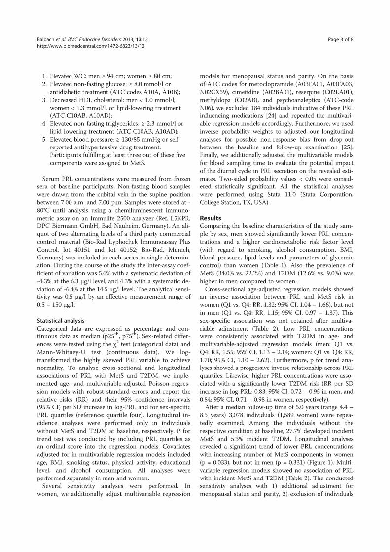
5. Elevated blood pressure: ≥ 130/85 mmHg or self-reported antihypertensive drug treatment.Participants fulfilling at least three out of these fivecomponents were assigned to MetS.
Serum PRL concentrations were measured from frozensera of baseline participants. Non-fasting blood sampleswere drawn from the cubital vein in the supine positionbetween 7.00 a.m. and 7.00 p.m. Samples were stored at -80°C until analysis using a chemiluminescent immuno-metric assay on an Immulite 2500 analyzer (Ref. L5KPR,DPC Biermann GmbH, Bad Nauheim, Germany). An ali-quot of two alternating levels of a third party commercialcontrol material (Bio-Rad Lyphochek Immunoassay PlusControl, lot 40151 and lot 40152; Bio-Rad, Munich,Germany) was included in each series in single determin-ation. During the course of the study the inter-assay coef-ficient of variation was 5.6% with a systematic deviation of-4.3% at the 6.3 μg/l level, and 4.3% with a systematic de-viation of -6.4% at the 14.5 μg/l level. The analytical sensi-tivity was 0.5 μg/l by an effective measurement range of0.5 – 150 μg/l.
Statistical analysisCategorical data are expressed as percentage and con-tinuous data as median (p25th, p75th). Sex-related differ-ences were tested using the χ2 test (categorical data) andMann-Whitney-U test (continuous data). We log-transformed the highly skewed PRL variable to achievenormality. To analyse cross-sectional and longitudinalassociations of PRL with MetS and T2DM, we imple-mented age- and multivariable-adjusted Poisson regres-sion models with robust standard errors and report therelative risks (RR) and their 95% confidence intervals(95% CI) per SD increase in log-PRL and for sex-specificPRL quartiles (reference: quartile four). Longitudinal in-cidence analyses were performed only in individualswithout MetS and T2DM at baseline, respectively. P fortrend test was conducted by including PRL quartiles asan ordinal score into the regression models. Covariatesadjusted for in multivariable regression models includedage, BMI, smoking status, physical activity, educationallevel, and alcohol consumption. All analyses wereperformed separately in men and women.Several sensitivity analyses were performed. In
women, we additionally adjust multivariable regression
models for menopausal status and parity. On the basisof ATC codes for metoclopramide (A03FA01, A03FA03,N02CX59), cimetidine (A02BA01), reserpine (C02LA01),methyldopa (C02AB), and psychoanaleptics (ATC-codeN06), we excluded 184 individuals indicative of these PRLinfluencing medications [24] and repeated the multivari-able regression models accordingly. Furthermore, we usedinverse probability weights to adjusted our longitudinalanalyses for possible non-response bias from drop-outbetween the baseline and follow-up examination [25].Finally, we additionally adjusted the multivariable modelsfor blood sampling time to evaluate the potential impactof the diurnal cycle in PRL secretion on the revealed esti-mates. Two-sided probability values < 0.05 were consid-ered statistically significant. All the statistical analyseswere performed using Stata 11.0 (Stata Corporation,College Station, TX, USA).
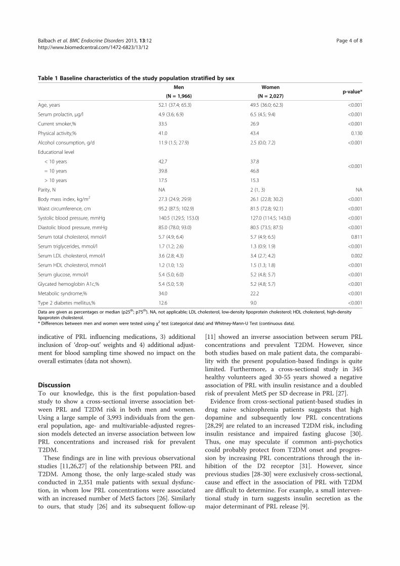
ResultsComparing the baseline characteristics of the study sam-ple by sex, men showed significantly lower PRL concen-trations and a higher cardiometabolic risk factor level(with regard to smoking, alcohol consumption, BMI,blood pressure, lipid levels and parameters of glycemiccontrol) than women (Table 1). Also the prevalence ofMetS (34.0% vs. 22.2%) and T2DM (12.6% vs. 9.0%) washigher in men compared to women.Cross-sectional age-adjusted regression models showed
an inverse association between PRL and MetS risk inwomen (Q1 vs. Q4: RR, 1.32; 95% CI, 1.04 – 1.66), but notin men (Q1 vs. Q4: RR, 1.15; 95% CI, 0.97 – 1.37). Thissex-specific association was not retained after multiva-riable adjustment (Table 2). Low PRL concentrationswere consistently associated with T2DM in age- andmultivariable-adjusted regression models (men: Q1 vs.Q4: RR, 1.55; 95% CI, 1.13 – 2.14; women: Q1 vs. Q4: RR,1.70; 95% CI, 1.10 – 2.62). Furthermore, p for trend ana-lyses showed a progressive inverse relationship across PRLquartiles. Likewise, higher PRL concentrations were asso-ciated with a significantly lower T2DM risk (RR per SDincrease in log-PRL: 0.83; 95% CI, 0.72 – 0.95 in men, and0.84; 95% CI, 0.71 – 0.98 in women, respectively).After a median follow-up time of 5.0 years (range 4.4 –
8.5 years) 3,078 individuals (1,589 women) were repea-tedly examined. Among the individuals without therespective condition at baseline, 27.7% developed incidentMetS and 5.3% incident T2DM. Longitudinal analysesrevealed a significant trend of lower PRL concentrationswith increasing number of MetS components in women(p = 0.033), but not in men (p = 0.331) (Figure 1). Multi-variable regression models showed no association of PRLwith incident MetS and T2DM (Table 2). The conductedsensitivity analyses with 1) additional adjustment formenopausal status and parity, 2) exclusion of individuals
Balbach et al. BMC Endocrine Disorders 2013, 13:12 Page 3 of 8http://www.biomedcentral.com/1472-6823/13/12
indicative of PRL influencing medications, 3) additionalinclusion of ‘drop-out’ weights and 4) additional adjust-ment for blood sampling time showed no impact on theoverall estimates (data not shown).
DiscussionTo our knowledge, this is the first population-basedstudy to show a cross-sectional inverse association bet-ween PRL and T2DM risk in both men and women.Using a large sample of 3,993 individuals from the gen-eral population, age- and multivariable-adjusted regres-sion models detected an inverse association between lowPRL concentrations and increased risk for prevalentT2DM.These findings are in line with previous observational
studies [11,26,27] of the relationship between PRL andT2DM. Among those, the only large-scaled study wasconducted in 2,351 male patients with sexual dysfunc-tion, in whom low PRL concentrations were associatedwith an increased number of MetS factors [26]. Similarlyto ours, that study [26] and its subsequent follow-up
[11] showed an inverse association between serum PRLconcentrations and prevalent T2DM. However, sinceboth studies based on male patient data, the comparabi-lity with the present population-based findings is quitelimited. Furthermore, a cross-sectional study in 345healthy volunteers aged 30-55 years showed a negativeassociation of PRL with insulin resistance and a doubledrisk of prevalent MetS per SD decrease in PRL [27].Evidence from cross-sectional patient-based studies in
drug naive schizophrenia patients suggests that highdopamine and subsequently low PRL concentrations[28,29] are related to an increased T2DM risk, includinginsulin resistance and impaired fasting glucose [30].Thus, one may speculate if common anti-psychoticscould probably protect from T2DM onset and progres-sion by increasing PRL concentrations through the in-hibition of the D2 receptor [31]. However, sinceprevious studies [28-30] were exclusively cross-sectional,cause and effect in the association of PRL with T2DMare difficult to determine. For example, a small interven-tional study in turn suggests insulin secretion as themajor determinant of PRL release [9].
Table 1 Baseline characteristics of the study population stratified by sex
Men Womenp-value*
(N = 1,966) (N = 2,027)
Age, years 52.1 (37.4; 65.3) 49.5 (36.0; 62.3) <0.001
Serum prolactin, μg/l 4.9 (3.6; 6.9) 6.5 (4.5; 9.4) <0.001
Current smoker,% 33.5 26.9 <0.001
Physical activity,% 41.0 43.4 0.130
Alcohol consumption, g/d 11.9 (1.5; 27.9) 2.5 (0.0; 7.2) <0.001
Educational level
<0.001< 10 years 42.7 37.8
= 10 years 39.8 46.8
> 10 years 17.5 15.3
Parity, N NA 2 (1, 3) NA
Body mass index, kg/m2 27.3 (24.9; 29.9) 26.1 (22.8; 30.2) <0.001
Waist circumference, cm 95.2 (87.5; 102.9) 81.5 (72.8; 92.1) <0.001
Systolic blood pressure, mmHg 140.5 (129.5; 153.0) 127.0 (114.5; 143.0) <0.001
Diastolic blood pressure, mmHg 85.0 (78.0; 93.0) 80.5 (73.5; 87.5) <0.001
Serum total cholesterol, mmol/l 5.7 (4.9; 6.4) 5.7 (4.9; 6.5) 0.811
Serum triglycerides, mmol/l 1.7 (1.2; 2.6) 1.3 (0.9; 1.9) <0.001
Serum LDL cholesterol, mmol/l 3.6 (2.8; 4.3) 3.4 (2.7; 4.2) 0.002
Serum HDL cholesterol, mmol/l 1.2 (1.0; 1.5) 1.5 (1.3; 1.8) <0.001
Serum glucose, mmol/l 5.4 (5.0; 6.0) 5.2 (4.8; 5.7) <0.001
Glycated hemoglobin A1c,% 5.4 (5.0; 5.9) 5.2 (4.8; 5.7) <0.001
Metabolic syndrome,% 34.0 22.2 <0.001
Type 2 diabetes mellitus,% 12.6 9.0 <0.001
Data are given as percentages or median (p25th; p75th). NA, not applicable; LDL cholesterol, low-density lipoprotein cholesterol; HDL cholesterol, high-densitylipoprotein cholesterol.* Differences between men and women were tested using χ2 test (categorical data) and Whitney-Mann-U Test (continuous data).
Balbach et al. BMC Endocrine Disorders 2013, 13:12 Page 4 of 8http://www.biomedcentral.com/1472-6823/13/12
In the search of potential explanations, two previousstudies related alterations of serotonergic pathwayscaused by low PRL concentrations to the pathogenesisof MetS [27,32]. Furthermore, PRL-receptor deficientmice show an impaired development of pancreatic ß-
cells, finally leading to a blunted insulin response andmild glucose intolerance [7]. In contrast to our find-ings, studies including pathological hyperprolactinemiacaused by PRL-secreting tumors (prolactinomas)showed that, after treatment with dopamine agonists,
Table 2 Cross-sectional and longitudinal associations of serum prolactin with metabolic syndrome and type 2 diabetesmellitus
Metabolic syndrome Type 2 diabetes mellitus
Cross-sectional Longitudinal Cross-sectional Longitudinal
Age-adjustedmodel
Multivariablemodel
Age-adjustedmodel
Multivariablemodel
Age-adjustedmodel
Multivariablemodel
Age-adjustedmodel
Multivariablemodel
Prolactin Men
per SDincrease
0.95 (0.89; 1.02) 0.95 (0.89; 1.01) 1.05 (0.95; 1.15) 1.04 (0.95; 1.14) 0.84 (0.73; 0.96)* 0.83 (0.72; 0.95)* 1.20 (0.98; 1.47) 1.19 (0.96; 1.47)
Quartiles(Q4 Ref.)
Q1 1.15 (0.97; 1.37) 1.15 (0.98; 1.35) 0.83 (0.64; 1.06) 0.87 (0.68; 1.10) 1.55 (1.12; 2.13)* 1.55 (1.13; 2.14)* 0.71 (0.41; 1.23) 0.78 (0.45; 1.35)
Q2 1.06 (0.89; 1.28) 1.03 (0.87; 1.22) 0.94 (0.74; 1.19) 0.94 (0.75; 1.18) 1.14 (0.81; 1.61) 1.11 (0.79; 1.56) 0.73 (0.42; 1.26) 0.78 (0.45; 1.33)
Q3 1.04 (0.87; 1.25) 1.02 (0.85; 1.21) 0.91 (0.72; 1.16) 0.94 (0.75; 1.18) 0.92 (0.63; 1.33) 0.92 (0.64; 1.33) 0.76 (0.44; 1.32) 0.72 (0.41; 1.27)
P for Trend 0.104 0.076 0.172 0.276 0.002 0.002 0.253 0.472
Women
per SDincrease
0.92 (0.85; 1.00) 0.97 (0.90; 1.05) 0.89 (0.79; 1.00) 0.92 (0.83; 1.02) 0.81 (0.68; 0.95)* 0.84 (0.71; 0.98)* 1.09 (0.82; 1.45) 1.13 (0.89; 1.44)
Quartiles(Q4 Ref.)
Q1 1.32 (1.04; 1.66)* 1.11 (0.89; 1.39) 1.26 (0.95; 1.68) 1.15 (0.86; 1.53) 1.89 (1.21; 2.96)* 1.70 (1.10; 2.62)* 0.88 (0.44; 1.78) 0.73 (0.37; 1.43)
Q2 1.13 (0.89; 1.44) 1.04 (0.83; 1.32) 1.22 (0.91; 1.63) 1.16 (0.87; 1.54) 1.82 (1.15; 2.86)* 1.69 (1.09; 2.63)* 0.81 (0.39; 1.68) 0.75 (0.37; 1.50)
Q3 1.11 (0.86; 1.44) 1.07 (0.83; 1.32) 0.73 (0.51; 1.04) 0.78 (0.55; 1.11) 1.29 (0.77; 2.16) 1.26 (0.76; 2.09) 1.00 (0.48; 2.06) 1.08 (0.53; 2.20)
P for Trend 0.016 0.399 0.013 0.106 0.001 0.007 0.633 0.224
* p < 0.05; SD, standard deviation. The multivariable model was adjusted for age, body mass index, smoking status (three categories), physical activity, educationallevel (three categories), and alcohol consumption.
Figure 1 Boxplots for median baseline prolactin concentrations (25th and 75th percentile) according to the follow-up number ofmetabolic syndrome (MetS) components, separately for men and women. One-way analysis of variance showed a significant inverse trendfor prolactin concentrations across number of MetS components at follow-up in women (p = 0.033), but not in men (p = 0.331).
Balbach et al. BMC Endocrine Disorders 2013, 13:12 Page 5 of 8http://www.biomedcentral.com/1472-6823/13/12
reduced PRL concentrations led to increased insulinsensitivity [33,34].These results, however, were collected in small patient-
based samples with clinically confirmed prolactinoma andserum PRL concentrations outside the reference range,thus limiting its transferability to the general population.On contrary, HOMA-IR, as a measure of insulin resis-tance, did not change after treatment with dopamine ago-nists [33], and PRL concentrations did not correlate withHOMA-IR change or decrease in glucose, respectively[8,34]. Interestingly, a previous cross-sectional study inobese non-prolactinoma patients showed similar absentcorrelations of PRL with insulin, HOMA-IR, and glucoselevels [10]. Available in-vitro studies rather suggest aninfluence of prolactin on ß-cell secretion via increased glu-cokinase activity [35], improved ß-cell specific survival[36,37], or inhibition of intrinsic ß-cell apoptosis [38].However, to provide the necessary level of detail to eluci-date the exact mechanisms of PRL on different tissues andits impact on cardiometabolic risk factors further experi-mental in-vitro research is needed.We previously published findings from our study sug-
gesting a positive association of PRL with biomarkers ofinflammation [39] and mortality (cardiovascular and all-cause mortality) [40], as well as an inverse association ofPRL with cardiac remodelling [41]. However, these ob-servational findings from our and other studies do notprovide consistent risk associations, suggesting the needfor further investigations into the potential role of PRLas cardiometabolic risk marker.For the interpretation of the present study, the follo-
wing strengths and limitations need to be considered.Major strengths include the large sample, the longitu-dinal study design, and a broad age range. Furthermore,comprehensive sensitivity analyses were conducted toassess the validity of our findings. An important limita-tion of this study includes the reliance on single serumPRL measurements based on non-fasting blood samples,which is not adapted to the pulsatile release of PRL. Butit was shown that the pulsatile secretion occurs mostlyduring the night and is relatively constant between9.00 a.m. and 5.00 p.m. [42]. Further limitations arisefrom the Caucasian study sample, which restricts thegeneralizability of our results. Finally, we did not excludeother endocrine diseases, which possibly trigger T2DMincluding hyperthyroidism [43,44], acromegaly and theCushing Syndrome [45].However, given the inherent limitations of observational
research [46], the definite role of serum PRL concentra-tions as a risk factor or risk marker can not be elucidatedbased on epidemiological data alone. Previous associationsbetween serum PRL concentrations and disease risk ob-served in clinical study samples, but not in the generalpopulation, suggest PRL rather as specific disease marker
than as causal risk factor for MetS and T2DM. Thus, itsapplication and significance in daily clinical practice isvery limited to date.
ConclusionsIn summary, this is the first population-based study toshow low PRL concentrations related to a higher T2DMrisk in both genders. But given the variable associationsbetween PRL and T2DM, but not with MetS, togetherwith the absent longitudinal associations with both out-comes, the present study does not support PRL as a causalcardiometabolic risk factor. Therefore, we hypothesizePRL rather as a marker of the complex multi-level alte-rations involved in the onset and progression of T2DM.However, to further elucidate the potential role of PRL inthe context of this complex interplay, further observa-tional as well as interventional research is needed.
Competing interestThere is no conflict of interest that could be perceived as prejudicing theimpartiality of the research reported.
Authors’ contributionsRH, LB, and HW contributed to the study design and ideas for the dataanalysis. HV, MD, MN, and HW organized the sample collection and datapreparation. Statistical analyses were performed by RH. LB, RH, and HWcontributed to the interpretation of the results and the discussion. LHdrafted the manuscript and wrote the final version together with all otherco-authors. All authors read, critically revised, and finally gave approval of theversion to be published.
AcknowledgementsThe contributions to data collection made by field workers, study physicians,ultrasound technicians, interviewers, and computer assistants are gratefullyacknowledged.
FundingSHIP is part of the Community Medicine Research net of the University ofGreifswald, Germany, which is funded by the Federal Ministry of Educationand Research (grants no. 01ZZ9603, 01ZZ0103, and 01ZZ0403), the Ministryof Cultural Affairs as well as the Social Ministry of the Federal State ofMecklenburg-West Pomerania. This work is also part of the research projectGreifswald Approach to Individualized Medicine (GANI_MED), funded by theFederal Ministry of Education and Research and the Ministry of CulturalAffairs of the Federal State of Mecklenburg – West Pomerania (03IS2061A).This study was also supported by the DZHK (German Centre forCardiovascular Research) and by the BMBF (German Ministry of Educationand Research). The used prolactin reagents were sponsored by Novo NordiskPharma GmbH, Mainz, Germany.
Author details1Institute of Clinical Chemistry and Laboratory Medicine, University MedicineGreifswald, Ferdinand-Sauerbruch-Straße, Greifswald 17475, Germany. 2DZHK(German Centre for Cardiovascular Research), partner site Greifswald,Greifswald, Germany. 3Institute for Community Medicine, University MedicineGreifswald, Walther-Rathenau-Straße 48, Greifswald 17475, Germany.4Department of Cardiology, University Medicine Greifswald,Ferdinand-Sauerbruch-Straße, Greifswald 17475, Germany.
Received: 1 October 2012 Accepted: 15 March 2013Published: 21 March 2013
References1. Cejkova P, Fojtikova M, Cerna M: Immunomodulatory role of prolactin in
diabetes development. Autoimmun Rev 2009, 9(1):23–27.
Balbach et al. BMC Endocrine Disorders 2013, 13:12 Page 6 of 8http://www.biomedcentral.com/1472-6823/13/12
2. Brandebourg T, Hugo E, Ben-Jonathan N: Adipocyte prolactin: regulationof release and putative functions. Diabetes Obes Metab 2007, 9(4):464–476.
3. Ben-Jonathan N, Hugo ER, Brandebourg TD, LaPensee CR: Focus onprolactin as a metabolic hormone. Trends in endocrinology andmetabolism. TEM 2006, 17(3):110–116.
4. Prasad H, Ryan DA, Celzo MF, Stapleton D: Metabolic syndrome: definitionand therapeutic implications. Postgrad Med 2012, 124(1):21–30.
5. Rosenzweig JL, Ferrannini E, Grundy SM, Haffner SM, Heine RJ, Horton ES,Kawamori R: Primary prevention of cardiovascular disease and type 2diabetes in patients at metabolic risk: an endocrine society clinicalpractice guideline. J Clin Endocrinol Metabol 2008, 93(10):3671–3689.
6. Brelje TC, Stout LE, Bhagroo NV, Sorenson RL: Distinctive roles for prolactinand growth hormone in the activation of signal transducer and activatorof transcription 5 in pancreatic islets of langerhans. Endocrinology 2004,145(9):4162–4175.
7. Freemark M, Avril I, Fleenor D, Driscoll P, Petro A, Opara E, Kendall W, OdenJ, Bridges S, Binart N, et al: Targeted deletion of the PRL receptor: effectson islet development, insulin production, and glucose tolerance.Endocrinology 2002, 143(4):1378–1385.
8. Serri O, Li L, Mamputu JC, Beauchamp MC, Maingrette F, Renier G: Theinfluences of hyperprolactinemia and obesity on cardiovascular riskmarkers: effects of cabergoline therapy. Clin Endocrinol (Oxf ) 2006,64(4):366–370.
9. Mingrone G, Manco M, Iaconelli A, Gniuli D, Bracaglia R, Leccesi L, CalvaniM, Nolfe G, Basu S, Berria R: Prolactin and insulin ultradian secretion andadipose tissue lipoprotein lipase expression in severely obese womenafter bariatric surgery. Obesity (Silver Spring) 2008, 16(8):1831–1837.
10. Ernst B, Thurnheer M, Schultes B: Basal serum prolactin levels in obesity–unrelated to parameters of the metabolic syndrome and unchangedafter massive weight loss. Obes Surg 2009, 19(8):1159–1162.
11. Corona G, Rastrelli G, Boddi V, Monami M, Melani C, Balzi D, Sforza A, FortiG, Mannucci E, Maggi M: Prolactin levels independently predict majorcardiovascular events in patients with erectile dysfunction. Int J Androl2011, 34(3):217–224.
12. Völzke H, Alte D, Schmidt CO, Radke D, Lorbeer R, Friedrich N, Aumann N,Lau K, Piontek M, Born G, et al: Cohort profile: the study of health inpomerania. Int J Epidemiol 2011, 40(2):294–307.
13. Haring R, Alte D, Volzke H, Sauer S, Wallaschofski H, John U, Schmidt CO:Extended recruitment efforts minimize attrition but not necessarily bias.J Clin Epidemiol 2009, 62(3):252–260.
14. Torkler S, Wallaschofski H, Baumeister SE, Volzke H, Dorr M, Felix S, Rettig R,Nauck M, Haring R: Inverse association between total testosteroneconcentrations, incident hypertension and blood pressure. Aging Male2011, 14(3):176–182.
15. Haring R, Hannemann A, John U, Radke D, Nauck M, Wallaschofski H, OwenL, Adaway J, Keevil BG, Brabant G: Age-specific reference ranges for serumtestosterone and androstenedione concentrations in women measuredby liquid chromatography-tandem mass spectrometry. J Clin EndocrinolMetabol 2012, 97(2):408–415.
16. Nauck M, Winkler K, Marz W, Wieland H: Quantitative determination of high-,low-, and very-low-density lipoproteins and lipoprotein(a) by agarose gelelectrophoresis and enzymatic cholesterol staining. Clin Chem 1995,41(12 Pt 1):1761–1767.
17. Haring R, Baumeister SE, Völzke H, Dorr M, Felix SB, Kroemer HK, Nauck M,Wallaschofski H: Prospective Association of Low Total TestosteroneConcentrations with an Adverse Lipid Profile and Increased IncidentDyslipidemia. Eur J Cardiovasc Prev Rehabil 2011, 18(1):86–96.
18. Alberti KG, Eckel RH, Grundy SM, Zimmet PZ, Cleeman JI, Donato KA,Fruchart JC, James WP, Loria CM, Smith SC Jr: Harmonizing the metabolicsyndrome: a joint interim statement of the International DiabetesFederation Task Force on Epidemiology and Prevention; National Heart,Lung, and Blood Institute; American Heart Association; World HeartFederation; International Atherosclerosis Society; and InternationalAssociation for the Study of Obesity. Circulation 2009, 120(16):1640–1645.
19. Hannemann A, Meisinger C, Bidlingmaier M, Doring A, Thorand B, Heier M,Belcredi P, Ladwig KH, Wallaschofski H, Friedrich N, et al: Association ofplasma aldosterone with the metabolic syndrome in two Germanpopulations. Eur J Endocrinol 2011, 164(5):751–758.
20. Schipf S, Alte D, Voelzke H, Friedrich N, Haring R, Lohmann T, Nauck M, FelixSB, Hoffmann W, John U, et al: Prävalenz des Metabolischen Syndroms in
Deutschland: Ergebnisse der Study of Health in Pomerania (SHIP).Diabetologie Stoffwechsel 2010, 5:161–168.
21. Haring R, Volzke H, Felix SB, Schipf S, Dorr M, Rosskopf D, Nauck M, Schofl C,Wallaschofski H: Prediction of metabolic syndrome by low serumtestosterone levels in men: results from the study of health inPomerania. Diabetes 2009, 58(9):2027–2031.
22. Haring R, Wallaschofski H, Nauck M, Felix SB, Schmidt CO, Dorr M, Sauer S,Wilmking G, Volzke H: Total and cardiovascular disease mortalitypredicted by metabolic syndrome is inferior relative to its components.Exp Clin Endocrinol Diabetes 2010, 118(10):685–691.
23. Lidfeldt J, Nyberg P, Nerbrand C, Samsioe G, Schersten B, Agardh CD:Socio-demographic and psychosocial factors are associated withfeatures of the metabolic syndrome. The Women's Health in the LundArea (WHILA) study. Diabetes Obes Metab 2003, 5(2):106–112.
24. Molitch ME: Drugs and prolactin. Pituitary 2008, 11(2):209–218.25. Afifi AA, Kotlerman JB, Ettner SL, Cowan M: Methods for improving
regression analysis for skewed continuous or counted responses. AnnuRev Public Health 2007, 28:95–111.
26. Corona G, Mannucci E, Jannini EA, Lotti F, Ricca V, Monami M, Boddi V, BandiniE, Balercia G, Forti G, et al: Hypoprolactinemia: a new clinical syndrome inpatients with sexual dysfunction. J Sex Med 2009, 6(5):1457–1466.
27. Muldoon MF, Mackey RH, Korytkowski MT, Flory JD, Pollock BG, Manuck SB:The metabolic syndrome is associated with reduced central serotonergicresponsivity in healthy community volunteers. J Clin Endocrinol Metabol2006, 91(2):718–721.
28. Meaney AM, O'Keane V: Prolactin and schizophrenia: clinicalconsequences of hyperprolactinaemia. Life Sci 2002, 71(9):979–992.
29. Rao ML, Gross G, Strebel B, Halaris A, Huber G, Braunig P, Marler M:Circadian rhythm of tryptophan, serotonin, melatonin, and pituitaryhormones in schizophrenia. Biol Psychiatry 1994, 35(3):151–163.
30. Ryan MC, Collins P, Thakore JH: Impaired fasting glucose tolerance in first-episode, drug-naive patients with schizophrenia. Am J Psychiatry 2003,160(2):284–289.
31. Oberweis B, Gragnoli C: Potential role of prolactin in antipsychotic-mediated association of schizophrenia and type 2 diabetes. J Cell Physiol2012, 227(8):3001–3006.
32. Muldoon MF, Mackey RH, Williams KV, Korytkowski MT, Flory JD, Manuck SB:Low central nervous system serotonergic responsivity is associated withthe metabolic syndrome and physical inactivity. J Clin Endocrinol Metabol2004, 89(1):266–271.
33. Berinder K, Nystrom T, Hoybye C, Hall K, Hulting AL: Insulin sensitivity andlipid profile in prolactinoma patients before and after normalization ofprolactin by dopamine agonist therapy. Pituitary 2011, 14(3):199–207.
34. dos Santos Silva CM, Barbosa FR, Lima GA, Warszawski L, Fontes R,Domingues RC, Gadelha MR: BMI and metabolic profile in patients withprolactinoma before and after treatment with dopamine agonists.Obesity (Silver Spring) 2011, 19(4):800–805.
35. Weinhaus AJ, Stout LE, Bhagroo NV, Brelje TC, Sorenson RL: Regulation ofglucokinase in pancreatic islets by prolactin: a mechanism for increasingglucose-stimulated insulin secretion during pregnancy. J Endocrinol 2007,193(3):367–381.
36. Yamamoto T, Ricordi C, Mita A, Miki A, Sakuma Y, Molano RD, Fornoni A,Pileggi A, Inverardi L, Ichii H: beta-Cell specific cytoprotection by prolactinon human islets. Transplant Proc 2008, 40(2):382–383.
37. Yamamoto T, Mita A, Ricordi C, Messinger S, Miki A, Sakuma Y, Timoneri F,Barker S, Fornoni A, Molano RD, et al: Prolactin supplementation to culturemedium improves beta-cell survival. Transplantation 2010, 89(11):1328–1335.
38. Terra LF, Garay-Malpartida MH, Wailemann RA, Sogayar MC, Labriola L:Recombinant human prolactin promotes human beta cell survival viainhibition of extrinsic and intrinsic apoptosis pathways. Diabetologia2011, 54(6):1388–1397.
39. Friedrich N, Schneider HJ, Spielhagen C, Markus MR, Haring R, Grabe HJ,Buchfelder M, Wallaschofski H, Nauck M: The association of serumprolactin concentration with inflammatory biomarkers - cross-sectionalfindings from the population-based Study of Health in Pomerania.Clin Endocrinol (Oxf ) 2011, 75(4):561–566.
40. Haring R, Friedrich N, Volzke H, Vasan RS, Felix SB, Dorr M, Meyer ZuSchwabedissen HE, Nauck M, Wallaschofski H: Positive association ofserum prolactin concentrations with all-cause and cardiovascularmortality. Eur Heart J 2012.
Balbach et al. BMC Endocrine Disorders 2013, 13:12 Page 7 of 8http://www.biomedcentral.com/1472-6823/13/12
41. Haring R, Volzke H, Vasan RS, Felix SB, Nauck M, Dorr M, Wallaschofski H:Sex-specific associations of serum prolactin concentrations with cardiacremodeling: longitudinal results from the Study of Health Pomerania(SHIP). Atherosclerosis 2012, 221(2):570–576.
42. Katznelson L, Riskind PN, Saxe VC, Klibanski A: Prolactin pulsatilecharacteristics in postmenopausal women. J Clin Endocrinol Metabol 1998,83(3):761–764.
43. Maratou E, Hadjidakis DJ, Peppa M, Alevizaki M, Tsegka K, Lambadiari V,Mitrou P, Boutati E, Kollias A, Economopoulos T, et al: Studies of insulinresistance in patients with clinical and subclinical hyperthyroidism.Eur J Endocrinol 2010, 163(4):625–630.
44. Mitrou P, Raptis SA, Dimitriadis G: Insulin action in hyperthyroidism: afocus on muscle and adipose tissue. Endocr Rev 2010, 31(5):663–679.
45. Resmini E, Minuto F, Colao A, Ferone D: Secondary diabetes associatedwith principal endocrinopathies: the impact of new treatmentmodalities. Acta Diabetol 2009, 46(2):85–95.
46. Rothman KJ, Greenland S: Causation and causal inference inepidemiology. Am J Public Health 2005, 95(Suppl 1):S144–150.
doi:10.1186/1472-6823-13-12Cite this article as: Balbach et al.: Serum prolactin concentrations as riskfactor of metabolic syndrome or type 2 diabetes?. BMC EndocrineDisorders 2013 13:12.
Submit your next manuscript to BioMed Centraland take full advantage of:
• Convenient online submission
• Thorough peer review
• No space constraints or color figure charges
• Immediate publication on acceptance
• Inclusion in PubMed, CAS, Scopus and Google Scholar
• Research which is freely available for redistribution
Submit your manuscript at www.biomedcentral.com/submit
Balbach et al. BMC Endocrine Disorders 2013, 13:12 Page 8 of 8http://www.biomedcentral.com/1472-6823/13/12
Eidesstattliche Erklärung Anhang
7.2 Eidesstattliche Erklärung
Hiermit erkläre ich, dass ich die vorgelegte Dissertation selbständig verfasst und
keine anderen als die angegebenen Hilfsmittel benutzt habe.
Die Dissertation ist bisher keiner anderen Fakultät, keiner anderen wissenschaftli-
chen Einrichtung vorgelegt worden.
Ich erkläre, dass ich bisher kein Promotionsverfahren erfolglos beendet habe und
dass eine Aberkennung eines bereits erworbenen Doktorgrades nicht vorliegt.
Greifswald, denLisa Balbach

Lebenslauf Anhang
7.3 Lebenslauf
Persönliche Daten:
Vorname, Name: Lisa Balbach
Geburtstag: 10. Juni 1988
Geburtsort: Berlin
Anschrift: Erich-Böhmke-Straÿe 22a, 17489 Greifswald
Familienstand: ledig
Nationalität: deutsch
Werdegang:
Schulische Ausbildung:
1994 � 2000 Stechlinsee-Grundschule, Berlin-Schöneberg
2000 � 2007 Rheingau-Gymnasium, Berlin-Schöneberg
28.06.2007 Abschluss: Abtiur (1,9)
Beru�iche Ausbildung:
01.02.2007 � 31.09.2009 Ausbildung zur medizinischen Fachangstellten
(MFA)
Studium:
seit 01.10.2009 Ernst-Moritz-Arndt-Universität Greifswald
Studiengang: Humanmedizin
Greifswald, denLisa Balbach

Danksagung Anhang
7.4 Danksagung
An dieser Stelle möchte ich mich bei allen Personen bedanken, die an der Verwirk-
lichung dieser Arbeit beteiligt waren.
Mein besonderer Dank gilt dabei Herrn Prof. Dr. H. Wallaschofski für die Überlas-
sung des Dissertationsthemas und für die Unterstützung in medizinischen Fragen,
sowie meinem Betreuer Herrn Dr. rer. med. Robin Haring, dem ich für die bio-
mathematische Auswertung, die graphische Darstellung und die Korrekturlesung
verp�ichtet bin.
Des Weiteren möchte ich mich bei den Co-Autoren für die gute Zusammenarbeit
und die vielfältigen Anregungen bedanken.
Darüber hinaus war mir Herr Oliver Bolm eine groÿe Hilfe bei der graphischen
Darstellung und Formatierung meiner Dissertation.
Nicht zuletzt danke ich meiner gesamten Familie, Frau Katharina-Marlene Schick-
Witte sowie Frau Tanja Testrich, die mich in der Zeit bis zur Fertigstellung der
Dissertation vor allem mit ihrer Liebe, Geduld und Motivation unterstützt haben.
Top Related