![Thyroid pathophysiology scintigraphy[1]](https://static.fdocuments.net/doc/165x107/588a7dc81a28abad628b4ebd/thyroid-pathophysiology-scintigraphy1.jpg)
Languages
Pages
Legal


Scintigraphy (Nuclear Medicine Imaging) in Patients with
Perplexing Abdominal Complaints
Mark Tulchinsky, MD, FACNM, CCD
Professor of Radiology and Medicine
Penn State University College of Medicine
Hershey, Pennsylvania

Learning Objectives:
Define …1. Nuclear Medicine and Its Basic Principles
Define Indications for …
2. Hepatobiliary Scintigraphy
– Functional Gallbladder Disorder
– Chronic Cholecystitis
– Related Conditions

Nuclear Medicine - Definition
A medical specialty that utilizes
radioactive isotopes or
pharmaceuticals labeled with
radioisotopes (called “radiotracers” or
”radiopharmaceuticals”) for diagnostic
and therapeutic purposes.
OK, but why “Nuclear” Medicine?
Why not .. say … “Radioisotopic” or
… “Radioactive” Medicine?!

Niels Bohr and Ernest Rutherford
Atomic Model, circa 1913
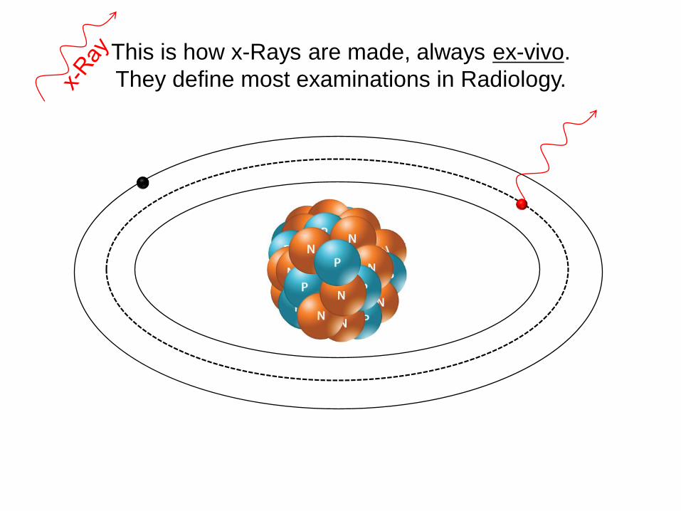
This is how x-Rays are made, always ex-vivo.
They define most examinations in Radiology.

Radioactive Isotopes have Unstable Nucleus
Thus, Nuclear Medicine!
Radiotracers emit excess energy, γ-Rays, from within patients.

Scintillator - Photomultiplier Tube (PMT):Ƴ-Photon → Electric Pulse
γ-Ray
From Latin word
Scintilla, "spark”, as
in spark of light
VonCount!
Scintigraphy = scan
obtained using this
method of imaging
Scintigraphically =
by the way of
scintigraphy

Gamma Camera = Images Gamma Emission

Gamma Camera = Images Gamma Emission

Gamma Camera = Images Gamma Emission

Gamma Camera = Images Gamma Emission
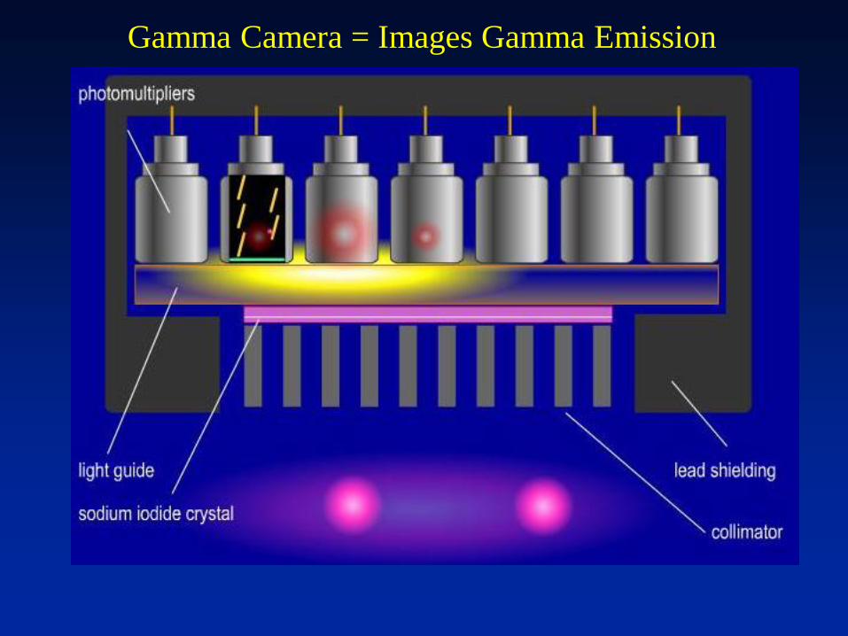
Gamma Camera = Images Gamma Emission

Gamma Camera = Images Gamma Emission
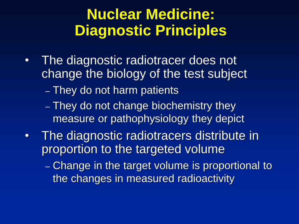
Nuclear Medicine:Diagnostic Principles
• The diagnostic radiotracer does not change the biology of the test subject
– They do not harm patients
– They do not change biochemistry they
measure or pathophysiology they depict
• The diagnostic radiotracers distribute in proportion to the targeted volume
– Change in the target volume is proportional to
the changes in measured radioactivity

Hepatobiliary Scintigraphy
Traces the clearance of bilirubin into bile and tracks the biliary flow

Functional Liver Unit:Hepatic Lobule (wedge - 1/4th)
Illustration courtesy of Mark Tulchinsky, MD.

Tc-99m-Mebrofenin ( ) IV, 25% via hepatic artery (H. a.) and 75% via portal vein (P. v.)
Radiotracer is Tc-99m-Mebrofenin
Illustration courtesy of Mark Tulchinsky, MD.
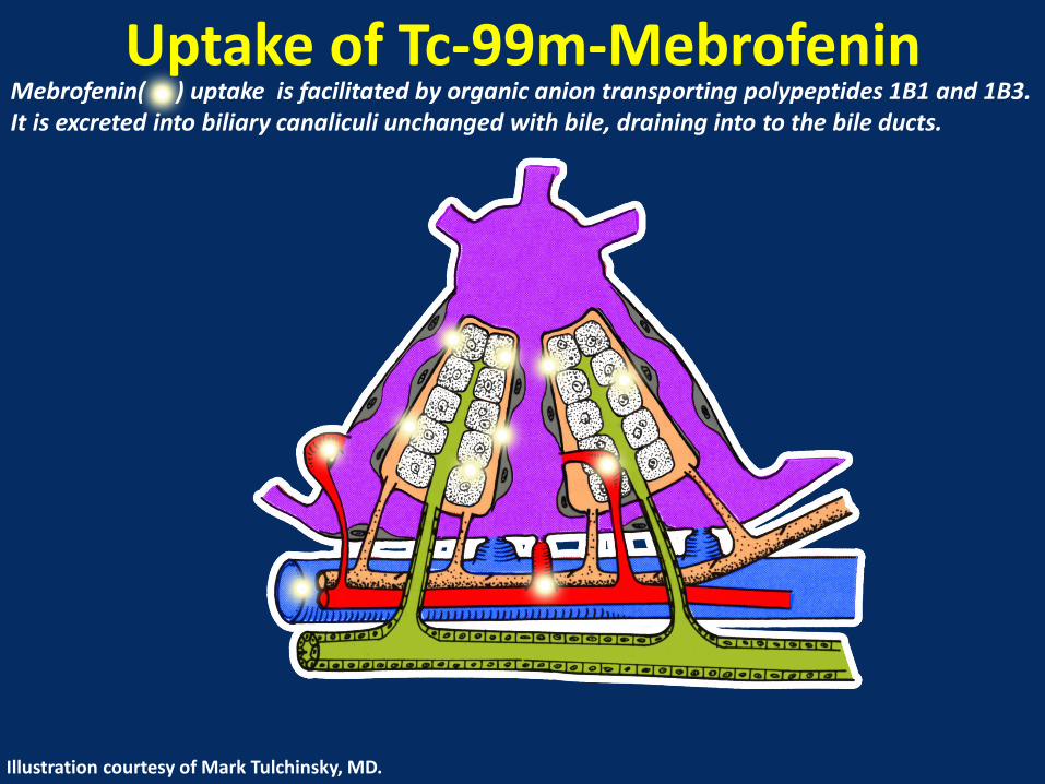
Uptake of Tc-99m-Mebrofenin Mebrofenin( ) uptake is facilitated by organic anion transporting polypeptides 1B1 and 1B3. It is excreted into biliary canaliculi unchanged with bile, draining into to the bile ducts.
Illustration courtesy of Mark Tulchinsky, MD.

Radiopharmaceutical extracted by hepatocytes and transported without modification.
Excretion of Tc-99m-Mebrofenin Mebrofenin( ) excretion is facilitated by multidrug resistance-associated protein 2.
Illustration courtesy of Mark Tulchinsky, MD.

The Gallbladder in Fasting State:Accommodation of Incoming Bile

The Gallbladder in Prolonged Fasting State (>24 Hours)

Postprandial Gallbladder:Set-up for a False-Positive

Postprandial Refilling of Gallbladder
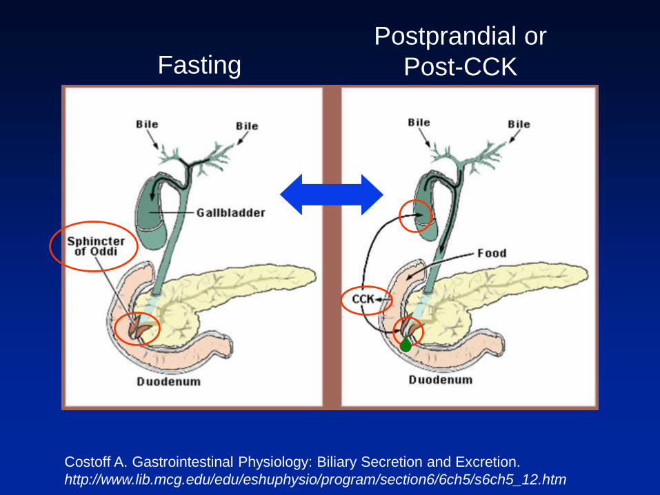
FastingPostprandial or
Post-CCK
Costoff A. Gastrointestinal Physiology: Biliary Secretion and Excretion.
http://www.lib.mcg.edu/edu/eshuphysio/program/section6/6ch5/s6ch5_12.htm

Patients With Chronic Abdominal Pain and Gallbladder Dysfunction
Why Is Diagnosis So Difficult?• Lack of a clear definition of biliary pain1
• Nonspecific nature of the symptom complex1
• Limited understanding of the natural history and pathophysiology of chronic biliary-type abdominal pain1
• Lack of standardization in performance of CCK-CS in terms of CCK dose, duration of administration, and definition of normal values1, 2
• Low percentage of US practices using the recommended CCK infusion protocol2
• Inappropriate referral for testing1
1. Dibaise JK, et al. Clin Gastroenterol Hepatol. 2011;9(5):376-384.
2. Tulchinsky M, et al. Poster presented at: 2012 Annual Meeting of the Society of Nuclear Medicine and Molecular
Imaging; June 9-13, 2012; Miami, FL. Poster 2117. Abstract available in: J Nucl Med. 2012;53(suppl 1):2117.

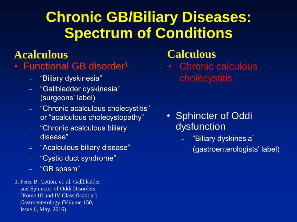
Chronic GB/Biliary Disease:Spectrum of Conditions
• Functional GB disorder– “Biliary dyskinesia”
– “Gallbladder dyskinesia” (surgeons’ label)
– “Chronic acalculous cholecystitis” or “acalculous cholecystopathy”
– “Chronic acalculous biliary disease”
– “Acalculous biliary disease”
– “Cystic duct syndrome”
– “GB spasm”
• Chronic calculous cholecystitis
Acalculous
Calculous
• Sphincter of Oddi dysfunction
– “Biliary dyskinesia”
(gastroenterologists’ label)

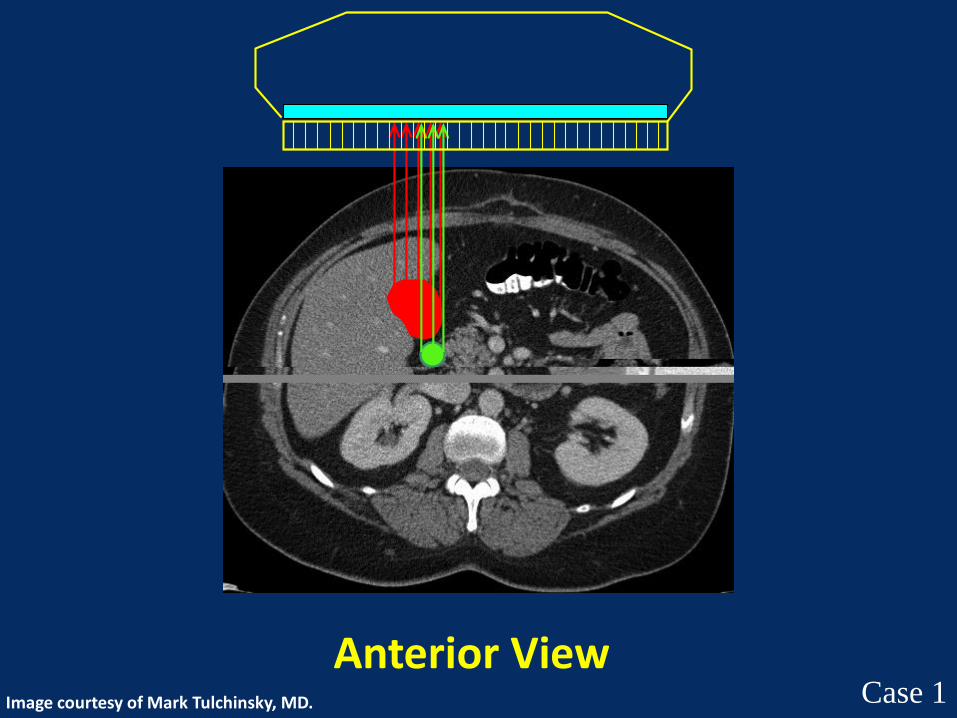
Anterior View
1 2 3 4
5 6 7 8
9 10 11 12
13 14 15
Images courtesy of Mark Tulchinsky, MD. Case 1

Anterior View
In the anterior view, the activity in the duodenum often contributes to (interferes with) activity in the gallbladder
region!
Image courtesy of Mark Tulchinsky, MD. Case 1

Anterior ViewImage courtesy of Mark Tulchinsky, MD. Case 1
Anterior ViewImage courtesy of Mark Tulchinsky, MD. Case 1

40o
LAO view separates the GB from duodenal activity –makes good anatomical sense!
Image courtesy of Mark Tulchinsky, MD.Case 1
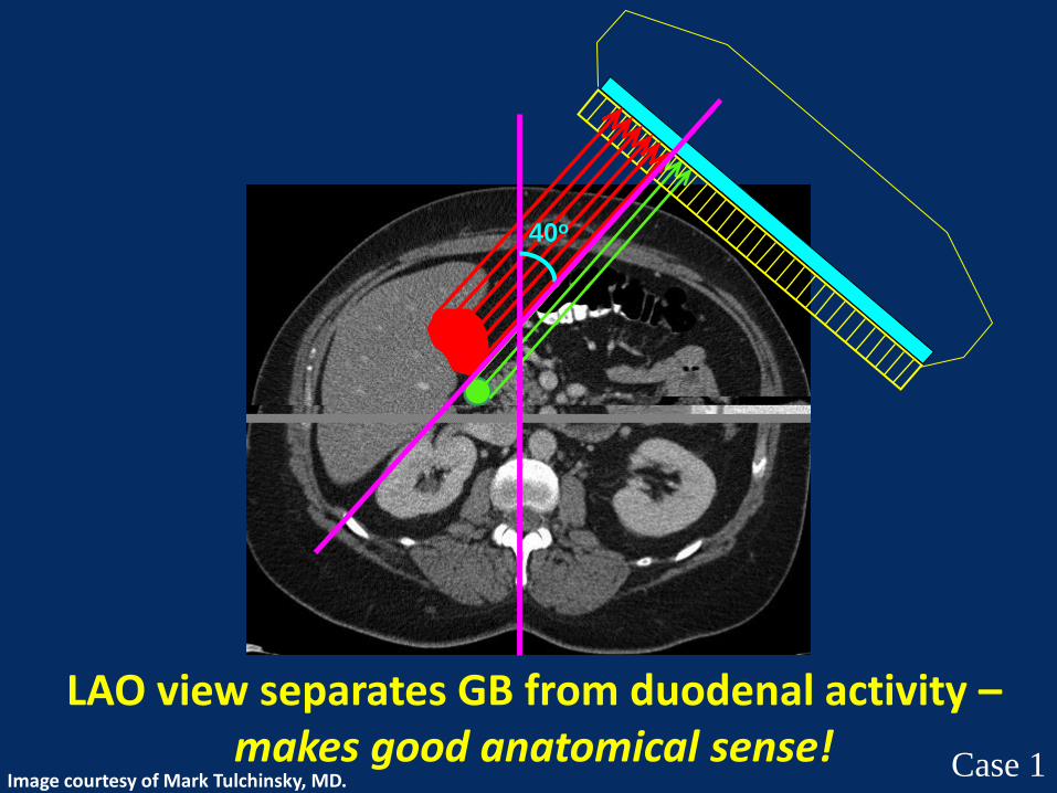
40o
LAO view separates GB from duodenal activity –makes good anatomical sense!
Image courtesy of Mark Tulchinsky, MD.Case 1

Post CCK
2 min/frame
40o LAO Projection
LAO view separates GB from duodenal activity –makes good functional imaging sense!
Image courtesy of Mark Tulchinsky, MD.
Case 1

Sincalide-Stimulated Cholescintigraphy: A Multicenter Investigation to Determine Optimal Infusion Methodology and Gallbladder Ejection
Fraction Normal Values
Min/µg per kg GBEF (M±S.D.) GBEF Range GBEF<35% CV(%)
15/0.02 56.9±29.4%* -2 to 98% 16/60 (27%) 52%
30/0.02 70.9±24.5%* 8 to 99% 6/60 (10%) 35%
60/0.02 84.3±15.5% * 38 to 100% 0/60 19%
* Significantly different from other 2 infusion rates, p < 0.0001
All 60 normal volunteers – GB viz by 60 min x 3! Therefore, if there
is GB non-viz by 60 min = abnormal GB function, test completed!
• 60 min sincalide infusion is the most specific test for GB function
• GBEF ≥ 38% is normal
Ziessman HA, Tulchinsky M, Lavely WC, et al. Sincalide-stimulated cholescintigraphy: a multicenter
investigation to determine optimal infusion methodology and gallbladder ejection fraction normal
values. J Nucl Med. 2010;51:277-281.
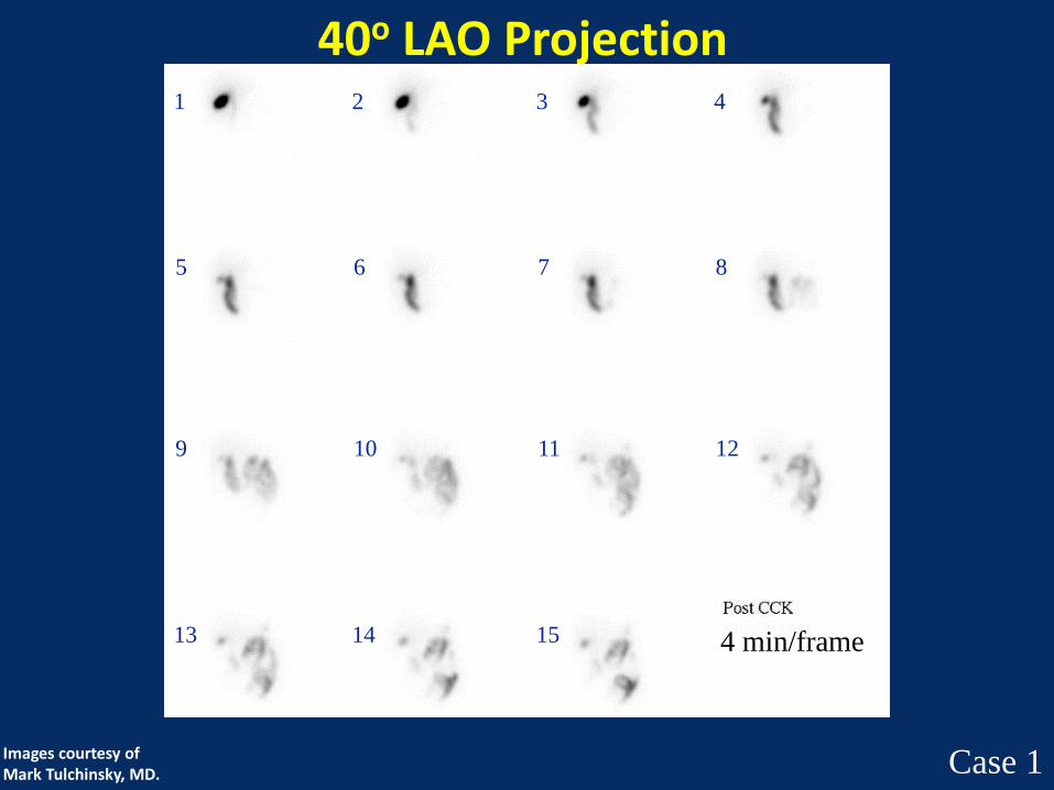
40o LAO Projection1 2 3 4
5 6 7 8
9 10 11 12
13 14 15
Images courtesy of Mark Tulchinsky, MD. Case 1
4 min/frame

40o LAO Separates GB From Duodenal Activity
This image is for quality control , showing
that the GB didn’t move outside the region of
interest, as can happen with patient motion – a
potential cause of a false-negative test.
Images courtesy of Mark Tulchinsky, MD.
Case 1
4 min/frame
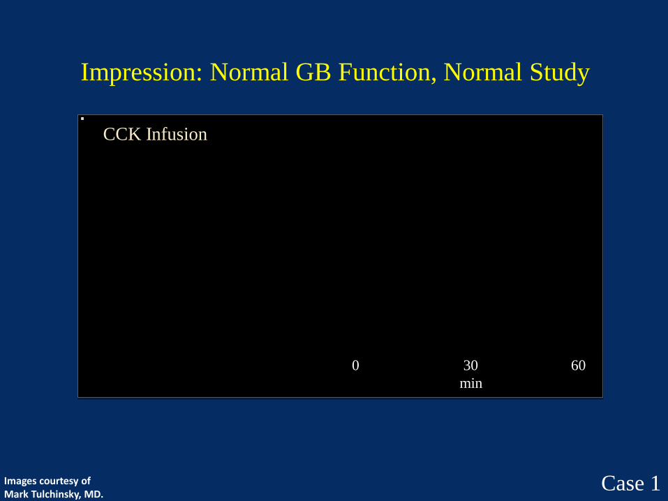
Impression: Normal GB Function, Normal Study
0 30 60
min
CCK Infusion
Case 1Images courtesy of Mark Tulchinsky, MD.
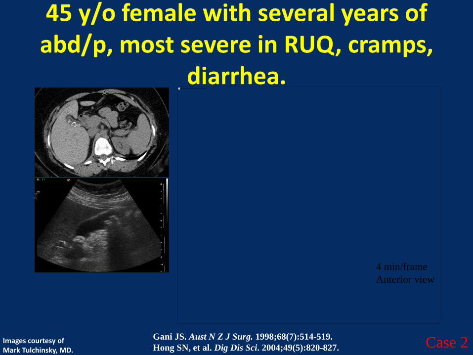
45 y/o female with several years of abd/p, most severe in RUQ, cramps,
diarrhea.
4 min/frame
Anterior view
Gani JS. Aust N Z J Surg. 1998;68(7):514-519.
Hong SN, et al. Dig Dis Sci. 2004;49(5):820-827.Images courtesy of Mark Tulchinsky, MD.
Case 2
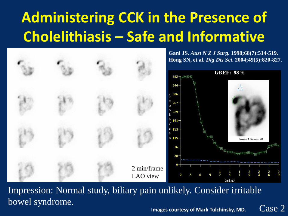
Administering CCK in the Presence of Cholelithiasis – Safe and Informative
2 min/frame
LAO view
Gani JS. Aust N Z J Surg. 1998;68(7):514-519.
Hong SN, et al. Dig Dis Sci. 2004;49(5):820-827.
Images courtesy of Mark Tulchinsky, MD. Case 2
Impression: Normal study, biliary pain unlikely. Consider irritable
bowel syndrome.
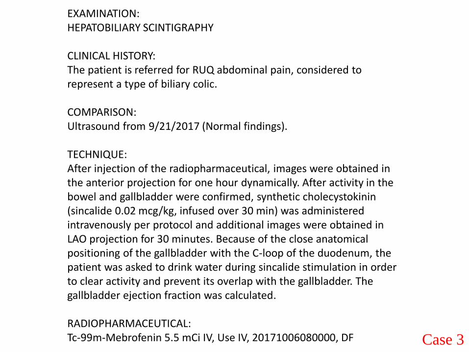
EXAMINATION:HEPATOBILIARY SCINTIGRAPHY
CLINICAL HISTORY: The patient is referred for RUQ abdominal pain, considered to represent a type of biliary colic.
COMPARISON: Ultrasound from 9/21/2017 (Normal findings).
TECHNIQUE:After injection of the radiopharmaceutical, images were obtained in the anterior projection for one hour dynamically. After activity in the bowel and gallbladder were confirmed, synthetic cholecystokinin (sincalide 0.02 mcg/kg, infused over 30 min) was administered intravenously per protocol and additional images were obtained in LAO projection for 30 minutes. Because of the close anatomical positioning of the gallbladder with the C-loop of the duodenum, the patient was asked to drink water during sincalide stimulation in order to clear activity and prevent its overlap with the gallbladder. The gallbladder ejection fraction was calculated.
RADIOPHARMACEUTICAL:Tc-99m-Mebrofenin 5.5 mCi IV, Use IV, 20171006080000, DF Case 3

First hour of dynamic imaging, obtained in anterior view at 15 seconds per frame, reformatted for display at 4 minutes per frame for visual inspection.
Static image, obtained for 2 minutes in right lateral projection.
Case 3

Dynamic imaging, obtained during sincalide infusion, in the left anterior oblique view at 1 minute per frame, reformatted for display at 2 minutes per frame. Pt drank 8 oz. H2O at time 0 sec and continued sipping additional 8 oz. through the test.
Case 3

The same dynamic imaging, obtained during sincalide infusion, in the left anterior oblique view. In addition shown are regions of interest encompassing the gallbladder in red outline and background in green outline.
Case 3

The image depicts combination of all frames obtained during sincalide infusion, in the left anterior oblique view with regions of interest encompassing the gallbladder in red outline and background in green outline.
Calculation of ejection fraction (EF) is based on background corrected counts (activity). Normal EF is ≥ 35%
Case 3
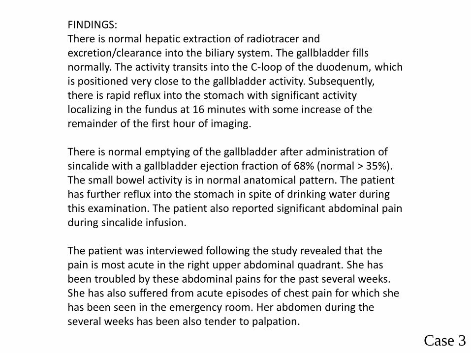
FINDINGS: There is normal hepatic extraction of radiotracer and excretion/clearance into the biliary system. The gallbladder fills normally. The activity transits into the C-loop of the duodenum, which is positioned very close to the gallbladder activity. Subsequently, there is rapid reflux into the stomach with significant activity localizing in the fundus at 16 minutes with some increase of the remainder of the first hour of imaging.
There is normal emptying of the gallbladder after administration of sincalide with a gallbladder ejection fraction of 68% (normal > 35%). The small bowel activity is in normal anatomical pattern. The patient has further reflux into the stomach in spite of drinking water during this examination. The patient also reported significant abdominal pain during sincalide infusion.
The patient was interviewed following the study revealed that the pain is most acute in the right upper abdominal quadrant. She has been troubled by these abdominal pains for the past several weeks. She has also suffered from acute episodes of chest pain for which she has been seen in the emergency room. Her abdomen during the several weeks has been also tender to palpation.
Case 3

IMPRESSION:1. Normal gallbladder filling and ejection fraction following sincalide stimulation of 68%. 2. There is prominent bile reflux into the stomach, consider bilious gastritis. 3. Given the patient's complaints of episodic chest pain associated with abdominal pain, gastroesophageal reflux with bilious contents should be considered. The patient did not have chest pain during this examination nor did the images show gastroesophageal reflux.
Case 3

ANT projection
4 min/frame
Case 4
If there is early and preferential GB filling, does that predict
normal GB function & obviates need for challenge? No!

LAO projection
2 min/frame
CCK Infusion Dynamic Images
Case 4

0 30 60
min
Now, what’s the
diagnosis?
Case 4
Chronic GB/Biliary Diseases:Spectrum of Conditions
• Functional GB disorder1
– “Biliary dyskinesia”
– “Gallbladder dyskinesia”
(surgeons’ label)
– “Chronic acalculous cholecystitis”
or “acalculous cholecystopathy”
– “Chronic acalculous biliary
disease”
– “Acalculous biliary disease”
– “Cystic duct syndrome”
– “GB spasm”
Acalculous Calculous• Chronic calculous
cholecystitis
• Sphincter of Oddidysfunction
– “Biliary dyskinesia”
(gastroenterologists’ label)
1. Peter B. Cotton, et. al. Gallbladder
and Sphincter of Oddi Disorders.
[Rome III and IV Classification.]
Gastroenterology (Volume 150,
Issue 6, May, 2016)
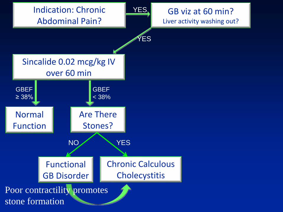
Indication: Chronic Abdominal Pain?
YES GB viz at 60 min?Liver activity washing out?
YES
NormalFunction
GBEF
≥ 38%
Sincalide 0.02 mcg/kg IV over 60 min
GBEF
< 38%
Chronic Calculous Cholecystitis
Are There Stones?
FunctionalGB Disorder
NO YES
Poor contractility promotes
stone formation

Case 5
• 35 y/o female with chronic abdominal pain, RUQ, characterized as biliary colic
• Referred for sincalide cholescintigraphy
• Ultrasound of the abdomen is normal
• You are brought this study to check

Anterior View
Rt. Lat.
@60 min
Case 5
ANT projection,
60 min baseline
Is the GB visualized?

Case 5
Is the GBEF normal?
Yes
No
Unsure
0 30 60
min
CCK Infusion Images

Case 5
What else do you need to make the final call?
4 min/frame
CCK Infusion Dynamic Images

Case 5

Take-Home Message: Look at the QA Image for motion!
Case 5
Post CCK
4 min./frame

Impression: Abnormal GBEF of 21%, consistent with diagnosis
of functional gallbladder disorder in the proper clinical setting.
Case 5 0 30 60
min
Cholecystokinin-cholescintigraphy in adults: Consensus
recommendations of an interdisciplinary panel.DiBaise JK, Richmond BK, Ziessman HH, Everson GT, Fanelli RD, Maurer A,
Ouyang A, Shamamian P, , Wall LA, , Tulchinsky M.
Clin Gastroenterol Hepatol. 2011 May;9(5):376-84.
doi: 10.1016/j.cgh.2011.02.013
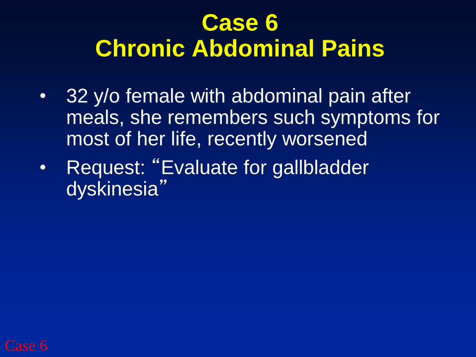
Case 6Chronic Abdominal Pains
• 32 y/o female with abdominal pain after meals, she remembers such symptoms for most of her life, recently worsened
• Request: “Evaluate for gallbladder dyskinesia”
Case 6

Case 6
Is there GB visualization?
Is there an abnormality on these images?
Should you give CCK?
ANT projection,
60 min baseline
Yes
No
Unsure
Yes
No
Unsure
Yes
No
Unsure
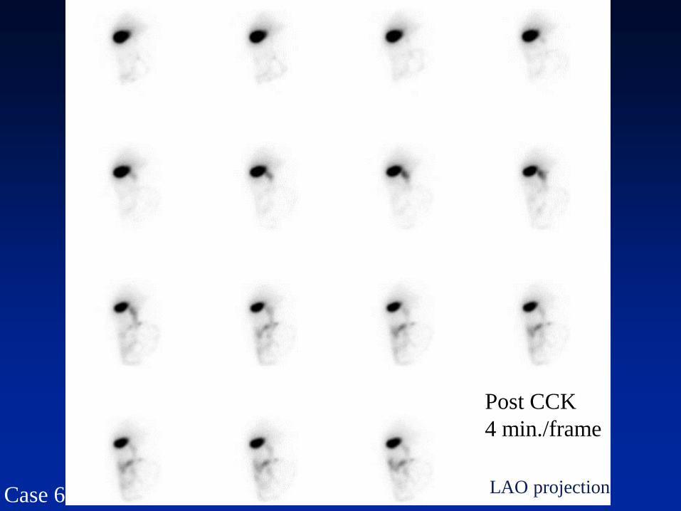
LAO projectionCase 6
Post CCK
4 min./frame

Normal Study.What is your final reading?
Case 6
Abnormal Study.
0 30 60
min
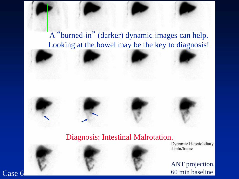
A “burned-in” (darker) dynamic images can help.
Looking at the bowel may be the key to diagnosis!
Diagnosis: Intestinal Malrotation.
Case 6
ANT projection,
60 min baseline
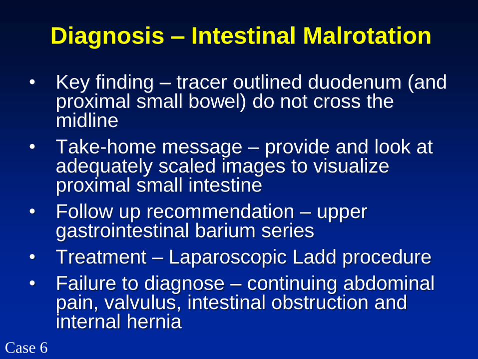
Diagnosis – Intestinal Malrotation
• Key finding – tracer outlined duodenum (and proximal small bowel) do not cross the midline
• Take-home message – provide and look at adequately scaled images to visualize proximal small intestine
• Follow up recommendation – upper gastrointestinal barium series
• Treatment – Laparoscopic Ladd procedure
• Failure to diagnose – continuing abdominal pain, valvulus, intestinal obstruction and internal hernia
Case 6
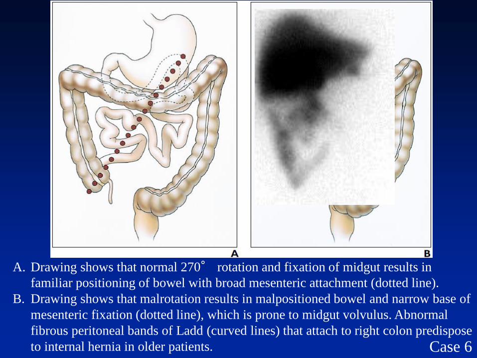
A. Drawing shows that normal 270° rotation and fixation of midgut results in
familiar positioning of bowel with broad mesenteric attachment (dotted line).
B. Drawing shows that malrotation results in malpositioned bowel and narrow base of
mesenteric fixation (dotted line), which is prone to midgut volvulus. Abnormal
fibrous peritoneal bands of Ladd (curved lines) that attach to right colon predispose
to internal hernia in older patients. Case 6
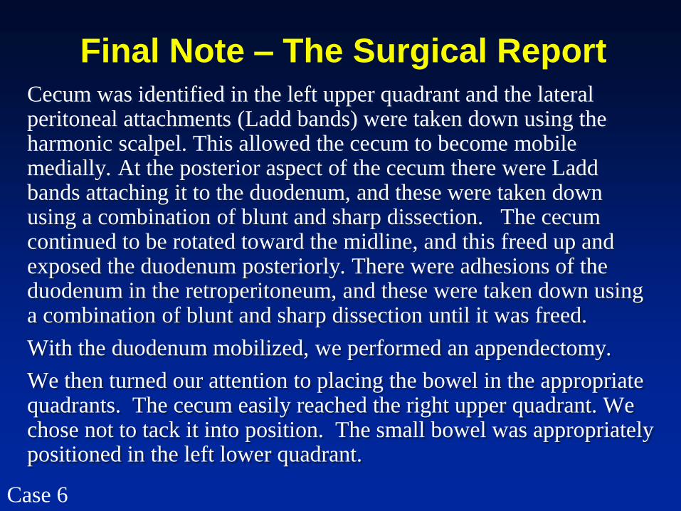
Final Note – The Surgical ReportCecum was identified in the left upper quadrant and the lateral peritoneal attachments (Ladd bands) were taken down using the harmonic scalpel. This allowed the cecum to become mobile medially. At the posterior aspect of the cecum there were Ladd bands attaching it to the duodenum, and these were taken down using a combination of blunt and sharp dissection. The cecum continued to be rotated toward the midline, and this freed up and exposed the duodenum posteriorly. There were adhesions of the duodenum in the retroperitoneum, and these were taken down using a combination of blunt and sharp dissection until it was freed.
With the duodenum mobilized, we performed an appendectomy.
We then turned our attention to placing the bowel in the appropriate quadrants. The cecum easily reached the right upper quadrant. We chose not to tack it into position. The small bowel was appropriately positioned in the left lower quadrant.
Case 6

Case 7
• Chronic Abdominal Pain
• Is the GB normal vs. abnormal (responsible for patient’s pains)?

Case 7
ANT projection,
60 min baseline
Is the GB acting as would be expected? Yes!
No!
GB spontaneously empting … or is it spontaneous?

ANT projection, 60 min baseline scintigraphy, activity of the GB showed:
Patient had nothing per os during the test.
What is going on? Any guesses?
Case 7
Patient was watching a movie and it was not too exciting …
But, at ab
out m
in 6

Cephalic Phase ResponsesSensory stimulation from foods lead to rapid activation of
physiologic processes at multiple sites that may optimize the
digestion, absorption, and use of ingested nutrients.
Case 7
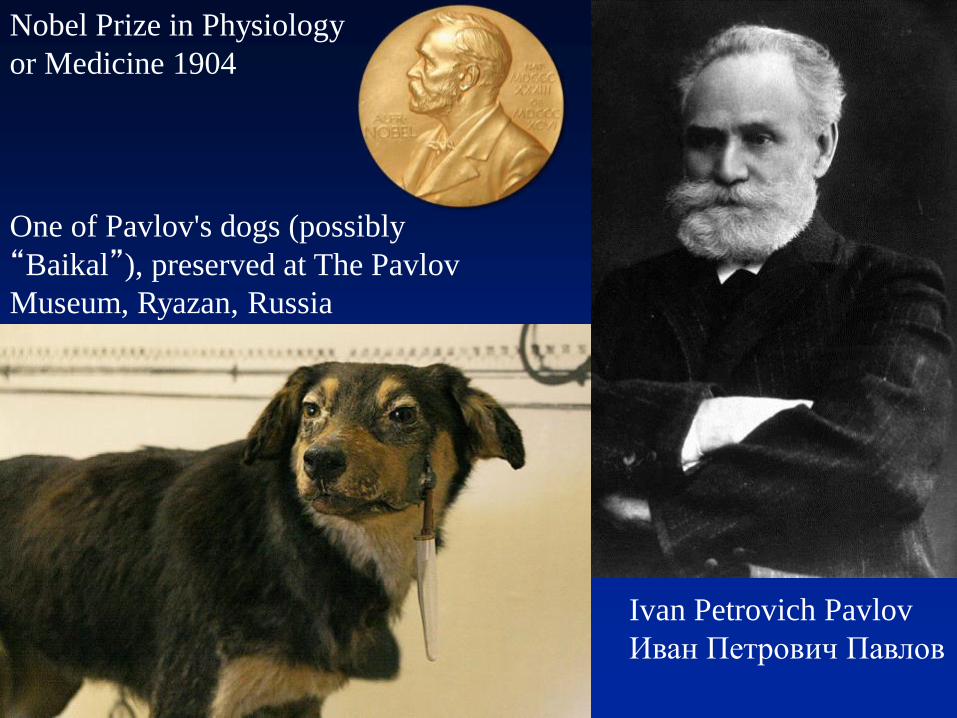
Ivan Petrovich Pavlov
Иван Петрович Павлов
Nobel Prize in Physiology
or Medicine 1904
One of Pavlov's dogs (possibly
“Baikal”), preserved at The Pavlov
Museum, Ryazan, Russia
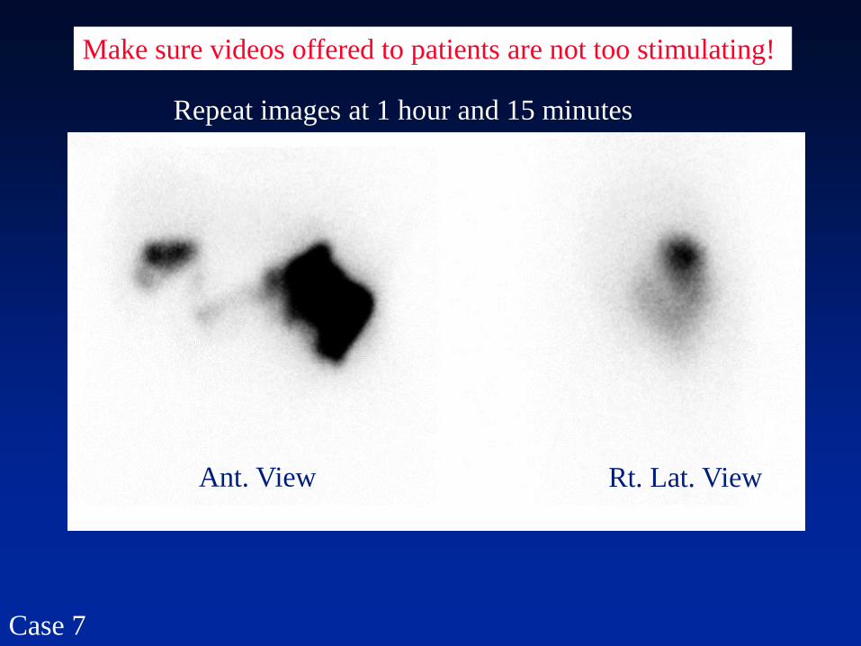
Repeat images at 1 hour and 15 minutes
Ant. View Rt. Lat. View
Case 7
Make sure videos offered to patients are not too stimulating!

Case 7
Sincalide Cholescintigraphy

25-year-old man was referred for chronic right upper quadrant abdominal pain for hepatobiliary
scintigraphy to evaluate (GB) function.
ANT projection

GB Curve: Baseline Imaging

25-year-old man was referred for chronic right upper quadrant abdominal pain for hepatobiliary
scintigraphy to evaluate (GB) function.
ANT projection

GB Curve, Sincalide Infusion

Copyright © 2015 by the American Osteopathic Association.
All rights reserved.
From: An Osteopathic Approach to Gastrointestinal Disease: Somatic Clues for Diagnosis and Clinical
Challenges Associated With Helicobacter pylori Antibiotic Resistance
J Am Osteopath Assoc. 2013;113(5):404-416. doi:10.7556/jaoa.2013.113.5.404
Figure Legend: Illustration of the
spinal cord in its role as a
“neurologic lens” for a variety of
stressors that can initiate somatic
or visceral symptoms.
Abbreviation: EENT, eye, ear,
nose, and throat. Reprinted with
permission from Kuchera ML,
McPartland JM. Myofascial
trigger points. In: Ward RC,
executive ed. Foundations for
Osteopathic Medicine. Baltimore,
MD: Lippincott Williams &
Wilkins; 1997:916.


Special Thanks to My Penn State Colleagues!
Joe Fotos Scott Winner Tom
Allen
Thank You For Your Attention!
Top Related