

Languages
Pages
Legal

Clinical Practice Guidelines
Role of endoscopy in primary sclerosing cholangitis: EuropeanSociety of Gastrointestinal Endoscopy (ESGE) and European
Association for the Study of the Liver (EASL) Clinical Guidelineq
European Society of Gastrointestinal Endoscopy, European Association for the Study of the Liver ⇑
Summary
This guideline is an official statement of the European Society of Gastrointestinal Endoscopy (ESGE) and of the European Association forthe Study of the Liver (EASL) on the role of endoscopy in primary sclerosing cholangitis. The Grading of Recommendations Assessment,Development and Evaluation (GRADE) system was adopted to define the strength of recommendations and the quality of evidence.
Main recommendations
1. ESGE/EASL recommend that, as the primary diagnostic modalitypreferred over endoscopic retrograde cholangiopancreatographyModerate quality evidence, strong recommendation.
2. ESGE/EASL suggest that ERCP can be considered if MRC plus livsisting clinical suspicion of PSC. The risks of ERCP have to be weightreatment recommendations.Low quality evidence, weak recommendation.
6. ESGE/EASL suggest that, in patients with an established diagnoWeak recommendation, low quality evidence.
7. ESGE/EASL suggest performing endoscopic treatment with concof suspected significant strictures identified at MRC in PSC patientscopic treatment.Strong recommendation, low quality evidence.
9. ESGE/EASL recommend weighing the anticipated benefits of bilcase basis. Strong recommendation, moderate quality evidence. Bcially after difficult cannulation.Strong recommendation, low quality evidence.
Biliary papillotomy/sphincterotomy should be considered especiaStrong recommendation, low quality evidence.
16. ESGE/EASL suggest routine administration of prophylactic anStrong recommendation, low quality evidence.
17. EASL/ESGE recommend that cholangiocarcinoma (CCA) shouldloss, raised serum CA19-9, and/or new or progressive dominant sStrong recommendation, moderate quality evidence.
Journal of Hepatology 20
Received 14 February 2017; accepted 14 February 2017q These Guidelines were developed by the EASL and the ESGE, and are published simuClinical practice guidelines panel: Chairs: Lars Aabakken (ESGE), Tom H. Karlsen (EASLMarc Dumonceau, Martti Färkkilä, Peter Fickert, Gideon M. Hirschfield, Andrea Laghi, MarCyriel Y. Ponsioen, Christoph Schramm, Fredrik Swahn, Andrea Tringali, Cesare Hassan.⇑ Corresponding author. Address: European Association for the Study of the Liver (EASGeneva, Switzerland. Tel.: +41 (0) 22 807 03 60; fax: +41 (0) 22 328 07 24.E-mail address: [email protected]
for PSC, magnetic resonance cholangiography (MRC) should be(ERCP).
er biopsy is equivocal or contraindicated in patients with per-ed against the potential benefit with regard to surveillance and
sis of PSC, MRC should be considered before therapeutic ERCP.
omitant ductal sampling (brush cytology, endobiliary biopsies)s who present with symptoms likely to improve following endo-
iary papillotomy/sphincterotomy against its risks on a case-by-iliary papillotomy/sphincterotomy should be considered espe-
lly after difficult cannulation.
tibiotics before ERCP in patients with PSC.
be suspected in any patient with worsening cholestasis, weighttricture, particularly with an associated enhancing mass lesion.
17 vol. 66 j 1265–1281
ltaneously in the Journal of Hepatology and Endoscopy.); Panel members: Jörg Albert, Marianna Arvanitakis, Olivier Chazouilleres, Jean-co Marzioni, Michael Fernandez, Stephen P. Pereira, Jürgen Pohl, Jan-Werner Poley,
L), The EASL Building – The Home of European Hepatology, 7 Rue Daubin, 1203
19. ESGE/EASL recommend ductal sampling (brush cytology, endobiliary biopsies) as part of the initial investigation for the diag-nosis and staging of suspected CCA in patients with PSC.Strong recommendation, high quality evidence.
Clinical Practice Guidelines
� 2017 Georg Thieme Verlag KG Stuttgart - New York andEuropean Association for the Study of the Liver. Published byElsevier B.V. All rights reserved.
Introduction
Primary sclerosing cholangitis(PSC) is a chronic bile duct diseasewith an estimated prevalence in the range of 1–16 per 100,000with significant regional differences across Europe. The preva-lence of PSC is increased in patients with ulcerative colitis andestimated to be in the range 1%–5% [1]. Magnetic resonanceimaging (MRI) studies have shown that the prevalence of imagingchanges compatible with PSC in ulcerative colitis is almost four-fold higher than that detected based on clinical assessments [2].PSC is more common in men (comprising 60%–70% of patients)and most patients present with pancolitis, often with a right-sided predominance [3–5]. A major challenge in the clinical man-agement of patients is a highly increased and unpredictable riskof biliary and colonic malignancies.
The diagnosis of PSC is based on the combination of clinical,laboratory, imaging, and histological findings. Briefly, a diagnosticwork-up for PSC should be performed in all patients with inflam-matory bowel disease (IBD) and abnormal liver biochemistry testfindings, especially elevated alkaline phosphatase (ALP) andgamma glutamyl transferase (GGT) values, as well as in non-IBD patients with elevated cholestatic liver enzymes not other-wise explained. A proposed algorithm for PSC diagnosis hasalready been presented by earlier European Association for theStudy of the Liver (EASL) guidelines [6], and comprehensive dis-cussion of issues unrelated to the use of endoscopy in PSC willnot be addressed in the present guideline.
Endoscopic retrograde cholangiopancreatography (ERCP)plays a significant role in the handling of PSC because of its highaccuracy and prognostic value as well as its sampling and thera-peutic possibilities. However, ERCP must be integrated withinwell-defined clinical algorithms together with less invasive ornon-invasive imaging and biochemical tests. In particular, thewidespread implementation of magnetic resonance cholangiog-raphy (MRC) has led to increasing restriction of the use of ERCPto cases where the diagnosis is equivocal or when sampling orendoscopic treatment are required.
The aim of this evidence- and consensus-based guideline, com-missioned by the European Society of Gastrointestinal Endoscopy(ESGE) and the EASL, is to provide practical advice on how to uti-lize ERCP and colonoscopy in PSC patients, in order to maximizetheir benefit and minimize their burden and adverse events.
Methods
The ESGE and the EASL commissioned this guideline andappointed panel representatives from both societies to participatein the project development. The guideline development processincluded meetings and online discussions among members ofthe guideline committee during January–April 2015 and July
1266 Journal of Hepatology 2017
2016. Key questions (see Supplementary material) were preparedby the coordinating team. A systematic literature search inPubMed/MEDLINE and the Cochrane Librarywas conducted, usingat a minimum the search terms ‘‘Primary Sclerosing Cholangitis”and ‘‘Endoscopy,” and ‘‘Colonoscopy” for the part related to thediagnosis and surveillance of IBD in PSC. Articleswerefirst selectedby title, their relevance was then assessed by review of full-textarticles, and publications with content that was considered irrele-vant were excluded. Aspects related to endoscopy in PSC patientsafter liver transplantation were omitted. Evidence tables weregenerated for each key question, summarizing the quality of theevidence of the available studies. The entire process was per-formed according to the Grading of Recommendations Assess-ment, Development and Evaluation (GRADE) system [7]. Draftproposals were presented to the entire group for general discus-sion and voting, during a plenarymeeting held in November 2015.
In May 2016, a compiled manuscript prepared by L.A. and T.H.K. was sent to all group members. After revisions and agreementon a final version, the manuscript was submitted for peer review.The revised manuscript was approved by all authors and the gov-erning boards of ESGE and EASL and was subsequently forwardedto Endoscopy and the Journal of Hepatology for publication.
Endoscopic diagnosis and surveillance of PSC
Diagnosis of PSC
Recommendation
v
1. ESGE/EASL recommend that, as the primary diagnosticmodality for PSC, magnetic resonance cholangiography(MRC) should be preferred over endoscopic retrogradecholangiopancreatography (ERCP).Moderate quality evidence, strong recommendation.
Although ERCP has been regarded as the standard of referencein diagnosing PSC, MRC is now recommended as a first-line non-invasive imaging method for patients with suspected PSC thatoffers comparable accuracy (except in early-stage PSC restrictedto intrahepatic bile ducts, and in the rare cases of contraindica-tions to MRC) [8–12]. A meta-analysis based on six studies usingERCP as a reference method concluded that MRC has high sensi-tivity and specificity (0.86 and 0.94, respectively) for the diagno-sis of PSC [13]. According to a decision model comparing differentapproaches in the work-up of patients with suspected PSC [14],the strategy of initial MRC, followed by ERCP only in selectedcases (e.g. ambiguous MRC findings), is the most cost-effectiveapproach [14,15].
The ductographic features defining PSC are described belowbut a number of other diseases of the biliary tree may presentsimilar features (Table 1). The specificity of the cholangiographicfeatures of PSC without the additional diagnostic clinical and bio-chemical clues is poor [16].
ol. 66 j 1265–1281
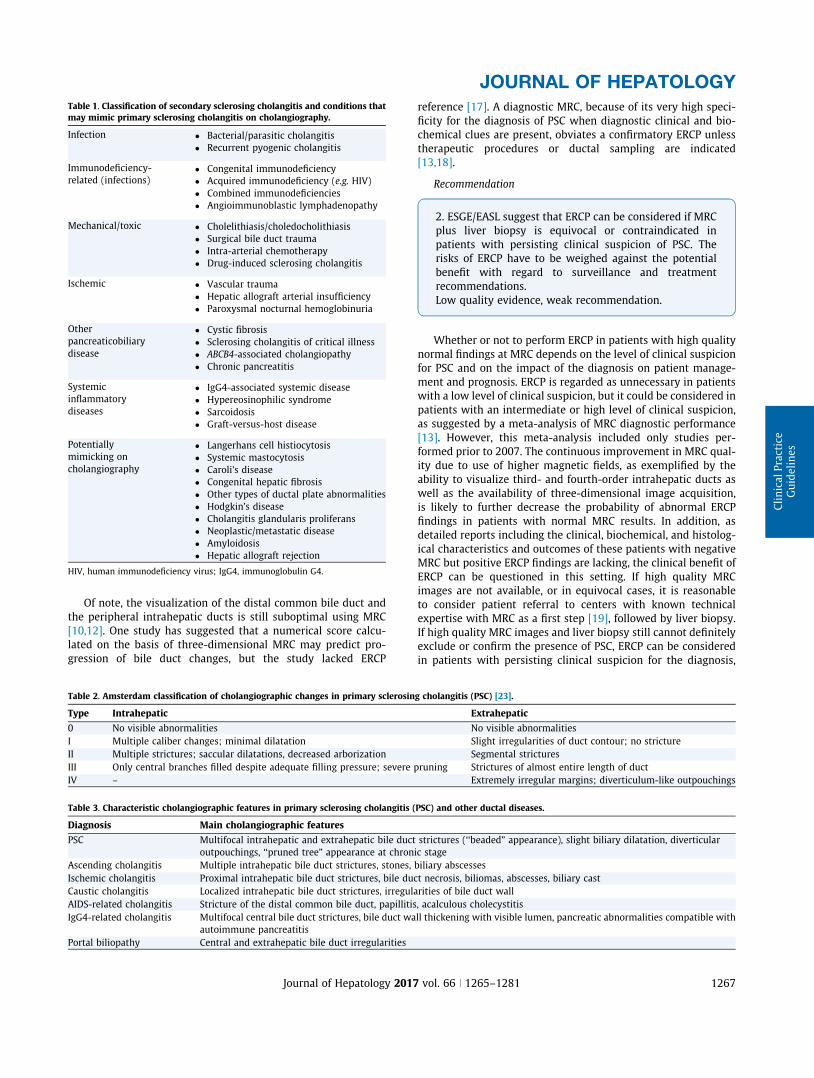
Table 1. Classification of secondary sclerosing cholangitis and conditions thatmay mimic primary sclerosing cholangitis on cholangiography.
Infection � Bacterial/parasitic cholangitis� Recurrent pyogenic cholangitis
Immunodeficiency-related (infections)
� Congenital immunodeficiency� Acquired immunodeficiency (e.g. HIV)� Combined immunodeficiencies� Angioimmunoblastic lymphadenopathy
Mechanical/toxic � Cholelithiasis/choledocholithiasis� Surgical bile duct trauma� Intra-arterial chemotherapy� Drug-induced sclerosing cholangitis
Ischemic � Vascular trauma� Hepatic allograft arterial insufficiency� Paroxysmal nocturnal hemoglobinuria
Otherpancreaticobiliarydisease
� Cystic fibrosis� Sclerosing cholangitis of critical illness� ABCB4-associated cholangiopathy� Chronic pancreatitis
Systemicinflammatorydiseases
� IgG4-associated systemic disease� Hypereosinophilic syndrome� Sarcoidosis� Graft-versus-host disease
Potentiallymimicking oncholangiography
� Langerhans cell histiocytosis� Systemic mastocytosis� Caroli’s disease� Congenital hepatic fibrosis� Other types of ductal plate abnormalities� Hodgkin’s disease� Cholangitis glandularis proliferans� Neoplastic/metastatic disease� Amyloidosis� Hepatic allograft rejection
HIV, human immunodeficiency virus; IgG4, immunoglobulin G4.
JOURNAL OF HEPATOLOGY
Of note, the visualization of the distal common bile duct andthe peripheral intrahepatic ducts is still suboptimal using MRC[10,12]. One study has suggested that a numerical score calcu-lated on the basis of three-dimensional MRC may predict pro-gression of bile duct changes, but the study lacked ERCP
Table 2. Amsterdam classification of cholangiographic changes in primary sclerosin
Type Intrahepatic
0 No visible abnormalitiesI Multiple caliber changes; minimal dilatationII Multiple strictures; saccular dilatations, decreased arborizationIII Only central branches filled despite adequate filling pressure; severe pIV –
Table 3. Characteristic cholangiographic features in primary sclerosing cholangitis (
Diagnosis Main cholangiographic features
PSC Multifocal intrahepatic and extrahepatic bile ductoutpouchings, ‘‘pruned tree” appearance at chron
Ascending cholangitis Multiple intrahepatic bile duct strictures, stones,Ischemic cholangitis Proximal intrahepatic bile duct strictures, bile ducCaustic cholangitis Localized intrahepatic bile duct strictures, irregulaAIDS-related cholangitis Stricture of the distal common bile duct, papillitisIgG4-related cholangitis Multifocal central bile duct strictures, bile duct wa
autoimmune pancreatitisPortal biliopathy Central and extrahepatic bile duct irregularities
Journal of Hepatology 2017
reference [17]. A diagnostic MRC, because of its very high speci-ficity for the diagnosis of PSC when diagnostic clinical and bio-chemical clues are present, obviates a confirmatory ERCP unlesstherapeutic procedures or ductal sampling are indicated[13,18].
Recommendation
g c
ru
PS
sticbiltri, all
v
2. ESGE/EASL suggest that ERCP can be considered if MRCplus liver biopsy is equivocal or contraindicated inpatients with persisting clinical suspicion of PSC. Therisks of ERCP have to be weighed against the potentialbenefit with regard to surveillance and treatmentrecommendations.Low quality evidence, weak recommendation.
Whether or not to perform ERCP in patients with high qualitynormal findings at MRC depends on the level of clinical suspicionfor PSC and on the impact of the diagnosis on patient manage-ment and prognosis. ERCP is regarded as unnecessary in patientswith a low level of clinical suspicion, but it could be considered inpatients with an intermediate or high level of clinical suspicion,as suggested by a meta-analysis of MRC diagnostic performance[13]. However, this meta-analysis included only studies per-formed prior to 2007. The continuous improvement in MRC qual-ity due to use of higher magnetic fields, as exemplified by theability to visualize third- and fourth-order intrahepatic ducts aswell as the availability of three-dimensional image acquisition,is likely to further decrease the probability of abnormal ERCPfindings in patients with normal MRC results. In addition, asdetailed reports including the clinical, biochemical, and histolog-ical characteristics and outcomes of these patients with negativeMRC but positive ERCP findings are lacking, the clinical benefit ofERCP can be questioned in this setting. If high quality MRCimages are not available, or in equivocal cases, it is reasonableto consider patient referral to centers with known technicalexpertise with MRC as a first step [19], followed by liver biopsy.If high quality MRC images and liver biopsy still cannot definitelyexclude or confirm the presence of PSC, ERCP can be consideredin patients with persisting clinical suspicion for the diagnosis,
holangitis (PSC) [23].
Extrahepatic
No visible abnormalitiesSlight irregularities of duct contour; no strictureSegmental strictures
ning Strictures of almost entire length of ductExtremely irregular margins; diverticulum-like outpouchings
C) and other ductal diseases.
rictures (‘‘beaded” appearance), slight biliary dilatation, diverticularstageiary abscessesnecrosis, biliomas, abscesses, biliary castties of bile duct wallcalculous cholecystitisthickening with visible lumen, pancreatic abnormalities compatible with
ol. 66 j 1265–1281 1267
Clinical Practice Guidelines
to take advantage of the filling pressure obtained by the balloonocclusion and the slight superiority as to visualization of theextrahepatic bile ducts.Ductographic criteria for PSC
The first ERCP criteria for ductographic changes in PSC were pub-lished in 1984 by Li-Yeng & Goldberg [20]. Typical changes seen inPSC consist of minor irregularities of duct contour and local nar-rowingwith pre-stenotic dilatation (type I), threadlike narrowingsalternating with normal caliber of bile ducts or slight dilatation(type II), multiple strictures with saccular dilatations (type III),and the most advanced changes consisting of advanced ductalnarrowing with resultant lack of filling of the peripheral ducts(type IV). The classification has later been modified by Majojieet al. [21] and Ponsioen et al. [22,23]. The classification of Ponsioenet al. [23] has been validated and shown to correlate with patientprognosis (Table 2). Another type of classification is based onevaluation of the grade, length, and extent of strictures, the degreeof bile duct dilatation, and the distribution of lesions [24].
None of the ductographic criteria published are specific forPSC and the findings must be interpreted in the context of patientdemographic data and the clinical features. Review by teams withexpertise in complex biliary disease is often useful, as multiplesecondary causes of sclerosing cholangitis must be considered[25] (Table 3).
Unusual cholangiographic features
Some PSC patients may present with cystic dilatations of intra-hepatic bile ducts simulating Caroli’s disease [10]. Of note, thefusiform and small cystic dilatations of intrahepatic (mostlyperipheral) bile ducts, as observed in patients with congenitalhepatic fibrosis and autosomal recessive polycystic kidney dis-ease, should not be misdiagnosed as PSC [11].
Another differential diagnosis is the peculiar cholangiographicphenotype of adult forms of ABCB4/MDR3 deficiency which maybe characterized by large unifocal or multifocal spindle-shapedintrahepatic bile ductdilatationswithorwithout apparent bile ductstenosis [12,26]. This diagnosis should be suspected on familialclustering of excessive gallstone disease and often a history of priorcholecystectomy at age\40 years and associated intrahepaticcholestasis of pregnancy, and is confirmed by ABCB4 genotyping.
Recommendation
1
3. For the diagnosis of PSC, ESGE/EASL do not suggest rou-tine use of endoscopic techniques other than endoscopicretrograde cholangiopancreatography (ERCP) (i.e., endo-scopic ultrasound including intraductal ultrasound[IDUS], cholangioscopy, confocal endomicroscopy).Weak recommendation, low quality evidence.
In the diagnosis of PSC there is no established role for endo-scopic techniques beyond ERCP, e.g. brush cytology, ductalbiopsy, cholangioscopy, or confocal laser endomicroscopy. Inselected cases with suspected extrahepatic disease and inconclu-sive MRC findings, endoscopic ultrasound (including IDUS) andelastography may add information on common bile duct stric-tures, wall thickening, and liver fibrosis stage [27–30].
268 Journal of Hepatology 2017
ERCP in established PSC
Recommendation
v
4. ESGE/EASL suggest that a dominant stricture at ERCPshould be defined as a stenosis with a diameterof 61.5 mm in the common bile duct and/or 61.0 mm inan hepatic duct within 2 cm of the main hepatic conflu-ence.Weak recommendation, low quality evidence.
Deciding on the clinical impact of a bile duct stricture maybe challenging. The ‘‘dominant stricture” denomination arosealongside the term ‘‘major stricture” early in the history ofendoscopic management of PSC [31]. The ‘‘major” or ‘‘dominant”stricture terms were initially used more broadly, pertaining tostrictures of the common bile duct and right and left bifurcationof the hepatic ducts (extrahepatic PSC lesions), since these werefound to be more prone to clinical events than intrahepaticstrictures [31,32]. The precise definition of a dominant stricturewas introduced by Stiehl et al. in 2002 for use in endoscopicstudies as a severity measure [33,34], although it employs asomewhat arbitrary value, depending, for example, on fillingpressure. A number of endoscopic studies, both before and after2002, do not apply the diameter criterion strictly when deter-mining a dominant stricture [35,36], and focus on suspectedclinical relevance. Determination of the clinical significanceand potential benefit from endoscopic interventions shouldtherefore not be based on this definition alone, and the decisionfor intervention rather considered as a compound clinicaldecision.
Multiple dominant strictures can be found in the same patient(12% in the study by Bjornsson et al.) [34].
Of note, the ERCP definition of a dominant stricture is usuallyconsidered to be not applicable to MRC, in particular in the extra-hepatic ducts, given the insufficient spatial resolution of MRC[10,17] and the lack of the hydrostatic pressure that is presentduring ERCP.
A complete occlusion cholangiogram should generally beobtained if an ERCP is performed, because it adds little risk tothe ERCP, decreases variability, and may reveal that a dominantstricture suspected at MRC is indeed not a stricture [37].
Recommendation
5. ESGE/EASL suggest ERCP and ductal sampling (brushcytology, endobiliary biopsies) should be considered inestablished PSC in the case of: (i) clinically relevant orworsening symptoms (jaundice, cholangitis, pruritus);(ii) rapid increase of cholestatic enzyme levels; or (iii)new dominant stricture or progression of existing domi-nant strictures identified at MRC in the context of appro-priate clinical findings.Weak recommendation, low quality evidence.
ERCP can be indicated in patients with a confirmed diagnosisof PSC when changes in clinical, laboratory, and radiological find-ings occur during the course of the disease. The purpose is to
ol. 66 j 1265–1281
JOURNAL OF HEPATOLOGY
make an assessment of the likelihood of the presence of biliarydysplasia as a risk factor for cholangiocarcinoma (CCA) and toidentify biliary strictures amenable to intervention.i. Clinical events:In the early stage of PSC, dominant biliary strictures are usu-
ally asymptomatic. Exacerbation of jaundice (not related to liverfailure), episodes of fever and chills suggestive of cholangitis, orworsening of pruritus are indications for ERCP for the treatmentof dominant strictures and to perform ductal brush sampling toexclude malignancy [8,38]. Worsening pain in the right upperabdominal quadrant, fatigue, and weight loss also need carefulevaluation.
ii. Laboratory results:Serum laboratory tests are neither sensitive nor specific
enough to evaluate PSC progression [38], but in the case ofrapid increase of serum bilirubin levels and/or cholestatic liverenzymes (serum ALP, serum GGT) ERCP is indicated [6], espe-cially in patients with a diagnosis of clinically significant hilaror extrahepatic strictures on MRC. Elevation of serum CA19-9in PSC patients has an unsatisfactory sensitivity (14%) andpositive predictive value (PPV) (67%) for the diagnosis ofCCA [36,38,39], and is not helpful in selecting patients forERCP.
iii. Progression/new-onset clinically significant strictures on MRC:Progressive intrahepatic or extrahepatic bile duct dilatation
on imaging studies (ultrasound or MRC) is an indication for ERCPwith ductal sampling [6]. A careful evaluation of new-onset dom-inant strictures in PSC is recommended, because of the increasedrisk of CCA in this situation.
In detail, a stricture that is disproportionately severe relativeto others, concomitant biliary filling defects, marked biliarydilatation (62 cm for the common bile duct, 61 cm for the rightor left intrahepatic ducts, 65mm for other intrahepatic ducts)suggests CCA [40]. Conversely, this risk was low in patients with-out dominant strictures according to a 25-year experience [41].Abnormal cytological findings, such as suspicion of malignancyor aneuploid DNA findings need a close follow-up by ERCP withrepeated sampling, unless urgent liver transplantation is consid-ered to be warranted.
The utility of ERCP in handling dominant strictures wasshown in a prospective study [42] on 171 PSC patients followedfor 20 years: repeated endoscopic therapy was associated witha transplant-free survival of 81% at 5 years and 52% at 10 yearsafter initial endoscopic therapy. In this population, a 6% CCA ratewas found in patients with dominant strictures.
Recommendation
6. ESGE/EASL suggest that, in patients with an establisheddiagnosis of PSC, MRC should be considered before thera-peutic ERCP.Weak recommendation, low quality evidence.
MRC may be useful to confirm the indication, to exclude focalparenchymal changes, and to give the clinicians performing theERCP imaging-based guidance to minimize the risk of complica-tions. Regarding MRC in established PSC, a retrospective single-center study reported a 76% accuracy of MRC in the diagnosisof CCA complicating PSC [40]. For these reasons, patients withan established diagnosis of PSC should have an MRC examinationin their clinical records [13,43].
Journal of Hepatology 2017
Recommendation
v
7. ESGE/EASL suggest performing endoscopic treatmentwith concomitant ductal sampling (brush cytology, endo-biliary biopsies) of suspected significant strictures identi-fied at MRC in PSC patients who present with symptomslikely to improve following endoscopic treatment.Strong recommendation, low quality evidence.
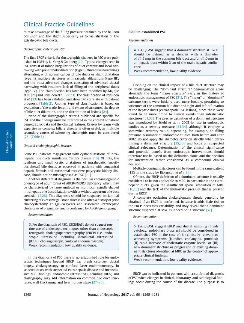
Selected series reporting on endoscopic treatment in PSCpatients are summarized in Table 4; none of these compared per-formance vs. no performance of endoscopic treatment for domi-nant stricture. The benefits reported following dilation ofdominant stricture included short-term improvement of symp-toms and of liver biochemical test results, as well as a longer livertransplantation-free survival compared to that predicted usingthe Mayo clinical risk model. Similar findings have also beenreported in several smaller case series [32,47–50].
The main criticisms of these studies are as follows:i. The Mayo clinical risk model was not designed to evaluate
patientswith dominant stricture; specifically,manypatientsunderwent therapeutic ERCP because of elevated bilirubin,which is part of the Mayo risk score and went down in mostpatients after the intervention. Hence, baseline Mayo riskscore was not determined in a steady-state situation.
ii. Serum test results for cholestasis may spontaneously fluc-tuate in patients with PSC complicated or not with a dom-inant stricture. In 125 PSC patients, Bjornsson et al.reported changes in serum ALP and serum bilirubin frombaseline up to 12 months following ERCP. As patients withdominant stricture received no stricture dilation, theauthors stated that ‘‘If our patients had been consequentlydilated or stented the decrease in bilirubin and clinical fea-tures at follow-up would have been attributed to endo-scopic therapy” [34]. However, in that study, thevariations reported in ALP and in total serum bilirubinafter vs. before ERCP were not significant, in contrast withvarious studies listed in Table 4 that used dominant stric-ture dilation/stenting. Also, it was not clear on what basisthese patients were treated conservatively, while othersdid receive endoscopic therapy.
Other limitations of most studies listed in Table 4 include ret-rospective design, selection bias, and reporting of results for amixture of treatments, namely dilation with and without stentingof dominant strictures as well as, in a minority of patients, treat-ment with ursodeoxycholic acid started during follow-up.
A critical issue is that potential benefits must be weighedagainst the certain risks of therapeutic ERCP in patients with noother therapeutic option except liver transplantation. Symptomslikely to improve following dominant stricture treatment generallyinclude pruritus, pain, cholangitis, and jaundice in patients with asignificant (620%) increase in cholestasis, while in patients withend-stage liver disease, only cholangitis is expected to improve.
Finally, patientswith advanced liver diseasewith cirrhosismaynot benefit from endoscopic treatment. Ahrendt et al. reported nochange in serum bilirubin at 1 year following endoscopic and/orpercutaneous stricture dilation in ten patients with cirrhosis anda baseline serum bilirubin 65 mg/dl [51]. Death following endo-scopic balloon dilation of dominant stricture has been reported
ol. 66 j 1265–1281 1269
Table 4. Selected series reporting on endoscopic treatment of dominant strictures in primary sclerosing cholangitis.
First author,Year [Ref.]
Study design Patients, n Intervention Outcomes Results
Dilation ± stentingGotthardt,2010 [42](Extensionof Stiehl 2002 study[33])
Prospective 96(ALP[2 � ULN)
Balloon dilation (8 mm in CBD,6–8 mm for IHBD), plus stentin 5 patients with severecholestasis and bacterialcholangitis
Short-term improvement incholestasisLiver transplantation-freesurvivalComplications
– At 2 weeks, mean bilirubin level significantly decreased(by 56%)
– Improvement in symptoms and liver transplantation-free survival
– Comparison with Mayo model not reported (5-year and10-year liver transplantation-free survival, 81% and52%)
– Overall complication rate, 3.8%Gluck,2008 [35]
Retrospective 84Symptomatic patients
Balloon dilation and stenting(70% and 51% of patients,respectively)
Liver transplantation-freesurvival
– Higher proportion of patients alive with no liver trans-plantation at 3 and 4 years than predicted using Mayomodel (p\0.05); at 1 and 2 years survival, similar toMayo prediction
– Adverse events in 21 therapeutic ERCPs (7.2% of 291procedures, 25% of patients)
Stiehl,2002 [33]
Prospective 52 (ALP[2 � ULN) Balloon dilation (8 mm in CBD,6–8 mm for IHBD), plus stentin 5 patients with severecholestasis and bacterialcholangitis
Bilirubin and liver enzymes2 weeks after dilationSymptomsLiver transplantation-freesurvival
– At 2 weeks, significant decrease in liver enzymes andbilirubin
– Improvement of jaundice in 24/24 and of pruritus in12/13 patients
– Longer liver transplantation-free survival than pre-dicted using 1992 Mayo model (p\0.0001)
Baluyut,2001 [44]
Retrospective 56 with symptoms7 without symptoms
Balloon dilation (4–12 mm,n = 61) Once per year, withstent if no significantradiological improvementfollowing dilation (n = 33)
Liver transplantation-freesurvivalComplication rate
– Longer liver transplantation-free survival than pre-dicted using 1999 Mayo model (p = 0.027)
– 12% complications
StentingPonsioen,1999 [36]
Retrospective 32Symptomatic patientswith successful stentingfor dominant stricture
1-week stenting (10-Fr stent)with no balloon dilation
2-month symptomatic andbiochemical improvement,Actuarial curve of re-intervention-free patients
– Improvement of symptoms in 83%– Significant decrease in bilirubin (44% had increased
conjugated bilirubin at baseline) and cholestasisenzymes
– Re-intervention-free patients (actuarial): 60% at 3 yearsvan Milligen de Wit,1996 [45]
Retrospective 25With symptoms orprogression of serum testsfor cholestasis
Stenting for a median of3 months (plus 8 mm dilationin 3 patients)
Change in symptoms andbiochemical tests within6 months following stentinsertionAdverse events
– Improvement of symptoms in 76%– Significant decrease in bilirubin (52% had increased
bilirubin at baseline) and serum tests for cholestasis– 32 episodes of cholangitis/jaundice related to stent
cloggingDilation vs. dilation + stentingKaya,2001 [46]
Retrospective 71with symptoms
Balloon dilation (4–8 mm,n = 34) vs. Balloon dilationwith 3–4-month stenting(n = 37)Intervention via PTBD in 0/34of balloon group vs. 23/37 ofstent group
Biochemical course up to24 months
– Both strategies improved liver biochemistry; feverresolved only in the dilation without stent group. Noadditional benefit of stenting after balloon dilation
– More complications in stent vs. dilation alone group(p = 0.001)
– More complications in PTBD vs. ERCP group (p\0.001)(No multivariate analysis)
ALP, alkaline phosphatases; ULN, upper limit of normal values; CBD, common bile duct; IHBD, intrahepatic bile duct; ERCP, endoscopic retrograde cholangiopancreatography; PTBD, percutaneous transhepatic biliarydrainage.
ClinicalPractice
Guidelines
1270Journal
ofHepatology
2017vol.66
j1265–1281
in a patient with PSC and end-stage liver disease [46]. DiagnosticERCPwas followed by deterioration of cholestasis in 7 of 8 patientswithmore advanced PSC at biopsy (Ludwig stage III or IV) vs. 1 of 7with less advanced disease (Ludwig stage I or II) [52].
Balloon dilation vs. stent therapy
Recommendation
8. ESGE/EASL suggest that the choice between stentingand balloon dilation should be left to the endoscopist’sdiscretion.Weak recommendation, low quality evidence.
Results from selected series reporting on endoscopic treat-ment of dominant strictures in PSC are summarized in Table 4.Of note: (i) in themajority of studies that reported on balloon dila-tion for dominant stricture, stents were inserted in a minority ofpatients; (ii) a significant improvement in liver transplantation-free survival compared with the Mayo model has been reportedonly with balloon dilation; and (iii) the perforation rate has beenhigher with stenting compared with balloon dilation.
A single retrospective study compared balloon dilation vs. bal-loon dilation combinedwith stenting for dominant stricture in PSCpatients (n = 34 and n = 37, respectively) [46]. The ‘‘balloon dila-tion alone” group was treated by endoscopic means only, while23 patients (62%) in the ‘‘stenting” group underwent percutaneoustreatment because of failed endoscopic access and/or dominantstricture dilation. Serum bilirubin decreased similarly in bothgroups of patients, but more procedures and more complicationswere recorded in the stent vs. the balloon dilation group (mediannumber of procedures per patient, 5.0 vs.2.1, respectively; patientswith complications, 54% vs. 15%, respectively). Complicationsincluded bile duct perforation in seven patients (10%), 5 of whomwere in the stent group. However, it is difficult to draw conclusionsbecause of the different access routes used (percutaneous in 62% inthe stent group vs. 0 in the balloon dilation group), a selection biasdue tomore severe stricture in the stent group, and the long stent-ing duration used (mean 3 months) putting the patient at high riskfor stent clogging and cholangitis. A short stenting duration (seerecommendation 13) is currently the standard of care.
The European multicenter randomized DILSTENT trial com-paring single-balloon dilatation vs. short-term stenting was pre-maturely stopped recently after a planned interim analysis.Preliminary results show no differences in outcome, but a signif-icantly higher serious adverse event rate in the stent group (Dr. C.Y. Ponsioen, personal communication).
Role of sphincterotomy
Recommendation
9. ESGE/EASL recommend weighing the anticipated bene-fits of biliary papillotomy/sphincterotomy against its riskson a case-by-case basis. Strong recommendation, moder-ate quality evidence. Biliary papillotomy/sphincterotomyshould be considered especially after difficult cannulation.Strong recommendation, low quality evidence.
Journal of Hepatology 2017
Biliary sphincterotomy was performed routinely as part of theendoscopic treatment of dominant stricture in some studies [46]
while its use was restricted to specific cases such as stone extrac-tion and difficulties in stent insertion in other studies. For exam-ple, in 32 PSC patients treated with stents for dominant stricture,sphincterotomy was performed in 12 patients (38%) [36] while inanother study of dominant stricture dilation with/without stent-ing, sphincterotomy was performed in 63% of 63 patients [44].Generally, biliary sphincterotomy is not recommended as aroutine procedure prior to biliary stenting because of the associ-ated risks as demonstrated in randomized controlled trials (RCTs)[53]. However, if cannulation is difficult, biliary sphincterotomyis advised, bearing in mind that these patients are likely torequire multiple procedures. Many endoscopists prefer a smallsphincterotomy in PSC in order to avoid ascending cholangitis.
Specifically in PSC, biliary sphincterotomy was independentlyassociated with an increased risk of short-term adverse events intwo retrospective studies (odds ratios [OR]: 4.7 and 5.0) [54,55]while previous biliary papillotomy/sphincterotomywas protectivefor subsequent ERCPs [54]. Therefore, experienced endoscopistsperform biliary sphincterotomy in patients with difficult cannula-tion in whom ERCP is likely to be repeated during follow-up.
Balloon dilation
Recommendations
JOURNAL OF HEPATOLOGY
v
10. ESGE/EASL suggest selecting a balloon caliber of up tothe maximum caliber of the ducts delimiting the stricture.Weak recommendation, low quality evidence.
11. ESGE/EASL suggest repeating dilation of relapsingdominant stricture if: (i) the dominant stricture isregarded as the cause of recurrent symptoms (cholangitis,pruritus) or of significant increase in cholestasis; and (ii)the patient’s response to previous dilations has been sat-isfactory.Weak recommendation, very low quality evidence.
There are no comparative data on the optimal dilation schemeor balloon diameter for treating dominant strictures. In the lar-gest prospective study (500 endoscopic balloon dilations in 96patients), the authors performed stepwise dominant stricturedilation up to diameters of 8 mm and 6–8 mm in the commonbile duct and the hepatic ducts, respectively [42]. Bile duct diam-eter upstream and downstream of the dominant stricture shouldbe taken into account for selecting the balloon diameter to avoiddilating to more than the duct diameter. Balloon dilations areusually repeated at intervals of 1 to 4 weeks up to technical suc-cess, for an average of 2–3 balloon dilations [33,42,50]. Technicalsuccess has been defined as complete balloon inflation within thedominant stricture with no waist observed fluoroscopically, fol-lowed by the unobstructed passage of contrast medium throughthe dilated biliary segment to the duodenum [42,50]. Using thistechnique, bile duct perforation was reported in 0.2% of dominantstricture dilations (1% of patients) [42]. In contrast, another studythat used balloons of diameter 4–12 mm for dilation reporteddilation-related biliary perforations in 3.5% of procedures [44].
Repeat balloon dilation during follow-up after initial treat-ment (usually consisting of several ERCPs) has been mentioned
ol. 66 j 1265–1281 1271
Clinical Practice Guidelines
in some studies, but no results of the repeat dilation, in terms ofclinical or biochemical improvement, have been reported [33,50].Stent therapy
Recommendation
1
12. ESGE/EASL suggest selecting a single 10-Fr stent fordominant stricture in the extrahepatic ducts or two 7-Frstents for hilar strictures extending into the left or righthepatic duct (final stent diameters in the case of stepwisestenting).Weak recommendation, very low quality evidence.
In all large studies of endoscopic treatment for dominantstricture, plastic stents measuring 7 to 10 Fr in diameter havebeen used, with no reported comparison of the results obtainedwith various stent diameters. Specifically, the Amsterdam groupaimed at inserting a single 10-Fr stent, and if this was not possi-ble at first attempt, it was preceded by 1-week stenting with a 7-Fr stent or insertion of a nasobiliary catheter [36,56]. The Mayogroup used 7–10-Fr stents at the endoscopist’s discretion [46].The Indianapolis group did not mention the diameter of stentsused [44]. Two 7-Fr stents have typically been used in patientswith multiple bilateral dominant strictures, and in patients witha hilar stricture extending into the left or right hepatic duct inorder to avoid temporary obstruction of the contralateral biliarysystem. In general, the stent caliber and length must be adaptedto the specific biliary tree configuration.
In other diseases, studies have shown that polyethylene stentsprovide better short-term (1-month) patency than Teflon modelsand that, in the long term, 10-Fr models provide longer biliarypatency compared with thinner ones (11.5-Fr models do not pro-vide longer patency) [53].
With respect to balloon dilation prior to stenting, it is cur-rently unclear whether balloon dilation is beneficial before stentplacement.
Duration of stenting
Recommendation
13. ESGE/EASL suggest that stents used for treating dom-inant stricture should be removed 1–2 weeks followinginsertion.Weak recommendation, low quality evidence.
No comparison of various stenting durations has been identi-fied in studies reporting on stenting for dominant stricture. Ashort stenting duration is currently favored because stents tendto clog rapidly in PSC patients and similar efficacy results havebeen reported with short (1–2 weeks) vs. standard (8–12 weeks)stenting duration. Specifically, a retrospective study of short-term stenting (mean duration 11 days) in 32 symptomatic PSCpatients with dominant stricture showed, at 2 months, a symp-tomatic improvement in 83% of the patients as well as a signifi-cant improvement of cholestasis test results; at 1 and 3 years,actuarial analysis showed that 80% and 60% of patients, respec-tively, would not require re-intervention [36]. Stent dysfunction
272 Journal of Hepatology 2017
was not reported in this study but two patients treated by stentremoval developed hydrops of the gallbladder. The same group ofauthors had previously reported similar efficacy results with 3-month stenting in 25 patients with symptomatic dominant stric-ture but, in that study, unscheduled stent exchange had to be per-formed on 32 occasions because of suspected stent clogging(cholangitis n = 23, jaundice n = 9) [45].
All studies mentioned focused on clinical and serum liver tests,not radiological data, to assess the short-term effect of therapeuticERCP [36,45,46,56]. Endoscopic treatment has been repeated in asizeable proportion of patients. For example, with long medianstentingperiods (3 months), themediannumber of repeated ERCPsper patient ranged between 3 and 5 during follow-up periods of 29and 22 months in two studies [45,46], while following a shortstenting period (mean 11 days) repeat ERCP rates at 1 and 3 yearsafter treatment were estimated at 20% and 40%, respectively [36].Other details about repeated treatments were not reported.
In many centers, stents are removed during an esophagogas-troduodenoscopy without biliary opacification in PSC patients.
Complications of endoscopic therapy
Recommendation
v
14. ESGE/EASL suggest that ERCP in PSC patients shouldbe undertaken by experienced pancreaticobiliary endo-scopists.Strong recommendation, very low quality evidence.
Several studies have evaluated the risk of complications in PSCpatients undergoing ERCP [33,35,44,49,54,55,57–62] (Table 5).ERCP carries an increased risk for complications in the context ofPSC, especially pancreatitis, cholangitis, and extravasation of con-trast, although not all studies have documented such an increasedrisk in PSC [59,62]. In a systematic survey [63] of post-ERCP com-plications associated with various indications for ERCP, including21 prospective studies and 16,855 patients, the total complicationrate was 6.85% (95% confidence interval [CI]: 6.46%–7.24%). Pan-creatitis occurred in 585 patients (3.47%, 95% CI: 3.19%–3.75%).In another large retrospective single-center study [47], with11,497 procedures over 12 years, the total complication rate was4.0% and pancreatitis occurred in 3.6%. The overall risk of adverseevents in patients with PSC has varied in different, much smallerstudies, from 1.8% to 18.4% [33,35,44,49,55,57–62], which ishigher than reported for other indications [47,63].
Retraction of the papilla and an altered, more difficult positionof the endoscope due to hypertrophy of the left liver lobe may beencountered during ERCP in PSC patients. Whether this actuallyinfluences cannulation success rates has not been investigatedby specific studies. Cohort studies describing PSC patients provideonly limited details on cannulation difficulties,with failure rates of0% to 6% [33,36,41,49,50,57,62,64–66]. Furthermore, there is likelya selection bias since most retrospective series describing theresults of endoscopic treatment have the initiation of therapy asprerequisite, therefore potentially excluding cannulation failures.
The largest series is the study by Ismail et al. [54]. In this ret-rospective review of 441 ERCP procedures over a 3-year time per-iod, primary cannulation success was 88.2%. Of note, in 137patients (37.8%) a previous biliary sphincterotomy had been per-formed. Pancreatic sphincterotomy as an access technique wasused in 11.8% and freehand needle-knife sphincterotomy in a
ol. 66 j 1265–1281
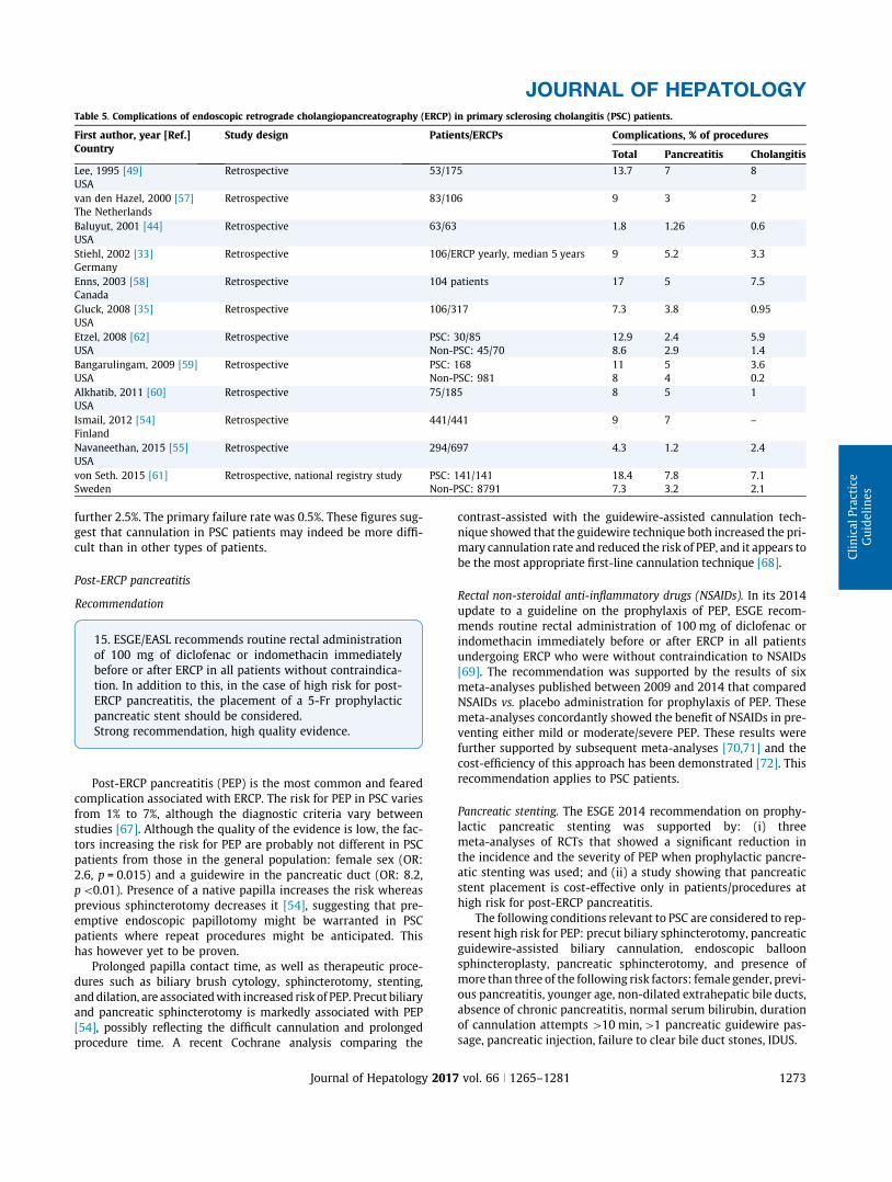
Table 5. Complications of endoscopic retrograde cholangiopancreatography (ERCP) in primary sclerosing cholangitis (PSC) patients.
First author, year [Ref.]Country
Study design Patients/ERCPs Complications, % of procedures
Total Pancreatitis Cholangitis
Lee, 1995 [49]USA
Retrospective 53/175 13.7 7 8
van den Hazel, 2000 [57]The Netherlands
Retrospective 83/106 9 3 2
Baluyut, 2001 [44]USA
Retrospective 63/63 1.8 1.26 0.6
Stiehl, 2002 [33]Germany
Retrospective 106/ERCP yearly, median 5 years 9 5.2 3.3
Enns, 2003 [58]Canada
Retrospective 104 patients 17 5 7.5
Gluck, 2008 [35]USA
Retrospective 106/317 7.3 3.8 0.95
Etzel, 2008 [62]USA
Retrospective PSC: 30/85Non-PSC: 45/70
12.98.6
2.42.9
5.91.4
Bangarulingam, 2009 [59]USA
Retrospective PSC: 168Non-PSC: 981
118
54
3.60.2
Alkhatib, 2011 [60]USA
Retrospective 75/185 8 5 1
Ismail, 2012 [54]Finland
Retrospective 441/441 9 7 –
Navaneethan, 2015 [55]USA
Retrospective 294/697 4.3 1.2 2.4
von Seth. 2015 [61]Sweden
Retrospective, national registry study PSC: 141/141Non-PSC: 8791
18.47.3
7.83.2
7.12.1
JOURNAL OF HEPATOLOGY
further 2.5%. The primary failure rate was 0.5%. These figures sug-gest that cannulation in PSC patients may indeed be more diffi-cult than in other types of patients.
Post-ERCP pancreatitis
Recommendation
15. ESGE/EASL recommends routine rectal administrationof 100 mg of diclofenac or indomethacin immediatelybefore or after ERCP in all patients without contraindica-tion. In addition to this, in the case of high risk for post-ERCP pancreatitis, the placement of a 5-Fr prophylacticpancreatic stent should be considered.Strong recommendation, high quality evidence.
Post-ERCP pancreatitis (PEP) is the most common and fearedcomplication associated with ERCP. The risk for PEP in PSC variesfrom 1% to 7%, although the diagnostic criteria vary betweenstudies [67]. Although the quality of the evidence is low, the fac-tors increasing the risk for PEP are probably not different in PSCpatients from those in the general population: female sex (OR:2.6, p = 0.015) and a guidewire in the pancreatic duct (OR: 8.2,p\0.01). Presence of a native papilla increases the risk whereasprevious sphincterotomy decreases it [54], suggesting that pre-emptive endoscopic papillotomy might be warranted in PSCpatients where repeat procedures might be anticipated. Thishas however yet to be proven.
Prolonged papilla contact time, as well as therapeutic proce-dures such as biliary brush cytology, sphincterotomy, stenting,anddilation, are associatedwith increased risk of PEP. Precut biliaryand pancreatic sphincterotomy is markedly associated with PEP[54], possibly reflecting the difficult cannulation and prolongedprocedure time. A recent Cochrane analysis comparing the
Journal of Hepatology 2017
contrast-assisted with the guidewire-assisted cannulation tech-nique showed that the guidewire technique both increased the pri-mary cannulation rate and reduced the risk of PEP, and it appears tobe the most appropriate first-line cannulation technique [68].
Rectal non-steroidal anti-inflammatory drugs (NSAIDs). In its 2014update to a guideline on the prophylaxis of PEP, ESGE recom-mends routine rectal administration of 100 mg of diclofenac orindomethacin immediately before or after ERCP in all patientsundergoing ERCP who were without contraindication to NSAIDs[69]. The recommendation was supported by the results of sixmeta-analyses published between 2009 and 2014 that comparedNSAIDs vs. placebo administration for prophylaxis of PEP. Thesemeta-analyses concordantly showed the benefit of NSAIDs in pre-venting either mild or moderate/severe PEP. These results werefurther supported by subsequent meta-analyses [70,71] and thecost-efficiency of this approach has been demonstrated [72]. Thisrecommendation applies to PSC patients.
Pancreatic stenting. The ESGE 2014 recommendation on prophy-lactic pancreatic stenting was supported by: (i) threemeta-analyses of RCTs that showed a significant reduction inthe incidence and the severity of PEP when prophylactic pancre-atic stenting was used; and (ii) a study showing that pancreaticstent placement is cost-effective only in patients/procedures athigh risk for post-ERCP pancreatitis.
The following conditions relevant to PSC are considered to rep-resent high risk for PEP: precut biliary sphincterotomy, pancreaticguidewire-assisted biliary cannulation, endoscopic balloonsphincteroplasty, pancreatic sphincterotomy, and presence ofmore than three of the following risk factors: female gender, previ-ous pancreatitis, younger age, non-dilated extrahepatic bile ducts,absence of chronic pancreatitis, normal serum bilirubin, durationof cannulation attempts [10 min,[1 pancreatic guidewire pas-sage, pancreatic injection, failure to clear bile duct stones, IDUS.
vol. 66 j 1265–1281 1273
Clinical Practice Guidelines
Recommendation1
16. ESGE/EASL suggest routine administration of prophy-lactic antibiotics before ERCP in patients with PSC.Strong recommendation, low quality evidence.
Bacterial cholangitis and bacteriobilia are a not infrequent find-ing among patients with PSC. In studies evaluating the complica-tions of ERCP in PSC the risk for cholangitis has varied from 0.25%to 8% [33,35,44,49,54,55,57–62] depending on, among other items,the criteria used to define cholangitis. The use of prophylacticantibioticsvariesmarkedlybetween studies, in termsof prevalence,type of antibiotic, and duration of administration (from one oraldose before the procedure to 1-week dosing afterwards). In aCochrane meta-analysis (9 RCTs, 1,573 patients), the prophylacticuse of antibiotics was shown to prevent cholangitis (relative risk[RR]: 0.54; 95% CI: 0.33–0.91), septicemia (RR: 0.35; 95% CI: 0.11–1.11), bacteremia (RR: 0.50; 95% CI: 0.33–0.78), and pancreatitis(RR: 0.54; 95% CI: 0.29–1.00). It was concluded that prophylacticantibiotics reduce bacteremia and seem to prevent cholangitisand septicemia in patients undergoing elective ERCP [73]. Our rec-ommendation is in linewith theAmerican Society forGastrointesti-nal Endoscopy recommendation to prescribe antibiotic prophylaxisin procedures where drainage achieved at ERCP is incomplete orachieved with difficulty, such as in PSC [74]. Bile fluid samplingcould be considered during ERCP, to guide antibiotic treatment incase cholangitis occurs despite the prophylaxis [75].
PSC and cholangiocarcinoma
Recommendations
17. EASL/ESGE recommend that cholangiocarcinoma(CCA) should be suspected in any patient with worseningcholestasis, weight loss, raised serum CA19-9, and/or newor progressive dominant stricture, particularly with anassociated enhancing mass lesion.Strong recommendation, moderate quality evidence.
18. A raised serum CA19-9 may support the diagnosis ofCCA, but has a poor specificity.Weak recommendation, low quality evidence.
PSC is associated with a markedly increased risk for CCA witha lifetime risk of 10%–20% [76,77], or up to 400-fold comparedwith the general population [78]. CCA represents a commoncause of death among PSC patients [79], whereby 27%–50% ofall CCAs are detected within 1 year of a PSC diagnosis[41,78,80] depending on the indications for ERCP.
CCA should be suspected in PSC patients experiencing rapiddeterioration of liver function test findings, increasing jaundice,weight loss, and abdominal pain. However, the development ofsuch a clinical trend may also suggest an advanced form ofCCA. An observational study performed in the US on 230 patientsaffected by PSC, 23 of whom had CCA, showed no major differ-ences in clinical features between patients without CCA andthose with CCA at an earlier stage [40].
Increased serum CA19-9 levels have been reported to indicatethe development of CCA in PSC patients. Cut-off levels of 129 or
274 Journal of Hepatology 2017
100 U/ml detected CCA with high sensitivity (nearly 80%) andspecificity (nearly 100%) [81], but only in advanced cases ofCCA. These data are in contrast with other observations thatshowed that one third of PSC patients with high CA19-9 levelsdid not have CCA [82,83]. In a recent study performed on 433PSC patients, 41 of whom had biliary malignancy, the use ofFUT2/3 genotype-dependent cut-off values for CA19-9 improvedsensitivity and reduced the number of false-positive results[84]. In a study screening for biliary dysplasia using ERCP andbrush cytology, serum CA19-9 had no prognostic value for biliarydysplasia or CCA [37].
Currently, there are no definite radiologic features that indi-cate CCA in a PSC patient, although the detection of a dominantstricture by MRC may be suggestive for CCA. However, 50% ofPSC patients experience a dominant stricture and its absencedoes not rule out CCA. In a cohort of 230 patients, ultrasound,computed tomography (CT), and MRCP were found to have highspecificity but low sensitivity (10%–32%) [40].
ERCP findings indicative of CCA
Dominant strictures are frequent in PSC [42] and do not per se indi-cate development of a malignancy. In a large single-center study,CCA was seen in 6/95 dominant strictures (6%). In general, it couldbe inferred that the chanceof anydominant strictureof harboringaCCA is around 5%. Most CCAs develop in the perihilar region or inextrahepatic bile ducts, and are reachablewith a cytological brush.In a large series of patients with CCA [85], 50% had perihilar can-cers, 42% had distal cancers, and only 8% were intrahepatic CCAs.No specific imaging features have been found to differentiatebenign strictures from malignant ones. Based on ERCP findingsonly, it is not possible to excludeCCA frombenign strictures causedby PSC, and the diagnosis always requires additional techniquessuch as imaging or biliary cytology or histology.
Recommendations
v
19. ESGE/EASL recommend ductal sampling (brush cytol-ogy, endobiliary biopsies) as part of the initial investiga-tion for the diagnosis and staging of suspected CCA inpatients with PSC.Strong recommendation, high quality evidence.
20. ESGE/EASL suggest that fluorescence in situ hybridiza-tion (FISH) or equivalent chromosomal assessments areconsidered in patients with suspected CCA when brushcytology results are equivocal.Weak recommendation, low quality evidence.
21. ESGE/EASL suggest that additional investigationssuch as cholangioscopy, endoscopic ultrasound, andprobe-based confocal laser endomicroscopy (pCLE) maybe useful in selected cases.Weak recommendation, low quality evidence.
Brush cytology
Bile duct brushing is the most common method for tissue sam-pling in patients with PSC for detecting inflammation, biliary dys-plasia or CCA (Tables 6 and 7). In a recent meta-analysis (11studies, 747 patients) [95], the pooled diagnostic values of bile
ol. 66 j 1265–1281
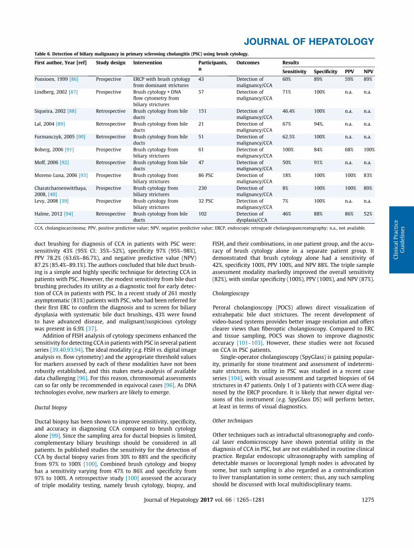
Table 6. Detection of biliary malignancy in primary sclerosing cholangitis (PSC) using brush cytology.
First author, Year [ref] Study design Intervention Participants,n
Outcomes Results
Sensitivity Specificity PPV NPV
Ponsioen, 1999 [86] Prospective ERCP with brush cytologyfrom dominant strictures
43 Detection ofmalignancy/CCA
60% 89% 59% 89%
Lindberg, 2002 [87] Prospective Brush cytology + DNAflow cytometry frombiliary strictures
57 Detection ofmalignancy/CCA
71% 100% n.a. n.a.
Siqueira, 2002 [88] Retrospective Brush cytology from bileducts
151 Detection ofmalignancy/CCA
46.4% 100% n.a. n.a.
Lal, 2004 [89] Retrospective Brush cytology from bileducts
21 Detection ofmalignancy/CCA
67% 94%, n.a. n.a.
Furmanczyk, 2005 [90] Retrospective Brush cytology from bileducts
51 Detection ofmalignancy/CCA
62.5% 100% n.a. n.a.
Boberg, 2006 [91] Prospective Brush cytology frombiliary strictures
61 Detection ofmalignancy/CCA
100% 84% 68% 100%
Moff, 2006 [92] Retrospective Brush cytology from bileducts
47 Detection ofmalignancy/CCA
50% 91% n.a. n.a.
Moreno Luna, 2006 [93] Prospective Brush cytology frombiliary strictures
86 PSC Detection ofmalignancy/CCA
18% 100% 100% 83%
Charatcharoenwitthaya,2008, [40]
Prospective Brush cytology frombiliary strictures
230 Detection ofmalignancy/CCA
8% 100% 100% 89%
Levy, 2008 [39] Prospective Brush cytology frombiliary strictures
32 PSC Detection ofmalignancy/CCA
7% 100% n.a. n.a.
Halme, 2012 [94] Retrospective Brush cytology from bileducts
102 Detection ofdysplasia/CCA
46% 88% 86% 52%
CCA, cholangiocarcinoma; PPV, positive predictive value; NPV, negative predictive value; ERCP, endoscopic retrograde cholangiopancreatography; n.a., not available.
JOURNAL OF HEPATOLOGY
duct brushing for diagnosis of CCA in patients with PSC were:sensitivity 43% (95% CI: 35%–52%), specificity 97% (95%–98%),PPV 78.2% (63.6%–86.7%), and negative predictive value (NPV)87.2% (85.4%–89.1%). The authors concluded that bile duct brush-ing is a simple and highly specific technique for detecting CCA inpatients with PSC. However, the modest sensitivity from bile ductbrushing precludes its utility as a diagnostic tool for early detec-tion of CCA in patients with PSC. In a recent study of 261 mostlyasymptomatic (81%) patients with PSC, who had been referred fortheir first ERC to confirm the diagnosis and to screen for biliarydysplasia with systematic bile duct brushings, 43% were foundto have advanced disease, and malignant/suspicious cytologywas present in 6.9% [37].
Addition of FISH analysis of cytology specimens enhanced thesensitivity for detecting CCA in patientswith PSC in several patientseries [39,40,93,94]. The ideal modality (e.g. FISH vs. digital imageanalysis vs. flow cytometry) and the appropriate threshold valuesfor markers assessed by each of these modalities have not beenrobustly established, and this makes meta-analysis of availabledata challenging [96]. For this reason, chromosomal assessmentscan so far only be recommended in equivocal cases [96]. As DNAtechnologies evolve, new markers are likely to emerge.
Ductal biopsy
Ductal biopsy has been shown to improve sensitivity, specificity,and accuracy in diagnosing CCA compared to brush cytologyalone [99]. Since the sampling area for ductal biopsies is limited,complementary biliary brushings should be considered in allpatients. In published studies the sensitivity for the detection ofCCA by ductal biopsy varies from 30% to 88% and the specificityfrom 97% to 100% [100]. Combined brush cytology and biopsyhas a sensitivity varying from 47% to 86% and specificity from97% to 100%. A retrospective study [100] assessed the accuracyof triple modality testing, namely brush cytology, biopsy, and
Journal of Hepatology 2017
FISH, and their combinations, in one patient group, and the accu-racy of brush cytology alone in a separate patient group. Itdemonstrated that brush cytology alone had a sensitivity of42%, specificity 100%, PPV 100%, and NPV 88%. The triple sampleassessment modality markedly improved the overall sensitivity(82%), with similar specificity (100%), PPV (100%), and NPV (87%).
Cholangioscopy
Peroral cholangioscopy (POCS) allows direct visualization ofextrahepatic bile duct strictures. The recent development ofvideo-based systems provides better image resolution and offersclearer views than fiberoptic cholangioscopy. Compared to ERCand tissue sampling, POCS was shown to improve diagnosticaccuracy [101–103]. However, these studies were not focusedon CCA in PSC patients.
Single-operator cholangioscopy (SpyGlass) is gaining popular-ity, primarily for stone treatment and assessment of indetermi-nate strictures. Its utility in PSC was studied in a recent caseseries [104], with visual assessment and targeted biopsies of 64strictures in 47 patients. Only 1 of 3 patients with CCA were diag-nosed by the ERCP procedure. It is likely that newer digital ver-sions of this instrument (e.g. SpyGlass DS) will perform better,at least in terms of visual diagnostics.
Other techniques
Other techniques such as intraductal ultrasonography and confo-cal laser endomicroscopy have shown potential utility in thediagnosis of CCA in PSC, but are not established in routine clinicalpractice. Regular endoscopic ultrasonography with sampling ofdetectable masses or locoregional lymph nodes is advocated bysome, but such sampling is also regarded as a contraindicationto liver transplantation in some centers; thus, any such samplingshould be discussed with local multidisciplinary teams.
vol. 66 j 1265–1281 1275
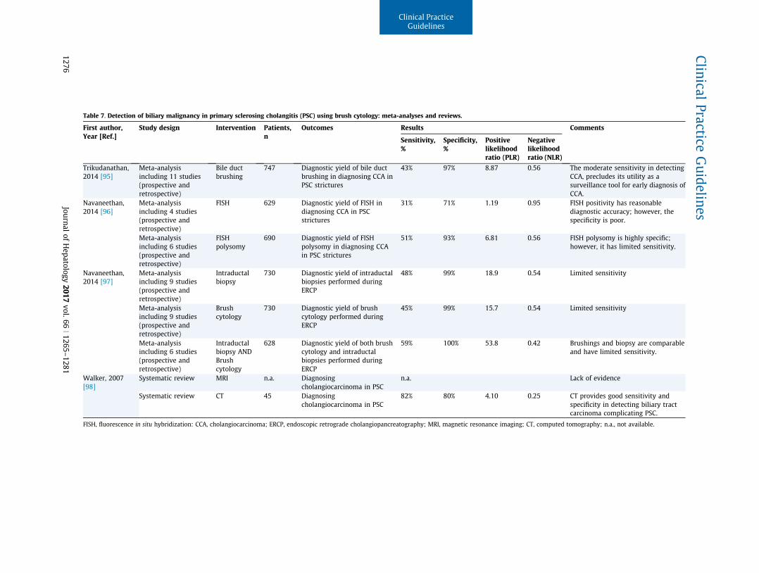
Table 7. Detection of biliary malignancy in primary sclerosing cholangitis (PSC) using brush cytology: meta-analyses and reviews.
First author,Year [Ref.]
Study design Intervention Patients,n
Outcomes Results Comments
Sensitivity,%
Specificity,%
Positivelikelihoodratio (PLR)
Negativelikelihoodratio (NLR)
Trikudanathan,2014 [95]
Meta-analysisincluding 11 studies(prospective andretrospective)
Bile ductbrushing
747 Diagnostic yield of bile ductbrushing in diagnosing CCA inPSC strictures
43% 97% 8.87 0.56 The moderate sensitivity in detectingCCA, precludes its utility as asurveillance tool for early diagnosis ofCCA.
Navaneethan,2014 [96]
Meta-analysisincluding 4 studies(prospective andretrospective)
FISH 629 Diagnostic yield of FISH indiagnosing CCA in PSCstrictures
31% 71% 1.19 0.95 FISH positivity has reasonablediagnostic accuracy; however, thespecificity is poor.
Meta-analysisincluding 6 studies(prospective andretrospective)
FISHpolysomy
690 Diagnostic yield of FISHpolysomy in diagnosing CCAin PSC strictures
51% 93% 6.81 0.56 FISH polysomy is highly specific;however, it has limited sensitivity.
Navaneethan,2014 [97]
Meta-analysisincluding 9 studies(prospective andretrospective)
Intraductalbiopsy
730 Diagnostic yield of intraductalbiopsies performed duringERCP
48% 99% 18.9 0.54 Limited sensitivity
Meta-analysisincluding 9 studies(prospective andretrospective)
Brushcytology
730 Diagnostic yield of brushcytology performed duringERCP
45% 99% 15.7 0.54 Limited sensitivity
Meta-analysisincluding 6 studies(prospective andretrospective)
Intraductalbiopsy ANDBrushcytology
628 Diagnostic yield of both brushcytology and intraductalbiopsies performed duringERCP
59% 100% 53.8 0.42 Brushings and biopsy are comparableand have limited sensitivity.
Walker, 2007[98]
Systematic review MRI n.a. Diagnosingcholangiocarcinoma in PSC
n.a. Lack of evidence
Systematic review CT 45 Diagnosingcholangiocarcinoma in PSC
82% 80% 4.10 0.25 CT provides good sensitivity andspecificity in detecting biliary tractcarcinoma complicating PSC.
FISH, fluorescence in situ hybridization: CCA, cholangiocarcinoma; ERCP, endoscopic retrograde cholangiopancreatography; MRI, magnetic resonance imaging; CT, computed tomography; n.a., not available.
ClinicalPractice
Guidelines
1276Journal
ofHepatology
2017vol.66
j1265–1281
Endoscopic surveillance of PSC-associated inflammatorybowel disease
The relationship between PSC and IBD is well established [105].The prevalence of IBD in patients with established PSC varieswidely, but is reported as 80% in Scandinavian countries [106].The often asymptomatic phenotype of IBD means that prevalencedata are strongly influenced by the level of proactive search forthe disease. The typical scenario was for IBD to precede the pre-sentation of PSC. However, the clinical presentation of IBD is vari-able, and the diseasemay be subclinical or asymptomatic for years[107] and is nowadays often diagnosed after the recognition of theliver disease. Notably, IBDmay have been present for an unknownperiod of time when PSC is diagnosed. The increased risk of coloncancer in PSC-associated IBD [108,109] hence makes it crucial toperform a full ileocolonoscopy at the time of PSC diagnosis in allpatients. As to the diagnosis of IBD per se, complete ileo-colonoscopy is critical since rectal sparing, as well as right-sidedinvolvement, is frequent in these patients [8].
Timing of screening
Recommendations
22. ESGE/EASL recommend screening ileocolonoscopy atthe time of PSC diagnosis. Strong recommendation, highquality evidence. If IBD is documented endoscopically orhistologically, annual surveillance colonoscopies are war-ranted.Strong recommendation, low quality evidence.
23. ESGE/EASL suggest that if no IBD is documented, thenext ileocolonoscopy should be considered at 5 years orwhenever bowel complaints suggestive of IBD occur.Weak recommendation, low quality evidence.
Based on initial screening, subsequent surveillance can beplanned. If IBD is documented, annual colonoscopies are war-ranted [6,110] since it has been shown that PSC-IBD patientswhose colorectal cancer (CRC) is detected in a surveillance pro-gram have a significantly lower risk of CRC-related mortality ascompared to non-surveilled patients [78]. If not, repeat colono-scopy should be donewith the occurrence of symptoms suggestiveof IBD, or of elevated F-calprotectin, or otherwise at 3–5 years[111], although this recommendation lacks any scientific evidencebeyond extrapolation from general IBD recommendations [112].
Endoscopic modality
Recommendations
24. For screening for the presence of IBD, EASL/ESGE rec-ommend ileocolonoscopy with four-quadrant biopsiesfrom all colonic segments and the terminal ileum.Strong recommendation, low quality evidence.
25. For dysplasia surveillance of PSC-associated IBD,EASL/ESGE recommend ileocolonoscopy with dye-basedchromoendoscopy with targeted biopsies.Strong recommendation, low quality evidence.
Journal of Hepatology 2017
PSC-associated colitis seems to be distinctive from other IBD:colitis is predominant in the right colon [113] and colon cancer is
typically right-sided [114]. Lack of inflammation in the rectum(‘‘rectal sparing”) is reported in some studies but less frequentlyobserved in others [3]. Endoscopic surveillance of PSC-associatedcolitis is presumed to increase the chance of early detection ofdysplasia or malignancy [115].Screening for IBD at diagnosis of PSC is best performed by highdefinition ileocolonoscopy with four-quadrant biopsies from allcolonic segments and the terminal ileum. Biopsies should betaken at the index endoscopy even without macroscopic signsof inflammation [111,116,117].
In established PSC-IBD, ileocolonoscopy with dye-based chro-moendoscopy (0.1% methylene blue or 0.1%–0.5% indigo carmine)with targeted biopsies is required for neoplasia surveillance ofPSC-associated IBD. In appropriately trained hands, in the situa-tion of quiescent disease activity and adequate bowel prepara-tion, non-targeted four-quadrant biopsies can be abandoned[118]. This approach is also endorsed by the European Crohn’sand Colitis Organisation (ECCO) [112]. It should be noted thatthere are no studies on colonic neoplasia surveillance specificallyin the setting of PSC-associated IBD.
Routine use of pancolonic chromoendoscopy with targetedbiopsies for neoplasia surveillance in patients with long-standing colitis (disease duration of[8 years) increased the pro-portion of patients found with dysplasia by a factor of 2.1–3.3compared to standard definition video-colonoscopy. For thedetection of patients with neoplasia, the pooled incremental yieldof conventional chromoendoscopy with random biopsies overstandard white-light endoscopy with random biopsies was 7%(95% CI: 3.2%–11.3%) [119]. The benefit of conventional chro-moendoscopy over white-light endoscopy with latest-generation high definition colonoscopes is unknown to date.
Handling of polyps and colorectal dysplasia
Recommendations
JOURNAL OF HEPATOLOGY
v
26. ESGE/EASL recommend endoscopic resection of anyvisible lesions and assessment of the surroundingmucosa. We recommend proctocolectomy in the case ofdysplasia in the surrounding mucosa, or when the lesioncannot be completely resected. Otherwise, repeat colono-scopy and close follow-up is warranted.Strong recommendation, low quality evidence.
27. In the case of invisible lesions with high grade dys-plasia (HGD) confirmed by two expert pathologists, proc-tocolectomy should be advised.Strong recommendation, low quality evidence.
28. In the case of invisible lesions with low grade dys-plasia (LGD) confirmed by two expert pathologists, repeatcolonoscopy after 3 months with chromoendoscopy isrecommended.Strong recommendation, low quality evidence.
Colorectal cancer (CRC) risk is significantly increased inpatients with coexisting IBD and PSC. A meta-analysis of 11 stud-ies concluded that patients with ulcerative colitis and PSC were atincreased risk of developing CRC compared to patients with
ol. 66 j 1265–1281 1277
Clinical Practice Guidelines
ulcerative colitis alone (OR: 4.09; 95% CI: 2.89–5.76) [109]. Arecent large population-based study in the Netherlands found a9-fold increased risk of developing CRC in PSC-ulcerative colitispatients, compared to the age- and gender-matched population(standardized incidence ratio [SIR]: 8.6; 95% CI: 3.5–17.7), anda 10-fold increased risk, compared to ulcerative colitis controls(ratio of SIRs: 9.8; 95% CI: 1.9–96.6) [78].Most dysplasia is visible at colonoscopy [120,121]. On theother hand, invisible dysplastic lesions can also be diagnosedby random biopsies during surveillance. According to the IBDDysplasia Morphology Study Group [122], dysplasia is subdividedinto LGD and HGD.
Recent ECCO guidelines state that a visible lesion with dys-plasia should be completely resected endoscopically irrespectiveof the grade of dysplasia or the location relative to the inflamedmucosal areas [112]. Subsequently, the surrounding mucosa(around the visible lesion) should be examined (withchromoendoscopy-guided targeted biopsies or random biopsiesif chromoendoscopy is not available). If endoscopic resection isincomplete or impossible, or if dysplasia is detected in the sur-rounding mucosa, total proctocolectomy is recommended.
In the case of invisible lesions with LGD, urgent repeat chro-moendoscopy should be performed, to eventually identify awell-circumscribed lesion and/or perform additional randombiopsies. If the presence of LGD is confirmed, there is no clearconsensus regarding management; proctocolectomy or surveil-lance could be recommended. Actually, two studies revealed asignificant 5-year progression rate (33%–54%) of LGD to HGD[123,124], whereas others showed low progression rates[125,126]. Finally, in the case of invisible lesions with HGD oradenocarcinoma, total proctocolectomy is indicated.
Disclaimer
This guideline from ESGE and EASL represents a consensus of bestpractice based on the available evidence at the time of prepara-tion. The recommendations might not apply in all situationsand should be interpreted in the light of specific clinical situa-tions and resource availability. Further controlled clinical studiesmay be needed to clarify aspects of the guideline, and revisionmay be necessary as new data appear. Clinical considerationsmay justify a course of action at variance to these recommenda-tions. This ESGE/EASL guideline is intended to be an educationaldevice to provide information that may assist endoscopists inproviding care to patients. It is not a set of rules and should notbe construed as establishing a legal standard of care or as encour-aging, advocating, requiring, or discouraging any particulartreatment.
Conflict of interest
J. Albert has received (from 2015 to 2016) speaker’s honorariafrom Fujifilm, the Falk Foundation, Covidien/Medtronic, andOlympus Europe, an honorarium from Covidien/Medtronic foradvisory services, and research support from Olympus Europe.P. Fickert has served on advisory boards for Dr. Falk Pharmaand Intercept; his department has received unrestricted researchgrants from the Falk Foundation (since 2010) and Gilead (since2012); he is listed as co-inventor in two patents filed by the
1278 Journal of Hepatology 2017
Medical University of Graz for the use of norUDCA in the treat-ment of liver diseases and arteriosclerosis (publication numbersWO2006119803 and WO20099013334). A. Laghi has received aspeaker fee from GE Healthcare (October 2016). J.-W. Poleyreceives consultancy, travel, and speaker fees from CookEndoscopy; his department receives financial support for consul-tancy, travel, and speaking from Boston Scientific; he receives tra-vel and consultancy fees from Pentax. C.Y. Ponsioen’s departmentis receiving research support from Olympus and Fujifilm.C. Schramm has served on an advisory board for InterceptPharmaceuticals (2016), and has given lectures sponsored byIntercept and the Falk Foundation. F. Swahn has served on a sci-entific advisory board for Rhenman & Partners, and has given lec-tures sponsored by Cook Medical Sweden and Boston ScientificNordic. L. Aabakken, M. Arvanitakis, O. Chazouilleres,J.-M. Dumonceau, M. Färrkkilä, C. Hassan, G.M. Hirschfield,T.H. Karlsen, M. Marzioni, M. Fernandez, S.P. Pereira, J. Pohl,and A. Tringali have no competing interests.
Supplementary data
Supplementary data associated with this article can be found, inthe online version, at http://dx.doi.org/10.1016/j.jhep.2017.02.013.
References
[1] Olsson R, Danielsson A, Jarnerot G, et al. Prevalence of primary sclerosingcholangitis in patients with ulcerative colitis. Gastroenterology1991;100:1319–1323.
[2] Lunder AK, Hov JR, Borthne A, et al. Prevalence of sclerosing cholangitis,detected by magnetic resonance cholangiography, in patients with long-term inflammatory bowel disease. Gastroenterology 2016;151:660–669,e4.
[3] Boonstra K, van Erpecum KJ, van Nieuwkerk KM, et al. Primary sclerosingcholangitis is associated with a distinct phenotype of inflammatory boweldisease. Inflamm Bowel Dis 2012;18:2270–2276.
[4] O’Toole A, Alakkari A, Keegan D, et al. Primary sclerosing cholangitis anddisease distribution in inflammatory bowel disease. Clin GastroenterolHepatol 2012;10:439–441.
[5] Loftus Jr EV, Harewood GC, Loftus CG, et al. PSC-IBD: a unique form ofinflammatory bowel disease associated with primary sclerosing cholangitis.Gut 2005;54:91–96.
[6] EASL Clinical Practice Guidelines. Management of cholestatic liver diseases.J Hepatol 2009;51:237–267.
[7] Atkins D, Eccles M, Flottorp S, et al. Systems for grading the quality ofevidence and the strength of recommendations I: critical appraisal ofexisting approaches The GRADE Working Group. BMC Health Serv Res2004;4:38.
[8] Chapman R, Fevery J, Kalloo A, et al. Diagnosis and management of primarysclerosing cholangitis. Hepatology 2010;51:660–678.
[9] Berstad AE, Aabakken L, Smith HJ, et al. Diagnostic accuracy of magneticresonance and endoscopic retrograde cholangiography in primary scleros-ing cholangitis. Clin Gastroenterol Hepatol 2006;4:514–520.
[10] Moff SL, Kamel IR, Eustace J, et al. Diagnosis of primary sclerosingcholangitis: a blinded comparative study using magnetic resonancecholangiography and endoscopic retrograde cholangiography. GastrointestEndosc 2006;64:219–223.
[11] Philpott C, Rosenbaum J, Moon A, et al. Paediatric MRCP: 10 year experiencewith 195 patients. Eur J Radiol 2013;82:699–706.
[12] Rossi G, Sciveres M, Maruzzelli L, et al. Diagnosis of sclerosing cholangitis inchildren: blinded, comparative study of magnetic resonance versus endo-scopic cholangiography. Clin Res Hepatol Gastroenterol 2013;37:596–601.
[13] Dave M, Elmunzer BJ, Dwamena BA, Higgins PD. Primary sclerosingcholangitis: meta-analysis of diagnostic performance of MR cholangiopan-creatography. Radiology 2010;256:387–396.
vol. 66 j 1265–1281
JOURNAL OF HEPATOLOGY
[14] Meagher S, Yusoff I, Kennedy W, et al. The roles of magnetic resonance andendoscopic retrograde cholangiopancreatography (MRCP and ERCP) in thediagnosis of patients with suspected sclerosing cholangitis: a cost-effectiveness analysis. Endoscopy 2007;39:222–228.
[15] Talwalkar JA, Angulo P, Johnson CD, et al. Cost-minimization analysis ofMRC versus ERCP for the diagnosis of primary sclerosing cholangitis.Hepatology 2004;40:39–45.
[16] Kalaitzakis E, Levy M, Kamisawa T, et al. Endoscopic retrograde cholan-giography does not reliably distinguish IgG4-associated cholangitis fromprimary sclerosing cholangitis or cholangiocarcinoma. Clin GastroenterolHepatol 2011;9(800–803):e2.
[17] Ruiz A, Lemoinne S, Carrat F, et al. Radiologic course of primary sclerosingcholangitis: assessment by three-dimensional magnetic resonance cholan-giography and predictive features of progression. Hepatology2014;59:242–250.
[18] Weber C, Kuhlencordt R, Grotelueschen R, et al. Magnetic resonancecholangiopancreatography in the diagnosis of primary sclerosing cholan-gitis. Endoscopy 2008;40:739–745.
[19] Eaton JE, Talwalkar JA, Lazaridis KN, et al. Pathogenesis of primarysclerosing cholangitis and advances in diagnosis and management. Gas-troenterology 2013;145:521–536.
[20] Li-Yeng C, Goldberg HI. Sclerosing cholangitis: broad spectrum of radio-graphic features. Gastrointest Radiol 1984;9:39–47.
[21] Majoie CB, Reeders JW, Sanders JB, et al. Primary sclerosing cholangitis: amodified classification of cholangiographic findings. AJR Am J Roentgenol1991;157:495–497.
[22] Ponsioen CY, Vrouenraets SM, Prawirodirdjo W, et al. Natural history ofprimary sclerosing cholangitis and prognostic value of cholangiography in aDutch population. Gut 2002;51:562–566.
[23] Ponsioen CY, Reitsma JB, Boberg KM, et al. Validation of a cholangiographicprognostic model in primary sclerosing cholangitis. Endoscopy2010;42:742–747.
[24] Craig DA, MacCarty RL, Wiesner RH, et al. Primary sclerosing cholangitis:value of cholangiography in determining the prognosis. AJR Am JRoentgenol 1991;157:959–964.
[25] Abdalian R, Heathcote EJ. Sclerosing cholangitis: a focus on secondarycauses. Hepatology 2006;44:1063–1074.
[26] Trauner M, Fickert P, Wagner M. MDR3 (ABCB4) defects: a paradigm forthe genetics of adult cholestatic syndromes. Semin Liver Dis 2007;27:77–98.
[27] Mesenas S, Vu C, Doig L, Meenan J. Duodenal EUS to identify thickening ofthe extrahepatic biliary tree wall in primary sclerosing cholangitis.Gastrointest Endosc 2006;63:403–408.
[28] Rustemovic N, Cukovic-Cavka S, Opacic M, et al. Endoscopic ultrasoundelastography as a method for screening the patients with suspectedprimary sclerosing cholangitis. Eur J Gastroenterol Hepatol2010;22:748–753.
[29] Lutz HH, Wasmuth HE, Streetz K, et al. Endoscopic ultrasound as an earlydiagnostic tool for primary sclerosing cholangitis: a prospective pilot study.Endoscopy 2012;44:934–939.
[30] EASL-ALEH Clinical Practice Guidelines. Non-invasive tests for evaluation ofliver disease severity and prognosis. J Hepatol 2015;63:237–264.
[31] Cotton PB, Nickl N. Endoscopic and radiologic approaches to therapy inprimary sclerosing cholangitis. Semin Liver Dis 1991;11:40–48.
[32] Johnson GK, Geenen JE, Venu RP, et al. Endoscopic treatment of biliary tractstrictures in sclerosing cholangitis: a larger series and recommendationsfor treatment. Gastrointest Endosc 1991;37:38–43.
[33] Stiehl A, Rudolph G, Klöters-Plachky P, et al. Development of dominant bileduct stenoses in patients with primary sclerosing cholangitis treated withursodeoxycholic acid: outcome after endoscopic treatment. J Hepatol2002;36:151–156.
[34] Bjornsson E, Lindqvist-Ottosson J, Asztely M, Olsson R. Dominant stricturesin patients with primary sclerosing cholangitis. Am J Gastroenterol2004;99:502–508.
[35] Gluck M, Cantone NR, Brandabur JJ, et al. A twenty-year experience withendoscopic therapy for symptomatic primary sclerosing cholangitis. J ClinGastroenterol 2008;42:1032–1039.
[36] Ponsioen CY, Lam K, van Milligen de Wit AW, et al. Four years experiencewith short term stenting in primary sclerosing cholangitis. Am J Gastroen-terol 1999;94:2403–2407.
[37] Boyd S, Tenca A, Jokelainen K, et al. Screening primary sclerosingcholangitis and biliary dysplasia with endoscopic retrograde cholangiog-raphy and brush cytology: risk factors for biliary neoplasia. Endoscopy2016;48:432–439.
Journal of Hepatology 2017
[38] Aljiffry M, Renfrew PD, Walsh MJ, et al. Analytical review of diagnosis andtreatment strategies for dominant bile duct strictures in patients withprimary sclerosing cholangitis. HPB (Oxford) 2011;13:79–90.
[39] Levy MJ, Baron TH, Clayton AC, et al. Prospective evaluation of advancedmolecular markers and imaging techniques in patients with indeterminatebile duct strictures. Am J Gastroenterol 2008;103:1263–1273.
[40] Charatcharoenwitthaya P, Enders FB, Halling KC, Lindor KD. Utility of serumtumor markers, imaging, and biliary cytology for detecting cholangiocar-cinoma in primary sclerosing cholangitis. Hepatology 2008;48:1106–1117.
[41] Chapman MH, Webster GJ, Bannoo S, et al. Cholangiocarcinoma anddominant strictures in patients with primary sclerosing cholangitis: a 25-year single-centre experience. Eur J Gastroenterol Hepatol2012;24:1051–1058.
[42] Gotthardt DN, Rudolph G, Kloters-Plachky P, et al. Endoscopic dilation ofdominant stenoses in primary sclerosing cholangitis: outcome after long-term treatment. Gastrointest Endosc 2010;71:527–534.
[43] Kaltenthaler E, Vergel YB, Chilcott J, et al. A systematic review andeconomic evaluation of magnetic resonance cholangiopancreatographycompared with diagnostic endoscopic retrograde cholangiopancreatogra-phy. Health Technol Assess 2004;8:1–89.
[44] Baluyut AR, Sherman S, Lehman GA, et al. Impact of endoscopic therapy onthe survival of patients with primary sclerosing cholangitis. GastrointestEndosc 2001;53:308–312.
[45] van Milligen de Wit AW, van Bracht J, Rauws EA, et al. Endoscopic stenttherapy for dominant extrahepatic bile duct strictures in primary scleros-ing cholangitis. Gastrointest Endosc 1996;44:293–299.
[46] Kaya M, Petersen BT, Angulo P, et al. Balloon dilation compared to stentingof dominant strictures in primary sclerosing cholangitis. Am J Gastroenterol2001;96:1059–1066.
[47] Cotton PB, Garrow DA, Gallagher J, Romagnuolo J. Risk factors forcomplications after ERCP: a multivariate analysis of 11,497 proceduresover 12 years. Gastrointest Endosc 2009;70:80–88.
[48] Johnson GK, Saeian K, Geenen JE. Primary sclerosing cholangitis treated byendoscopic biliary dilation: review and long-term follow-up evaluation.Curr Gastroenterol Rep 2006;8:147–155.
[49] Lee JG, Schutz SM, England RE, et al. Endoscopic therapy of sclerosingcholangitis. Hepatology 1995;21:661–667.
[50] Wagner S, Gebel M, Meier P, et al. Endoscopic management of biliary tractstrictures in primary sclerosing cholangitis. Endoscopy 1996;28:546–551.
[51] Ahrendt SA, Pitt HA, Kalloo AN, et al. Primary sclerosing cholangitis: resect,dilate, or transplant? Ann Surg 1998;227:412–423.
[52] Beuers U, Spengler U, Sackmann M, et al. Deterioration of cholestasis afterendoscopic retrograde cholangiography in advanced primary sclerosingcholangitis. J Hepatol 1992;15:140–143.
[53] Dumonceau JM, Tringali A, Blero D, et al. Biliary stenting: indications,choice of stents and results: European Society of Gastrointest Endosc(ESGE) clinical guideline. Endoscopy 2012;44:277–292.
[54] Ismail S, Kylänpää L, Mustonen H, et al. Risk factors for complications ofERCP in primary sclerosing cholangitis. Endoscopy 2012;44:1133–1138.
[55] Navaneethan U, Jegadeesan R, Nayak S, et al. ERCP-related adverse eventsin patients with primary sclerosing cholangitis. Gastrointest Endosc2015;81:410–419.
[56] van Milligen deWit AW, Rauws EA, van Bracht J, et al. Lack of complicationsfollowing short-term stent therapy for extrahepatic bile duct strictures inprimary sclerosing cholangitis. Gastrointest Endosc 1997;46:344–347.
[57] van den Hazel SJ, Wolfhagen EH, van Buuren HR, et al. Prospective riskassessment of endoscopic retrograde cholangiography in patients withprimary sclerosing cholangitis. Dutch PSC Study Group. Endoscopy2000;32:779–782.
[58] Enns R, Eloubeidi MA, Mergener K, et al. Predictors of successful clinical andlaboratory outcomes in patients with primary sclerosing cholangitisundergoing endoscopic retrograde cholangiopancreatography. Can J Gas-troenterol 2003;17:243–248.
[59] Bangarulingam SY, Gossard AA, Petersen BT, et al. Complications ofendoscopic retrograde cholangiopancreatography in primary sclerosingcholangitis. Am J Gastroenterol 2009;104:855–860.
[60] Alkhatib AA, Hilden K, Adler DG. Comorbidities, sphincterotomy, andballoon dilation predict post-ERCP adverse events in PSC patients: operatorexperience is protective. Dig Dis Sci 2011;56:3685–3688.
[61] von Seth E, Arnelo U, Enochsson L, Bergquist A. Primary sclerosingcholangitis increases the risk for pancreatitis after endoscopic retrogradecholangiopancreatography. Liver Int 2015;35:254–262.
[62] Etzel JP, Eng SC, Ko CW, et al. Complications after ERCP in patients withprimary sclerosing cholangitis. Gastrointest Endosc 2008;67:643–648.
vol. 66 j 1265–1281 1279
Clinical Practice Guidelines
[63] Andriulli A, Loperfido S, Napolitano G, et al. Incidence rates of post-ERCPcomplications: a systematic survey of prospective studies. Am J Gastroen-terol 2007;102:1781–1788.
[64] Lombard M, Farrant M, Karani J, et al. Improving biliary-enteric drainage inprimary sclerosing cholangitis: experience with endoscopic methods. Gut1991;32:1364–1368.
[65] Stiehl A, Rudolph G, Sauer P, et al. Efficacy of ursodeoxycholic acid treatmentand endoscopic dilation of major duct stenoses in primary sclerosingcholangitis. An 8-year prospective study. J Hepatol 1997;26:560–566.
[66] Linder S, Soderlund C. Endoscopic therapy in primary sclerosing cholan-gitis: outcome of treatment and risk of cancer. Hepatogastroenterology2001;48:387–392.
[67] Moreno Luna LE, Gores GJ. Advances in the diagnosis of cholangiocarci-noma in patients with primary sclerosing cholangitis. Liver Transpl2006;12:S15–S19.
[68] Tse F, Yuan Y, Moayyedi P, Leontiadis GI. Guidewire-assisted cannulation ofthe common bile duct for the prevention of post-endoscopic retrogradecholangiopancreatography (ERCP) pancreatitis. Cochrane Database Syst Rev2012;12:CD009662. http://dx.doi.org/10.1002/14651858.CD009662.pub2.
[69] Dumonceau JM, Andriulli A, Elmunzer BJ, et al. Prophylaxis of post-ERCPpancreatitis: European Society of Gastrointest Endosc (ESGE) Guideline –updated June 2014. Endoscopy 2014;46:799–815.
[70] Shi N, Deng L, Altaf K, et al. Rectal indomethacin for the prevention of post-ERCP pancreatitis: a meta-analysis of randomized controlled trials. Turk JGastroenterol 2015;26:236–240.
[71] Andrade-Davila VF, Chavez-Tostado M, Davalos-Cobian C, et al. Rectalindomethacin versus placebo to reduce the incidence of pancreatitis afterendoscopic retrograde cholangiopancreatography: results of a controlledclinical trial. BMC Gastroenterol 2015;15:85.
[72] Nicolas-Perez D, Castilla-Rodriguez I, Gimeno-Garcia AZ, et al. Prevention ofpost-endoscopic retrograde cholangiopancreatography pancreatitis: a cost-effectiveness analysis. Pancreas 2015;44:204–210.
[73] Brand M, Bizos D, O’Farrell Jr P. Antibiotic prophylaxis for patientsundergoing elective endoscopic retrograde cholangiopancreatography.Cochrane Database Syst Rev 2010;10:CD007345. http://dx.doi.org/10.1002/14651858.CD007345.pub2.
[74] Khashab MA, Chithadi KV, Acosta RD, et al. Antibiotic prophylaxis for GIendoscopy. Gastrointest Endosc 2015;81:81–89.
[75] Negm AA, Schott A, Vonberg RP, et al. Routine bile collection formicrobiological analysis during cholangiography and its impact on themanagement of cholangitis. Gastrointest Endosc 2010;72:284–291.
[76] Razumilava N, Gores GJ, Lindor KD. Cancer surveillance in patients withprimary sclerosing cholangitis. Hepatology 2011;54:1842–1852.
[77] Weismüller TJ, Trivedi PJ, Bergquist A, et al. Gastroenterolog 2017. http://dx.doi.org/10.1053/j.gastro.2017.02.038, [Epub ahead of print], PMID:28274849.
[78] Boonstra K, Weersma RK, van Erpecum KJ, et al. Population-basedepidemiology, malignancy risk, and outcome of primary sclerosing cholan-gitis. Hepatology 2013;58:2045–2055.
[79] de Valle MB, Bjornsson E, Lindkvist B. Mortality and cancer risk related toprimary sclerosing cholangitis in a Swedish population-based cohort. LiverInt 2012;32:441–448.
[80] Boberg KM, Bergquist A, Mitchell S, et al. Cholangiocarcinoma in primarysclerosing cholangitis: risk factors and clinical presentation. Scand JGastroenterol 2002;37:1205–1211.
[81] Levy C, Lymp J, Angulo P, et al. The value of serum CA 19-9 in predictingcholangiocarcinomas in patients with primary sclerosing cholangitis. DigDis Sci 2005;50:1734–1740.
[82] Sinakos E, Saenger AK, Keach J, et al. Many patients with primary sclerosingcholangitis and increased serum levels of carbohydrate antigen 19-9 do nothave cholangiocarcinoma. Clin Gastroenterol Hepatol 2011;9:434–439, e1.
[83] Venkatesh PG, Navaneethan U, Shen B, McCullough AJ. Increased serumlevels of carbohydrate antigen 19-9 and outcomes in primary sclerosingcholangitis patients without cholangiocarcinoma. Dig Dis Sci2013;58:850–857.
[84] Wannhoff A, Hov JR, Folseraas T, et al. FUT2 and FUT3 genotype determinesCA19-9 cut-off values for detection of cholangiocarcinoma in patients withprimary sclerosing cholangitis. J Hepatol 2013;59:1278–1284.
[85] DeOliveira ML, Cunningham SC, Cameron JL, et al. Cholangiocarcinoma:thirty-one-year experience with 564 patients at a single institution. AnnSurg 2007;245:755–762.
[86] Ponsioen CY, Vrouenraets SM, van Milligen de Wit AW, et al. Value of brushcytology for dominant strictures in primary sclerosing cholangitis. Endo-scopy 1999;31:305–309.
1280 Journal of Hepatology 2017
[87] Lindberg B, Arnelo U, Bergquist A, et al. Diagnosis of biliary strictures inconjunction with endoscopic retrograde cholangiopancreaticography, withspecial reference to patients with primary sclerosing cholangitis. Endo-scopy 2002;34:909–916.
[88] Siqueira E, Schoen RE, Silverman W, et al. Detecting cholangiocarcinoma inpatients with primary sclerosing cholangitis. Gastrointest Endosc2002;56:40–47.
[89] Lal A, Okonkwo A, Schindler S, et al. Role of biliary brush cytology inprimary sclerosing cholangitis. Acta Cytol 2004;48:9–12.
[90] Furmanczyk PS, Grieco VS, Agoff SN. Biliary brush cytology and thedetection of cholangiocarcinoma in primary sclerosing cholangitis: evalu-ation of specific cytomorphologic features and CA19-9 levels. Am J ClinPathol 2005;124:355–360.
[91] Boberg KM, Jebsen P, Clausen OP, et al. Diagnostic benefit of biliary brushcytology in cholangiocarcinoma in primary sclerosing cholangitis. J Hepatol2006;45:568–574.
[92] Moff SL, Clark DP, Maitra A, et al. Utility of bile duct brushings for the earlydetection of cholangiocarcinoma in patients with primary sclerosingcholangitis. J Clin Gastroenterol 2006;40:336–341.
[93] Moreno Luna LE, Kipp B, Halling KC, et al. Advanced cytologic techniquesfor the detection of malignant pancreatobiliary strictures. Gastroenterology2006;131:1064–1072.
[94] Halme L, Arola J, Numminen K, et al. M. Biliary dysplasia in patients withprimary sclerosing cholangitis: additional value of DNA ploidity. Liver Int2012;32:783–789.
[95] Trikudanathan G, Navaneethan U, Njei B, et al. Diagnostic yield of bile ductbrushings for cholangiocarcinoma in primary sclerosing cholangitis: asystematic reviewandmeta-analysis.Gastrointest Endosc2014;79:783–789.
[96] Navaneethan U, Njei B, Venkatesh PG, et al. Fluorescence in situ hybridiza-tion for diagnosis of cholangiocarcinoma in primary sclerosing cholangitis:a systematic review and meta-analysis. Gastrointest Endosc 2014;79(943–950):e3.
[97] Navaneethan U, Njei B, Lourdusamy V, et al. Comparative effectiveness ofbiliary brush cytology and intraductal biopsy for detection of malignantbiliary strictures: a systematic review and meta-analysis. GastrointestEndosc 2015;81:168–176.
[98] Walker SL, McCormick PA. Diagnosing cholangiocarcinoma in primarysclerosing cholangitis: an ‘‘evidence based radiology” review. AbdomImaging 2008;33:14–17.
[99] Karlsen TH, Vesterhus M, Boberg KM. Review article: controversies in themanagement of primary biliary cirrhosis and primary sclerosing cholangi-tis. Aliment Pharmacol Ther 2014;39:282–301.
[100] Nanda A, Brown JM, Berger SH, et al. Triple modality testing by endoscopicretrograde cholangiopancreatography for the diagnosis of cholangiocarci-noma. Therap Adv Gastroenterol 2015;8:56–65.
[101] Fukuda Y, Tsuyuguchi T, Sakai Y, et al. Diagnostic utility of peroralcholangioscopy for various bile-duct lesions. Gastrointest Endosc2005;62:374–382.
[102] Kawakami H, Kuwatani M, Etoh K, et al. Endoscopic retrograde cholan-giography versus peroral cholangioscopy to evaluate intraepithelial tumorspread in biliary cancer. Endoscopy 2009;41:959–964.
[103] Nishikawa T, Tsuyuguchi T, Sakai Y, et al. Comparison of the diagnosticaccuracy of peroral video-cholangioscopic visual findings and cholan-gioscopy-guided forceps biopsy findings for indeterminate biliary lesions: aprospective study. Gastrointest Endosc 2013;77:219–226.
[104] Arnelo U, von Seth E, Bergquist A. Prospective evaluation of the clinicalutility of single-operator peroral cholangioscopy in patients with primarysclerosing cholangitis. Endoscopy 2015;47:696–702.
[105] Smith MP, Loe RH. Sclerosing cholangitis; review of recent case reports andassociated diseases and four new cases. Am J Surg 1965;110:239–246.
[106] Broome U, Lindberg G, Lofberg R. Primary sclerosing cholangitis inulcerative colitis – a risk factor for the development of dysplasia andDNA aneuploidy? Gastroenterology 1992;102:1877–1880.
[107] Broome U, Lofberg R, Lundqvist K, Veress B. Subclinical time span ofinflammatory bowel disease in patients with primary sclerosing cholan-gitis. Dis Colon Rectum 1995;38:1301–1305.
[108] Broome U, Lofberg R, Veress B, Eriksson LS. Primary sclerosing cholangitisand ulcerative colitis: evidence for increased neoplastic potential. Hepa-tology 1995;22:1404–1408.
[109] Soetikno RM, Lin OS, Heidenreich PA, et al. Increased risk of colorectalneoplasia in patients with primary sclerosing cholangitis and ulcerativecolitis: a meta-analysis. Gastrointest Endosc 2002;56:48–54.
[110] Broome U, Bergquist A. Primary sclerosing cholangitis, inflammatory boweldisease, and colon cancer. Semin Liver Dis 2006;26:31–41.
vol. 66 j 1265–1281
JOURNAL OF HEPATOLOGY
[111] Lindor KD, Kowdley KV, Harrison ME. American College of Gastroenterol-ogy. ACG Clinical Guideline: Primary sclerosing cholangitis. Am J Gas-troenterol 2015;110:646–659, quiz 60.
[112] Annese V, Daperno M, Rutter MD, et al. European evidence based consensusfor endoscopy in inflammatory bowel disease. J Crohns Colitis2013;7:982–1018.
[113] Tsaitas C, Semertzidou A, Sinakos E. Update on inflammatory bowel diseasein patients with primary sclerosing cholangitis. World J Hepatol2014;6:178–187.
[114] Claessen MM, Lutgens MW, van Buuren HR, et al. More right-sided IBD-associated colorectal cancer in patients with primary sclerosing cholangi-tis. Inflamm Bowel Dis 2009;15:1331–1336.
[115] Vera A, Gunson BK, Ussatoff V, et al. Colorectal cancer in patients withinflammatory bowel disease after liver transplantation for primarysclerosing cholangitis. Transplantation 2003;75:1983–1988.
[116] Jorgensen KK, Grzyb K, Lundin KE, et al. Inflammatory bowel disease inpatients with primary sclerosing cholangitis: clinical characterization inliver transplanted and nontransplanted patients. Inflamm Bowel Dis2012;18:536–545.
[117] Fausa O, Schrumpf E, Elgjo K. Relationship of inflammatory bowel diseaseand primary sclerosing cholangitis. Semin Liver Dis 1991;11:31–39.
[118] Kaminski MF, Hassan C, Bisschops R, et al. Advanced imaging for detectionand differentiation of colorectal neoplasia: European Society of Gastroin-testinal Endoscopy (ESGE) Guideline. Endoscopy 2014;46:435–449.
Journal of Hepatology 2017
[119] Subramanian V, Mannath J, Ragunath K, Hawkey CJ. Meta-analysis: thediagnostic yield of chromoendoscopy for detecting dysplasia in patientswith colonic inflammatory bowel disease. Aliment Pharmacol Ther2011;33:304–312.
[120] Rutter MD, Saunders BP, Wilkinson KH, et al. Most dysplasia in ulcerativecolitis is visible at colonoscopy. Gastrointest Endosc 2004;60:334–339.
[121] Rubin DT, Rothe JA, Hetzel JT, et al. Are dysplasia and colorectal cancerendoscopically visible in patients with ulcerative colitis? GastrointestEndosc 2007;65:998–1004.
[122] Riddell RH, Goldman H, Ransohoff DF, et al. Dysplasia in inflammatorybowel disease: standardized classification with provisional clinical appli-cations. Human Pathol 1983;14:931–968.
[123] Connell WR, Lennard-Jones JE, Williams CB, et al. Factors affecting theoutcome of endoscopic surveillance for cancer in ulcerative colitis.Gastroenterology 1994;107:934–944.
[124] Ullman TA, Loftus Jr EV, Kakar S, et al. The fate of low grade dysplasia inulcerative colitis. Am J Gastroenterol 2002;97:922–927.
[125] Befrits R, Ljung T, Jaramillo E, Rubio C. Low-grade dysplasia in extensive,long-standing inflammatory bowel disease: a follow-up study. Dis ColonRectum 2002;45:615–620.
[126] Lim CH, Dixon MF, Vail A, et al. Ten year follow up of ulcerative colitispatients with and without low grade dysplasia. Gut 2003;52:1127–1132.
vol. 66 j 1265–1281 1281
Top Related