

![Diagnosis and Management of Rhinitis: Complete Guidelines ... · different forms of rhinitis (allergic, non-allergic, occupational rhinitis, hormonal rhinitis [pregnancy and hypothyroidism],](https://static.fdocuments.net/doc/165x107/5d61f07588c993197b8b51b8/diagnosis-and-management-of-rhinitis-complete-guidelines-different-forms.jpg)


Languages
Pages
Legal

RhinitisRhinitisRhinitisRhinitisRhinitisRhinitisRhinitisRhinitisRhinitisRhinitisProf. Dr. Samy Elwany
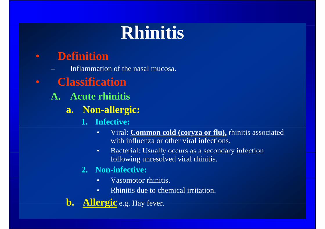
RhinitisRhinitis• Definition
– Inflammation of the nasal mucosa.
• ClassificationA. Acute rhinitis
a. Non-allergic:1. Infective:
• Viral: Common cold (coryza or flu), rhinitis associated with influenza or other viral infections.
• Bacterial: Usually occurs as a secondary infectionBacterial: Usually occurs as a secondary infection following unresolved viral rhinitis.
2. Non-infective:V hi i i• Vasomotor rhinitis.
• Rhinitis due to chemical irritation.
b Allergic e g Hay feverb. Allergic e.g. Hay fever.
B. Chronic rhinitis:B. Chronic rhinitis:a. Non-allergic:
1 Non specific:1. Non-specific:• Chronic catarrhal rhinitis.• Chronic hypertrophic rhinitis.yp p• Chronic atrophic rhinitis.• Rhinitis medicamentosa (drug-induced rhinitis).
2. Specific:• Scleroma.
S hili b l i l d l• Rare types: Syphilis, tuberculosis, lupus and, leprosy.
b. Allergic: Perennial allergic rhinitis.
Common Cold (Coryza)Common Cold (Coryza)
• Common cold is acute viral rhinosinusitis and is Co o co d s acute v a os us t s a d scurrently the commonest cause of nasal obstruction.
• Pathology:Pathology:– Rhinovirus.– Droplet infectionDroplet infection.– The disease is self-limiting and spontaneous resolution
usually takes place within 7-10 days unless secondary y p y ybacterial infection occurs.
• Clinical picture1. Stage of invasion (few hours):g ( )
Sneezing, burning sensation in the nasopharynx, nasal obstruction, and headache.
2. Stage of secretion (few days):Low grade fever, malaise, arthralgia, nasal g , , g ,obstruction, and profuse watery rhinorrhea.
3 St f l ti R l ti ithi 5 7 d i th3. Stage of resolution: Resolution within 5-7 days is the natural course of an uncomplicated disease.
Symptoms lasting beyond 7 days, or worsening instead of improving suggest that secondary bacterial infection is being establishedbacterial infection is being established.
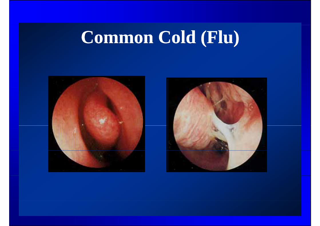
Common Cold (Flu)Common Cold (Flu)
Common Cold (Coryza)Common Cold (Coryza)
• ComplicationsComplications1. Acute sinusitis.2 Acute otitis media2. Acute otitis media.3. Chest infection.T t t• Treatment
1. Supportive treatment: bed rest, analgesics, nasal d t t (l l i d d t i ) ddecongestants (local i.e. drops and systemic), and occasionally steam inhalations.
2 A tibi ti h ld b d f t t t f2. Antibiotics should be reserved for treatment of secondary bacterial infections.
Chronic Catarrhal and Chronic Catarrhal and Hypertrophic RhinitisHypertrophic RhinitisHypertrophic RhinitisHypertrophic Rhinitis
• Definition– Chronic catarrhal orhypertrophic rhinitis is a chronic
nonspecific inflammation of the nasal mucosa h i d b l i d d dcharacterized by mucosal congestion and edema and
swelling or hypertrophy of the inferior turbinates.
– It may be caused by many etiologic factors.
– It is an important cause of chronic nasal obstruction.
– In the early stage the pathological changes are t e ea y stage t e pat o og ca c a ges a ereversible (chronic catarrhal rhinitis or simple chronic rhinitis).
– Later on fibrosis starts in the submucosa and the h l i l h l ibl ( h ipathological changes are no longer reversible (chronic
hypertrophic rhinitis).
• Clinical pictureClinical picture
A.Symptoms:A.Symptoms:1.Nasal obstruction: This is the predominant
symptom. It is usually bilateral orsymptom. It is usually bilateral or alternating between the two sides.
2.Mucoid nasal and post nasal discharge.2.Mucoid nasal and post nasal discharge.3.Hyposmia (may be – proportionate to nasal
obstruction ).obstruction ).
Chronic Hypertrophic RhinitisChronic Hypertrophic Rhinitis
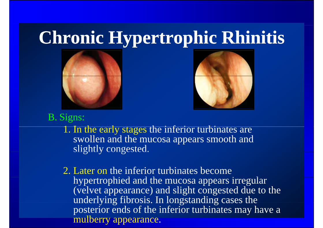
B. Signs:1 I th l t th i f i t bi t1. In the early stages the inferior turbinates are
swollen and the mucosa appears smooth and slightly congested.slightly congested.
2. Later on the inferior turbinates become h hi d d h i lhypertrophied and the mucosa appears irregular (velvet appearance) and slight congested due to the underlying fibrosis. In longstanding cases theunderlying fibrosis. In longstanding cases the posterior ends of the inferior turbinates may have a mulberry appearance.
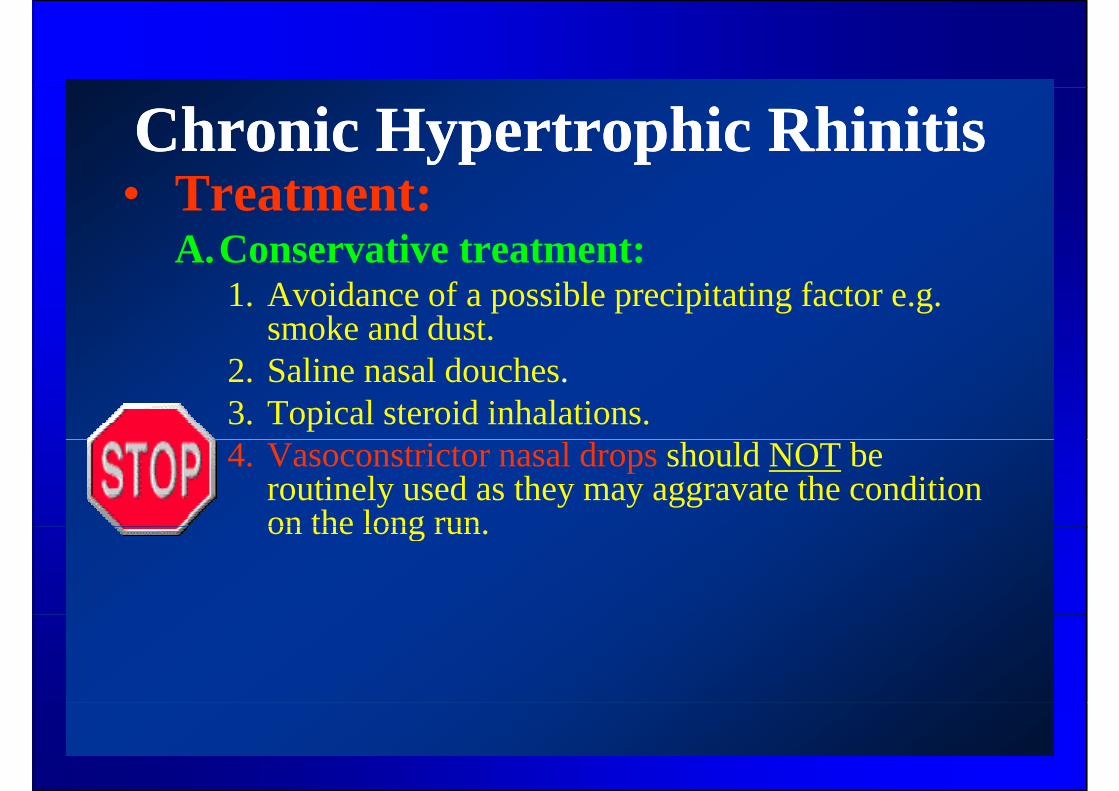
Chronic Hypertrophic RhinitisChronic Hypertrophic RhinitisT• Treatment:A.Conservative treatment:
1. Avoidance of a possible precipitating factor e.g. smoke and dust.
2. Saline nasal douches.3. Topical steroid inhalations.4. Vasoconstrictor nasal drops should NOT be
routinely used as they may aggravate the condition on the long runon the long run.
B. Surgical treatment:. Su g ca t eat e t:• Surgical reduction of the size of the inferior
turbinate may be indicated to relieveturbinate may be indicated to relieve annoying nasal obstruction NOT relieved by prolonged medical treatment.p g
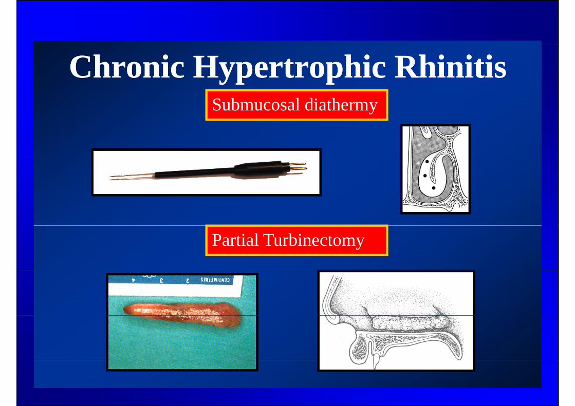
Chronic Hypertrophic RhinitisChronic Hypertrophic RhinitisSubmucosal diathermy
Partial Turbinectomy
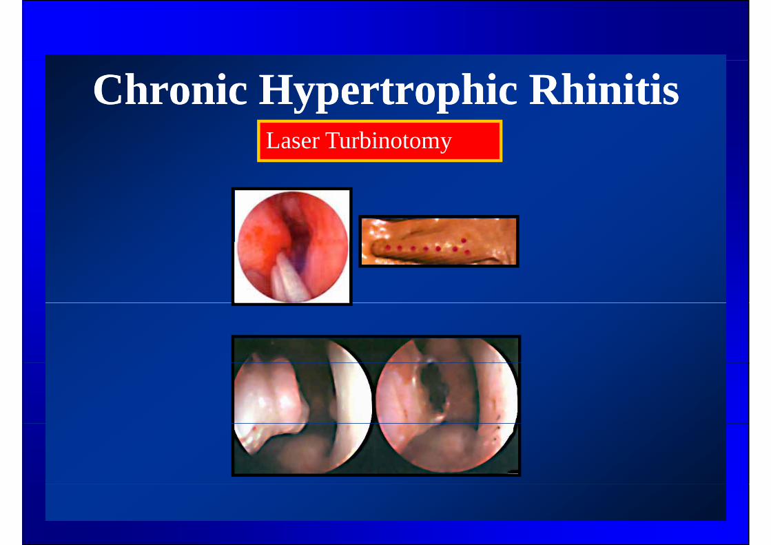
Chronic Hypertrophic RhinitisChronic Hypertrophic RhinitisLaser Turbinotomy
Primary Atrophic Rhinitis Primary Atrophic Rhinitis (O )(O )(Ozaena) (Ozaena)
• DefinitionDefinition – Primary atrophic rhinitis is a clinical condition
characterized by atrophy of the nasal mucosa, y p y ,offensive odor, roomy nasal cavities , crust formation, and anosmia.
I id• Incidence– The disease is more common in females around the age
f b tof puberty.
• EtiologyEtiology– The etiology of the disease is still unknown and various
theories (hormonal, infective…etc) have been ( , )proposed.
B t i l i lt f th l it f tl– Bacteriologic cultures from the nasal cavity frequently reveal bacillus foetidis ozaenae which is thought to be responsible for the offensive odor which is not pperceived by the patient due to atrophy of the olfactory nerve fibers.
Primary Atrophic Rhinitis Primary Atrophic Rhinitis (Ozaena)(Ozaena)(Ozaena) (Ozaena)
• Clinical pictureClinical picture1. The typical patient is a young adult female
presenting anosmia and an offensive odor whichpresenting anosmia, and an offensive odor which is NOT perceived by the patient due to the associated anosmia.
2. Nasal obstruction (sometimes)
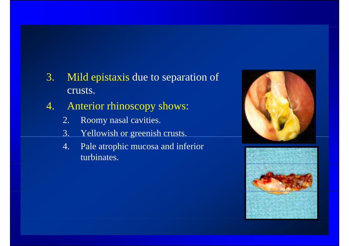
3. Mild epistaxis due to separation of p pcrusts.
4. Anterior rhinoscopy shows:4. Anterior rhinoscopy shows:2. Roomy nasal cavities.3. Yellowish or greenish crusts.g4. Pale atrophic mucosa and inferior
turbinates.
Primary Atrophic Rhinitis Primary Atrophic Rhinitis (Ozaena) (Ozaena)
• TreatmentTreatment1. Frequent cleaning of the nose by saline washes is
practically the safest and most effective symptomaticpractically the safest and most effective symptomatic treatment.
2. Lubricant drops e.g. menthol paraffin drops and 25% glucose in glycerin may be also used.
3. Surgical treatment is also of limited value.
Secondary Atrophic RhinitisSecondary Atrophic Rhinitis
• Causes:Causes:1. Excessive surgical resection of the inferior turbinates.2 R di th f th h d2. Radiotherapy of the head.3. Marked deviation of the nasal septum (on the roomy
id )side).4. Granulomata e.g. Scleroma.
• Treatment:1. Treatment or avoidance of the cause.2. Nasal washes.
Rhinitis MedicamentosaRhinitis Medicamentosa• Definition
– Rhinitis due to inappropriate use of vasoconstrictor nasal drops to relieve nasal pcongestion. Rhinitis medicamentosa may develop as early as 7 days of medication usedevelop as early as 7 days of medication use.
• PathophysiologyPathophysiology– As the effect of vasoconstrictor drops wears
ff d dil i ioff, secondary vasodilatation occurs causing the sense of obstruction to return (vicious circle).
Rhinitis MedicamentosaRhinitis Medicamentosa
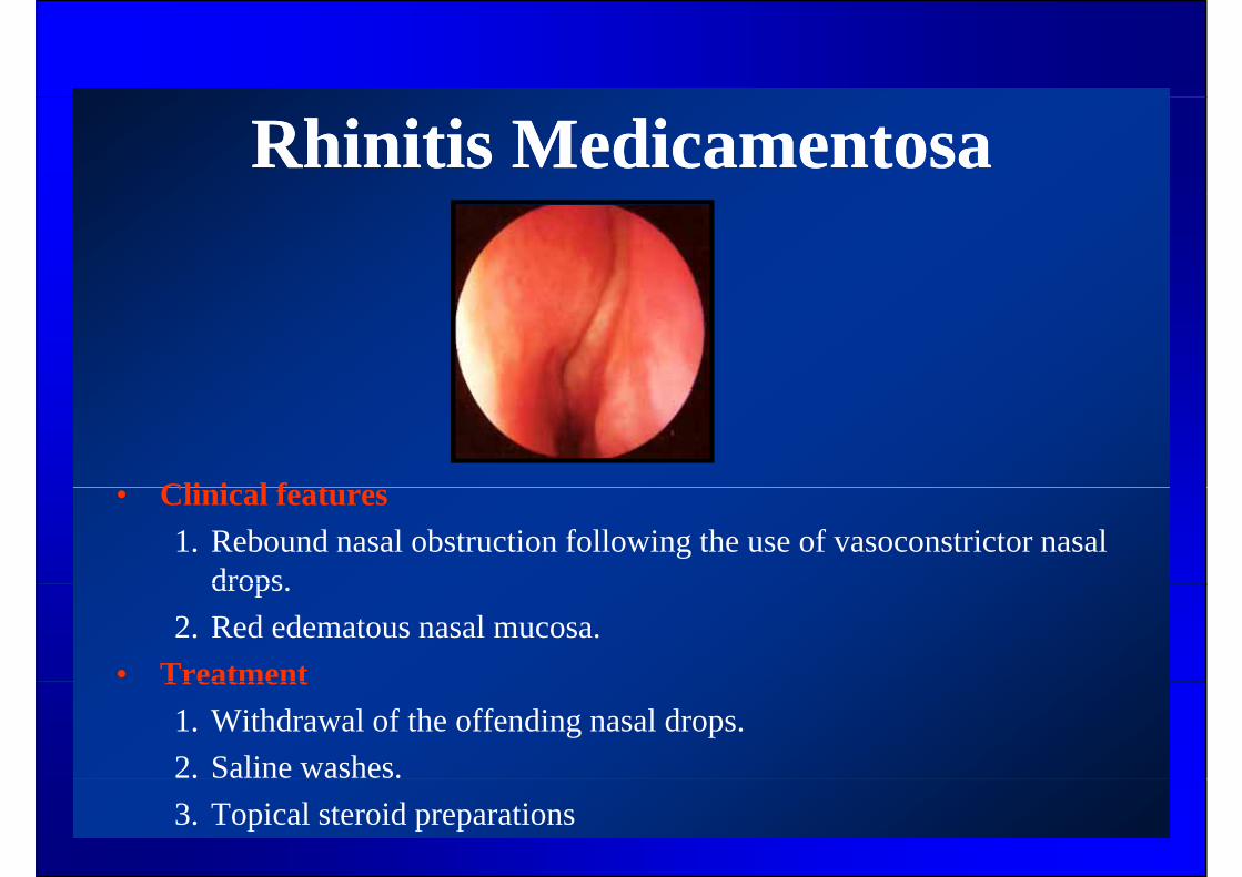
Cli i l f t• Clinical features1. Rebound nasal obstruction following the use of vasoconstrictor nasal
dropsdrops.2. Red edematous nasal mucosa.
• TreatmentTreatment1. Withdrawal of the offending nasal drops.2. Saline washes.3. Topical steroid preparations
BackgroundBackgroundScleroma = Hard swellinggSynonyms: Mickulicz disease, rhinosclerosis.
Chronic specific inflammation of the upperrespiratory tract caused by Frisch bacillus orKlebsiella rhinoscleromatis and is characterizedKlebsiella rhinoscleromatis and is characterized,in the typical case, with granulomatous lesions withh t i ti h d h thcharacteristic hardness hence the name
“scleroma” .
BackgroundBackgroundThe disease almost allways commences in theThe disease almost allways commences in the nose (Rhinoscleroma) and later extends into nasophar n & orophar n the lar n tracheanasopharynx & oropharynx , the larynx, trachea, bronchi, and other areas. (*)
For this reason , the disease was sometimesFor this reason , the disease was sometimes designated as Respiratory Scleroma , rather than rhinoscleroma (**)than rhinoscleroma. (**)
Incidence Incidence • The disease is endemic in some areas in Egypt .It is
also seen in central and south-eastern Europe, NorthAfrica, the Indian subcontinent, Indonesia and, ,Central and South America (scleroma belt).
• Epidemiologic associations: Poor hygiene, badcrowded living conditions, and malnutrition.
IncidenceIncidence
• It may occur in all races and either sex• It may occur in all races and either sex.• Typically it starts in patients aged 10-30 years.• Slightly more common in females .
Bacteriology Bacteriology
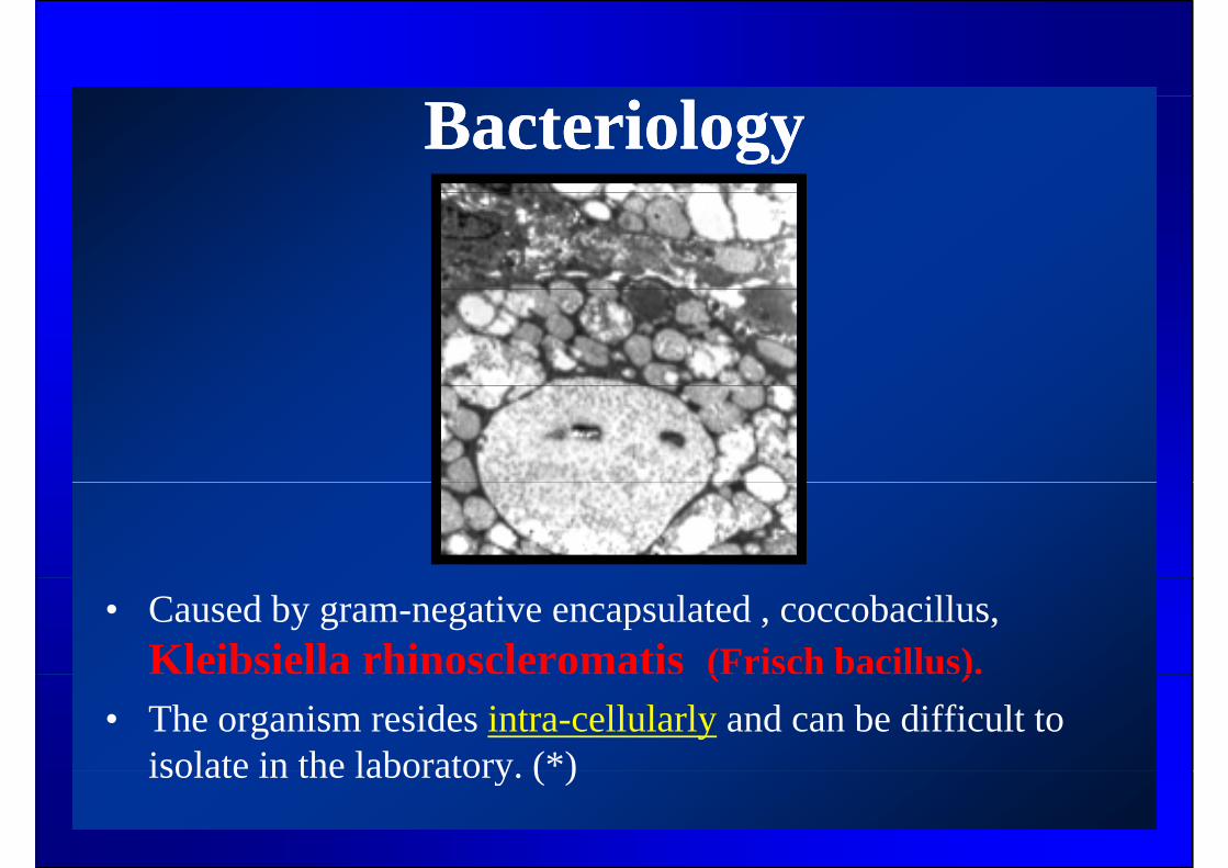
• Caused by gram-negative encapsulated , coccobacillus, Kleibsiella rhinoscleromatis (Frisch bacillus).e bs e osc e o s ( )
• The organism resides intra-cellularly and can be difficult to isolate in the laboratory (*)isolate in the laboratory. ( )
PathogenesisPathogenesis
The disease is not highly contagious. It is probablyThe disease is not highly contagious. It is probablytransmitted by droplets or by contamination ofmaterial that is subsequently inhaled.material that is subsequently inhaled.
Cellular immunity is impaired in scleroma patientsCellular immunity is impaired in scleroma patientsbut humoral immunity is preserved.
The disease usually begins in areas of epithelialt iti h th tib l f th d thtransition such as the vestibule of the nose and thesubglottic region.
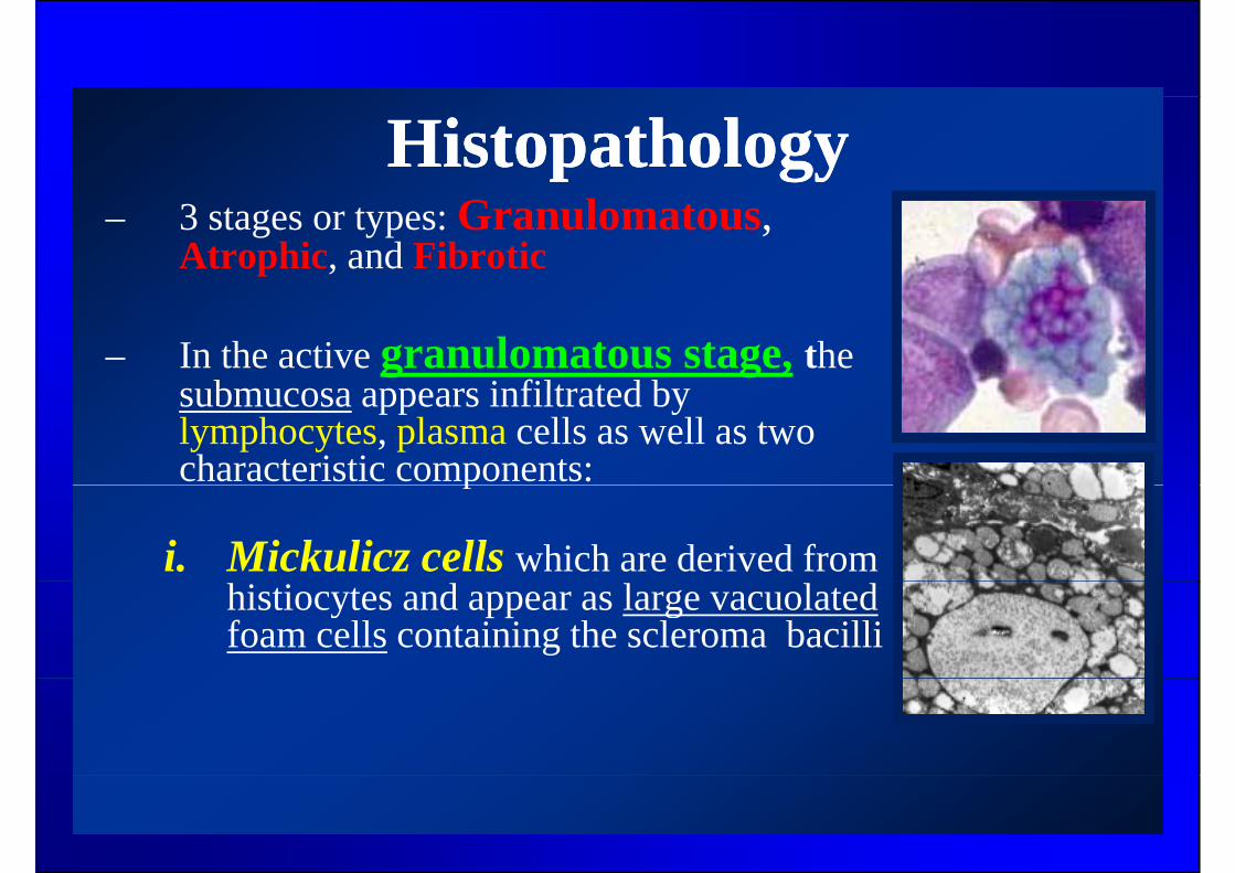
HistopathologyHistopathology– 3 stages or types: Granulomatous,
Atrophic, and Fibrotic
– In the active granulomatous stage, the b i filt t d bsubmucosa appears infiltrated by
lymphocytes, plasma cells as well as two characteristic components:p
i. Mickulicz cells which are derived from histiocytes and appear as large vacuolated foam cells containing the scleroma bacilli
HistopathologyHistopathology
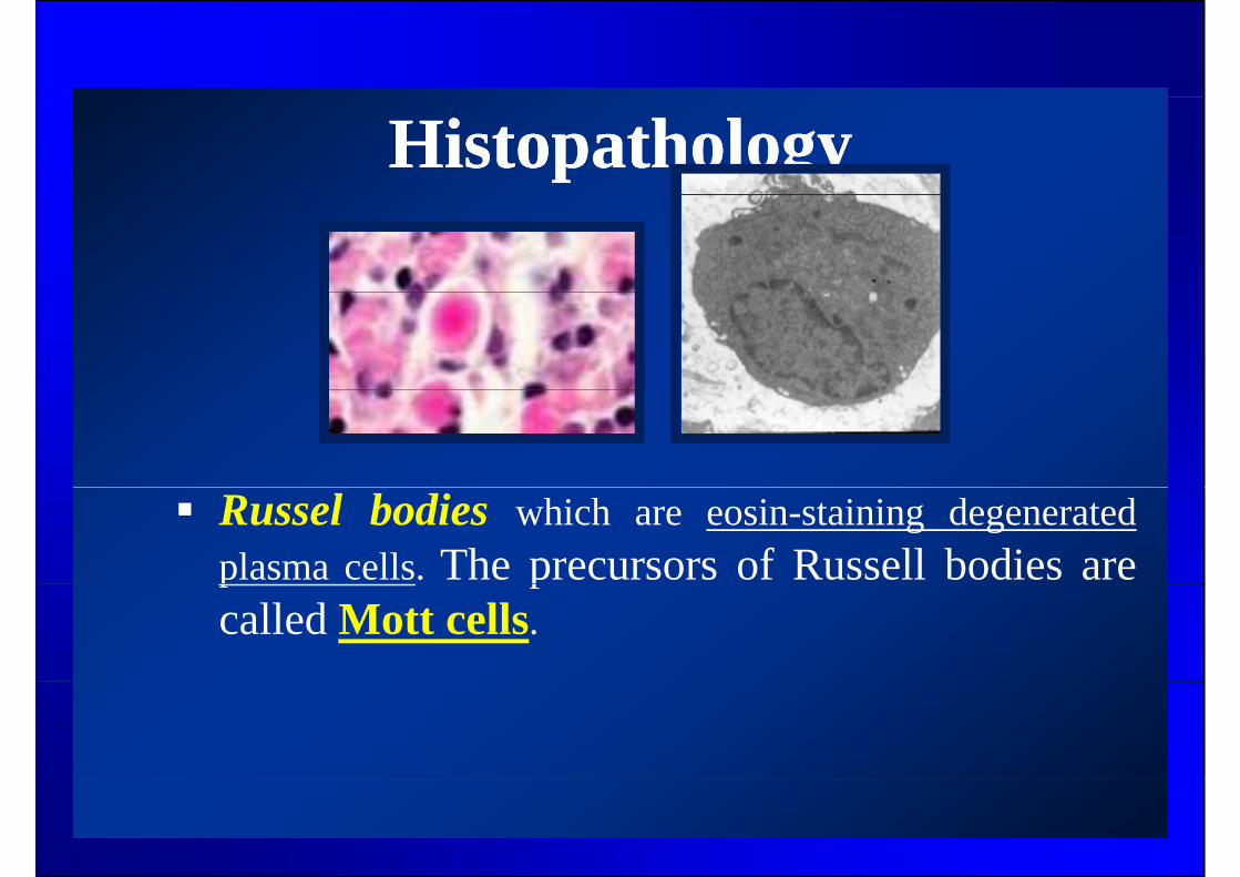
Russel bodies which are eosin-staining degeneratedplasma cells. The precursors of Russell bodies arep pcalled Mott cells.
Clinical pictureClinical picture
• Most often the presentation is non specific• Most often the presentation is non-specificresembling chronic rhinitis.
• The most common complaint is nasal• The most common complaint is nasal obstruction.
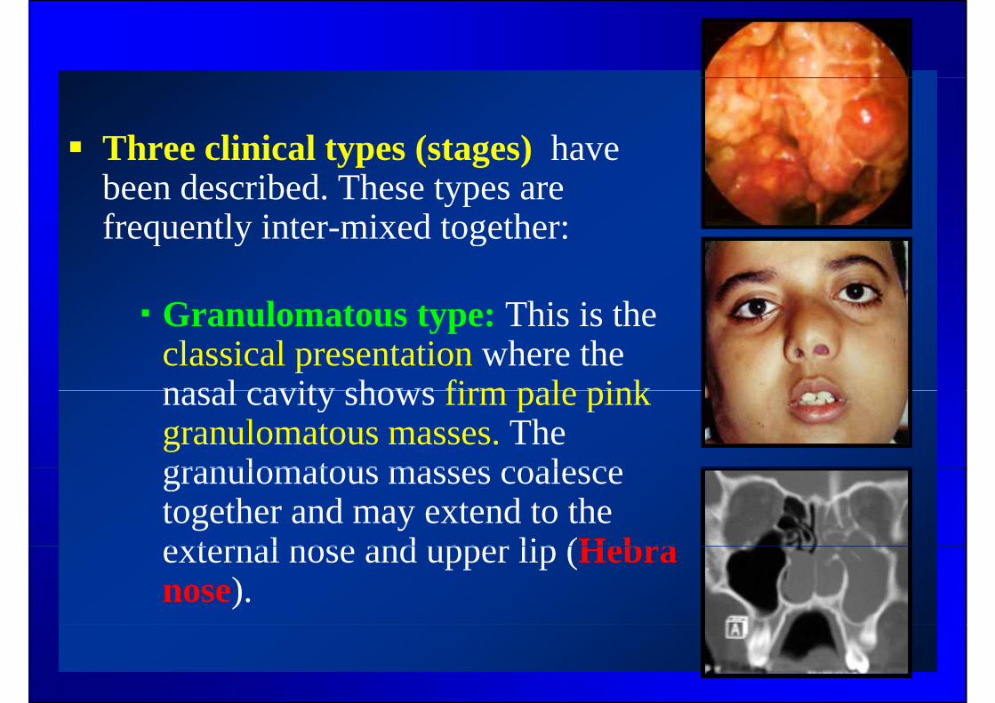
Three clinical types (stages) haveThree clinical types (stages) have been described. These types are frequently inter-mixed together:frequently inter mixed together:
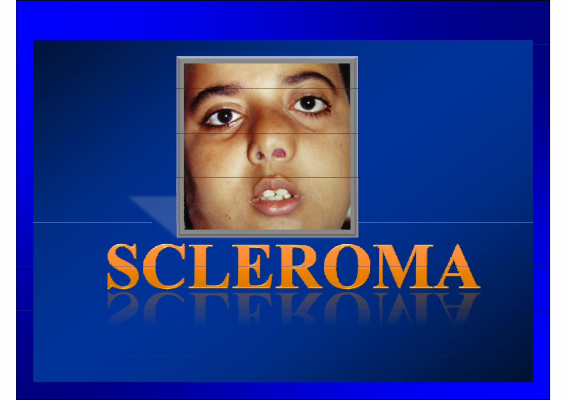
Granulomatous type: This is theGranulomatous type: This is the classical presentation where the nasal cavity shows firm pale pinknasal cavity shows firm pale pink granulomatous masses. The granulomatous masses coalescegranulomatous masses coalesce together and may extend to the external nose and upper lip (Hebraexternal nose and upper lip (Hebranose).
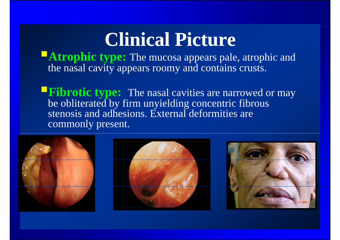
Clinical PictureClinical PictureAt hi t h l hi dAtrophic type: The mucosa appears pale, atrophic and the nasal cavity appears roomy and contains crusts.
Fibrotic type: The nasal cavities are narrowed or may be obliterated by firm unyielding concentric fibrousbe obliterated by firm unyielding concentric fibrous stenosis and adhesions. External deformities are commonly present.
Extensions of the diseaseExtensions of the disease
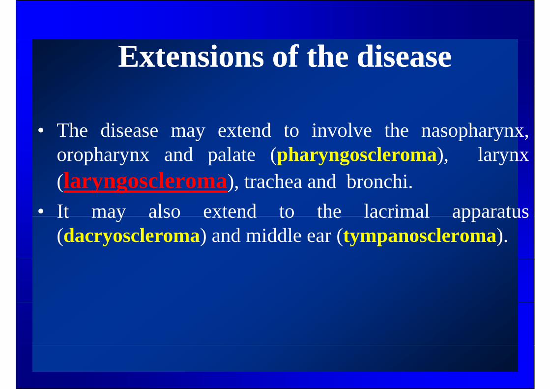
• The disease may extend to involve the nasopharynx• The disease may extend to involve the nasopharynx,oropharynx and palate (pharyngoscleroma), larynxl l(laryngoscleroma), trachea and bronchi.
• It may also extend to the lacrimal apparatusIt may also extend to the lacrimal apparatus(dacryoscleroma) and middle ear (tympanoscleroma).
LaryngoscleromaLaryngoscleroma
Usually secondary to nasal involvementUsually secondary to nasal involvement (15 – 40%). Occasionally primary.More common in females.U ll ff h b l i iUsually affect the subglottic region.
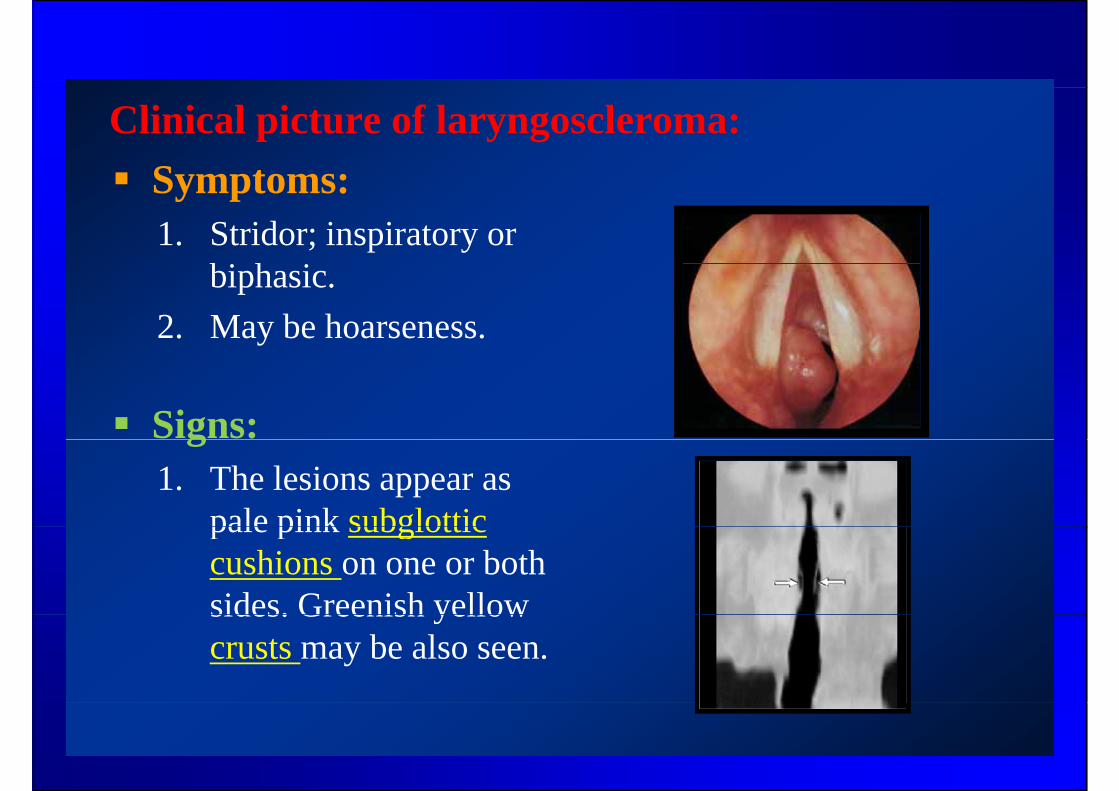
Clinical picture of laryngoscleroma:Symptoms:Symptoms:1. Stridor; inspiratory or
bi h ibiphasic.2. May be hoarseness.
Signs:g1. The lesions appear as
pale pink subglotticpale pink subglottic cushions on one or both sides. Greenish yellowsides. Greenish yellow crusts may be also seen.
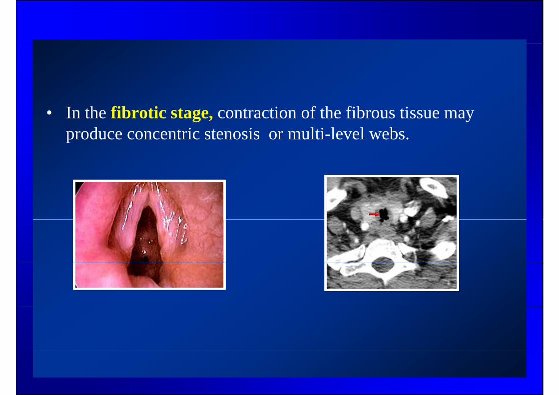
• In the fibrotic stage, contraction of the fibrous tissue may produce concentric stenosis or multi level websproduce concentric stenosis or multi-level webs.
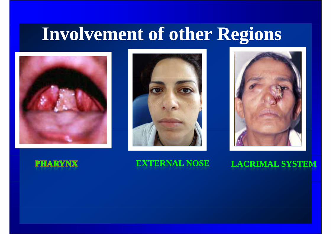
Involvement of other RegionsInvolvement of other Regions
EXTERNAL NOSEEXTERNAL NOSE LACRIMAL SYSTEMLACRIMAL SYSTEM
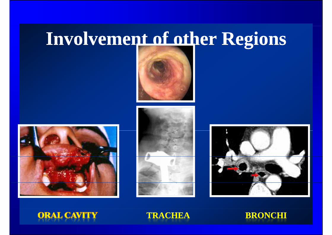
Involvement of other RegionsInvolvement of other Regions
TRACHEATRACHEA BRONCHIBRONCHI
Diagnosis Diagnosis
1. Biopsy is the best choice to demonstrate the p ycharacteristic histological features.
2. Bacteriological identification of the organism is ll NOTusually NOT necessary.
3. Multislice CT scans are useful especially for larynx and trachea (crypt-like irregularities)larynx and trachea (crypt like irregularities).
Differential diagnosisDifferential diagnosis
• Infectious granulomatous diseases :• Infectious granulomatous diseases : tuberculosis, syphilis , leprosy, and fungal infections.
• Lymphomas and neoplasms.
Treatment Treatment
Once the diagnosis has been confirmed by biopsy ,Once the diagnosis has been confirmed by biopsy ,prolonged antibiotic treatment must be started.
The traditional antibiotics used were streptomycin(ototoxic) and tetracycline (useful for young(ototoxic) and tetracycline (useful for youngpatients).
Better results are obtained now with oral therapyith i l ( i fl i ) d if i iwith quinolones (ciprofloxacin). and rifampicin
(both inhibit bacterial DNA synthesis).
TreatmentTreatment• Surgical debulking may be done for
gran lomato s lesionsgranulomatous lesions.• In late cases where disease has been
eradicated, further plastic andreconstructive nasal surgery may bereconstructive nasal surgery may berequired.
Treatment of LaryngoscleromaTreatment of Laryngoscleroma
1. Steroids and humidified inhalations may ybe needed to alleviate stridor.
2. Tracheostomy may be needed for severe stridor.stridor.
3. Carbon dioxide Laser vaporization may be performed for granulomatous lesions and websand webs.
Treatment of laryngoscleromaTreatment of laryngoscleroma
• Endoscopic dilatation +/ stenting• Endoscopic dilatation +/- stenting .• External procedures may be needed to p y
deal with stenotic areas. These are usually difficult for long stenotic segmentsdifficult for long stenotic segments.
Top Related