


Languages
Pages
Legal


1Cardiovascular Division, Department of Medicine, Brigham and Women’s Hospital, Harvard Medical School, 77 Avenue Louis Pasteur, Boston, Massachusetts 02115, USA. 2Division of Cardiovascular Medicine, Division of Preventive Medicine, Department of Medicine, Brigham and Women’s Hospital, Harvard Medical School, Boston, Massachusetts 02115, USA. 3Department of Medicine and Center for Molecular Medicine, Karolinska University Hospital, Karolinska Institutet, Stockholm SE-17176, Sweden.
Progress and challenges in translating the biology of atherosclerosisPeter Libby1, Paul M Ridker1,2 & Göran K. Hansson3
Atherosclerosis is a chronic disease of the arterial wall, and a leading cause of death and loss of productive life years worldwide. Research into the disease has led to many compelling hypotheses about the pathophysiology of atherosclerotic lesion formation and of complications such as myocardial infarction and stroke. Yet, despite these advances, we still lack definitive evidence to show that processes such as lipoprotein oxidation, inflammation and immunity have a crucial involvement in human atherosclerosis. Experimental atherosclerosis in animals furnishes an important research tool, but extrapolation to humans requires care. Understanding how to combine experimental and clinical science will provide further insight into atherosclerosis and could lead to new clinical applications.
Powerful laboratory research in the past decade has led to many reviews that describe the biological and genetic bases of athero-sclerosis1–3. Despite this progress, the leap from experimental
animal findings to human atherosclerosis and clinical application pre-sents challenges. The laboratory literature and experimental community sometimes assume that the results obtained in cultured cells or animals closely correspond to humans. Although experimental work has helped to unravel some of the principles of atherosclerosis pathophysiology, gaps remain in translation to the clinic, and these breeches require bridging to achieve the full promise of scientific advances in atherosclerosis.
This Review summarizes the burgeoning biological understanding of atherosclerosis. Instead of celebrating the astounding advances already achieved, we highlight some of the challenges to the clinical application of these advances. We also offer possible ways to move forward and overcome these obstacles.
Current concepts of atherogenesisAtherogenesis refers to the development of atheromatous plaques in the inner lining of the arteries. On the basis of animal experiments and observations in human specimens, most contemporary schemes of atherogenesis posit an initial qualitative change in the monolayer of endothelial cells that lines the inner arterial surface (Fig. 1a). Arterial endothelial cells, which normally resist attachment of the white blood cells streaming past them, express adhesion molecules that capture leukocytes on their surfaces (Fig. 1b) when subjected to irritative stimuli (such as dyslipidaemia, hypertension or pro-inflammatory mediators). Parallel changes in endothelial permeability and the composition of the extracellular matrix beneath the endothelium promote the entry and retention of cholesterol-containing low-density lipoprotein (LDL) particles in the artery wall4. Biochemically modified components of these particles may induce leukocyte adhesion, and intact but modified particles undergo endocytosis by monocyte-derived macrophages, leading to intracellular cholesterol accumulation. Chemoattractant mediators direct the migration of the bound leukocytes into the innermost layer of the artery, the tunica intima (Figs 1b and 2). The localized distribution of atheromatous lesions in the arterial tree, despite a systemic rise in risk factors such as increased LDL levels or blood pressure, probably reflects differing haemodynamics in different segments of the arterial tree, distinction in the regional development
of arteries5 and the ability of normal laminar shear stress to elicit an atheroprotective program of gene expression by the endothelium6. Once resident in the artery wall, monocytes — the most numerous white blood cells in plaques — differentiate into tissue macrophages. In the nascent atheroma, these mononuclear phagocytes engulf lipoprotein particles and become foam cells — a term that reflects the microscopic appearance of these lipid-laden macrophages.
In mice, a pro-inflammatory subset of monocytes induced by hyperlipidaemia may preferentially furnish the precursors of lesional foam cells, but the fates and functions of this monocyte subset and its human equivalent remain under intense exploration7,8. Macrophages in the atheroma may also have a pro-inflammatory palette of functions, characteristic of M1 macrophages9, which produce high levels of effectors such as the cytokines interleukin-1β (IL-1β) and tumour-necrosis factor (TNF). Some mononuclear phagocytes in plaques have the characteristics, and probably the antigen-presenting functions, of dendritic cells. Other leukocyte classes (such as lymphocytes) and mast cells also accumulate in atheromata, but less abundantly than phagocytes. Lesional T cells, although far fewer in number than macrophages, probably have key regulatory functions in plaques.
Atheroma formation also involves the recruitment of smooth muscle cells (SMCs) from the tunica media — the middle layer of the artery wall — into the tunica intima (Fig. 1c). Unlike that of most experimental animals used to study atherosclerosis, the intima of human arteries (including the coronary arteries) contains resident SMCs. During atherogenesis, other SMCs migrate from the media into the intima, and proliferate in response to mediators such as platelet-derived growth factor. In the intima, the SMCs produce extracellular matrix molecules, including interstitial collagen and elastin, and form a fibrous cap that covers the plaque. This cap typically overlies a collection of macrophage-derived foam cells, some of which die (for example, by apoptosis) and release lipids that accumulate extracellularly. The inefficient clearance of dead cells — a process known as efferocytosis — can promote the accumulation of cellular debris and extracellular lipids, forming a lipid-rich pool called the necrotic core of the plaque10.
Plaques generally cause clinical manifestations by producing flow-limiting stenoses that lead to tissue ischaemia, or by provoking thrombi that can interrupt blood flow locally or embolize and lodge
REVIEWdoi:10.1038/nature10146
1 9 M A Y 2 0 1 1 | V O L 4 7 3 | N A T U R E | 3 1 7© 2011 Macmillan Publishers Limited. All rights reserved

in distal arteries. Paradoxically, thrombotic complications do not always occur at the sites of the most severe arterial narrowing by plaques. Instead, thrombi often arise after physical disruption of the plaque, most commonly a fracture of the fibrous cap that exposes pro-coagulant material in the plaque’s core to coagulation proteins in the blood, triggering thrombosis (Fig. 1d). Plaques that rupture typically have thin, collagen-poor fibrous caps with few SMCs but abundant macrophages. The inflammatory cells may hasten plaque disruption by elaborating collagenolytic enzymes that can degrade collagen, and by generating mediators that provoke the death of SMCs, the source of arterial collagen11. Plaque macrophages also produce the pro-coagulant tissue factor that renders the lipid core thrombogenic. Thus, the infiltrating inflammatory cells interact with the intrinsic arterial cells (smooth muscle and endothelium), promoting lesion formation and complications.
The risk factors for atherosclerosis act at several points on this pathogenic pathway. Hypertension is a major risk factor for atheromata, and can increase arterial wall tension, leading to disturbed repair processes and aneurysm formation. Angiotensin II, a major
pressor hormone, can alter endothelial function, inciting leukocyte adhesion. Cigarette smoking and diabetes also affect vascular biology, but through less well understood mechanisms. The role of cholesterol has been investigated in great detail, yielding success in cardiovascular prevention strategies.
Lipids and atherosclerosisLipids have a central role in the pathogenesis of plaques, but the mechanistic links between lipids and atherogenesis remain unclear. Observational data support a strong association between plasma lipid levels and the risk of cardiovascular disease12. In particular, LDL levels satisfy modified Koch’s postulates — criteria for judging whether a specific microbe is the cause of a disease — for causality of atherosclerosis13. LDL levels correlate with the risk of cardiovascular events in human populations, and augment individual susceptibility to atherosclerosis and its complications. Monogenic disorders that raise plasma levels of LDL heighten cardiovascular risk. Several interventions that lower LDL levels by independent mechanisms diminish the likelihood of atherosclerotic events.
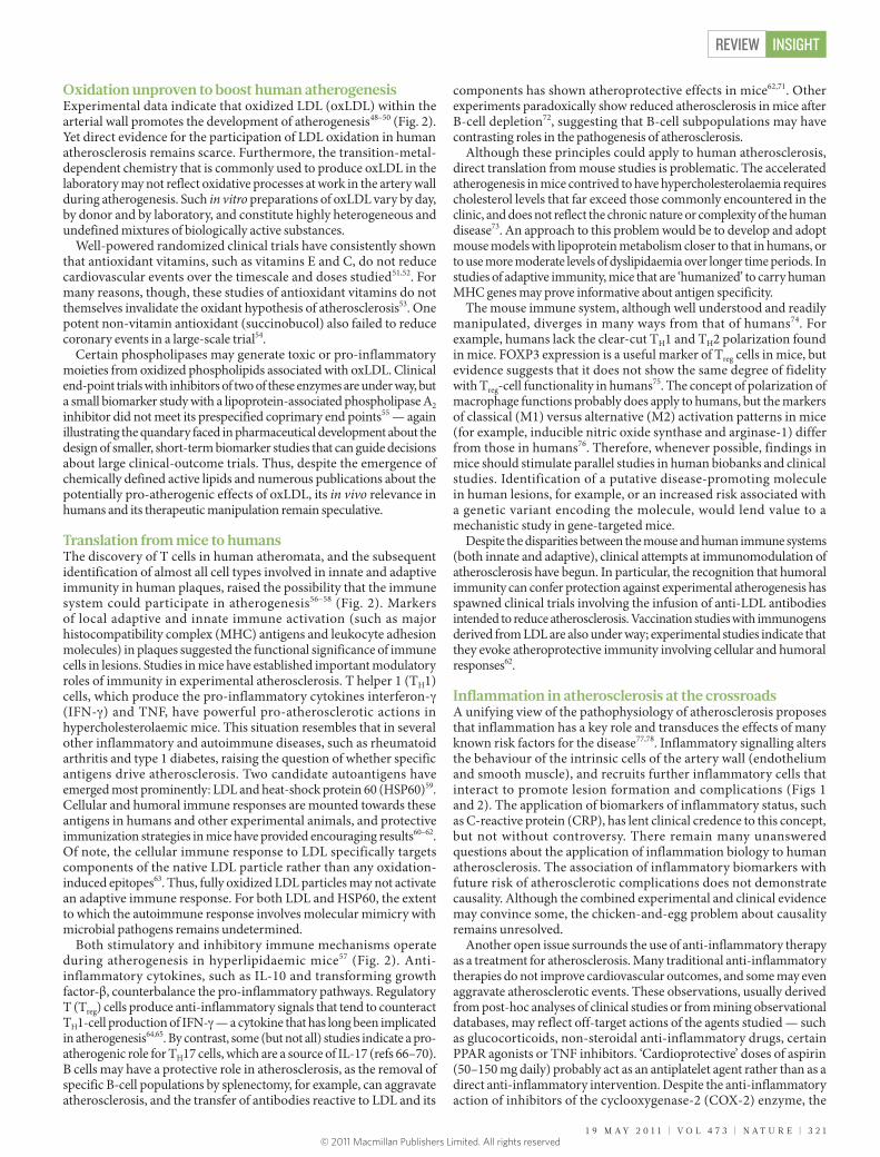
Figure 1 | Stages in the development of atherosclerotic lesions. The normal muscular artery and the cell changes that occur during disease progression to thrombosis are shown. a, The normal artery contains three layers. The inner layer, the tunica intima, is lined by a monolayer of endothelial cells that is in contact with blood overlying a basement membrane. In contrast to many animal species used for atherosclerosis experiments, the human intima contains resident smooth muscle cells (SMCs). The middle layer, or tunica media, contains SMCs embedded in a complex extracellular matrix. Arteries affected by obstructive atherosclerosis generally have the structure of muscular arteries. The arteries often studied in experimental atherosclerosis are elastic arteries, which have clearly demarcated laminae in the tunica media, where layers of elastin lie between strata of SMCs. The adventitia, the outer layer of arteries, contains mast cells, nerve endings and microvessels. b, The initial steps of atherosclerosis include adhesion of blood leukocytes to the activated endothelial monolayer, directed migration of the bound
leukocytes into the intima, maturation of monocytes (the most numerous of the leukocytes recruited) into macrophages, and their uptake of lipid, yielding foam cells. c, Lesion progression involves the migration of SMCs from the media to the intima, the proliferation of resident intimal SMCs and media-derived SMCs, and the heightened synthesis of extracellular matrix macromolecules such as collagen, elastin and proteoglycans. Plaque macrophages and SMCs can die in advancing lesions, some by apoptosis. Extracellular lipid derived from dead and dying cells can accumulate in the central region of a plaque, often denoted the lipid or necrotic core. Advancing plaques also contain cholesterol crystals and microvessels. d, Thrombosis, the ultimate complication of atherosclerosis, often complicates a physical disruption of the atherosclerotic plaque. Shown is a fracture of the plaque’s fibrous cap, which has enabled blood coagulation components to come into contact with tissue factors in the plaque’s interior, triggering the thrombus that extends into the vessel lumen, where it can impede blood flow.
REVIEWINSIGHT
3 1 8 | N A T U R E | V O L 4 7 3 | 1 9 M A Y 2 0 1 1
Cholesterolcrystal
Vasavasorum
a
SMCsIntima
Media
b
Foam cell
AdventitiaFibroblast
MonocyteT cell
Endothelial cell
Dendritic cell
Apoptoticbodies
Apoptoticmacrophage
Dividing SMC
Collagen
Thrombusformation
Fibrous caprupture
Lipid core
MigratingSMC
c d
Foam cell
Mast cell
Macrophage
Platelet
© 2011 Macmillan Publishers Limited. All rights reserved

The LDL success story lacks a final chapterThe determination of the LDL pathway and therapy with inhibitors of hydroxymethyl glutaryl coenzyme A reductase (collectively known as statins), which regulate this pathway, are conspicuous victories of cardiovascular science and medicine14. But even in patients treated with statins, a considerable residual burden of cardiovascular risk remains15. More than 20% of patients will have a recurrent event within 30 months of an acute coronary syndrome, despite receiving high-dose statin treatment16. These findings indicate that treatments to decrease LDL levels even further, beyond the targets currently mandated by various national guidelines, could provide further clinical benefit. Unfortunately, at least one-quarter of high-risk patients who receive intensive statin therapy have LDL levels above current guideline-mandated goals17. New biological targets have emerged that may yield incremental lowering of LDL levels to a greater degree than that achieved by high-dose statin therapy (Box 1).
HDL as a frustrating next frontierConsistent evidence has shown that levels of high-density lipoprotein (HDL) correlate inversely with cardiovascular risk. Numerous approaches to increase HDL exist or are in development. Because of the heterogeneity in HDL particles, the complicated pathways of cholesterol flux mediated by HDL and the association of HDL with many proteins that may modify atherosclerosis, the steady-state levels of HDL cholesterol in blood reflect HDL function poorly. HDL particles can effect reverse cholesterol transport, and transfer cholesterol from peripheral tissues to the liver for excretion. This process involves the unloading of cholesterol from lipid-laden macrophages in atheromata by means of membrane-bound ATP-binding cassette transporters. Mature HDL interacts with one ATP-binding cassette transporter (ABCG1), and nascent HDL with another (ABCA1)18,19 (Fig. 2).
In addition to mediating reverse cholesterol transport, HDL can exert anti-inflammatory actions both in vitro and in vivo19. HDL particles associate with dozens of proteins, many with biological activities that have relevance to atherogenesis20. The lipid content of HDL particles can be remodelled — for example, the plasma protein cholesteryl ester transfer protein (CETP) facilitates the exchange of
cholesteryl esters in HDL for triglycerides from apolipoprotein-B-containing lipoproteins21,22 (Fig. 2). The protein content of HDL particles can also be remodelled — for example, when plasma levels of the acute-phase reactant serum amyloid A increase during inflammatory states23. Typical clinical assays for HDL do not reflect this high degree of heterogeneity of the particles that influence plaque biology24. Thus, the mere increase in HDL levels in response to some interventions may not necessarily confer clinical benefit, owing to qualitative changes in the particles. By contrast, the lowering of LDL levels usually reduces cardiovascular event rates. Of the approaches to increase HDL under study, the potential of CETP inhibition to improve outcomes remains unclear. The CETP inhibitor torcetrapib failed in the clinic, probably owing to off-target effects18,25,26, and two other CETP inhibitors, dalcetrapib and anacetrapib, have entered clinical evaluation. The safety of anacetrapib was recently affirmed by a phase III clinical trial, which provided preliminary evidence for reduced clinical events27. Ultimately, the results of continuing large end-point trials should settle the CETP controversy.
Apolipoprotein A-I (Apo-AI), the major protein component of HDL, has received much attention as a possible therapeutic target for atherosclerosis28–30. But difficulties have plagued the development of protein therapeutics and mimetics. Despite small biomarker studies that suggest possible efficacy of some such agents, various limitations have stalled their entry to trials that could show efficacy in cardiovascular event reduction.
Manipulation of the transcription of APOA1 has proven elusive, with only one agent in development for this purpose. Stimulation of the nuclear receptor peroxisome proliferator-activated receptor-α (PPAR-α) moderately increases Apo-AI levels. Moreover, preclinical and biomarker studies have suggested beneficial vascular actions of PPAR-α agonism that do not depend on Apo-AI31. Clinical trials of one agent with PPAR-α-stimulating activity, gemfibrozil, have shown a reduction in cardiovascular events32,33. Unfortunately, the combination of gemfibrozil with statins raises major safety concerns, owing to a well-defined drug–drug interaction34. Another agent with relatively weak PPAR-α-agonist action, fenofibrate, has not reduced events in several large clinical trials35,36.
Here we consider biological targets that may reduce LDL levels to a greater extent than that obtained by high-dose statin therapy.
Niemann–Pick C1-like protein 1Inhibition of the intestinal cholesterol transporter Niemann–Pick C1-like protein 1 (NPC1L1) by the agent ezetimibe can reduce LDL levels by almost 20% in individuals already being treated with statins89. Although combined therapy with statins and ezetimibe can help more individuals to reach mandated LDL targets for their level of risk, no clinical trial data have so far shown that this strategy will lower cardiovascular event rates beyond the drop produced by statin monotherapy. Studies of biomarkers such as the thickness of the carotid artery intima media, flow-mediated vasodilation, or inflammation cannot supplant lacking of data on clinical events. This example emphasizes three important points: (1) the need to choose biomarkers carefully to be pursued in clinical development; (2) the ultimate requirement for clinical end-point studies to determine the efficacy of interventions; and (3) the value of starting such definitive studies early in drug-development programmes.
Proprotein convertase subtilisin/kexin type 9Genetic studies have shown that mutations in the gene that encodes
the enzyme proprotein convertase subtilisin/kexin type 9 (PCSK9)augment LDL receptor levels on cell surfaces, boosting LDL clearance and yielding lower LDL concentrations in the blood90. The enzymatic activity of PCSK9 — autocatalysis — does not directly degrade LDL receptors. Although enzymes generally make good drug targets, the autocatalytic activity of PCSK9 has proven difficult to inhibit by conventional medicinal chemistry approaches, and does not necessarily reflect its regulation of LDL receptor levels, spurring the development of biological agents that seek to limit PCSK9 action.
Individuals with loss-of-function variants in PCSK9, who are exposed to lower levels of LDL from childhood than those with the common genotype for this enzyme, seem protected from atherosclerotic events even when they have other cardiovascular risk factors91. This observation suggests that lowering LDL levels for longer periods than those encompassed by typical clinical trials should continue to provide benefit, and supports a pivotal, perhaps permissive, role for LDL in atherosclerosis. Such genetic data also help to clarify the importance of LDL lowering compared with other potential mechanisms of the benefits of statins (for example, statins interfere with prenylation of small G proteins, modulates lipid-raft organization and activates of Krüppel-like factor 2)18,92,93.
BOX 1
New biological targets for lowering LDL levels
REVIEW INSIGHT
1 9 M A Y 2 0 1 1 | V O L 4 7 3 | N A T U R E | 3 1 9© 2011 Macmillan Publishers Limited. All rights reserved

Nicotinic acid raises HDL levels effectively and has shown some event reduction in clinical trials, but tolerability issues have limited its use. The recognition of G-protein-coupled receptors for nicotinic acid, and of β-hydroxybutyrate as an endogenous ligand of one such receptor, has not yet led to a therapeutic approach37. Clinical end-point trials are testing whether adding nicotinic acid to standard care (usually including statin treatment) improves cardiovascular outcomes38. One trial is using an extended-release preparation of nicotinic acid combined with a prostaglandin D2 receptor antagonist, intended to reduce the cutaneous flushing that limits the acceptability of high-dose nicotinic acid for many patients39.
Thus, despite considerable understanding of HDL and its metabolism, none of the pharmacological agents tested so far has offered a practical and proven way to reduce cardiovascular events. We must await the results of ongoing trials of approaches to raise HDL levels to reach this elusive goal.
Triglycerides on trialFasting or non-fasting triglyceride levels can predict cardiovascular events, but adjustment for other risk factors considerably weakens or even abolishes the association40. Lifestyle changes such as weight loss, physical activity or low-carbohydrate diets can lower blood-circulating levels of triglycerides. The clinical benefits of
such lifestyle modifications probably result from a combination of mechanisms, so they cannot affirm triglycerides as a causal risk factor for atherosclerosis. Strict control of diabetes can also lessen hypertriglyceridaemia, yet tight glycaemic control may increase, rather than prevent, clinical complications of atherosclerosis in people with type 2 diabetes41,42. Fibrates effectively decrease triglyceride levels, but trials of these drugs have proven disappointing in reducing clinical events, and have not shown reductions in mortality rates43. Omega-3 fatty acids, prominent constituents of certain fish oils, reduce triglyceride levels and can limit cardiovascular events in some populations. These fatty acids also have anti-arrhythmic action, can mute inflammation and impair platelet aggregability — precluding a conclusion about the extent to which their clinical benefit arises from lowered triglyceride levels44.
Recent evidence suggests that fractions of triglyceride-rich lipoproteins, particularly those that contain Apo-CIII, confer risk not conveyed by the total triglyceride level. Indeed, Apo-CIII acts as a pro-inflammatory mediator and an endogenous ligand of the Toll-like receptor 2 signalling pathway, which is implicated in the aggravation of mouse atherosclerosis45,46 (Fig. 2). In addition, very-low-density lipoprotein (VLDL) promotes transcription of the plasminogen activator-1 gene, leading to a reduced capacity to lyse thrombi47.
Figure 2 | The intersection of inflammation and lipid metabolism modulates atherosclerosis and provides potential targets for therapeutic manipulation. Atherogenesis begins with the recruitment of inflammatory cells to the intima. Activated endothelial cells express leukocyte adhesion molecules that capture blood monocytes, including (but not exclusively) the pro-inflammatory subset marked by high expression levels of the cell-surface protein Ly6C in mice. After inflammatory activation, monocytes recruited to the intima express scavenger receptors that permit the uptake of modified LDL particles, such as oxidized LDL (oxLDL). Cholesterol loading leads to the formation of foam cells, and ultimately leads to the mature lipid-laden macrophages of the plaque’s core. These cells can produce pro-inflammatory mediators, reactive oxygen species, and tissue factor pro-coagulants that amplify local inflammation and promote thrombotic complications. Although fewer in number than the mononuclear phagocytes, T cells also enter the intima and send decisive regulatory signals. After antigen-specific activation, T helper 1 (TH1) cells secrete the signature cytokine interferon-g (IFN-g), which can activate vascular wall cells and macrophages, and magnify and sustain the inflammatory response in the intima. Regulatory T (Treg) cells produce interleukin-10 (IL-10) and transforming growth factor-β (TGF-β), two cytokines considered to exert anti-inflammatory actions. Although not numerically prominent in the plaque, B cells accumulate and
organize in the perivascular tissue surrounding atherosclerotic arteries. They produce circulating antibodies that may limit inflammation and mute atherogenesis. In addition to modified LDL, triglyceride-rich lipoproteins such as very-low-density lipoprotein (VLDL) — particularly those particles that bear apolipoprotein C-III (Apo-CIII) or apolipoprotein B (Apo-B) — can instigate vascular inflammation through Toll-like receptor 2 (TLR2) signalling. Macrophage foam cells can efflux cholesterol (chol) through ATP-binding cassette (ABC) transporters, which work in tandem. ABCA1 loads cholesterol-poor nascent high-density lipoprotein (HDL) particles (pre-β HDL) with cholesterol. ABCG1 can load more mature HDL particles with cholesterol. Having taken up cholesterol through interaction with the ABC transporters in the artery wall, HDL particles can exit through the bloodstream, contributing to reverse cholesterol transport from lesional macrophages to the periphery. VLDL and LDL particles bearing ApoB can unload cholesterol from HDL particles through the action of cholesteryl ester transfer protein (CETP). Blockade of CETP can thus augment HDL levels, a process not yet known to produce clinical benefit. The ApoB-containing lipoproteins can promote clearance of cholesterol through capture by peripheral LDL receptors. Loss-of-function mutations in the enzyme PCSK9 (not shown) can increase the number of LDL receptors on peripheral cells, thereby augmenting the clearance of LDL.
Vasavasorum
Lipid core
B cell
Antigen-presenting cell
T cell
oxLDL
Scavengerreceptor
Apo-CIII
VLDL
LDL
VLDL
TLR2
TLR2
Antibody
LDL
Pro-in�ammatory monocyte
Endothelial cell
IFN-γ
Treg cell
TH1 cell
TGF-β
IL-10
ABCA1
Apo-AI
HDL Chol
Chol
Chol
Lipid-ladenmacrophage
Pre-β HDL
CETPABCG1
ng
IFN-γTH1cell
CABCG1
Foam-cell formation
REVIEWINSIGHT
3 2 0 | N A T U R E | V O L 4 7 3 | 1 9 M A Y 2 0 1 1© 2011 Macmillan Publishers Limited. All rights reserved
Oxidation unproven to boost human atherogenesisExperimental data indicate that oxidized LDL (oxLDL) within the arterial wall promotes the development of atherogenesis48–50 (Fig. 2). Yet direct evidence for the participation of LDL oxidation in human atherosclerosis remains scarce. Furthermore, the transition-metal-dependent chemistry that is commonly used to produce oxLDL in the laboratory may not reflect oxidative processes at work in the artery wall during atherogenesis. Such in vitro preparations of oxLDL vary by day, by donor and by laboratory, and constitute highly heterogeneous and undefined mixtures of biologically active substances.
Well-powered randomized clinical trials have consistently shown that antioxidant vitamins, such as vitamins E and C, do not reduce cardiovascular events over the timescale and doses studied51,52. For many reasons, though, these studies of antioxidant vitamins do not themselves invalidate the oxidant hypothesis of atherosclerosis53. One potent non-vitamin antioxidant (succinobucol) also failed to reduce coronary events in a large-scale trial54.
Certain phospholipases may generate toxic or pro-inflammatory moieties from oxidized phospholipids associated with oxLDL. Clinical end-point trials with inhibitors of two of these enzymes are under way, but a small biomarker study with a lipoprotein-associated phospholipase A2 inhibitor did not meet its prespecified coprimary end points55 — again illustrating the quandary faced in pharmaceutical development about the design of smaller, short-term biomarker studies that can guide decisions about large clinical-outcome trials. Thus, despite the emergence of chemically defined active lipids and numerous publications about the potentially pro-atherogenic effects of oxLDL, its in vivo relevance in humans and its therapeutic manipulation remain speculative.
Translation from mice to humansThe discovery of T cells in human atheromata, and the subsequent identification of almost all cell types involved in innate and adaptive immunity in human plaques, raised the possibility that the immune system could participate in atherogenesis56–58 (Fig. 2). Markers of local adaptive and innate immune activation (such as major histocompatibility complex (MHC) antigens and leukocyte adhesion molecules) in plaques suggested the functional significance of immune cells in lesions. Studies in mice have established important modulatory roles of immunity in experimental atherosclerosis. T helper 1 (TH1) cells, which produce the pro-inflammatory cytokines interferon-g (IFN-g) and TNF, have powerful pro-atherosclerotic actions in hypercholesterolaemic mice. This situation resembles that in several other inflammatory and autoimmune diseases, such as rheumatoid arthritis and type 1 diabetes, raising the question of whether specific antigens drive atherosclerosis. Two candidate autoantigens have emerged most prominently: LDL and heat-shock protein 60 (HSP60)59. Cellular and humoral immune responses are mounted towards these antigens in humans and other experimental animals, and protective immunization strategies in mice have provided encouraging results60–62. Of note, the cellular immune response to LDL specifically targets components of the native LDL particle rather than any oxidation-induced epitopes63. Thus, fully oxidized LDL particles may not activate an adaptive immune response. For both LDL and HSP60, the extent to which the autoimmune response involves molecular mimicry with microbial pathogens remains undetermined.
Both stimulatory and inhibitory immune mechanisms operate during atherogenesis in hyperlipidaemic mice57 (Fig. 2). Anti-inflammatory cytokines, such as IL-10 and transforming growth factor-β, counterbalance the pro-inflammatory pathways. Regulatory T (Treg) cells produce anti-inflammatory signals that tend to counteract TH1-cell production of IFN-g — a cytokine that has long been implicated in atherogenesis64,65. By contrast, some (but not all) studies indicate a pro-atherogenic role for TH17 cells, which are a source of IL-17 (refs 66–70). B cells may have a protective role in atherosclerosis, as the removal of specific B-cell populations by splenectomy, for example, can aggravate atherosclerosis, and the transfer of antibodies reactive to LDL and its
components has shown atheroprotective effects in mice62,71. Other experiments paradoxically show reduced atherosclerosis in mice after B-cell depletion72, suggesting that B-cell subpopulations may have contrasting roles in the pathogenesis of atherosclerosis.
Although these principles could apply to human atherosclerosis, direct translation from mouse studies is problematic. The accelerated atherogenesis in mice contrived to have hypercholesterolaemia requires cholesterol levels that far exceed those commonly encountered in the clinic, and does not reflect the chronic nature or complexity of the human disease73. An approach to this problem would be to develop and adopt mouse models with lipoprotein metabolism closer to that in humans, or to use more moderate levels of dyslipidaemia over longer time periods. In studies of adaptive immunity, mice that are ‘humanized’ to carry human MHC genes may prove informative about antigen specificity.
The mouse immune system, although well understood and readily manipulated, diverges in many ways from that of humans74. For example, humans lack the clear-cut TH1 and TH2 polarization found in mice. FOXP3 expression is a useful marker of Treg cells in mice, but evidence suggests that it does not show the same degree of fidelity with Treg-cell functionality in humans75. The concept of polarization of macrophage functions probably does apply to humans, but the markers of classical (M1) versus alternative (M2) activation patterns in mice (for example, inducible nitric oxide synthase and arginase-1) differ from those in humans76. Therefore, whenever possible, findings in mice should stimulate parallel studies in human biobanks and clinical studies. Identification of a putative disease-promoting molecule in human lesions, for example, or an increased risk associated with a genetic variant encoding the molecule, would lend value to a mechanistic study in gene-targeted mice.
Despite the disparities between the mouse and human immune systems (both innate and adaptive), clinical attempts at immunomodulation of atherosclerosis have begun. In particular, the recognition that humoral immunity can confer protection against experimental atherogenesis has spawned clinical trials involving the infusion of anti-LDL antibodies intended to reduce atherosclerosis. Vaccination studies with immunogens derived from LDL are also under way; experimental studies indicate that they evoke atheroprotective immunity involving cellular and humoral responses62.
Inflammation in atherosclerosis at the crossroadsA unifying view of the pathophysiology of atherosclerosis proposes that inflammation has a key role and transduces the effects of many known risk factors for the disease77,78. Inflammatory signalling alters the behaviour of the intrinsic cells of the artery wall (endothelium and smooth muscle), and recruits further inflammatory cells that interact to promote lesion formation and complications (Figs 1 and 2). The application of biomarkers of inflammatory status, such as C-reactive protein (CRP), has lent clinical credence to this concept, but not without controversy. There remain many unanswered questions about the application of inflammation biology to human atherosclerosis. The association of inflammatory biomarkers with future risk of atherosclerotic complications does not demonstrate causality. Although the combined experimental and clinical evidence may convince some, the chicken-and-egg problem about causality remains unresolved.
Another open issue surrounds the use of anti-inflammatory therapy as a treatment for atherosclerosis. Many traditional anti-inflammatory therapies do not improve cardiovascular outcomes, and some may even aggravate atherosclerotic events. These observations, usually derived from post-hoc analyses of clinical studies or from mining observational databases, may reflect off-target actions of the agents studied — such as glucocorticoids, non-steroidal anti-inflammatory drugs, certain PPAR agonists or TNF inhibitors. ‘Cardioprotective’ doses of aspirin (50–150 mg daily) probably act as an antiplatelet agent rather than as a direct anti-inflammatory intervention. Despite the anti-inflammatory action of inhibitors of the cyclooxygenase-2 (COX-2) enzyme, the
REVIEW INSIGHT
1 9 M A Y 2 0 1 1 | V O L 4 7 3 | N A T U R E | 3 2 1© 2011 Macmillan Publishers Limited. All rights reserved

pro-thrombotic effect of inhibiting prostacyclin production may contribute to increased cardiovascular morbidity79.
Statins effectively lower LDL and CRP levels in humans. Analyses of several large studies of statins in primary- and secondary-prevention populations suggest that some of their clinical benefit accrues from an anti-inflammatory action distinct from LDL lowering80,81. The hypothesis that an anti-inflammatory intervention can reduce cardiovascular events independent of lipoprotein effects still requires rigorous testing. Thus, despite hundreds of studies affirming a role for inflammation in atherosclerosis in mice, and many intriguing observations in humans, Koch’s postulates remain unfulfilled.
Ultimately, testing the inflammatory hypothesis of atherothrombosis will require a series of randomized, placebo-controlled trials that evaluate proven anti-inflammatory agents without confounding effects on cholesterol or platelet function as cardiovascular therapeutic agents. At least two such trials (Box 2) should begin soon, targeting a high-risk population with persistent inflammation, thus limiting the intervention to those most likely to benefit.
Animal experiments versus human diseaseWhat lessons can we learn from the frustrations in clinical application of advances in atherosclerosis biology, and how can we tighten the coupling between scientific advances and clinical practice? Animal experiments have proven indispensable to studies of disease mechanisms, but we must not forget their limitations. Too often, the pharmaceutical or biotechnology sector adopts or abandons targets or strategies on the basis of uncritical acceptance of the results of animal studies. The recognition of animal preparations as ‘models’ of human disease requires considerable scepticism. For example, atherosclerotic lesions in the commonly used genetically modified mice seldom develop plaque disruption with thrombosis — a mechanism that commonly complicates the human disease. Mouse studies generally focus on the aorta and proximal great vessels, whereas the most important clinical consequences of atherosclerosis in humans arise from lesions in the coronary, carotid and cerebral arteries. The structure and hydrodynamics of these smaller muscular arteries, and even the embryonic origin of their SMCs, differ markedly from the large elastic arteries usually analysed in mouse studies. The proximal left anterior descending coronary artery in humans, a frequent site of lesion formation, characteristically contains a considerable population of intimal SMCs, even in early life
— a major difference from mouse arteries. Coronary arterial SMCs arise from the proepicardial organ, not from the splanchnic mesoderm as do those in the descending aorta5. In contrast to the aorta, in which flow predominates in systole, flow in the coronary arteries occurs mainly in diastole, and the heart’s arteries experience compressive forces during systole. Human coronary arteries usually lie in an extensive pedicle of perivascular fat that may provoke outside-in signalling.
These distinctions by no means indicate that we should discard animal studies of atherosclerosis, or forgo the immense power of mouse genetics to pose questions about pathophysiology. But animal studies do require judicious interpretation, and recognition of their limitations, when extrapolating to human disease. Experimental atherosclerosis in animals allows the rigorous testing of mechanistic hypotheses, but does not mimic the human condition entirely.
The importance of biomarkers Advances in proteomic, metabolomic and genetic technologies have led to the accelerated identification of putative biomarkers of disease and risk factors for complications, and the targeting or improved efficacy of therapies. Selective harnessing of biomarkers can help to gauge the relevance of experimental results to human disease. For example, a highly sensitive assay to measure CRP has helped to translate to the clinic the results of decades of laboratory studies that implicated inflammatory pathways in the pathogenesis of atherosclerosis.
Genome-wide association studies have reproducibly identified and validated regions of the human genome that associate with the risk of myocardial infarction. For example, the chromosome 9p21 region, which consistently associates with a greater risk of myocardial infarction, has begun to yield new biological insight82, as have several variants associated with lipoprotein disorders83,84. This unbiased approach will identify therapeutic targets that have eluded the classical model of drug development. Most identified genetic risk factors contribute moderately to disease and do not yet justify population screening85,86. Because many risk-conferring genes may make only small contributions to risk, common variants (compared with rarer mutations) may not prove useful in risk prediction.
In addition to bridging laboratory and human studies, the application of biomarkers could help to advance the treatment of people with, or at risk of, atherosclerosis by improving prognostication, by assessing the need for and intensity of therapy, by individualizing the use of specific
Here we describe two randomized, placebo-controlled trials that assess the efficacy of proven anti-inflammatory agents as cardiovascular therapeutic agents.
The cardiovascular inflammation reduction trialThis trial proposes to randomly allocate stable patients with post-myocardial infarction, who are receiving a complete standard-care regimen (including high-dose statin therapy), to either low-dose methotrexate (10–15 mg per week) or placebo94. The treatment of rheumatoid arthritis routinely uses low-dose methotrexate, which has anti-inflammatory efficacy and an acceptable safety record among patients with similar age and co-morbidity status as individuals with stable coronary disease. Data from seven non-randomized observational cohorts of patients with rheumatoid arthritis or psoriatic arthritis demonstrate significant reductions in vascular event rates and cardiovascular death among individuals taking low-dose methotrexate rather than other disease-modifying agents. As low-dose methotrexate is a generic drug, a successful outcome for the trial would provide a simple, cost-effective method to address residual risk related to inflammation.
The Canakinumab Anti-inflammatory Thrombosis Outcomes StudyThis study proposes to address directly whether, compared with placebo, IL-1β inhibition can reduce the rates of recurrent myocardial infarction, stroke and cardiovascular-associated death among stable patients with coronary artery disease on a background of standard-care therapy (P.M.R., T. Thuren, A. Zalewski and P.L., manuscript in preparation). Canakinumab, a human monoclonal antibody, neutralizes the pro-inflammatory cytokine IL-1β, which is implicated in atherothrombosis. Cholesterol crystals stimulate the NLRP3 inflammasome, which generates the active form of IL-1β (refs 95, 96) (Fig. 1c). Canakinumab significantly reduces levels of inflammatory biomarkers such as CRP, and is currently used to treat inherited IL-1β-driven inflammatory diseases such as Muckle–Wells syndrome. Because IL-1β may participate in autoimmune processes related to pancreatic dysfunction and insulin resistance, this study also has a secondary prespecified end point of new-onset diabetes. If successful, the trial would support the inflammatory hypothesis of atherothrombosis, and provide a new cytokine-based therapy for the secondary prevention of cardiovascular disease and new-onset diabetes.
BOX 2
Clinical trials evaluating anti-inflammatory agents
REVIEWINSIGHT
3 2 2 | N A T U R E | V O L 4 7 3 | 1 9 M A Y 2 0 1 1© 2011 Macmillan Publishers Limited. All rights reserved

therapies, and by helping to develop new therapeutics. For example, including CRP with conventional risk factors improves risk prediction for atherosclerotic events, both in people with and without established disease. Sensitive cardiac troponin measurements can detect levels of ischaemic damage far beneath the clinical threshold and convey incremental risk information. The serum protein cystatin-C and brain natriuretic peptides may also have clinical use in risk prediction.
A randomized, multicentre trial that finished in 2008 illustrates how the inflammation biomarker CRP can identify individuals who are not eligible for therapy according to traditional approaches, but who could benefit from treatment with a statin that has potent LDL-lowering and anti-inflammatory effects87. Yet this study could not, nor was it designed to, determine the mechanism of event reduction, reinforcing the need for future trials of anti-inflammatory agents that do not alter lipid levels.
Beyond risk prediction and targeting of therapy, the application of biomarkers may help the field of cardiovascular therapeutics to confront the enormous challenge it faces — to discover and develop therapeutics for modulating atherosclerosis. Owing to the success of the current standard of care, clinical end-point trials now generally involve patient populations with lower event rates. Consequently, clinical trials that pit new strategies for reducing atherosclerotic events against the current standard of care will require greater numbers of participants and longer study durations than in previous eras, with attendant greater expense. Better validation methods are needed for the targets arising from the burgeoning basic science of atherosclerosis in humans. Genome-wide association studies are identifying numerous new targets for drug development. Application of this knowledge in humans will require methods to determine whether interventions will affect their intended targets, to optimize doses, and to obtain early signals compatible with clinical benefit in pilot studies of fewer subjects and shorter duration. Such methods could inform decisions about which agents should move forward into increasingly expensive and arduous large clinical trials.
There is no single optimum biomarker for reporting the possible clinical efficacy of new therapeutics. Biomarker selection for these purposes should reflect the mechanisms under scrutiny. Atheroma volume measured by intravascular ultrasound, for example, might be an appropriate biomarker for an intervention designed to unload lipid from plaques, such as an Apo-AI mimetic; the level of lipoprotein-associated phospholipase A2 in peripheral blood leukocytes would be an appropriate biomarker for determining the dose range of an inhibitor of that enzyme (in the absence of direct access to the relevant tissue — the human plaque itself); and CRP measurement could serve as a marker to assess an anti-inflammatory intervention that may not affect plaque size.
Biomarkers include traditional analytes in body fluids and anthropometric measurements, and can include, by some definitions, structural variables measured by imaging. Biomarkers are unlikely to provide surrogate end points of efficacy that prove acceptable to regulatory agencies for the registration of new therapeutics in the foreseeable future, but they should assist in bridging the translational gap.
In addition to anatomical imaging, harnessing biological processes to provide imaging targets (molecular imaging) may help to test mechanistic hypotheses in humans, and provide early signals about the efficacy of interventions in small and short pilot studies88. With respect to atherosclerosis, molecular imaging using different platforms has proven promising in visualizing adhesion molecules, integrins, phagocytosis, proteases, reactive oxygen species and modified lipoproteins88. The modalities that have shown potential in this regard include isotope-tagged ligands, paramagnetic agents visualized by magnetic resonance imaging, contrast-enhanced ultrasound and near-infrared fluorescent probes. Such methodologies might result in crucial information for phase II drug development, including ascertainment of in vivo targeting in humans (not just in the blood, but in the atheroma itself), and provide human data about doses for clinical end-point studies.
Although molecular imaging of atherosclerosis shows promise in animals, it faces great hurdles to clinical translation. The production
of molecular probes for human use requires good manufacturing processes, toxicology evaluation and, often, the extension of innovative imaging platforms beyond the pilot stage. Overcoming these barriers requires resources beyond the reach of most academic groups, necessitating governmental, industrial or philanthropic support.
Clinical trials as a laboratory for discoveryClinical trials should be used more often as an early scientific probe, not just as a pathway to the commercialization of pharmaceuticals or for evaluating comparative efficacy of established agents. Although daunting to design, fund and conduct, clinical trials constitute the ultimate translational tool. The publications reporting many laboratory studies convey an optimistic speculation about clinical extrapolation. A deep and wide chasm separates the promises in these sentences and a randomized, prospective clinical trial that tests the conjecture. Prohibitive practical limitations impose themselves, and not many hypotheses arising from laboratory studies will undergo such rigorous clinical evaluation; hence, it is necessary to harness biomarkers more effectively to identify strategies that have the most promise for clinical translation. Clinical trialists should strive to archive biobanks and build biomarker sub-studies into clinical trials whenever possible, to allow post-hoc data mining, generate new hypotheses, and test those mechanistic hypotheses already specified.
The increasing expense of clinical end-point trials, driven by the considerations explained above, constitutes a major limitation to the translation of biological advances to atherosclerosis treatment. The daunting costs of cardiovascular clinical trials have diverted investments of the pharmaceutical industry to other therapeutic areas, reducing the discovery effort and limiting the number of approaches that will undergo clinical evaluation. Models for public support of trials to test crucial hypotheses, including those that may have little commercial appeal, for funding of ancillary mechanistic studies or sub-studies, and for improvements in trial designs to render them less costly would help to surmount these barriers.
The biological insights and experimental progress in understanding the mechanisms of atherosclerosis and its complications have advanced markedly. But full understanding of the applicability of laboratory findings to humans and the realization of therapeutic promise require another investigative dimension. We must reach beyond the tools available in the laboratory to probe pathophysiology, and more urgently strive to bridge the gap to human disease. ■
1. Glass, C. K. & Witztum, J. L. Atherosclerosis: the road ahead. Cell 104, 503–516 (2001).
2. Lusis, A. J. Atherosclerosis. Nature 407, 233–241 (2000).3. Libby, P. Vascular biology of atherosclerosis: overview and state of the art. Am. J.
Cardiol. 91, 3–6 (2003).4. Tabas, I., Williams, K. J. & Boren, J. Subendothelial lipoprotein retention as the
initiating process in atherosclerosis: update and therapeutic implications. Circulation 116, 1832–1844 (2007).
5. Majesky, M. W. Developmental basis of vascular smooth muscle diversity. Arterioscler. Thromb. Vasc. Biol. 27, 1248–1258 (2007).
6. Gimbrone, M. A. Jr, Topper, J. N., Nagel, T., Anderson, K. R. & Garcia-Cardeña, G. Endothelial dysfunction, hemodynamic forces, and atherogenesis. Ann. NY Acad. Sci. 902, 230–240 (2000).
7. Swirski, F. K. et al. Ly-6Chi monocytes dominate hypercholesterolemia-associated monocytosis and give rise to macrophages in atheromata. J. Clin. Invest. 117, 195–205 (2007).
8. Tacke, F. et al. Monocyte subsets differentially employ CCR2, CCR5, and CX3CR1 to accumulate within atherosclerotic plaques. J. Clin. Invest. 117, 185–194 (2007).
9. Bouhlel, M. A. et al. PPARg activation primes human monocytes into alternative M2 macrophages with anti-inflammatory properties. Cell Metab. 6, 137–143 (2007).
10. Tabas, I. Macrophage death and defective inflammation resolution in atherosclerosis. Nature Rev. Immunol. 10, 36–46 (2010).
11. Libby, P. Molecular and cellular mechanisms of the thrombotic complication of atherosclerosis. J. Lipid Res. 50, S352–S357 (2009).
12. Steinberg, D. The Cholesterol Wars: the Skeptics vs. the Preponderance of Evidence 1st edn (Elsevier, 2007).
13. Goldstein, J. L. & Brown, M. S. The LDL receptor. Arterioscler. Thromb. Vasc. Biol. 29, 431–438 (2009).
14. Brown, M. S. & Goldstein, J. L. Heart attacks: gone with the century? Science 272, 629 (1996).
REVIEW INSIGHT
1 9 M A Y 2 0 1 1 | V O L 4 7 3 | N A T U R E | 3 2 3© 2011 Macmillan Publishers Limited. All rights reserved
15. Libby, P. The forgotten majority: unfinished business in cardiovascular risk reduction. J. Am. Coll. Cardiol. 46, 1225–1228 (2005).
16. Cannon, C. P. et al. Intensive versus moderate lipid lowering with statins after acute coronary syndromes. N. Engl. J. Med. 350, 1495–1504 (2004).
This study demonstrates the residual risk for patients who have survived an acute coronary syndrome despite intensive statin treatment.
17. Nissen, S. E. et al. Effect of very high-intensity statin therapy on regression of coronary atherosclerosis: the ASTEROID trial. J. Am. Med. Assoc. 295, 1556–1565 (2006).
18. Tall, A. R., Yvan-Charvet, L., Terasaka, N., Pagler, T. & Wang, N. HDL, ABC transporters, and cholesterol efflux: implications for the treatment of atherosclerosis. Cell Metab. 7, 365–375 (2008).
19. Rye, K. A., Bursill, C. A., Lambert, G., Tabet, F. & Barter, P. J. The metabolism and anti-atherogenic properties of HDL. J. Lipid Res. 50, S195–S200 (2009).
20. Vaisar, T. et al. Shotgun proteomics implicates protease inhibition and complement activation in the antiinflammatory properties of HDL. J. Clin. Invest. 117, 746–756 (2007).
21. Brewer, H. B. Jr. High-density lipoproteins: a new potential therapeutic target for the prevention of cardiovascular disease. Arterioscler. Thromb. Vasc. Biol. 24, 387–391 (2004).
22. Chapman, M. J., Le Goff, W., Guerin, M. & Kontush, A. Cholesteryl ester transfer protein: at the heart of the action of lipid-modulating therapy with statins, fibrates, niacin, and cholesteryl ester transfer protein inhibitors. Eur. Heart J. 31, 149–164 (2010).
23. Jahangiri, A. et al. HDL remodeling during the acute phase response. Arterioscler. Thromb. Vasc. Biol. 29, 261–267 (2009).
24. Asztalos, B. F. et al. Differential effects of HDL subpopulations on cellular ABCA1- and SR-BI-mediated cholesterol efflux. J. Lipid Res. 46, 2246–2253 (2005).
25. Barter, P. J. et al. Effects of torcetrapib in patients at high risk for coronary events. N. Engl. J. Med. 357, 2109–2122 (2007).
26. Barter, P. J. & Kastelein, J. J. Targeting cholesteryl ester transfer protein for the prevention and management of cardiovascular disease. J. Am. Coll. Cardiol. 47, 492–499 (2006).
27. Cannon, C. P. et al. Safety of anacetrapib in patients with or at high risk for coronary heart disease. N. Engl. J. Med. 363, 2406–2415 (2010).
28. Nissen, S. E. et al. Effect of recombinant ApoA-I Milano on coronary atherosclerosis in patients with acute coronary syndromes: a randomized controlled trial. J. Am. Med. Assoc. 290, 2292–2300 (2003).
29. Tardif, J. C. et al. Effects of reconstituted high-density lipoprotein infusions on coronary atherosclerosis: a randomized controlled trial. J. Am. Med. Assoc. 297, 1675–1682 (2007).
30. Navab, M. et al. Human apolipoprotein AI mimetic peptides for the treatment of atherosclerosis. Curr. Opin. Investig. Drugs 4, 1100–1104 (2003).
31. Staels, B. Fibrates in CVD: a step towards personalised medicine. Lancet 375, 1847–1848 (2010).
32. Frick, M. H. et al. Helsinki Heart Study: primary-prevention trial with gemfibrozil in middle-aged men with dyslipidemia. N. Engl. J. Med. 317, 1237–1245 (1987).
33. Robins, S. J. et al. Relation of gemfibrozil treatment and lipid levels with major coronary events. VA-HIT: a randomized controlled trial. J. Am. Med. Assoc. 285, 1585–1591 (2001).
34. Jones, P. H. & Davidson, M. H. Reporting rate of rhabdomyolysis with fenofibrate + statin versus gemfibrozil + any statin. Am. J. Cardiol. 95, 120–122 (2005).
35. Keech, A. et al. Effects of long-term fenofibrate therapy on cardiovascular events in 9795 people with type 2 diabetes mellitus (the FIELD study): randomised controlled trial. Lancet 366, 1849–1861 (2005).
36. Ginsberg, H. N. et al. Effects of combination lipid therapy in type 2 diabetes mellitus. N. Engl. J. Med. 362, 1563–1574 (2010).
37. Taggart, A. K. et al. (d)-β-Hydroxybutyrate inhibits adipocyte lipolysis via the nicotinic acid receptor PUMA-G. J. Biol. Chem. 280, 26649–26652 (2005).
This paper identifies a target of nicotinic acid action that may provide mechanistic insight into its mode of action.
38. Brown, B. G. & Zhao, X. Q. Nicotinic acid, alone and in combinations, for reduction of cardiovascular risk. Am. J. Cardiol. 101, 58B–62B (2008).
39. Duffy, D. & Rader, D. J. Update on strategies to increase HDL quantity and function. Nature Rev. Cardiol. 6, 455–463 (2009).
40. Sarwar, N. et al. Triglycerides and the risk of coronary heart disease: 10,158 incident cases among 262,525 participants in 29 Western prospective studies. Circulation 115, 450–458 (2007).
41. Gerstein, H. C. et al. Effects of intensive glucose lowering in type 2 diabetes. N. Engl. J. Med. 358, 2545–2559 (2008).
42. Duckworth, W. et al. Glucose control and vascular complications in veterans with type 2 diabetes. N. Engl. J. Med. 360, 129–139 (2009).
43. Jun, M. et al. Effects of fibrates on cardiovascular outcomes: a systematic review and meta-analysis. Lancet 375, 1875–1884 (2010).
44. Siscovick, D. S., Lemaitre, R. N. & Mozaffarian, D. The fish story: a diet–heart hypothesis with clinical implications: n-3 polyunsaturated fatty acids, myocardial vulnerability, and sudden death. Circulation 107, 2632–2634 (2003).
45. Kawakami, A. et al. Toll-like receptor 2 mediates apolipoprotein CIII-induced monocyte activation. Circ. Res. 103, 1402–1409 (2008).
46. Mullick, A. E., Tobias, P. S. & Curtiss, L. K. Modulation of atherosclerosis in mice by Toll-like receptor 2. J. Clin. Invest. 115, 3149–3156 (2005).
47. Eriksson, P., Nilsson, L., Karpe, F. & Hamsten, A. Very-low-density lipoprotein response element in the promoter region of the human plasminogen activator inhibitor-1 gene implicated in the impaired fibrinolysis of hypertriglyceridemia. Arterioscler. Thromb. Vasc. Biol. 18, 20–26 (1998).
48. Berliner, J. A. & Watson, A. D. A role for oxidized phospholipids in atherosclerosis. N. Engl. J. Med. 353, 9–11 (2005).
49. Steinberg, D. The LDL modification hypothesis of atherogenesis: an update. J. Lipid. Res. 50, S376–S381 (2009).
A balanced weighing of the oxidized-lipid hypothesis of atherosclerosis from a pioneer in the field.
50. Steinberg, D. & Witztum, J. L. Oxidized low-density lipoprotein and atherosclerosis. Arterioscler. Thromb. Vasc. Biol. 30, 2311–2316 (2010).
51. Lonn, E. et al. Effects of long-term vitamin E supplementation on cardiovascular events and cancer: a randomized controlled trial. J. Am. Med. Assoc. 293, 1338–1347 (2005).
52. Lonn, E. et al. Homocysteine lowering with folic acid and B vitamins in vascular disease. N. Engl. J. Med. 354, 1567–1577 (2006).
53. Steinberg, D. & Witztum, J. L. Is the oxidative modification hypothesis relevant to human atherosclerosis? Circulation 105, 2107–2111 (2002).
54. Tardif, J. C. et al. Effects of succinobucol (AGI-1067) after an acute coronary syndrome: a randomised, double-blind, placebo-controlled trial. Lancet 371, 1761–1768 (2008).
55. Serruys, P. W. et al. Effects of the direct lipoprotein-associated phospholipase A2
inhibitor darapladib on human coronary atherosclerotic plaque. Circulation 118, 1172–1182 (2008).
56. Jonasson, L., Holm, J., Skalli, O., Bondjers, G. & Hansson, G. K. Regional accumulations of T cells, macrophages, and smooth muscle cells in the human atherosclerotic plaque. Arteriosclerosis 6, 131–138 (1986).
57. Hansson, G. K. & Libby, P. The immune response in atherosclerosis: a double-edged sword. Nature Rev. Immunol. 6, 508–519 (2006).
58. Hartvigsen, K. et al. The role of innate immunity in atherogenesis. J. Lipid. Res. 50, S388–S393 (2008).
59. Andersson, J., Libby, P. & Hansson, G. K. Adaptive immunity and atherosclerosis. Clin. Immunol. 134, 33–46 (2010).
60. Palinski, W., Miller, E. & Witztum, J. L. Immunization of low density lipoprotein (LDL) receptor-deficient rabbits with homologous malondialdehyde-modified LDL reduces atherogenesis. Proc. Natl Acad. Sci. USA 92, 821–825 (1995).
61. Maron, R. et al. Mucosal administration of heat shock protein-65 decreases atherosclerosis and inflammation in aortic arch of low-density lipoprotein receptor-deficient mice. Circulation 106, 1708–1715 (2002).
62. Hansson, G. K. & Nilsson, J. Vaccination against atherosclerosis? Induction of atheroprotective immunity. Semin. Immunopathol. 31, 95–101 (2009).
63. Hermansson, A. et al. Inhibition of T cell response to native low-density lipoprotein reduces atherosclerosis. J. Exp. Med. 207, 1081–1093 (2010).
64. Robertson, A. K. et al. Disruption of TGF-β signaling in T cells accelerates atherosclerosis. J. Clin. Invest. 112, 1342–1350 (2003).
65. Ait-Oufella, H. et al. Natural regulatory T cells control the development of atherosclerosis in mice. Nature Med. 12, 178–180 (2006).
66. van Es, T. et al. Attenuated atherosclerosis upon IL-17R signaling disruption in LDLr deficient mice. Biochem. Biophys. Res. Commun. 388, 261–265 (2009).
67. Taleb, S., Tedgui, A. & Mallat, Z. Interleukin-17: friend or foe in atherosclerosis? Curr. Opin. Lipidol. 21, 404–408 (2010).
68. Taleb, S. et al. Loss of SOCS3 expression in T cells reveals a regulatory role for interleukin-17 in atherosclerosis. J. Exp. Med. 206, 2067–2077 (2009).
69. Madhur, M. S. et al. Role of interleukin 17 in inflammation, atherosclerosis, and vascular function in apolipoprotein E-deficient mice. Arterioscler. Thromb. Vasc. Biol. doi:10.1161/ATVBAHA.111.227629 2011 (7 April 2011).
70. Cheng, X. et al. Inhibition of IL-17A in atherosclerosis. Atherosclerosis 215, 471–474 (2011).
71. Caligiuri, G., Nicoletti, A., Poirier, B. & Hansson, G. K. Protective immunity against atherosclerosis carried by B cells of hypercholesterolemic mice. J. Clin. Invest. 109, 745–753 (2002).
72. Ait-Oufella, H. et al. B cell depletion reduces the development of atherosclerosis in mice. J. Exp. Med. 207, 1579–1587 (2010).
73. Bentzon, J. F. & Falk, E. Atherosclerotic lesions in mouse and man: is it the same disease? Curr. Opin. Lipidol. 21, 434–440 (2010).
74. Nussenblatt, R. B. et al. National Institutes of Health Center for Human Immunology Conference, September 2009. Ann. NY Acad. Sci. 1200, E1–E23 (2010).
A recent compilation of some of the distinctions between the human and mouse immune systems.
75. Tran, D. Q., Ramsey, H. & Shevach, E. M. Induction of FOXP3 expression in naive human CD4+FOXP3– T cells by T-cell receptor stimulation is transforming growth factor-β-dependent but does not confer a regulatory phenotype. Blood 110, 2983–2990 (2007).
76. Raes, G., Van den Bergh, R., De Baetselier, P. & Ghassabeh, G. H. Arginase-1 and Ym1 are markers for murine, but not human, alternatively activated myeloid cells. J. Immunol. 174, 6561–6562 (2005).
77. Hansson, G. K. Inflammation, atherosclerosis, and coronary artery disease. N. Engl. J. Med. 352, 1685–1695 (2005).
78. Libby, P. & Ridker, P. M. Inflammation and atherothrombosis. J. Am. Coll. Cardiol. 48, 33–46 (2006).
79. Grosser, T., Fries, S. & FitzGerald, G. A. Biological basis for the cardiovascular consequences of COX-2 inhibition: therapeutic challenges and opportunities. J. Clin. Invest. 116, 4–15 (2006).
80. Ridker, P. M. et al. C-reactive protein levels and outcomes after statin therapy. N. Engl. J. Med. 352, 20–28 (2005).
81. Ridker, P. M. et al. Reduction in C-reactive protein and LDL cholesterol and cardiovascular event rates after initiation of rosuvastatin: a prospective study of the JUPITER trial. Lancet 373, 1175–1182 (2009).
82. Harismendy, O. et al. 9p21 DNA variants associated with coronary artery disease impair interferon-γ signalling response. Nature 470, 264–268 (2011).
REVIEWINSIGHT
3 2 4 | N A T U R E | V O L 4 7 3 | 1 9 M A Y 2 0 1 1© 2011 Macmillan Publishers Limited. All rights reserved

83. Schunkert, H. et al. Repeated replication and a prospective meta-analysis of the association between chromosome 9p21.3 and coronary artery disease. Circulation 117, 1675–1684 (2008).
84. Kathiresan, S. et al. Six new loci associated with blood low-density lipoprotein cholesterol, high-density lipoprotein cholesterol or triglycerides in humans. Nature Genet. 40, 189–197 (2008).
85. Morgan, T. M., Krumholz, H. M., Lifton, R. P. & Spertus, J. A. Nonvalidation of reported genetic risk factors for acute coronary syndrome in a large-scale replication study. J. Am. Med. Assoc. 297, 1551–1561 (2007).
This paper describes the lack of reproducibility of associations between single-nucleotide polymorphisms and atherosclerotic outcomes.
86. Paynter, N. P. et al. Association between a literature-based genetic risk score and cardiovascular events in women. J. Am. Med. Assoc. 303, 631–637 (2010).
This study shows that a panel of reproducible genetic variants from genome-wide association studies does not improve cardiovascular risk prediction.
87. Ridker, P. M. et al. Rosuvastatin to prevent vascular events in men and women with elevated C-reactive protein. N. Engl. J. Med. 359, 2195–2207 (2008).
88. Libby, P., Di Carli, M. F. & Weissleder, R. The vascular biology of atherosclerosis and imaging targets. J. Nucl. Med. 51 (suppl. 1), 33S–37S (2010).
89. Altmann, S. W. et al. Niemann-Pick C1 like 1 protein is critical for intestinal cholesterol absorption. Science 303, 1201–1204 (2004).
90. Cohen, J. C., Boerwinkle, E., Mosley, T. H. Jr & Hobbs, H. H. Sequence variations in PCSK9, low LDL, and protection against coronary heart disease. N. Engl. J. Med. 354, 1264–1272 (2006).
This study identifies a new target for LDL lowering and provides evidence
that life-long low LDL levels confer considerable cardiovascular protection, reinforcing and extending the results of short-term drug-intervention trials.
91. Brown, M. S. & Goldstein, J. L. Lowering LDL — not only how low, but how long? Science 311, 1721–1723 (2006).
92. Wang, C. Y., Liu, P. Y. & Liao, J. K. Pleiotropic effects of statin therapy: molecular mechanisms and clinical results. Trends Mol. Med. 14, 37–44 (2008).
93. Parmar, K. M. et al. Integration of flow-dependent endothelial phenotypes by Kruppel-like factor 2. J. Clin. Invest. 116, 49–58 (2006).
94. Ridker, P. M. Testing the inflammatory hypothesis of atherothrombosis: scientific rationale for the cardiovascular inflammation reduction trial (CIRT). J. Thromb. Haemost. 7 (suppl. s1), 332–339 (2009).
95. Duewell, P. et al. NLRP3 inflammasomes are required for atherogenesis and activated by cholesterol crystals. Nature 464, 1357–1361 (2010).
96. Rajamaki, K. et al. Cholesterol crystals activate the NLRP3 inflammasome in human macrophages: a novel link between cholesterol metabolism and inflammation. PLoS ONE 5, e11765 (2010).
Acknowledgements We thank S. Karwacki for editorial assistance.
Author Information Reprints and permissions information is available at www.nature.com/reprints. The authors declare competing financial interests: details accompany the full-text HTML version of the paper at www.nature.com/nature. Readers are welcome to comment on the online version of this article at www.nature.com/nature. Correspondence should be addressed to P.L. ([email protected]).
REVIEW INSIGHT
1 9 M A Y 2 0 1 1 | V O L 4 7 3 | N A T U R E | 3 2 5© 2011 Macmillan Publishers Limited. All rights reserved
Top Related