
![Gustatory Interface: The Challenges of `How' to Stimulate ...users.sussex.ac.uk/~cv90/files/2017.Vi.GustatoryInterfaces.pdf · Ranasinghe et al. [17] designed a lollipop shaped gustatory](https://static.fdocuments.net/doc/165x107/5ff2b7c8c4fc492119702c40/gustatory-interface-the-challenges-of-how-to-stimulate-users-cv90files2017vigustatoryinterfacespdf.jpg)


Languages
Pages
Legal


Regulation of Gustatory Phy
Current Biology 20, 300–309, February 23, 2010 ª2010 Elsevier Ltd All rights reserved DOI 10.1016/j.cub.2009.12.055
Articlesiology
and Appetitive Behaviorby the Drosophila Circadian Clock
Abhishek Chatterjee,1 Shintaro Tanoue,1 Jerry H. Houl,1
and Paul E. Hardin1,*1Department of Biology and Center for Biological ClocksResearch, Texas A&M University, College Station,TX 77843-3258, USA
Summary
Background: Circadian regulation of chemosensory pro-cesses is common in animals, but little is known about howcircadian clocks control chemosensory systems or the conse-quences of rhythms in chemosensory system function. Taste isa major chemosensory gate used to decide whether or not ananimal will eat, and the main taste organ in Drosophila, theproboscis, harbors autonomous circadian oscillators. Herewe examine gustatory physiology, tastant-evoked appetitivebehavior, and food ingestion to understand clock-dependentregulation of the Drosophila gustatory system.Results: Here we report that single-unit responses fromlabellar gustatory receptor neurons (GRNs) to attractive andaversive tastants show diurnal and circadian rhythms in spikeamplitude, frequency, and duration across different classes ofgustatory sensilla. Rhythms in electrophysiological responsesparallel behavioral rhythms in proboscis extension reflex.Molecular oscillators in GRNs are necessary and sufficientfor rhythms in gustatory responses and drive rhythms in Gprotein-coupled receptor kinase 2 (GPRK2) expression thatmediate rhythms in taste sensitivity. Eliminating clock functionin certain GRNs increases feeding and locomotor activity,mimicking a starvation response.Conclusions: Circadian clocks in GRNs control neuronaloutput and drive behavioral rhythms in taste responses thatpeak at a time of day when feeding is maximal in flies. Ourresults argue that oscillations in GPRK2 levels drive rhythmsin gustatory physiology and behavior and that GRN clocksrepress feeding. The similarity in gustatory system organiza-tion and feeding behavior in flies and mammals, as well asdiurnal changes in taste sensitivity in humans, suggest thatour results are relevant to the situation in humans.
Introduction
In animals, plants, fungi, and some prokaryotes, endogenouscircadian clocks drive daily rhythms in gene expression, phys-iology, metabolism, and behavior, thus enabling organismsto anticipate daily environmental changes. At the molecularlevel, the circadian timekeeping mechanism in eukaryotes iscomprised of core and interlocked transcriptional feedbackloops [1]. In Drosophila, CLOCK-CYCLE (CLK-CYC) hetero-dimers bind E boxes to activate transcription of period (per)and timeless (tim), and then PER and TIM proteins nucleatethe formation of protein complexes that feed back to represstranscription of per, tim, and other CLK-CYC-activated geneswithin these feedback loops [2]. Feedback repression is
*Correspondence: [email protected]
released when PER and TIM are degraded, thus initiating thenext cycle of transcription [2].
Circadian clocks are present in both the central nervoussystem and peripheral tissues [1]. A circuit of w150 brainneurons controls locomotor activity rhythms in Drosophila[3, 4], whereas peripheral clocks in antenna, epidermis, oeno-cytes, and testis regulate local physiology [5–8]. In contrast tomammals, peripheral oscillators in Drosophila maintain syn-chrony in the absence of rhythmic input from the brain[9–12]. Although autonomous, light-entrainable oscillatorsare known to be present in many Drosophila tissues, includingMalphighian tubules, proboscis, leg, and wing [12, 13], rela-tively little is known about the rhythms that they control.Perhaps the best-understood peripheral oscillators in Dro-sophila reside in the antenna. These oscillators drive rhythmsin spontaneous and odor-induced physiological responses inolfactory receptor neurons (ORNs) and are thought to controlodor-driven chemotactic behavior in adult flies [11, 14, 15].Circadian rhythms in odor-evoked physiological responseshave also been described in humans, mice, cockroaches,and moths, which implies a conserved and important functionfor circadian regulation of smell [16–19].
Drosophila senses taste via gustatory receptors (GRs)expressed in gustatory receptor neurons (GRNs) on the pro-boscis, leg, wing margins, and ovipositor [20]. At the tip of themain gustatory organ in Drosophila, the proboscis, is thelabellum, which contains 31 pairs of taste hairs, each housingtwo or fourGRNs [21]. In insects, feeding is regulated byexternalsignals such as gustatory stimuli and olfactory cues [22, 23]and internal signals such as feeding status and metabolicneeds [23]. A set of conserved peptide hormones, Drosophilainsulin-like peptides (DILPs) and the glucagon analog adipoki-netic hormone (AKH), function reciprocally to control energyhomeostasis in fruit flies and other animals [24]. Drosophiladisplay daily rhythms in feeding that are regulated in part bycircadian clocks in ORNs and the fat body [25]. Food intake isincreased in Clock mutant mice [26, 27], demonstrating a con-served role for the circadian clock in the control of feeding.
Given the remarkable mechanistic and structural similaritiesbetween the Drosophila gustatory and olfactory systemsand experiments demonstrating that the proboscis contains aself-sustaining oscillator [12], we reasoned that the probos-cis clock controls rhythms in gustatory physiology andbehavior. Here we show that GRN clocks control sugar-and caffeine-induced physiological and behavioral responsesin the proboscis and identify G protein-coupled receptorkinase 2 (GPRK2) as a key signal transduction molecule thatunderlies these rhythms. Disrupting clock function in GRNsincreases feeding, implying that GRN oscillators restrict foodconsumption.
Results
The Amplitude, Frequency, and Duration of Gustatory
Receptor Neuron Spikes Are Controlled by the CircadianClock
Based on their size and location, labellar taste hairs are dividedinto large (l-type), intermediate (i-type), and small (s-type)

Figure 1. S Spikes Are Under Circadian Clock Control in the L-Type Sensilla
(A–F) Spike amplitudes (A and D), frequencies (B and E), and durations (C and F) were measured in wild-type (WT) flies collected at the indicated time points
during light:dark (LD) cycles (A–C) or on the second day of constant darkness (D–F). The overall effects of time of day are significant (p % 0.02) by one-way
analysis of variance (ANOVA) (A and C–F) and are also significant (p = 0.002; heteroscedastic data set) by one-way Welch’s ANOVA (B). Asterisks indicate
significant (p < 0.05) changes in spike parameters at a given time point compared to all other times of day.
(G–L) Spike amplitudes (G and J), frequencies (H and K), and durations (I and L) were measured in cyc01 (G–I) and per01 (J–L) flies collected at Zeitgeber time
1 (ZT1) and ZT17. The differences in mean amplitudes, frequencies, and durations of spikes at ZT1 and ZT17 are not significant (p > 0.18).
Each time point represents amplitudes calculated from a minimum of 30 individual spikes (in A, D, G, and J), frequencies calculated from a minimum of
10 individual gustatory receptor neurons (GRNs) (in B, E, H, and K), and spike durations calculated from a minimum of 20 individual spikes (in C, F, I,
and L). All values are mean 6 standard error of the mean (SEM). Representative traces of single unit recordings are shown in Figure S1.
Gustatory Rhythms in Drosophila301
sensilla [20, 28]. GRNs housed in l-type and s-type sensilla areclassified as S neurons (responsive to sugar), W neurons(responsive to water and low osmolarity), L1 neurons (respon-sive to low salt concentration), and L2 neurons (responsive tobitter compounds and high salt concentration) based on theirelectrophysiological response spectra [20, 29]. Recordingsfrom single l-type sensillae were made in wild-type flies col-lected during 12 hr light:12 hr dark (LD) cycles. A different pop-ulation of flies (n R 6) was recorded at each time point. Thesweet-sensitive S neuron was stimulated by application of100 mM sucrose [30]. An w3.5-fold rhythm in S spike ampli-tude was detected with a peak at Zeitgeber time 1 (ZT1) anda trough at ZT17 (Figure 1A; see Figure S1 available online).The extent of diurnal influence on spiking activity of S neuronswas determined by recording the rate of firing in response to100 mM sucrose. An w1.5-fold rhythm in spike frequencywas detected, which showed a sharp trough at ZT17 (Fig-ure 1B). Because the waveforms of action potentials canencode biological information [31], we investigated changesin spike duration as a function of time of day. An w2-fold
rhythm in S spike duration was found, with a peak at ZT1and a trough at ZT17 (Figure 1C). These rhythms in spikeamplitude, frequency, and duration persisted in constant dark-ness (DD) (Figures 1D–1F), thereby demonstrating that therhythms are not a passive response to LD cycles but are drivenby circadian clocks. These electrophysiological responses areconstantly low in per01 and cyc01 null mutants, even in LDcycles (Figures 1G–1L; Figure S1), thus demonstrating thatthe clock is required for the daily increase in responses fromS neurons.
To determine whether other classes of GRNs and othertypes of sensillae exhibit circadian rhythms in spike activity,we measured single-unit responses to the bitter compoundcaffeine (10 mM) in L2 neurons from s-type sensilla duringDD. Rhythms in spike amplitude, frequency, and durationwere detected that peaked at circadian time 1 (CT1), in whichCT0 was subjective lights-on and CT12 was subjective lights-off (Figures 2A, 2C,and 2E). These rhythms were abolished incyc01 mutants in DD (Figures 2B, 2D, and 2F), in which spikeamplitude and frequency were near the wild-type trough and
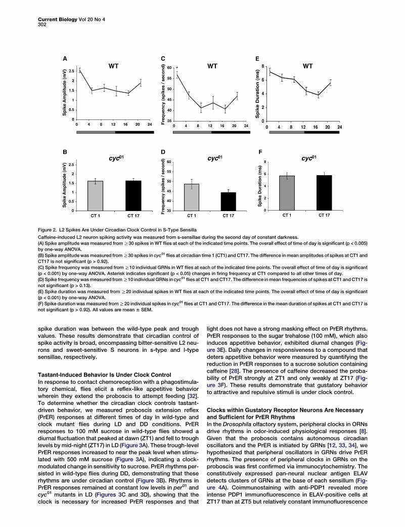
Figure 2. L2 Spikes Are Under Circadian Clock Control in S-Type Sensilla
Caffeine-induced L2 neuron spiking activity was measured from s-sensillae during the second day of constant darkness.
(A) Spike amplitude was measured from R30 spikes in WT flies at each of the indicated time points. The overall effect of time of day is significant (p < 0.005)
by one-way ANOVA.
(B) Spike amplitude was measured from R30 spikes in cyc01 flies at circadian time 1 (CT1) and CT17. The difference in mean amplitudes of spikes at CT1 and
CT17 is not significant (p > 0.92).
(C) Spike frequency was measured from R10 individual GRNs in WT flies at each of the indicated time points. The overall effect of time of day is significant
(p < 0.001) by one-way ANOVA. Asterisk indicates significant (p < 0.05) changes in firing frequency at CT1 compared to all other times of day.
(D) Spike frequency was measured from R10 individual GRNs in cyc01 flies at CT1 and CT17. The difference in mean frequencies of spikes at CT1 and CT17 is
not significant (p > 0.13).
(E) Spike duration was measured from R20 individual spikes in WT flies at each of the indicated time points. The overall effect of time of day is significant
(p < 0.001) by one-way ANOVA.
(F) Spike duration was measured from R20 individual spikes in cyc01 flies at CT1 and CT17. The difference in the mean duration of spikes at CT1 and CT17 is
not significant (p > 0.92). All values are mean 6 SEM.
Current Biology Vol 20 No 4302
spike duration was between the wild-type peak and troughvalues. These results demonstrate that circadian control ofspike activity is broad, encompassing bitter-sensitive L2 neu-rons and sweet-sensitive S neurons in s-type and l-typesensillae, respectively.
Tastant-Induced Behavior Is Under Clock Control
In response to contact chemoreception with a phagostimula-tory chemical, flies elicit a reflex-like appetitive behaviorwherein they extend the proboscis to attempt feeding [32].To determine whether the circadian clock controls tastant-driven behavior, we measured proboscis extension reflex(PrER) responses at different times of day in wild-type andclock mutant flies during LD and DD conditions. PrERresponses to 100 mM sucrose in wild-type flies showed adiurnal fluctuation that peaked at dawn (ZT1) and fell to troughlevels by mid-night (ZT17) in LD (Figure 3A). These trough-levelPrER responses increased to near the peak level when stimu-lated with 500 mM sucrose (Figure 3A), indicating a clock-modulated change in sensitivity to sucrose. PrER rhythms per-sisted in wild-type flies during DD, demonstrating that theserhythms are under circadian control (Figure 3B). Rhythms inPrER responses remained at constant low levels in per01 andcyc01 mutants in LD (Figures 3C and 3D), showing that theclock is necessary for increased PrER responses and that
light does not have a strong masking effect on PrER rhythms.PrER responses to the sugar trehalose (100 mM), which alsoinduces appetitive behavior, exhibited diurnal changes (Fig-ure 3E). Daily changes in responsiveness to a compound thatdeters appetitive behavior were measured by quantifying thereduction in PrER responses to a sucrose solution containingcaffeine [28]. The presence of caffeine decreased the proba-bility of PrER strongly at ZT1 and only weakly at ZT17 (Fig-ure 3F). These results demonstrate that gustatory behaviorto attractive and repulsive stimuli is under clock control.
Clocks within Gustatory Receptor Neurons Are Necessaryand Sufficient for PrER Rhythms
In the Drosophila olfactory system, peripheral clocks in ORNsdrive rhythms in odor-induced physiological responses [8].Given that the proboscis contains autonomous circadianoscillators and the PrER is initiated by GRNs [12, 33, 34], wehypothesized that peripheral oscillators in GRNs drive PrERrhythms. The presence of peripheral clocks in GRNs on theproboscis was first confirmed via immunocytochemistry. Theconstitutively expressed pan-neural nuclear antigen ELAVdetects clusters of GRNs at the base of each sensillum (Fig-ure 4A). Coimmunostaining with anti-PDP1 revealed moreintense PDP1 immunofluorescence in ELAV-positive cells atZT17 than at ZT5 but relatively constant immunofluorescence

Figure 3. Drosophila Display Circadian Rhythms in Gusta-
tory Behavioral Responses
(A and B) Proboscis extension reflex (PrER) responses to
100 mM sucrose (black line) or 500 mM sucrose (filled
square) were measured in WT flies during LD cycles (A) or
the first day of constant darkness (B). The overall effects of
time of day in LD (A) and constant darkness (B) are significant
(p < 0.001) by one-way ANOVA. Asterisks indicate significant
(p < 0.05) changes in PrER behavior at ZT1 and ZT17 (A) or
CT1 (B) compared to all other times of day.
(C and D) PrER responses to 100 mM sucrose were
measured in cyc01 (C) and per01 (D) flies at ZT1 and ZT17.
The difference in mean PrER responses at ZT1 and ZT17
are not significant (p > 0.30) in cyc01 or per01 flies.
(E) PrER responses to 100 mM trehalose were measured in
WT flies at ZT1 and ZT17. Asterisks indicate a significant
(p < 0.001) reduction in PrER responses at ZT17 compared
to ZT1.
(F) Decrease in PrER responses to a 100 mM sucrose solu-
tion containing 10 mM caffeine versus 100 mM sucrose alone
in WT flies at ZT1 and ZT17. Asterisk indicates significant
(p = 0.025) decrease in PrER inhibition by caffeine at ZT17
compared to ZT1. All values are mean 6 SEM.
Gustatory Rhythms in Drosophila303
intensity in ELAV-negative cells (Figure 4A). All PDP1 immu-nostaining was eliminated in the PDP13-specific mutantPdp13135 [35], indicating that only PDP13 is expressed in theseELAV-positive and -negative cells. Rhythmic PDP13 stainingin ELAV-positive cells is consistent with PDP13 cycling inbrain and peripheral oscillator cells [35–37] and demonstratesthat the GRNs within gustatory sensilla contain circadianoscillators.
To test the idea that local oscillators within GRNs are neces-sary for PrER rhythms, we expressed a dominant-negativeform of CYC (CYCDN) to abolish clock function in the sweet-sensitive S neurons that elicit PrER behavior in response tosucrose [33]. Under LD conditions, PrER responses were abol-ished in flies containing both the Gr5a-Gal4 driver, which isexpressed in S neurons [34], and UAS-cycDN responder, butnot in control flies containing the Gr5a-Gal4 driver or UAS-cycDN responder alone (Figure 4B). This result demonstratesthat circadian oscillators in GRNs are required for PrERrhythms.
We then sought to determine whether local clocks in GRNsare sufficient for PrER rhythms by generating flies with circa-dian oscillators only in S neurons. For this, oscillator functionwas rescued exclusively in S neurons by using Gr5a-Gal4 todrive UAS-cyc expression in cyc01 flies. PrER behavior incyc01 flies containing both Gr5a-Gal4 and UAS-cyc wasrhythmic, whereas cyc01 flies containing Gr5a-Gal4 or UAS-cyc alone were arrhythmic (Figure 4C). These data demon-strate that clocks in GRNs are sufficient for PrER rhythms.Because clocks are not present elsewhere in cyc01 flies con-taining Gr5a-Gal4 and UAS-cyc, these data also show thatcentral clocks in the brain are not necessary for PrER rhythms.
Taken together, these results demonstrate thatGRN clocks are necessary and sufficient tocontrol rhythms in gustatory behavior.
Cycling GPRK2 Levels Drive PrER Behavior
RhythmsBecause circadian oscillators in GRNs are suffi-cient for PrER rhythms, the clock output pathwaythat controls this rhythm must also reside inGRNs. To identify a clock-controlled molecule
involved in gustatory signal transduction, we focused ourattention on GPRK2, which is required for rhythms in olfactoryresponses in Drosophila [14, 38]. Western blot analysis showsthat GPRK2 protein is expressed in the proboscis of wild-typeflies but that GPRK2 levels are reduced in the Gprk206936
mutant (Figure 5A) [39]. In contrast to the two GPRK2 isoformsthat are detected in antennae [38], only one GPRK2 band isseen in the proboscis of wild-type and Gprk206936 flies (Fig-ure 5A). The levels of GPRK2 cycled w2-fold in wild-typeproboscises with a peak at ZT17 and a trough at ZT1 (Fig-ure 5B). GPRK2 cycling was abolished in per01 and cyc01 flies(data not shown), indicating circadian clock control. GPRK2immunostaining was detected in the cell body of GRNs atthe base of taste hairs that were coimmunostained withELAV (Figure 5C). GPRK2 was also detected in the shaft ofthe sensillar hair, which contains GRN dendritic projectionsand possibly support cells closely associated with GRNs(Figure 5C).
The levels of GPRK2 in the proboscis are lowest when PrERresponses peak and peak when PrER responses are lowest.This antiphasic relationship suggests that GPRK2 levels maycontrol rhythmic PrER behavior. Consistent with this possi-bility, PrER responses to sucrose and trehalose were con-stantly repressed when GPRK2 was overexpressed but werealways high in the Gprk206936 mutant (Figures 5D and 5E).Thus, these experiments argue that cycling GPRK2 levels driverhythms in PrER behavior. Given that PrER responses areconstantly high in Gprk206936 flies and that spike amplitude,frequency, and duration in GRNs cycle in parallel to PrERresponses, we reasoned that these spike activity parametersshould be constant and relatively high in the GRNs of

Figure 4. Oscillators within Gustatory Receptor Neurons Are Necessary and Sufficient for PrER Rhythms
(A) PDP1 and ELAV immunostaining in GRNs of Pdp13135 mutant flies collected at ZT17 and wild-type flies collected at CT5 and CT17. Anti-ELAV immunos-
taining (ELAV) is shown in red, anti-PDP1 immunostaining (PDP1) is shown in green, and colocalized PDP1 and ELAV immunostaining (ELAV + PDP1) is
shown in yellow.
(B) PrER responses were measured at ZT1 and ZT17 in wild-type flies bearing the Gr5a-Gal4, UAS-cycDN, or Gr5a-Gal4 + UAS-cycDN transgenes. The differ-
ences in mean PrER responses at ZT1 and ZT17 are significant (p < 0.001) in flies containing Gr5a-Gal4 or UAS-cycDN alone but are not significant (p < 0.30)
in flies carrying Gr5a-Gal4 + UAS-cycDN.
(C) PrER responses were measured at ZT1 and ZT17 in cyc01 flies carrying the Gr5a-Gal4, UAS-cyc, or UAS-cyc + Gr5a-Gal4 transgenes. There are no signif-
icant (p > 0.30) differences in PrER responses at ZT17 and ZT1 in cyc01 flies carrying either UAS-cyc or Gr5a-Gal4. The differences in mean PrER responses at
ZT1 and ZT17 are significant (p < 0.001) in cyc01 flies carrying UAS-cyc + Gr5a-Gal4. Asterisks denote a significant (p < 0.05) change in PrER responses
between ZT17 and ZT1. All values are mean 6 SEM.
Current Biology Vol 20 No 4304
Gprk206936 flies. Rhythms in spike amplitude, frequency, andduration were all abolished in Gprk206936 flies, in which spikefrequency was close to the wild-type peak, but spike ampli-tude was midway between the wild-type peak and trough,and spike duration was only modestly higher than the wild-type trough (Figure S2). These results suggest that certainaspects of GRN cell activity, particularly spike frequencyand, to a lesser extent, spike amplitude, correlate with PrERbehavior.
GPRK2 mediates circadian rhythms in the subcellular local-ization of Drosophila odorant receptors (ORs) [38]. BecauseDrosophila ORs and GRs belong to the same family of insectchemoreceptor proteins, we wished to determine whetherGPRK2-dependent regulation of rhythmic PrER responsesrelies on GRs. A mutant that removes all six Drosophila Gr64genes (DGr64) shows drastically reduced PrER responsesto most sugars [33]. When DGr64 flies were stimulated with100 mM sucrose at ZT1 and ZT17, their PrER responseswere not rhythmic, but DGr64 mutants rescued by a transgenecontaining the entire Gr64 gene cluster [33] recovered PrERrhythms (Figure 5F). Overexpression of the sucrose receptorGr64a resulted in arrhythmic PrER responses that were nearthe circadian peak value (Figure 5F). Likewise, deletion ofGr5a, which is required for responses to trehalose [40, 41],resulted in constant low PrER responses to trehalose, whereasGR5a overexpression resulted in constant high responses to
trehalose (data not shown). These results imply that GRs arenot only required to detect tastants but are also necessaryfor sustaining rhythms in tastant-evoked appetitive behavior.
Gustatory Receptor Neuron Clocks Regulate FeedingBoth external sensory cues and internal metabolic statecontribute to the regulation of feeding [23]. Recent work inDrosophila has shown that loss of clock function in fat bodyincreases feeding by altering metabolic state [25]. We soughtto determine whether GRN oscillators also regulate feedingbecause they modulate taste sensitivity. Food ingestion wasmeasured with a blue food dye that can be quantified spectro-photometrically and via the capillary feeder (CAFE) assay[25, 42]. Under LD conditions, flies that express CYCDN insweet-sensitive Gr5a neurons consumed significantly morefood over 24 hr than controls carrying the driver or respondertransgenes (Figures 6A and 6B). Moreover, food intake washigher in the morning (ZT0–4) than in the evening (ZT12–16),demonstrating that increased consumption is not uniformduring a diurnal cycle (Table S1). This result shows that circa-dian clocks in a subset of GRNs act to limit the amount of foodintake.
Although flies that lack clocks in Gr5a neurons eat more,they do not gain weight compared to controls carryingthe driver or responder transgenes alone (Figure 6C). Never-theless, loss of clock function in Gr5a neurons led to

Figure 5. G Protein-Coupled Receptor Kinase 2 and Gustatory Receptor Expression Levels Control Rhythms in PrER Behavior
(A) Western blot showing G protein-coupled receptor kinase 2 (GPRK2) expression in proboscises from WT and Gprk206936 mutant (Gprk2 Mut) flies and
antennae (Ant) from WT flies. GPRK2 runs as two isoforms in antennae and one isoform in proboscises. ACTIN was used as a loading control.
(B) Western blot showing GPRK2 levels in proboscises of WT flies collected at the indicated times during an LD cycle. The GPRK2:ACTIN values at ZT1, ZT5,
ZT9, ZT13, and ZT21 are relative to the value at ZT17, which was set to 1.0. Each time point represents the mean of three independent experiments. The
overall effect of time of day is significant (p < 0.005) by one-way ANOVA.
(C) GPRK2 and ELAV immunostaining in labellar GRNs from WT and Gprk2 mutant flies. Anti-GPRK2 immunoreactivity is shown in green, and anti-ELAV
signal is shown in red. Scale bars represent 10 mm. Gray arrows represent GPRK2 localization in the cytosol; white arrows represent GPRK2 immunostaining
in the shaft of a sensillar hair.
(D and E) PrER responses to sucrose and trehalose were measured at ZT1 and ZT17 in WT flies carrying Gr5a-Gal4 and UAS-Gprk2, which overexpress
GPRK2 in S neurons (GPRK2 OE), and in Gprk206936 mutants (Gprk2 mutant). Mean PrER responses to sucrose (D) and trehalose (E) at ZT1 and ZT17
were not significant (p > 0.16) and remained at constant low levels in Gprk2 mutant flies and constant high levels in GPRK2 OE flies. For each genotype,
three or more groups of R10 flies were tested for PrER responses to sucrose and trehalose at each time point. Asterisks denote a significant (p < 0.05)
change in PrER responses between ZT17 and ZT1.
(F) PrER responses to 100 mM sucrose in Gr64 mutant (R1/+;R2/+;DGr64/DGr64), Gr64 rescue (R1/+;R2/+;DGr64/DGr64 carrying one copy of the UAS-
Gr64abcd_GFP_f reporter), and GR64a-overexpressing flies at ZT1 and ZT17. The differences in mean responses at ZT1 and ZT17 are not significant in
Gr64 mutants (p > 0.90) or GR64a-overexpressing flies (p > 0.05) but are significant (p < 0.001) in Gr64 rescue flies. All values are mean 6 SEM. As with
PrER responses, lower GPRK2 expression in Gprk2 mutant flies disrupts rhythms in GRN spike activity (Figure S2).
Gustatory Rhythms in Drosophila305
a considerable increase in triglyceride and glycogen content(Figures 6D and 6E). Increased triglyceride and glycogencontent in flies lacking clocks in Gr5a neurons was associatedwith higher levels of locomotor activity over a 24 hr period(Figure 6F), in which increased activity levels coincided withincreased feeding (Table S1). Thus, flies lacking clocks inGr5a neurons eat more and store triglycerides and glycogeneven though they expend more energy to fuel increased loco-motor activity.
Discussion
Circadian Rhythms in Gustatory Physiology and BehaviorThe ability to detect and discriminate tastants provides asurvival advantage to animals ranging from flies to humansbecause chemosensation is universally employed to identifyfood sources and reject harmful substances [34]. Our resultsdemonstrate for the first time that this fundamental sensoryprocess is controlled by the circadian clock. The amplitude,frequency, and duration of voltage spikes evoked by attractive
and aversive tastants peak around dawn in multiple classes ofGRNs and different types of taste sensilla (Figure 1; Figure 2).Ventrolateral clock neurons (LNvs) in the fly brain showrhythms in firing rate that also peak during the day [43–45],but whether a common mechanism controls rhythms in theelectrical properties of GRNs, LNvs, and ORNs is not known.
The PrER is a direct, robust, and all-or-none indicator ofa fly’s attraction and motivation to ingest a substance [32].PrER response levels change as a function of time of day(Figure 3), in which the phase of this taste-behavior rhythmmirrors rhythms in the rate, amplitude, and duration of GRNimpulses in wild-type flies under LD and DD conditions. Theseresults suggest that spike amplitude and duration, in additionto spike frequency, are dynamic neuronal response propertiescapable of influencing sensitivity to chemical cues. Circadianrhythms in spike amplitude are also seen in the olfactorysystem of flies [14], where the phases of these electrophysio-logical rhythms coincide with rhythms in odor-dependentchemotactic behavior that peak during mid-night [15]. Ourdata suggest that rhythms in spike properties of GRNs tune

Figure 6. Circadian Clocks in Gr5a Neurons Regulate Feeding, Food Storage, and Activity
(A and B) Relative food intake was measured after 24 hr of feeding by quantifying the amount of blue food dye ingested (A) or by measuring the food
consumed via the capillary feeder assay (B) (see Experimental Procedures). Flies carrying both the Gr5a-Gal4 and UAS-cycDN transgenes show significantly
(p < 0.02) increased feeding compared to control flies containing either the Gr5a-Gal4 or the UAS-cycDN transgene.
(C) The body weight of flies carrying both the Gr5a-Gal4 and UAS-cycDN transgenes was not different (p > 0.50) from control flies bearing the Gr5a-Gal4 or
UAS-cycDN transgenes.
(D) Overall activity was measured as the number of times flies crossed an infrared light beam during a 24 hr period. Flies carrying both the Gr5a-Gal4 and
UAS-cycDN transgenes show significantly (p < 0.001) increased activity compared to control flies containing either the Gr5a-Gal4 or the UAS-cycDN trans-
gene.
(E) Glycogen levels are significantly (p < 0.04) higher in flies carrying both the Gr5a-Gal4 and UAS-cycDN transgenes than control flies bearing the Gr5a-Gal4
or UAS-cycDN transgenes.
(F) Triglyceride levels are significantly (p < 0.001) higher in flies carrying both the Gr5a-Gal4 and UAS-cycDN transgenes than control flies bearing the Gr5a-
Gal4 or UAS-cycDN transgenes. Error bars represent mean 6 SEM. Increased feeding and activity in flies lacking clocks in Gr5a neurons is not uniform over
the circadian cycle (Table S1).
Current Biology Vol 20 No 4306
the activity of downstream neurons in such a way that behav-ioral responses to the same stimulus show clock-regulatedplasticity. Given that PrER behavior likely involves localcircuitry with limited processing [20], it is surprising that this‘‘hardwired’’ behavior is subjected to daily functional remodel-ing by the clock and that a straightforward predictive relation-ship emerges between rhythms in GRN responses andrhythms in tastant-driven appetitive behavior.
Control of Proboscis Extension Reflex RhythmsWe show that circadian oscillators in GRNs are necessary andsufficient for PrER rhythms (Figure 4). To our knowledge, this isthe first example in which a single population of peripheraloscillator neurons is shown to generate behavioral rhythms.GRNs from the proboscis project primarily into the centralportion of the subesophageal ganglion (SOG) [46]. A numberof SOG motor neurons are known to innervate muscles inproboscis and pharynx [47], which may be indirectly controlledby the GRN clock on a daily basis.
In Drosophila, GRNs express GPRK2, and rhythms inGPRK2 abundance are antiphase relative to PrER rhythms(Figures 5A–5F). Analyses of Gprk2 mutant and GPRK2-over-expression flies suggest that GPRK2 levels drive rhythms in
PrER responses and correspond to GRN spike frequencyand, to a lesser extent, spike amplitude (Figures 5D and 5E;Figure S2). Rhythms in PrER responses are also abolishedby altering GR levels; increasing or decreasing GR64 orGR5a levels results in constant high or low PrER responses,respectively (Figure 5F; data not shown). PrER responses areconstantly low in Gr64 and Gr5a deletion mutants andGPRK2-overexpression flies but are constantly high in GR64-and GR5a-overexpression flies and Gprk2 mutants, whichargues that the balance between GR and GPRK2 abundancedetermines PrER response levels. Although the phase ofGPRK2 cycling is the same in ORNs and GRNs, olfactoryresponses (e.g., spike amplitude) peak when gustatoryresponses are low, and gustatory responses peak when olfac-tory responses are low [14] (Figure 1; Figure 2). The differencein gustatory and olfactory response phases implies thatGPRK2 has distinctly different activities in the olfactory andgustatory systems.
In ORNs, GPRK2 rhythmically promotes dendritic localiza-tion of ORs [38]. ORs and GRs are both seven transmembranedomain proteins that belong to the same superfamily of insectchemoreceptor proteins [20]. It is tempting to speculate thatGPRK2 directly phosphorylates GRs, thereby controlling the

Gustatory Rhythms in Drosophila307
abundance or activity of GR-dependent channels or ligand-gated GR channels in GRNs. This rhythmic regulation of neu-ronal excitability may be translated into rhythms in spikeamplitude, frequency, and duration. Thus, the PrER rhythm islikely a behavioral correlate of certain features of electrophys-iological rhythms such as spike frequency and perhaps spikeamplitude.
Functional Significance of Gustatory RhythmsPeripheral oscillators may play widespread roles in sensoryprocessing, such that the perceived meaning of a sensoryinput is determined not just by the modality of the signal orits intensity but also by the circadian time when the signal isregistered. Our results indicate that the clock tunes thegustatory system to a higher gain level in the morning. Thismay allow the fly to temporally couple the morning bout ofactivity with food-detection machinery that works betterat dawn, leading to increased feeding. This strategy can mini-mize energy expenditure by shutting down hardwired tasteresponses to weak stimuli (behavioral noise) at times whenflies are resting and selectively boosting acuity at timeswhen they are wakeful. Interestingly, the acrophase of feedingrhythms coincides with the early morning peak in gustatoryresponse rhythms [25].
Social experience, which can influence behavior in Dro-sophila [48], is communicated by chemosensory cues suchas pheromones. Moreover, circadian clocks in oenocytesregulate rhythms in the abundance of male pheromones,including 7-tricosene [7], that are detected by GRNs [29, 49].Thus, local clocks in oenocytes temporally gate the productionof male pheromones, and local clocks in GRNs may temporallygate pheromone reception and signaling. Such a system couldfunction to define a time window for social interactions andmating, and the resulting social experience may in turn influ-ence clocks that control pheromone production and/or gusta-tory sensitivity.
Feeding Is Modulated by the Gustatory ClockIncreased feeding in flies that lack circadian clock function inGr5a neurons suggests that clocks in these cells act to restrictfood consumption (Figure 6). Given that clock function wascompromised in GRNs that detect sugars [33, 50], increasedfood consumption may be due to a change in taste sensitivity.However, loss of clock function in Gr5a neurons decreasesPrER responses at ZT1 and increases PrER responses atZT17 (Figure 4), indicating that the clock increases taste sensi-tivity in the morning and decreases it at night. When feedingwas measured under the same conditions as PrER responses,food intake increased as PrER responses decreased in themorning, and food intake decreased as PrER responsesincreased in the evening (Table S1). In control genotypesthat have clocks in Gr5a neurons, CAFE assays show thatfood intake is similar in the morning and the evening, consis-tent with estimates of food intake at different times of day insingle flies via a novel assay that measures the proportion oftime flies were observed extending their proboscis to feed[51]. Our results suggest that increased feeding is not due toaltered taste sensitivity in flies lacking clocks in Gr5a neurons.It is possible that the increased food intake at ZT0–4 and thedecreased food intake at ZT16–20 in flies lacking Gr5a neuronoscillators is due to metabolic feedback on tissues with func-tional clocks. Consistent with this possibility, food intake inClkJrk and cyc01 mutant flies is the same as that in wild-typeflies [25].
Flies lacking clocks in Gr5a neurons do not show a measur-able gain in weight (Figure 6C), presumably because of a highfixed level of cuticle, protein, and water weight. However, theirtriglyceride and glycogen content increases substantially(Figures 6D and 6E), indicating that the higher amounts offood consumed are being stored. Given this increase in foodstorage, it was surprising that flies lacking clocks in Gr5aneurons were also more active (Figure 6F). Increased activityis typically observed when starved flies are searching forfood [52], yet flies lacking clocks in Gr5a neurons consumemore food than wild-type flies (Figures 6A and 6B). The lossof clocks in Gr5a neurons may mimic starvation conditions,particularly during the day, when feeding is increased andPrER responses are relatively low compared to wild-type flies(Table S1; Figure 4). At night, even though PrER responses arehigher in flies lacking clocks in Gr5a neurons than in wild-typeflies, activity is already low, and feeding is even lower than inwild-type flies. Thus, loss of clock function in Gr5a neuronsmay produce starvation signals during the day, therebyincreasing activity and feeding, but not at night, becausefeeding is decreased and flies are already inactive.
Experimental Procedures
Fly Strains
Zero- to seven-day-old flies reared on standard cornmeal media were
entrained for 3 days in 12 hr light:12 hr dark cycles at 25�C. Lights were
turned on at ZT0 and off at ZT12. Canton-S was used as our wild-type strain.
The Gr5a-Gal4 driver [34] and the UAS-Gr64a [53], UAS-Gr5a [40], UAS-
cycDN [8], UAS-cyc [8], and UAS-Gprk2 [38] responders were described
previously. These experiments also employed the P element insertion
mutant Gprk206936 [39], the Pdp13-specific deletion mutant Pdp13135 [35],
the Gr5a deletion mutant DEP(X)-5 [40], the DGr64 mutant (R1/+;R2/+;
DGr64/DGr64) that lacks all six Gr64 genes [33], and the transgenically
rescued DGr64 mutant (R1/+;R2/+;DGr64/DGr64 flies carrying one copy of
the UAS-Gr64abcd_GFP_f reporter) [33].
Western Blotting and Immunostaining
Thirty to thirty-five proboscises were dissected from flies entrained for at
least three LD cycles. Western blots were processed as described [38].
Blots were probed with anti-GPRK2 antibody (1:1000 dilution) and anti-actin
antibody (Sigma-Aldrich, 1:10000 dilution) and visualized via enhanced
chemiluminescence (Amersham). Immunostaining was carried out on
cryosectioned proboscises as detailed in the Supplemental Experimental
Procedures.
Single-Sensillum Recording
Male flies (3–10 days old) entrained to LD cycles for R3 days were collected
during LD or the second day of DD and mounted, and the proboscis was
immobilized. Individual labellar sensillae were observed under 12003
magnification. Recordings in the dark were made with a <600 nm filter.
The indifferent electrode was inserted into the eye. The recording electrode
contained tastant dissolved in 1 mM KCl and was used to stimulate
a sensillum by physical contact with the tip of that sensillum. All recordings
with a given genotype and tastant were performed at least six times per time
point for R6 flies. A new group of flies was recorded at each time point.
Sucrose (100 mM) was used to stimulate S cells in accessible l-type sensilla,
which respond to sugars in an identical manner [30]. Caffeine (10 mM) was
used to stimulate s6 and s2 sensilla, whose L2 neurons are responsive to
bitter compounds [30]. The number of spikes initiated by the tastant was
counted manually over 500 ms duration beginning 50 ms after the onset
of stimulation. Spike traces were analyzed with Axoscope (Axon) software
in offline mode, in which the peak and trough values of individual spikes
were used to compute amplitude. The time elapsed between the peak
and trough values for an activity spike was used as a measure of spike dura-
tion [54].
Proboscis Extension Reflex Assay
Three- to seven-day-old male flies that had been entrained to LD cycles
for R3 days were starved for 24 hr, collected at different times during LD

Current Biology Vol 20 No 4308
or the first day of DD, mounted on a slide, and allowed to recover for 30 min.
Proboscis extension in response to 100 mM sucrose and 100 mM trehalose
was recorded as described [33], with minor modifications detailed in the
Supplemental Experimental Procedures.
Feeding Assays
Three- to ten-day-old male flies entrained for at least 3 LD cycles were given
food containing 5% sucrose, 1% low-melting-point agarose, and 0.5% bril-
liant blue FCF (Wako) for 24 hr starting at ZT12. Flies were then collected and
prepared for quantification of blue dye ingestion as described [25]. For
a given genotype, at least six independent experiments, each set consisting
of 10 flies, were carried out. CAFE assays were used to measure feeding
behavior of grouped fruit flies [25, 42]. For each genotype, CAFE assays
were conducted as described [25], except that flies were habituated to
feeding from glass capillaries for 24 hr and feeding was measured over
4 hr. CAFE assays were repeated at least five times for each data point.
Levels of glycogen and triglycerides were measured as previously
described [25].
Activity Measurement
For each line, 7- to 10-day-old male flies were entrained for at least 3 days in
LD cycles and placed in Drosophila activity monitors (Trikinetics). Activity
was measured by counting the number of infrared beam breaks every
10 min and was analyzed with Clocklab software.
Statistical Analysis
Statistical analysis was done with Statistica (Statsoft). Analysis of the
effects of time of day was examined by one-way analysis of variance
(ANOVA). Welch’s ANOVA was used for heteroscedastic data set, provided
Levene’s test indicated unequal variances. Post hoc comparisons were
done with Scheffe’s test (a = 0.05). Unpaired Student’s t test (two-tailed)
was used to compare values at peak and trough time points.
Supplemental Information
Supplemental Information includes Supplemental Experimental Proce-
dures, two figures, and one table and can be found with this article online
at doi:10.1016/j.cub.2009.12.055.
Acknowledgments
We are thankful to H. Amrein, C. Montell, and A. Dahanukar for generously
providing fly strains. We would also like to thank T. Tanimura and K. Ueno
for their valuable technical suggestions. This work was supported by funds
from Texas A&M University to P.E.H.
Received: July 27, 2009
Revised: December 18, 2009
Accepted: December 22, 2009
Published online: February 11, 2010
References
1. Bell-Pedersen, D., Cassone, V.M., Earnest, D.J., Golden, S.S., Hardin,
P.E., Thomas, T.L., and Zoran, M.J. (2005). Circadian rhythms from
multiple oscillators: Lessons from diverse organisms. Nat. Rev. Genet.
6, 544–556.
2. Hardin, P.E. (2005). The circadian timekeeping system of Drosophila.
Curr. Biol. 15, R714–R722.
3. Nitabach, M.N., and Taghert, P.H. (2008). Organization of the Drosophila
circadian control circuit. Curr. Biol. 18, R84–R93.
4. Sheeba, V., Kaneko, M., Sharma, V.K., and Holmes, T.C. (2008). The
Drosophila circadian pacemaker circuit: Pas de deux or Tarantella?
Crit. Rev. Biochem. Mol. Biol. 43, 37–61.
5. Beaver, L.M., Gvakharia, B.O., Vollintine, T.S., Hege, D.M., Stanewsky,
R., and Giebultowicz, J.M. (2002). Loss of circadian clock function
decreases reproductive fitness in males of Drosophila melanogaster.
Proc. Natl. Acad. Sci. USA 99, 2134–2139.
6. Ito, C., Goto, S.G., Shiga, S., Tomioka, K., and Numata, H. (2008).
Peripheral circadian clock for the cuticle deposition rhythm in
Drosophila melanogaster. Proc. Natl. Acad. Sci. USA 105, 8446–8451.
7. Krupp, J.J., Kent, C., Billeter, J.C., Azanchi, R., So, A.K., Schonfeld, J.A.,
Smith, B.P., Lucas, C., and Levine, J.D. (2008). Social experience
modifies pheromone expression and mating behavior in male
Drosophila melanogaster. Curr. Biol. 18, 1373–1383.
8. Tanoue, S., Krishnan, P., Krishnan, B., Dryer, S.E., and Hardin, P.E.
(2004). Circadian clocks in antennal neurons are necessary and suffi-
cient for olfaction rhythms in Drosophila. Curr. Biol. 14, 638–649.
9. Emery, I.F., Noveral, J.M., Jamison, C.F., and Siwicki, K.K. (1997).
Rhythms of Drosophila period gene expression in culture. Proc. Natl.
Acad. Sci. USA 94, 4092–4096.
10. Giebultowicz, J.M., Stanewsky, R., Hall, J.C., and Hege, D.M. (2000).
Transplanted Drosophila excretory tubules maintain circadian clock
cycling out of phase with the host. Curr. Biol. 10, 107–110.
11. Krishnan, B., Dryer, S.E., and Hardin, P.E. (1999). Circadian rhythms in
olfactory responses of Drosophila melanogaster. Nature 400, 375–378.
12. Plautz, J.D., Kaneko, M., Hall, J.C., and Kay, S.A. (1997). Independent
photoreceptive circadian clocks throughout Drosophila. Science 278,
1632–1635.
13. Giebultowicz, J.M., and Hege, D.M. (1997). Circadian clock in Malpigh-
ian tubules. Nature 386, 664.
14. Krishnan, P., Chatterjee, A., Tanoue, S., and Hardin, P.E. (2008). Spike
amplitude of single-unit responses in antennal sensillae is controlled
by the Drosophila circadian clock. Curr. Biol. 18, 803–807.
15. Zhou, X., Yuan, C., and Guo, A. (2005). Drosophila olfactory response
rhythms require clock genes but not pigment dispersing factor or lateral
neurons. J. Biol. Rhythms 20, 237–244.
16. Granados-Fuentes, D., Tseng, A., and Herzog, E.D. (2006). A circadian
clock in the olfactory bulb controls olfactory responsivity. J. Neurosci.
26, 12219–12225.
17. Merlin, C., Lucas, P., Rochat, D., Francois, M.C., Maıbeche-Coisne, M.,
and Jacquin-Joly, E. (2007). An antennal circadian clock and circadian
rhythms in peripheral pheromone reception in the moth Spodoptera
littoralis. J. Biol. Rhythms 22, 502–514.
18. Nordin, S., Lotsch, J., Murphy, C., Hummel, T., and Kobal, G. (2003).
Circadian rhythm and desensitization in chemosensory event-related
potentials in response to odorous and painful stimuli. Psychophysi-
ology 40, 612–619.
19. Page, T.L., and Koelling, E. (2003). Circadian rhythm in olfactory
response in the antennae controlled by the optic lobe in the cockroach.
J. Insect Physiol. 49, 697–707.
20. Vosshall, L.B., and Stocker, R.F. (2007). Molecular architecture of smell
and taste in Drosophila. Annu. Rev. Neurosci. 30, 505–533.
21. Stocker, R.F. (1994). The organization of the chemosensory system in
Drosophila melanogaster: A review. Cell Tissue Res. 275, 3–26.
22. Christiensen, T. (2004). Methods in Insect Sensory Neuroscience (Boca
Raton, FL: CRC Press).
23. Melcher, C., Bader, R., and Pankratz, M.J. (2007). Amino acids, taste
circuits, and feeding behavior in Drosophila: Towards understanding
the psychology of feeding in flies and man. J. Endocrinol. 192, 467–472.
24. Lee, G., and Park, J.H. (2004). Hemolymph sugar homeostasis and star-
vation-induced hyperactivity affected by genetic manipulations of the
adipokinetic hormone-encoding gene in Drosophila melanogaster.
Genetics 167, 311–323.
25. Xu, K., Zheng, X., and Sehgal, A. (2008). Regulation of feeding and
metabolism by neuronal and peripheral clocks in Drosophila. Cell
Metab. 8, 289–300.
26. Bray, M.S., and Young, M.E. (2007). Circadian rhythms in the develop-
ment of obesity: Potential role for the circadian clock within the adipo-
cyte. Obes. Rev. 8, 169–181.
27. Turek, F.W., Joshu, C., Kohsaka, A., Lin, E., Ivanova, G., McDearmon, E.,
Laposky, A., Losee-Olson, S., Easton, A., Jensen, D.R., et al. (2005).
Obesity and metabolic syndrome in circadian clock mutant mice.
Science 308, 1043–1045.
28. Amrein, H., and Thorne, N. (2005). Gustatory perception and behavior in
Drosophila melanogaster. Curr. Biol. 15, R673–R684.
29. Ebbs, M.L., and Amrein, H. (2007). Taste and pheromone perception in
the fruit fly Drosophila melanogaster. Pflugers Arch. 454, 735–747.
30. Hiroi, M., Marion-Poll, F., and Tanimura, T. (2002). Differentiated
response to sugars among labellar chemosensilla in Drosophila. Zoo-
log. Sci. 19, 1009–1018.
31. Hochner, B., Klein, M., Schacher, S., and Kandel, E.R. (1986). Action-
potential duration and the modulation of transmitter release from the
sensory neurons of Aplysia in presynaptic facilitation and behavioral
sensitization. Proc. Natl. Acad. Sci. USA 83, 8410–8414.
32. Dethier, V.G. (1976). The Hungry Fly (Cambridge, MA: Harvard University
Press).

Gustatory Rhythms in Drosophila309
33. Slone, J., Daniels, J., and Amrein, H. (2007). Sugar receptors in
Drosophila. Curr. Biol. 17, 1809–1816.
34. Wang, Z., Singhvi, A., Kong, P., and Scott, K. (2004). Taste representa-
tions in the Drosophila brain. Cell 117, 981–991.
35. Zheng, X., Koh, K., Sowcik, M., Smith, C.J., Chen, D., Wu, M.N., and Seh-
gal, A. (2009). An isoform-specific mutant reveals a role of PDP1 epsilon
in the circadian oscillator. J. Neurosci. 29, 10920–10927.
36. Benito, J., Zheng, H., and Hardin, P.E. (2007). PDP1epsilon functions
downstream of the circadian oscillator to mediate behavioral rhythms.
J. Neurosci. 27, 2539–2547.
37. Cyran, S.A., Buchsbaum, A.M., Reddy, K.L., Lin, M.C., Glossop, N.R.,
Hardin, P.E., Young, M.W., Storti, R.V., and Blau, J. (2003). vrille,
Pdp1, and dClock form a second feedback loop in the Drosophila circa-
dian clock. Cell 112, 329–341.
38. Tanoue, S., Krishnan, P., Chatterjee, A., and Hardin, P.E. (2008). G
protein-coupled receptor kinase 2 is required for rhythmic olfactory
responses in Drosophila. Curr. Biol. 18, 787–794.
39. Schneider, L.E., and Spradling, A.C. (1997). The Drosophila G-protein-
coupled receptor kinase homologue Gprk2 is required for egg morpho-
genesis. Development 124, 2591–2602.
40. Dahanukar, A., Foster, K., van der Goes van Naters, W.M., and Carlson,
J.R. (2001). A Gr receptor is required for response to the sugar trehalose
in taste neurons of Drosophila. Nat. Neurosci. 4, 1182–1186.
41. Ueno, K., Ohta, M., Morita, H., Mikuni, Y., Nakajima, S., Yamamoto, K.,
and Isono, K. (2001). Trehalose sensitivity in Drosophila correlates
with mutations in and expression of the gustatory receptor gene Gr5a.
Curr. Biol. 11, 1451–1455.
42. Ja, W.W., Carvalho, G.B., Mak, E.M., de la Rosa, N.N., Fang, A.Y., Liong,
J.C., Brummel, T., and Benzer, S. (2007). Prandiology of Drosophila and
the CAFE assay. Proc. Natl. Acad. Sci. USA 104, 8253–8256.
43. Cao, G., and Nitabach, M.N. (2008). Circadian control of membrane
excitability in Drosophila melanogaster lateral ventral clock neurons.
J. Neurosci. 28, 6493–6501.
44. Park, D., and Griffith, L.C. (2006). Electrophysiological and anatomical
characterization of PDF-positive clock neurons in the intact adult
Drosophila brain. J. Neurophysiol. 95, 3955–3960.
45. Sheeba, V., Gu, H., Sharma, V.K., O’Dowd, D.K., and Holmes, T.C.
(2008). Circadian- and light-dependent regulation of resting membrane
potential and spontaneous action potential firing of Drosophila circa-
dian pacemaker neurons. J. Neurophysiol. 99, 976–988.
46. Thorne, N., Chromey, C., Bray, S., and Amrein, H. (2004). Taste percep-
tion and coding in Drosophila. Curr. Biol. 14, 1065–1079.
47. Rajashekhar, K.P., and Singh, R.N. (1994). Neuroarchitecture of the tri-
tocerebrum of Drosophila melanogaster. J. Comp. Neurol. 349,
633–645.
48. Levine, J.D., Funes, P., Dowse, H.B., and Hall, J.C. (2002). Resetting the
circadian clock by social experience in Drosophila melanogaster.
Science 298, 2010–2012.
49. Lacaille, F., Hiroi, M., Twele, R., Inoshita, T., Umemoto, D., Maniere, G.,
Marion-Poll, F., Ozaki, M., Francke, W., Cobb, M., et al. (2007). An inhib-
itory sex pheromone tastes bitter for Drosophila males. PLoS ONE 2,
e661.
50. Marella, S., Fischler, W., Kong, P., Asgarian, S., Rueckert, E., and Scott,
K. (2006). Imaging taste responses in the fly brain reveals a functional
map of taste category and behavior. Neuron 49, 285–295.
51. Wong, R., Piper, M.D., Wertheim, B., and Partridge, L. (2009). Quantifi-
cation of food intake in Drosophila. PLoS ONE 4, e6063.
52. Sarov-Blat, L., So, W.V., Liu, L., and Rosbash, M. (2000). The Drosophila
takeout gene is a novel molecular link between circadian rhythms and
feeding behavior. Cell 101, 647–656.
53. Jiao, Y., Moon, S.J., and Montell, C. (2007). A Drosophila gustatory
receptor required for the responses to sucrose, glucose, and maltose
identified by mRNA tagging. Proc. Natl. Acad. Sci. USA 104, 14110–
14115.
54. Gur, M., Beylin, A., and Snodderly, D.M. (1999). Physiological properties
of macaque V1 neurons are correlated with extracellular spike ampli-
tude, duration, and polarity. J. Neurophysiol. 82, 1451–1464.
Top Related