


Languages
Pages
Legal


UNIVERSIDADE FEDERAL DE SANTA MARIA CENTRO DE CIÊNCIAS RURAIS
PROGRAMA DE PÓS-GRADUAÇÃO EM MEDICINA VETERINÁRIA
REGULAÇÃO DA EXPRESSÃO DO RECEPTOR AT2 E EFEITO DA ANGIOTENSINA II SOBRE A EXPRESSÃO DE GENES ENVOLVIDOS NO DESENVOLVIMENTO FOLICULAR E
OVULAÇÃO EM CÉLULAS DA GRANULOSA DE BOVINOS
TESE DE DOUTORADO
Valério Valdetar Marques Portela Junior
Santa Maria, RS, Brasil
2007

REGULAÇÃO DA EXPRESSÃO DO RECEPTOR AT2 E EFEITO
DA ANGIOTENSINA II SOBRE A EXPRESSÃO DE GENES ENVOLVIDOS NO DESENVOLVIMENTO FOLICULAR E
OVULAÇÃO EM CÉLULAS DA GRANULOSA DE BOVINOS
por
Valerio Valdetar Marques Portela Junior
Tese apresentada ao Curso de Mestrado do Programa de Pós-Graduação em Medicina Veterinária, Área de Concentração em
Fisiopatologia da Reprodução Animal, da Universidade Federal de Santa Maria (UFSM, RS), como requisito parcial para obtenção do grau de
Doutor em Medicina Veterinária.
Orientador: Prof. Paulo Bayard Dias Gonçalves
Santa Maria, RS, Brasil
2007

Universidade Federal de Santa Maria Centro de Ciências Rurais
Programa de Pós-Graduação em Medicina Veterinária
A Comissão Examinadora, abaixo assinada, aprova a Tese de Doutorado
REGULAÇÃO DA EXPRESSÃO DO RECEPTOR AT2 E EFEITO DA ANGIOTENSINA II SOBRE A EXPRESSÃO DE GENES ENVOLVIDOS NO
DESENVOLVIMENTO FOLICULAR E OVULAÇÃO EM CÉLULAS DA GRANULOSA DE BOVINOS
elaborada por Valerio Valdetar Marques Portela Junior
como requisito parcial para obtenção do grau de Doutor em Medicina Veterinária
COMISÃO EXAMINADORA:
Dr. Paulo Bayard Dias Gonçalves (Presidente/Orientador)
Dr. José Buratini Junior(UNESP)
Dra. Magda Vieira Benavides (EMBRAPA)
Dra Danila Barreiro Campos (USP)
Dr. José Carlos Ferrugem Moraes (EMBRAPA)
Santa Maria, 27 de setembro de 2007.

Agradecimentos
Ao Grande Arquiteto do Universo pela oportunidade
de vida e realização dos meus sonhos.
Aos meus pais pelo apoio incondicional e por acreditar
na realização deste trabalho.
À Angela pela cumplicidade e carinho dedicados
durante todo o curso.
Ao orientador e amigo Prof. Paulo Bayard pelos
ensinamentos profissionais, pessoais e pela oportunidade que
me concedeu.
Ao Prof. Christopher Price por ter proporcionado a
oportunidade de realizar estes experimentos, orientação, pelos
ensinamentos da língua inglesa e pela amizade.
Aos co-orientadores Prof. João Francisco e Paulo
Lovato pela amizade, ensinamentos, sugestões e pelos debates
que ajudaram a aguçar meu senso crítico.
Ao Dr. Edmir Nicola pela incondicional ajuda na
realização desta tese e agradável convívio.
As Dras. Danila Campos e Flávia Lópes pela amizade e
por terem tornado a minha estada no Canadá um momento
agradável e inesquecível.
Aos colegas Paolete, Gabriel, Siqi, Fatiha pelas
sugestões e por tornarem agradável o convívio no decorrer do
curso.
Ao laboratório de Biotecnologia e Reprodução animal
(BioRep) e ao Centre de Recherche en Reprodution Animale
(CRRA) por oportunizarem todas as condições para a
realização desse trabalho.
Ao CNPq pela bolsa de estudos.

SUMÁRIO
1. INTRODUÇÃO ............................................................................................................. 10
2. REVISÃO BIBLIOGRÁFICA ....................................................................................... 11
2.1 – Angiotensina e seus receptores......................................................................... 11
2.2 – Desenvolvimento folicular .............................................................................. 13
2.3 – Desenvolvimento folicular e o remodelamento da matriz extracelular .............. 14
2.4 – Fatores de crescimento......................................................................................... 16
2.5 – Papel da AngII na maturação de oócitos ............................................................. 17
2.6 – Papel da AngII na maturação de oócitos e no desenvolvimento folicular ............18
3. CAPITULO 1 – Regulation of angiotensin type 2 receptor in bovine granulosa cells in
vitro…………………………………………………………………………………………. 21
4. CAPITULO 2 – Angiotensin II regulates protease-nexin 1 expression in bovine granulosa
cells in vitro..............................................................................................................................41
5. CAPITULO 3 – The expression of genes involved in ovulation are regulated by
angiotensin II in granulosa cells in vitro..................................................................................66
6. DISCUSSÃO... .............................................................................................................. .83
7. CONCLUSÃO............................................................................................................... 86
8. REFERÊNCIAS BIBLIOGRÁFICAS.................................................................................87

LISTA DE ABREVIATURAS
AGTR1 Receptor de angiotensina tipo 1 AGTR2 Receptor de angiotensina tipo 2 AngII Angiotensina II AP Ativador do plasminogênio BMP Proteína morfogenética óssea FGF Fatores de crescimento fribroblástico FSH Hormônio folículo estimulante GnRH Hormônio liberador de gonadotrofinas H2AFZ Histona H2 IAP Inibidor do ativador do plasminogênio IGF Fator de crescimento semelhante a insulina LH Hormônio luteinizante MEC Matriz extracelular MMP Metelo proteínases de matriz PN-1 Proteína nexin 1 Ptgs Prostaglandina Endoperoxidase Sintetase 2 RME Remodelamento da matriz extracelular TGF Fator de crescimento transformante

RESUMO
Tese de Doutorado Programa de Pós-Graduação em Medicina Veterinária
Universidade Federal de Santa Maria
REGULAÇÃO DA EXPRESSÃO DO RECEPTOR AT2 E EFEITO DA ANGIOTENSINA II SOBRE A EXPRESSÃO DE GENES ENVOLVIDOS NO
DESENVOLVIMENTO FOLICULAR E OVULAÇÃO EM CÉLULAS DA GRANULOSA DE BOVINOS
AUTOR: VALÉRIO VALDETAR MARQUES PORTELA JR ORIENTADOR: PAULO BAYARD DIAS GONÇALVES
Data e Local da Defesa: Santa Maria, 27 de setembro de 2007.
O objetivo deste trabalho foi estabelecer o controle de expressão dos receptores de angiotencina II (AngII) e determinar a ação fisiológica da AngII em células da granulosa (CG) cultivadas in vitro. Os receptores de AngII tipo 1 (AGTR1) e tipo 2 (AGTR2) foram localizados em folículos de bovinos de diferentes tamanhos. Verificou-se que as CG de bovinos provenientes de folículos entre 2- 5 mm e cultivadas com FSH, IGF-1, BMP-7 apresentaram aumento na expressão do receptor AGTR2 (P<0,05) em relação ao grupo controle (GC), bem como aumento da secreção de estradiol (E2; P<0,05). Em contraste, as CG tratadas com 10 ng/ml de FGF-2 ou 10 e 100 ng/ml de FGF-7 e FGF-10 apresentaram uma redução na secreção de E2 (P<0,05), porém somente os grupos FGF-7 e 10 nas doses 10 e 100 ng/ml reduziram (P<0,05) a expressão do receptor AGTR2. Para os dois experimentos, não houve diferença na expressão do receptor AGTR1 entre o GC e os grupos tratados. As CG e células da teca (CT) foram coletadas de ovários provenientes de abatedouro para extração de RNA e o fluido folicular para dosagem de E2 e progesterona P4. Esses folículos foram classificados como dominantes (FD; P4<100ng/ml e E2>100ng/ml) e atrésicos (FA; P4>40ng/ml). A expressão dos receptores AGTR1 e AGTR2 foi mensurada por RT-PCR. Não houve diferença na expressão do receptor AGTR1 entre FD e FA. No entanto, o receptor AGTR2 apresentou um aumento (P<0,05) na expressão em CG de FD em relação a FA. A expressão do receptor AGTR1 se manteve constante em CG e CT de FD e FA. Para determinar os afeitos da AngII através da ativação de seus receptores CG foram cultivadas em meio livre de soro com FSH e/ou AngII. A AngII não apresentou efeito na secreção de E2 ou P4, mas inibiu (P<0.05) mRNA e proteína para a protease nexin-1 (PN-1). Considerando a redução de expressão da PN-1 envolvida no controle do remodelamento da matriz extracelular (RME), é possível especular um efeito de AngII sobre RME durante o desenvolvimento folicular. Em um terceiro experimento, as CG de folículos grandes (>10mm) foram cultivadas durante 6h, 12h e 24h na presença de Ang (10-5) com ou sem LH (100ng/ml). A combinação de AngII e LH aumentou significativamente (P<0,05) a expressão de mRNA e proteína para COX-2, ativador do plasminogênio tipo U e T, bem como para PN-1. Entretanto, AngII ou LH não aumentaram a expressão de COX-2. O aumento da expressão destes gene indica uma função de AngII no processo de ovulação através das CG. Em um segundo momento, verificou-se através de qual receptor a AngII atua para controlar a expressão desses genes. As CG foram cultivadas por 6h com LH e/ou AngII com ou sem inibidores específicos para o receptor AGTR1 (losartan) e AGTR2 (PD123,319). Os resultados demonstraram que a presença do inibidor de AGTR2 bloqueou o efeito da associação de LH e AngII em relação ao grupo controle (P<0.05), demonstrado que a

ação da AngII é mediada pelo receptor AGTR2 em CG. Em conclusão, o receptor AGTR2 está presente nas células da granulosa de bovinos e o mRNA para o receptor AGTR2 é regulado durante o crescimento folicular. Além disso, a expressão do mRNA e a tradução da proteína para o AGTR2 são reguladas por FSH, IGF-1, BMP-7, FGF-7 e FGF-10 em CG de bovinos cultivadas in vitro. Os dados também sugerem que AngII regula a proteína PN-1 em CG e age como um co-fator fisiológico necessário para a ovulação.
Palavras-chave: angiotensina II, remodelamento da matriz extra celular, desenvolvimento folicular, células da granulosa.

ABSTRACT
Tese de Doutorado Programa de Pós-Graduação em Medicina Veterinária
Universidade Federal de Santa Maria
REGULATION OF AT2 RECEPTORS IN BOVINE GRANULOSA CELLS, AND EFFECTS OF ANGIOTENSIN II ON GENES INVOLVED IN FOLLICLE
DEVELOPMENT AND OVULATION AUTHOR: VALÉRIO VALDETAR MARQUES PORTELA JR
ADVISOR: PAULO BAYARD DIAS GONÇALVES Date of Defese: Santa Maria, 27 de setembro de 2007.
The objective of this study was to investigate the factors controling the
expression of angiotensin II (AngII) receptors and to determine the physiological role of AngII in granulosa cells. The AGTR2 receptor was localized in granulosa (and theca) cells from follicles of different sizes. Bovine ovaries were collected at a local abattoir and small follicles (2-5mm) were isolated for harvesting granulosa cells. The cells were cultured in free medium serum in non-luteinizing conditions without FSH (control group) or with graded doses of FSH or IGF1. In other cultures, cells were cultured with or without IGF1 and bone morphogenetic protein-7 (BMP-7), and fibroblast growth factor-2 (FGF-2). Treatment with FSH, IGF1 and BMP-7 increased (P<0.05) estradiol secretion and AGTR2 mRNA expression relative to control cultures. In contrast, none of these treatments affected AGTR1 receptor expression. Addition of FGF-2 significantly decreased estradiol secretion but did not affect AGTR1 or AGTR2 expression. Cells were cultured with FSH plus graded doses of FGF-7 or FGF-10, and the effects of these factors on AGTR2 protein levels were measured by Western blot. AGTR2 protein levels decreased in the groups treated with FGF-7 (10 and 100ng/ml) and FGF-10 (all concentrations; P<0.05), and estradiol secretion was significantly inhibited by the highest dose of each FGF (P<0.05). Bovine follicles greater than 5 mm diameter were dissected and granulosa and theca cells were separated for RNA extraction, and follicle fluid assayed for estradiol (E2) and progesterone (P) content. Non-atretic follicles (P less than 100ng/ml) were classed as estrogenic (E2 greater than 100ng/ml) or non-estrogenic (E2 less than 40ng/ml). There were no differences in AGTR1 receptor expression in theca and granulosa cells between estrogenic and non-estrogenic follicles. Likewise, there were no changes in AGTR2 receptor expression in theca cells with follicle state. However, AGTR2 receptor mRNA levels were significantly higher in granulosa cells of estrogenic compared to non-estrogenic follicles (P<0.01), and AGTR2 receptor mRNA was correlated with E2 concentrations in follicular fluid. To determine the physiological consequences of AT activation in granulosa cells, cells from small (2-5mm) bovine follicles were cultured in serum-free medium with FSH ± AngII. The addition of AngII had no effect on estradiol or progesterone secretion, but significantly inhibited protease nexin-1 (PN-1) mRNA levels and protein secretion (P<0.05). PN-1 is an inhibitor of proteases involved in extracellular matrix remodeling and follicle rupture. Bovine granulosa cells from large (>10 mm) follicles were cultured for 6h, 12h and 24h with LH (100ng/ml) or AngII, with or without angiotensin receptor blocker (losartan for AGTR1 and PD123,319 for AGTR2). These cells expressed Ptgs2 under basal culture conditions, which was not upregulated by either LH or AngII alone. However, LH and AngII in combination significantly enhanced Ptgs2 (P<0.05) mRNA and protein accumulation. Similarly,

expression of the proteolytic enzymes uPA and tPA, and their inhibitor, PN-1, were upregulated by the combination of LH and AngII but not by either factor alone. The addition of AGTR blockers inhibited the effect of AngII. In conclusion, AGTR2 receptor is present in granulosa bovine cells, and mRNA and protein are regulated by FSH, IGF-1, BMP-7, FGF-7 and FGF-10 in bovine granulosa cells in vitro, AGTR2 but not AGTR1 receptor mRNA levels are regulated during follicular growth in cattle, and that AngII regulates granulosa PN-1 secretion. These data suggest that AngII is a physiological co-factor necessary for the expression of genes in granulosa cells that are critical for ovulation. Key words: angiotensin II, matrix extracelular remodeling, follicular development, granulosa cells.
11
1. INTRODUÇÃO
Em bovinos crescimento dos folículos ovarianos inicia-se com o recrutamento
dos folículos primordiais e início da fase antral levando um grande número de folículos
a crescer (FORTUNE et al., 2000). O futuro folículo pré-ovulatório passa por diversas
fases de crescimento denominadas de seleção, divergência e dominância (GINTHER et
al., 1996; FORTUNE et al., 2001) sendo que no final da ultima onda de crescimento um
folículo culmina com o processo ovolatório, liberação de um oócito maduro e início da
formação lútea a partir das células foliculares (MURPHY, 2004). Entretanto a maioria
dos folículos degeneram durante a fase de formação de antro (MARKSTRÖM et al.,
2002). Embora se conheça elemento que estão envolvidos no recrutamento, divergência
e ovulação folicular este mecanismo ainda não está bem esclarecido.
Durante o crescimento folicular os folículos crescem em relação ao seu tamanho
até 400 vezes entre o estádio pré-antral e pré-ovulátorio (LUSSIER et al., 1987). A área
de superfície de um folículo pré-ovulatório dobra 19 vezes comparado com folículos
primordiais (RODGERS et al., 1999). Também durante o desenvolvimento folicular
intenso aumento da lâmina basal e mudanças na composição folicular bem como da
matriz extracelular (MEC). As mudanças que ocorrem na parede folicular durante o
crescimento do folículo são relatadas como efeito de uma cascata de enzimas
proteolíticas reguladas pelos ativadores do plasminogênio (AP) e seus inibidores.
Os AP são proteases que convertem um grande volume de plasminogênio
extracelular em plasmina, levando a degradação dos componentes da MEC, bem como
ativando as metaloproteinases de matriz (MMP).
Neste sistema, especialmente em células da granulosa, também estão envolvidas
proteínas reguladoras como a proteína nexin-1 (PN-1). A expressão desta proteína é
durante o crescimento folicular é pouco entendida. Porém foi demonstrado que a
expressão de PN-1 mRNA em células da granulosa de bovinos apresenta-se alta em
folículos dominantes quando comparada com folículos pequenos (BÉDARD et al.,
2003). A AngII que é um peptídeo envolvido na regulação de pressão sanguínea e tem
seus receptores distribuídos em diferentes tecidos do organismo, regula negativamente a
proteína PN-1 e o inibidor do AP tipo 1 em células de Schwann (BLEUEL et al., 1995).
O crescimento e desenvolvimento folicular é composto por processos que são
ativados por gonadotrofina, sinais extra ovarianos e fatores intra ovarianos. O FSH é
12
essencial para o desenvolvimento de folículos primários e dominantes. Também se sabe
que outro elemento com o IGF-1, BMPs, FGFs e EGF estão envolvidos.
13
2. REVISÃO BIBLIOGRÁFICA
Estudos sobre os fatores que atuam na diferenciação e no desenvolvimento
folicular vêm sendo realizados há muitos anos, evidenciando que elementos como IGF e
suas proteínas de ligação, níveis de estrógeno e progesterona, FSH, LH e seus
receptores, atuam de forma crucial para o recrutamento, desenvolvimento e dominância
folicular. No entanto, esses hormônios não são os únicos elementos envolvidos no
desenvolvimento folicular; outros candidatos já conhecidos, como o ativador do
plasminogênio (AP) e a angiotensina (Ang) II, podem estar envolvidos diretamente no
crescimento folicular ou através do remodelamento da matriz extracelular (RME)
regulando os elementos que controlam esse processo.
2.1 Angiotensina II e seus receptores
A AngII é um octapeptídeo derivado das porções terminais da AngI, e é um
hormônio ativo do sistema renina-angiotensina que tem uma ação bastante conhecida na
vasoconstrição arterial, angiogênese e síntese de aldosterona (HUSAIN et al., 1987;
YOSHIMURA et al., 1994a; ACOSTA et al., 2000a). Baseado na presença de
receptores nas células foliculares em diversas espécies, alguns autores sugerem que a
AngII também desempenhe um papel na regulação da função ovariana (HUSAIN et al.,
1987; AGUILERA et al., 1989; BRUNSWIG-SPICKENHEIER &
MUKHOPADHYAY, 1992a; ACOSTA et al., 1999; SCHAUSER et al., 2001a). Em
folículos pré-antrais de suínos, a AngII tem importante ação na formação do antro,
proliferação celular e secreção de esteróides (SHUTTLEWORTH et al., 2002). A
presença de receptores para a AngII já foi descrita nas células da teca e granulosa em
ratas, coelhas (YOSHIMURA et al., 1996), e nas células da teca em vacas e macacas
(AGUILERA et al., 1989; ACOSTA et al., 1999). Baseado em suas diferenças
farmacológicas e funcionais, os receptores de AngII são classificados em dois tipos:
AGTR1(também conhecido como AT1), responsável pela maioria dos efeitos
conhecidos da AngII, como vasoconstrição arterial, angiogênese e secreção de
aldosterona; e AGTR2 (també conhecido como AT2), relacionado com efeitos
relacionados, principalmente induzindo apoptose e mediando funções reprodutivas
(CHIU et al., 1989; WHITEBREAD et al., 1989).

14
A expressão do mRNA para o receptor AGTR1 e AGTR2 tem sido demonstrada
em ovários de camundongos, coelhos, suínos e humanos (KONISHI et al., 1994;
YOSHIMURA et al., 1996; KOTANI et al., 1999; LI et al., 2004). Em folículos de ratos
o receptor AGTR2 está presente em células da granulosa e teca interna de folículos
atrésicos (KOTANI et al., 1999; DE GOOYER et al., 2004). Em ovários de coelhos os
dois tipos de receptores de AngII estão presentes tanto na célula da granulosa quanto na
teca, porém somente o AGTR1 está presente em células da teca de folículos pré-
ovulatórios (ACOSTA et al., 1999). Em ovários de camundongos, alguns estudos têm
demonstrado que existe um predomínio da expressão do AGTR2 sobre AGTR1
sugerindo uma função mais efetiva para este receptor (BRUNSWIG-SPICKENHEIER
& MUKHOPADHYAY, 1992b). No folículo bovinos, a expressão do AGTR2 é
descrita somente nas células da teca, indicando que os mecanismos que envolvem os
efeitos da AngII são via o receptor AGTR1 e não AGTR2 nesta espécie (ACOSTA et
al., 1999; SCHAUSER et al., 2001b).
No ovário, a atividade de AngII tem sido descrita em algumas espécies e com
diferentes ações. Em coelhas, sua atividade foi relacionada à maturação do oócito,
ovulação e esteroidogênese (YOSHIMURA et al., 1992; YOSHIMURA et al., 1993;
FÉRAL et al., 1995; TANAKA et al., 1995; HAYASHI et al., 2000). Em bovinos, a
atividade de AngII está relacionada com o crescimento folicular, com a reversão da
inibição maturação nuclear in vitro causada por células foliculares e com a
esteroidogênese (NIELSEN et al., 1994; ACOSTA et al., 2000a; GIOMETTI et al.,
2005b). Através da técnica de microdiálise, foi mostrado que a AngII, juntamente com
outros peptídeos, também tem um papel vascular na ovulação, formação e regressão do
corpo lúteo em bovinos (ACOSTA et al., 2000a). No entanto, outros autores têm
demonstrado que além da função vascular, a AngII tem papel indispensável na ovulação
através da produção de prostaglandinas induzidas por gonadotrofinas (YOSHIMURA et
al., 1992; KUJI et al., 1996).
Algumas evidências sugerem que o sistema renina-angiotensina tem um efeito
importante no processo de ovulação em bovinos. Estudos in vitro demonstraram que a
AngII atua como um intermediário na ovulação induzida por gonadotrofinas em
coelhas, ratas e vacas (KUO et al., 1991; YOSHIMURA et al., 1992; PETERSON et al.,
1993; KOTANI et al., 1999; FERREIRA et al., 2007). Em bovinos, a presença de
receptores para AngII nas células foliculares e o aumento nas concentrações de AngII
no fluido folicular após a liberação pré-ovulatória de gonadotrofinas, sugerem uma

15
atividade biológica deste peptídeo também nessa espécie (BRUNSWIG-
SPICKENHEIER & MUKHOPADHYAY, 1992a; ACOSTA et al., 2000b;
SCHAUSER et al., 2001a). Recentemente, nosso grupo demonstrou que a AngII
participa da maturação nuclear de oócitos bovinos, evento síncrono, porém
independente da ovulação, e que aplicação de bloqueadores dos receptores de AngII em
bovinos resulta no bloqueio da ovulação nesta espécie (GIOMETTI et al., 2005a;
FERREIRA et al., 2007).
2.2 Desenvolvimento folicular
Até a formação do antro, os folículos são ativados pela ação de fatores
intrínsecos e estão comprometidos a crescer (ou entrar em atresia), não podendo voltar
ao estádio quiescente. Na maioria das espécies, somente a partir de folículos
secundários e, em algumas espécies, de folículos primários, há uma dependência de
FSH para o seu crescimento. Após a formação do antro, um grupo de folículos cresce
por ação, principalmente, de baixos níveis de gonadotrofinas. Eles possuem receptores
para FSH nas células da granulosa e receptores para LH nas células da teca (MARTIN
et al., 1988; GINTHER et al., 1989; EVANS & FORTUNE, 1997). À medida que os
folículos se desenvolvem, aumenta a resposta a gonadotrofinas, até atingirem
aproximadamente 4-5 mm, quando passam a ser dependentes de elevados níveis de
gonadotrofinas. Nesse estádio, os folículos dobram de tamanho a cada quatro dias até
que um começa a se diferenciar, resultando em seleção do futuro folículo dominante e
divergência folicular (momento em que o folículo dominante continua seu crescimento
e o segundo maior folículo inicia o processo de regressão (EVANS & FORTUNE,
1997; FORTUNE et al., 2001).
A seleção e dominância folicular ocorrem por regulação endócrina, autócrina e
parácrina. O conhecimento atual permite estabelecer que inibina, ativina, fator de
crescimento semelhante à insulina I (IGF-I) e suas proteínas de ligação (IGFBP) estão
envolvidas e atuam diretamente nas células da teca e granulosa, modulando o
desenvolvimento folicular e a esteroidogênese (MAZERBOURG et al., 2001). O
crescimento do folículo dominante e o incremento da produção de estradiol (E2) e
inibina estão acompanhados pela diminuição nos níveis de ativina e IGFBP,
simultaneamente com o aumento nas concentrações de IGF-I livre. No entanto, não há
um aumento nos níveis totais de IGF-I. Há evidências de que a inibina e IGFBPs de

16
baixo peso molecular (IGFBP-2, -4 e -5) têm efeito negativo na ação das gonadotrofinas
no folículo e que os diferentes padrões de secreção de FSH e LH regulam esses fatores
na seleção, dominância e atresia do folículo dominante. A diminuição nas
concentrações foliculares dessas IGFBPs, com o conseqüente aumento de IGF-I livre, é
importante à dominância folicular e é conseqüência do nível de expressão gênica,
degradação por atividade proteolítica ou por ambos os fatores (RIVERA & FORTUNE,
2003).
2.3 Desenvolvimento folicular e o remodelamento da matriz extracelular
Existem evidências que o AP pode estar envolvido no desenvolvimento de
pequenos folículos antrais (CAO et al., 2004). A expressão do AP é variável durante o
desenvolvimento folicular possuindo um inibidor específico nas células da granulosa
PN-1 (CAO et al., 2006a). Em células endoteliais a AngII tem se mostrado um potente
estimulador do inibidor do AP tipo 1 (SKURK et al., 2001). Já em células de Schwann a
AngII tem demonstrado regular negativamente a expressão de SERPINE2 (BLEUEL et
al., 1995), que é também um inibidor do AP e que no ovário esta presente em células da
granulosa de ratos e bovinos (HAGGLUND et al., 1996; HASAN et al., 2002;
BÉDARD et al., 2003).
A parede folicular é constituída de uma matriz rica em colágeno, onde,
geralmente, ocorre a ação de enzimas proteolíticas que atuam no tecido conjuntivo no
momento da ovulação. O AP e as MMPs parecem serem responsáveis por mecanismos
celulares e pelo RME (CAMPBELL et al., 1987). Ativadores do plasminogênio são
serinas que convertem o plasminogênio zimógeno extracelular em plasmina, uma
protease ativa que degrada componentes da MEC (BLASI et al., 1987).
Para ocorrer o crescimento e desenvolvimento folicular, é necessário que ocorra
não somente a liberação de gonadotrofinas e fatores de crescimento, é preciso que
ocorra no folículo, desde o recrutamento até a ovulação, um extenso remodelamento
tecidual, possibilitando a proliferação e diferenciação celular (SMITH et al., 1999). O
remodelamento de tecido envolve várias proteases e uma cascata de enzimas, inclusive
metaloproteinase de matriz e o AP (MURPHY et al., 1999). Concomitantemente, o AP
ativa o plasminogênio e, conseqüentemente, a plasmina, que está envolvida na migração
celular e estimula as contrações ovarianas, as quais facilitam a ruptura folicular. A
hipótese de que as prostaglandinas estariam envolvidas no processo da ovulação implica

17
no seguinte mecanismo: AngII, prostaglandinas, E1 e E2 estimulariam a síntese da
enzima ativadora do plasminogênio que forma plasmina nas células da granulosa,
imediatamente antes, próximo a ovulação a plasmina ocasionaria o enfraquecimento da
parede folicular e posteriormente levaria a sua ruptura (ALWACHI et al., 1981;
ACOSTA et al., 1999). A AngII aumentada concentração folicular pré-ovulatória de
PGF2 (ACOSTA et al., 1999; ACOSTA et al., 2000a), produzida pelas células da
granulosa e estimularia a síntese de enzimas como a colagenase e a elastase, nas células
foliculares.
Células da granulosa de ratas, in vivo, são capazes de sintetizar quantidades
crescentes da enzima AP, à medida que o momento da ovulação se aproxima. As células
da granulosa podem ser estimuladas a sintetizar o AP mediante gonadotrofinas, porém o
FSH parece ser mais ativo que o LH (CAO et al., 2004; CAO et al., 2006b). Tanto a
PGE1, PGE2 como os análogos estáveis do AMPc estimulam a síntese desta enzima nas
células da granulosa. Entre os elementos responsáveis pelo RME temos o AP, uma
proteína que converte o plasminogênio extracelular em plasmina, uma protease ativa
que degrada componentes da matriz extracelular. Duas formas de AP foram descritas
em mamíferos: o AP tecidual (APt) e o urocinase (APu) (MACCHIONE et al., 2000).
Esses dois elementos são produtos de dois genes, Plat e Plau, respectivamente. O tipo
de AP produzido entre as espécies é célula-específica no rato (CANIPARI et al., 1987;
GALWAY et al., 1989; POLITIS et al., 1990). As células da granulosa secretam
predominantemente APt em folículos pré-ovulatórios de bovinos, RNAm de APt foi
detectado principalmente em células da granulosa, considerando que RNAm de APu
foi detectado em células da granulosa e da teca (DOW et al., 2002a). Um mecanismo
para a regulação e ativação do plasminogênio tem sido sugerido através da produção de
inibidores do AP (IAP) e PN-1, controlados por fatores de crescimento e FSH em
células foliculares (CAO et al., 2004). Os três IAP são IAP-1, IAP-2 e a protease nexin
- 1 (PN-1) (KRUITHOF, 1988; ROBERTS et al., 1995), expressos pelos genes
SERPINE1, SERPINEB2 e SERPINE2 (SILVERMAN et al., 2001). Em roedores e
bovinos, o gene que codifica IAP-1 é expresso, predominantemente, nas células
intersticiais da teca (LIU et al., 1997; DOW et al., 2002b). Já o gene que codifica para o
IAP-2 é expresso na teca de ratos tratados com hCG e em células da granulosa de
ovários humanos estimulados por hCG (LEONARDSSON et al., 1995). Em contraste, o
gene que codifica PN-1 é expresso fortemente em células da granulosa de ratos e
bovinos (HAGGLUND et al., 1996; BÉDARD et al., 2003).

18
É amplamente aceito que o AP é importante durante o processo de ovulação e
durante a degradação proteolítica da parede de folículo. No período final do
desenvolvimento folicular, quando o folículo torna-se pré-ovulatório em ratos e
primatas, ocorre nas células de granulosa um concomitante aumento da expressão do
gene Plat que codifica para APt. O aumento da atividade do APt inicia o processo
proteolítico que degrada a parede do folículo pré-ovulatório (PENG et al., 1993; LIU,
2004). Esta mesma degradação da MEC e remodelamento tecidual que ocorre na
ovulação também é importante para o crescimento e desenvolvimento de folículos
pequenos (LI et al., 1997b). Como os folículos bovinos sempre aumentam em tamanho
várias vezes desde o estádio de pré-antral até pré-ovulatório, é necessário que ocorram
modificações na MEC para que as células se diferenciem ao longo do crescimento.
Estudos em ratos mostraram que APu é o AP predominante em folículos pequenos e
em crescimento, enquanto que APt é predominante em folículos pré-ovulatórios
(KARAKJI & TSANG, 1995; LI et al., 1997a). Não está evidente como a atividade do
AP, regulada através de inibidores, interfere nas fases de desenvolvimento folicular,
porém se sabe que a AngII é um potente estimulador em outros tecidos do gene que
codifica para o IAP-1 (CHEN & FEENER, 2004), porém sabe-se é um elemento
importante para o desenvolvimento de folículos pré-antrais (SHUTTLEWORTH et al.,
2002). Em ratos, a expressão de SERPINE1 é baixa em folículos pequenos em
crescimento aumentando em folículos diferenciados (LI et al., 1997a), o que ocorre
semelhantemente durante a ovulação. Expressão de SERPINE2 não foi evidenciada em
folículos bovinos pré-ovulatórios, mesmo antes do pico pré-ovilatório de GnRH (DOW
et al., 2002a). Curiosamente, o gene SERPINE2 é altamente expresso em folículos
pequenos de ratos (HAGGLUND et al., 1996; HASAN et al., 2002), e em folículos pré-
antrais e antrais durante o crescimento em bovinos (BÉDARD et al., 2003). O PN-1
parece ser o principal inibidor de AP expresso em células da granulosa de folículos em
crescimento.
2.4 Fatores de crescimento (BMPs e FGFs)
As proteínas morfogenéticas ósseas (BMPs) são membros da família do fator de
crescimento transformante beta (TGF ) são responsáveis por vários sinais
extracelulares envolvidos com o crescimento, diferenciação e apoptose de vários tipos
celulares. As BMPs já foram descritas como expressas nas células da teca, granulosa e

19
oócito de diversas espécies (ELVIN et al., 1999; SHIMASAKI et al., 1999; ELVIN et
al., 2000). Também já foi demonstrado o envolvimento do BMP-4, 6 e 7 na regulação
da esteroidogênese bem como a presença de seus receptores nas células da teca e
granulosa (GLISTER et al., 2004; CAO et al., 2006c).
Já os fatores de crescimento fribroblásticos (FGFs), especialmente o FGF
básico, estimula a expressão do APt e a atividade desta enzima em células da granulosa
de ratos cultivadas in vitro em tempos e doses dependentes, sugerindo um efeito intra-
ovariano induzido pelo APt (LAPOLT et al., 1990). Células da granulosa de bovinos
cultivadas com FGF básico, apresentam um aumento na ploliferação celular e na
expressão da proteína inibidora de metaloproteinases (HOSHI et al., 1995). Além disso,
a expressão de FGF-3 e 4 em células da teca e granulosa de bovinos é modulada durante
o desenvolvimento folicular pelos níveis de FSH (BURATINI et al., 2005).
Recentemente, foi evidenciado que células da granulosa cultivadas com FGF-7 e 10
inibem a secreção de estradiol nas células da granulosa cultivadas in vitro, podendo ser
um importante marcador da diferenciação celular (BURATINI et al., 2007).
2.5 Papel da Ang II na maturação de oócitos
Os oócitos permanecem em estádio de vesícula germinativa (VG) durante o
desenvolvimento folicular até próximo da ovulação. In vitro, os oócitos reiniciam a
meiose espontaneamente e progridem até o estádio de metáfase II (MII) quando são
removidos de seus folículos e cultivados sob condições adequadas. Entretanto, células
da teca (mas não da granulosa) são capazes de manter oócitos bovinos em estádio de
VG quando cultivados in vitro (RICHARD & SIRARD, 1996; GIOMETTI et al.,
2005b). Por outro lado, in vivo, a maturação meiótica dos oócitos bovinos ocorre dentro
do folículo, levantando ao questionamento a respeito do envolvimento de sinal(is)
positivo(s) que induzam o reinício da meiose.
Quando oócitos bovinos são cultivados diretamente com AngII ou saralasina
(antagonista dos receptores de AngII), não há efeito na maturação. Entretanto, em
oócitos cultivados com células foliculares, a AngII reverte o efeito inibitório das células
da teca, promovendo a maturação nuclear do oócito bovino, inibindo ou estimulando a
produção de algumas substâncias pelas células da teca [30]. Em folículos bovinos,
receptores de AngII foram detectados principalmente nas células da teca e em menor
quantidade nas células da granulosa (BRUNSWIG-SPICKENHEIER &

20
MUKHOPADHYAY, 1992a; ACOSTA et al., 1999; SCHAUSER et al., 2001a). Isso
explica o porquê do efeito da AngII ocorrer somente quando as células foliculares
estavam presentes no sistema de cultivo de oócitos. A AngII também reverteu a inibição
da maturação nuclear in vitro causada pela presença de células foliculares. No entanto,
parece que a AngII é um dos fatores positivos envolvidos na maturação de oócitos
bovinos.
2.6 Papel da Angiotensina II na Ovulação e Desenvolvimento Folicular
Evidências sugerem que o sistema reniana Angiotensina tem um importante
papel no processo de ovulação em bovinos. Estudos in vitro têm demonstrado que a
AngII atua como mediador na ovulação induzida por gonadotrofinas em coelhas (KUO
et al., 1991; YOSHIMURA et al., 1992) e ratas (PETERSON et al., 1993). Em bovinos,
a presença de receptores de AngII nas células foliculares (BRUNSWIG-
SPICKENHEIER & MUKHOPADHYAY, 1992a; ACOSTA et al., 1999; SCHAUSER
et al., 2001a) e o aumento nas concentrações de AngII após o pico de LH (ACOSTA et
al., 2000a) sugerem uma atividade biológica deste peptídeo nessa espécie.
Recentemente, nosso grupo demonstrou que a AngII atua como mediador na ovulação
induzida por gonadotrofinas em bovinos (FERREIRA et al., 2007). Foi adaptado um
modelo in vivo o qual permite estudar o papel da AngII na ovulação, injetando
antagonistas dos receptores de AngII em folículos pré-ovulatórios. Esse modelo in vivo,
possibilita estudos em diversas áreas da reprodução sem alterar o crescimento folicular
e a fisiologia da ovulação.
Nesses estudos, as vacas receberam injeções intrafoliculares de acordo com cada
tratamento quando os folículos atingiram um diâmetro mínimo de 12 mm, e foram
desafiadas com uma aplicação IM de análogo do GnRH. A aplicação intrafolicular de
100 mM de saralasina (inibidor dos receptores de AngII) bloqueou a ovulação somente
antes do estro, portanto, antes do pico de LH (14.3% e 83.3% das vacas ovularam nos
grupos saralasina e controle, respectivamente)(FERREIRA et al., 2007). O pico
ovulatório de LH ocorre cerca de uma hora após o início do estro e a AngII aumenta no
fluído folicular após este evento (ACOSTA et al., 2000b). Uma vez que a AngII se liga
aos seus receptores e inicia o mecanismo de ovulação, não há efeito da saralasina na
taxa de ovulação, o que explica porquê a inibição da AngII não bloqueia a ovulação
após o início do estro. Baseado nesses resultados, outro experimento foi conduzido para

21
determinar o momento em que a AngII desempenha seu papel na ovulação. A saralasina
bloqueou a ovulação somente quando aplicada no momento e 6 horas após o tratamento
com análogo do GnRH, mas não quando este inibidor foi administrado 12 horas após o
GnRH. As concentrações de AngII permanecem elevadas durante todo o processo de
ovulação (ACOSTA et al., 2000a). Entretanto, resultados in vivo demonstraram que a
AngII desempenha uma função fundamental somente no início do mecanismo de
ovulação em bovinos (FERREIRA et al., 2007). Yoshimura et al. (YOSHIMURA et al.,
1992) induziram a ovulação com AngII na ausência de gonadotrofinas em um modelo
utilizando ovários de coelhas perfundidos in vitro. Esses dados mostram que a AngII
participa como iniciador do processo de ovulação induzido por gonadotrofinas. Em
ovários de coelhas perfundidos, as concentrações de AngII no fluído folicular
aumentam quatro horas após exposição a gonadotrofinas. Isso provavelmente ocorre
devido a um aumento na atividade intrafolicular de renina (YOSHIMURA et al.,
1994b).
Essas descobertas estão de acordo com nossos resultados, nos quais foi
observado um bloqueio parcial da ovulação quando a AngII foi inibida 6 horas após a
injeção de análogo do GnRH. Para determinar qual subtipo de receptor de AngII está
envolvido na ovulação induzida por LH, uma injeção intrafolicular de losartan
(antagonista dos receptores AGTR1), PD123,319 (antagonista AGTR2),
losartan+PD123,319 ou solução salina foi realizada no momento em que as vacas foram
desafiadas com análogo de GnRH. A ovulação foi inibida pela aplicação de PD123,319
e losartan+PD123,319, mas não pela aplicação de losartan ou solução salina. Portanto, a
injeção intrafolicular do antagonista AGTR2 PD123,319 bloqueou a ovulação
independente da presença do antagonista do receptor AGTR1 losartan, mostrando que
somente o receptor AGTR2 desempenha uma função indispensável no processo de
ovulação. Foi observado aumento na expressão de receptor AGTR2 em folículos pré-
ovulatórios, sugerindo uma participação deste receptor nos estádios finais de
crescimento e ovulação destes folículos (SCHAUSER et al., 2001a). Tem sido
demonstrado que a AngII aumenta as concentrações de prostaglandina em folículos pré-
ovulatórios e que a ovulação é inibida quando receptores de AngII são bloqueados
(PELLICER et al., 1988; KUJI et al., 1996; ACOSTA et al., 1998; FERREIRA et al.,
2007). A Ang II pode atuar diminuindo a secreção de IAP (BROWN et al., 2000;
PORTELA et al., 2006), portanto melhorando o remodelamento da matriz extracelular.
Há evidência de que o AP pode desempenhar funções no desenvolvimento de pequenos

22
folículos antrais. A expressão do AP altera-se durante o crescimento folicular de
bovinos, assim como ocorre com a expressão de um inibidor específico do AP das
células da granulosa, a protease nexin-1 (PN-1; (CAO et al., 2004).

23
3. CAPÍTULO 1
REGULATION OF ANGIOTENSIN TYPE 2 RECEPTOR IN
BOVINE GRANULOSA CELLS IN VITRO
Valério M Portela, Paulo BD Gonçalves, Angela M Veiga, Edmir Nicola, Gustavo
Zamberlam , José Buratini, Jr., Christopher A Price.

24
REGULATION OF ANGIOTENSIN TYPE 2 RECEPTOR IN BOVINE
GRANULOSA CELLS IN VITRO
Valério M Portela 1,4, Paulo BD Gonçalves 1, Angela M Veiga 2, Edmir Nicola 1,4,
Gustavo Zamberlam 1, José Buratini, Jr.3, Christopher A Price4,5.
Laboratório de Biotecnologia e Reprodução Animal1, Universidade Federal de Santa
Maria, Santa Maria, RS, Brazil; Universidade Federal do Rio Grande do Sul2, Brazil;
Departamento de Fisiologia3, Instituto de Biociências, Universidade Estadual Paulista,
Botucatu, São Paulo, CEP 18618-000, Brazil; Centre de Recherche en Reproduction
Animale4, Faculty of Veterinary Medicine, University of Montreal, St-Hyacinthe, QC,
Canada.
Key words: angiotensin receptor, granulosa cell, follicle
Running head: Angiotensin receptors in bovine follicles
5 Correspondance: C.A.Price, CRRA, Faculte de médecine vétérinaire, C.P. 5000, St-
Hyacinthe QC, J2S 7C6 Canada. Email: [email protected]

25
Abstract
Angiotensin II (AngII) is best known for its role in blood pressure regulation,
but it also has documented actions in the reproductive system. There are two AngII
receptors, type 1 (AGTR1) and type 2 (AGTR2). AGTR2 mediates the non-
cardiovascular effects of AngII, including induction of ovulation. In cattle, expression
of AGTR2 mRNA is reported to occur only in theca cells, whereas granulosa cells also
express AGTR2 in rodents. The objectives of the present study were to determine if
AngII receptors are expressed in granulosa cells and whether expression is regulated by
gonadotropins and growth factors. RT-PCR demonstrated AGTR1 and AGTR2 mRNA
in both granulosa and theca cells of bovine follicles. AGTR1 expression did not differ
between healthy and atretic follicles, whereas granulosa but not theca AGTR2
expression was lower in atretic compared to healthy follicles. The presence of AGTR2
protein in bovine follicles was confirmed by immunohistochemistry. Granulosa cells
were cultured in free serum medium, and treatment with FSH, IGF1 and BMP-7
increased (P<0.05) estradiol secretion and AGTR2 mRNA and protein levels relative to
control cultures. In contrast, none of these treatments affected AGTR1 receptor
expression. FGF-7 and FGF-10 inhibited estradiol secretion and AGTR2 protein levels.
In conclusion, AGTR2 receptor is present in granulosa bovine cells, and mRNA and
protein are regulated by FSH, IGF-1, BMP-7, FGF-7 and FGF-10 in bovine granulosa
cells in vitro.

26
Introduction
The renin-angiotensin system (RAS) consists of the enzyme renin that converts
angiotensinogen to angiotensin I (AngI). Angiotensin converting enzyme (ACE) then
cleaves the decapeptide AngI to the octapeptide AngII, which is considered to be the
major bioactive peptide of the RAS (reviewed in [1]). Local RAS have been described
in several organ systems, including the ovary [1]. To date, most data point to a role for
AngII in the induction of ovulation. AngII infusion induced ovulation in perfused rabbit
ovaries and AngII antagonists inhibited ovulation in rabbits and rats [2-5]. AngII
stimulated prostaglandin (PG) secretion from rabbit ovaries [2, 4] and bovine
preovulatory follicles [6, 7]; this may be the mechanism through which AngII induces
ovulation, as mice deficient in PG synthase 2 fail to ovulate [8].
AngII acts through two distinct transmembrane receptors. The effects of AngII
on blood pressure regulation are mediated through the type 1 receptor (AGTR1, also
known as AT1) whereas other actions are generally mediated through the type 2
receptor (AGTR2, also known as AT2) [1]. There appears to be considerable species
differences in the function and location of AngII receptors in the ovary. In rabbits,
receptors are mostly AGTR2 and expressed in granulosa cells of preovulatory follicles,
consistent with the role of AngII in ovulation [2]. In rats, AGTR2 is the predominant
receptor and is mostly expressed in granulosa cells, but only in atretic follicles [9-11].
This suggests that AngII is involved in the process of atresia, which is supported by in
vitro data. Culturing rat granulosa cells without FSH induced atresia and apoptosis, and
markedly increased AngII binding to AGTR2 [12], and AngII inhibited estradiol
secretion from cultured rat and rabbit granulosa cells [13, 14]. In cattle, only theca cells
contain AngII binding sites, predominantly AGTR2 [15, 16] although AGTR1 is also
expressed [6]. The role of AngII in bovine follicle growth/atresia is unknown.

27
There may be a role for the RAS in follicle atresia in cattle as atretic follicles
contain 4 – 5 times more prorenin that healthy follicles [17]. As bovine granulosa cells
do not express AGTR2, this is likely to involve AngII signaling through the theca cell
layer, in contrast to the mechanism in rats in which AngII signals through granulosa
cells. Therefore, the objectives of the present study were 1) to measure AGTR1 and
AGTR2 expression in bovine granulosa and theca cells in atretic and nonatretic
follicles, and 2) to determine whether AGTR1 and AGTR2 expression are regulated.
Materials and methods
Follicles
Ovaries were obtained from an abattoir and transported to the laboratory in
saline on ice. Follicles greater than 6 mm in diameter were dissected from the ovaries,
and follicular fluid was aspirated, centrifuged and frozen for steroid assay. The antral
cavity was flushed repeatedly with cold saline and granulosa cells recovered by
centrifugation at 1200 g for 1 minute, and pooled with the follicular fluid pellet. The
remaining granulosa cells adhering to the follicle wall were removed by gently scraping
with a blunt Pasteur pipette, and the theca layer removed with forceps and washed in
saline by passing repeatedly through a 1mL syringe. The samples were collected into
Trizol (Invitrogen; São Paulo, Brazil) and homogenized with a Polytron. Total RNA
was extracted immediately according to the Trizol protocol.
Follicles were grouped by estradiol:progesterone (E:P) ratio of >1 (healthy) or
<1 (atretic) as described [18]. Cross-contamination of theca and granulosa cells was
tested by detection of mRNA encoding cytochromes P450 aromatase (Cyp19) and 17 -
hydroxylase (Cyp17) in each sample by PCR [19].

28
Granulosa cell culture
The granulosa cell culture system was based on that described by [20] with
slight modifications [21]. All materials were obtained from Invitrogen Life
Technologies (Burlington, ON, Canada) except where otherwise stated. Briefly, bovine
ovaries were collected from adult cows, irrespective of stage of the estrous cycle, at a
local abattoir, and were transported to the laboratory in PBS at 35°C containing
penicillin (100 IU/ml), streptomycin (100 μg/ml) and fungizone (1 μg/ml). Cells were
collected from small (2-5 mm diameter), medium (6-8 mm) or large (>8mm) follicles
by repeatedly passing bisected follicle walls through a pipette, and were washed twice
by centrifugation at 219 g for 20 min each, and suspended in -MEM containing
Hepes (20 mM), sodium bicarbonate (10 mM), sodium selenite (4 ng/ml), BSA (0.1%;
Sigma-Aldrich Canada, Oakville, ON, Canada), penicillin (100 IU/ml), streptomycin
(100 μg/ml), transferrin (2.5 μg/ml), non-essential amino acid mix (1.1 mM),
androstenedione (10-7 M at start of culture, and 10-6 M at each medium change) and
insulin (10 ng/ml). Cell viability was estimated with 0.4% Trypan Blue Stain. Cells
were seeded into 24-well tissue culture plates (Sarstedt, Montreal, QC) at a density of 1
x 106 viable cells per well in 1 ml medium. Cultures were maintained at 37°C in 5%
CO2 in air for 6 days, with 700 μl medium being replaced every 2 days.
To assess hormone regulation of AGTR1 and AGTR2 expression, cells from
small follicles were cultured with graded doses of FSH or insulin-like growth factor 1
(IGF1). In separate cultures, cells were cultured with IGF1 (10 ng/ml) with or without
bone morphogenetic protein-7 (BMP7; 50 ng/ml), epidermal growth factor (EGF; 10
ng/ml) or fibroblast growth factor-2 (FGF-2; 10 ng/ml). These treatments have been
demonstrated to alter estradiol secretion in this model [22]. We then tested the effect of
graded doses of FGF-7 or FGF-10, as these factors have also been shown to alter

29
estradiol secretion from granulosa cells [23] [24]. Medium samples were collected on
day 6, and stored at -20°C until steroid assay. For PCR assay, cells were lysed in Trizol
for extraction of RNA and DNA. To measure AGTR2 protein, cells were collected into
TES buffer (10mM Tris HCl, 1 mM EDTA, 250mM sucrose) containing a protease
inhibitor cocktail (Complete mini, Roche) for Western blotting.
Nucleic acid extraction & semi-quantitative RT-PCR
Total RNA and DNA were extracted using Trizol according to the
manufacturer’s instructions. Total RNA was quantified by absorbance at 260 nm. Total
RNA (1 μg) was first treated with 1 U DNase (Promega, Madison, WI) at 37°C for 30
min to digest any contaminating DNA, followed by adding 1 μl of EDTA stop buffer at
65°C for 10 min. The RNA was reverse transcribed in the presence of 1 mM oligo(dT)
primer and 4 U Omniscript RTase (Omniscript RT Kit, Qiagen, Mississauga, ON), 0.25
mM dideoxy-nucleotide triphosphate (dNTP) mix, and 19.33 U RNase Inhibitor
(Amersham Biosciences, Baie D’Urfé, QC) in a volume of 20 μl at 42°C for 2 h. The
reaction was terminated by incubation at 93°C for 5 min.
Bovine-specific primers were used for amplifying AGTR1 (sense: 5’-
AAATACATTCCCCCAAAGGC -3’, antisense: 5’- TGTGGCTTTGCTTTGTTGAG -
3’, [6]) and AGTR2 (sense: 5`- TTTGGCTACTCTTCCTCTCTGG -3’, antisense: 5’-
CATACTTCTCAGGTGGGAAAGC -3’, [6]). Variability in mRNA amounts was
assessed by amplifying cyclophilin A (PPIA) [25] or histone H2AFZ (H2AFZ; sense:
5’-AGCGTATTACCCCTGGTCAC -3’, antisense: 5’-
CCAGGCATCCTTTAGACAGT -3’). An aliquot (2 μl) of the cDNA template was
amplified by PCR using 0.25 μl (2.5 U) Taq Polymerase (Amersham Pharmacia
Biotech Inc., Oakville, ON, Canada) in a 20-μl PCR buffer (Amersham Pharmacia

30
Biotech Inc.) containing 0.1 mM dNTP mix, and 0.2 μM specific primers. After an
initial denaturation step for 3 min at 94°C, target cDNA was amplified with a
denaturation step at 94°C for 30 sec (AGTR1 & AGTR2) or 45 sec (H2AFZ), annealing
for 45 sec at 60°C (AGTR1, PPIA), 64°C (AGTR2) or at 58°C (H2AFZ) for 30 sec, and
elongation at 72°C for 1 min. All reactions were terminated with a final elongation at
72°C for 5 min. Semiquantitative RT-PCR was validated for each gene product.
Reactions were performed for 28 cycles for AGTR1 and 30 cycles for AGTR2 and
H2AFZ.
The PCR products were separated on 2 % agarose gels with 0.001% ethidium
bromide, and visualized under UV light. Quantification of band intensity was performed
with NIH Image software. Target gene mRNA abundance was expressed relative to H2a
mRNA abundance.
Western Blot
AGTR2 protein abundance in cell lysates was analyzed by Western blot.
Samples were subjected to electrophoresis in 12% denaturing polyacrylamide gels and
electrotransferred onto nitrocellulose membranes. After blocking for 1 h in TTBS plus
0.5% skim milk, blots were incubated with 1:5000 rabbit anti-sheep AGTR2 (cat.
19134; Abcam Inc, Cambridge, MA, USA) for 2 h with agitation, followed by three
washes with 0.2% TTBS. The blots were then incubated with 1:10000 anti-rabbit-HRP
(Amersham, Oakville, ON, Canada) for 1 h with agitation, followed by three washes
with 0.2% TTBS. Finally, the blots were incubated in Immobilon Western
Chemiluminescent HRP (Millipore, Billerica, USA) for 5 min and were exposed to X-
ray film for image analysis.

31
Immunohistochemistry
To confirm AGTR2 protein localization in bovine follicles in vivo we performed
immunohistochemistry with the same antibody used in Western blotting. Ovaries were
collected at a local abattoir and fixed in 4% paraformaldehyde, 0.25% picric acid in 0.1
M phosphate buffer, pH 7.3. After deparaffinization and antigen retrieval (boiling in
0.01M sodium citrate and 0.1 M citric acid for 5 min), sections were blocked in 10%
normal goat serum for 1 h and then AGTR2 antibody (1:300) was applied for 18 h at 4
C. After the primary antibody, slides were washed in PBS and then incubated with
biotinylated second antibody for 45 min. After two 5-min washes in PBS, a complex of
avidin-peroxidase was applied for 45 min. Positive reactions were revealed by NovaRed
staining. Sections were then washed in distilled water and lightly counterstained with
hematoxylin for 10s. Control sections were subjected to the same procedure, except that
dilute rabbit serum replaced the first antibody.
Further sections were examined by immunofluorescence, performed as above
but using Cy3-conjugated second antibody (Jackson ImmunoResearch,West Grove, PA)
and counterstaining with DAPI. Negative controls were performed in the absence of
primary antibody.
Steroid assay
Estradiol was measured in follicular fluid and conditioned medium in duplicate
as described [26], without solvent extraction. Intra- and inter-assay coefficients of
variation were 8.5% and 6.3%, respectively. Progesterone was measured in duplicate as
described [27] with mean intra- and inter-assay coefficients of variation were 7.2% and
18%, respectively. The sensitivity of these assays were 10 pg and 4 pg per tube for

32
estradiol and progesterone, equivalent to 0.3 and 20 ng/ g protein, respectively. Steroid
concentrations in culture medium were corrected for cell number by expressing per unit
mass of total DNA.
Statistical Analysis
Data that did not follow a normal distribution (Shapiro-Wilk test) were
transformed to logarithms. Homogeneity of variance was tested with O’Brien and
Brown-Forsythe tests. Analysis of data was performed with JMP software (SAS
Institute) with treatment or follicle group as main effect and culture replicate (where
appropriate) as a random variable in the F-test. For gene expression data, housekeeping
gene abundance was included as a covariate in the main effects model. Differences
between means were tested with the Tukey-Kramer HSD test. Data are presented as
means ± SEM.
Results
AGTR1 and AGTR2 were first surveyed in theca and in granulosa cells from
healthy and atretic follicles of abattoir origin. Healthy follicles contained 1027 ± 529
and 29 ± 3 ng/ml estradiol and progesterone, respectively, and atretic follicles contained
3 ± 1 and 37 ± 7 ng/ml, respectively. Relative AGTR1 and AGTR2 mRNA abundance in
theca cells was not different between healthy and atretic follicles (Fig 1). In granulosa
cells, AGTR2 mRNA abundance was significantly higher in healthy compared with
atretic follicles, whereas AGTR1 mRNA levels did not differ (Fig 1). AGTR2 protein
was detected by immunohistochemistry in theca and granulosa cells of antral follicles of
different sizes (Fig. 2). Strong staining was also observed in the wall of blood vessels,
but staining was weak or absent in ovarian stroma or in sections processed without

33
AGTR2 antibody. Staining was specific for AGTR2, as the same antibody recognized a
single band of the correct size in Western blot (Fig 2).
The above data suggest that AGTR2 expression in granulosa cells may be
regulated during follicle development, therefore we employed a non-luteinizing
granulosa cell culture model to examine the regulation of AGTR2 expression. When
cells from small, medium and large follicles were cultured for 6 days in serum-free
medium supplemented with 1 ng/ml FSH, the cells from large follicles expressed
greater abundance of AGTR2 mRNA compared with cells from small follicles, but at
levels that did not vary with time in culture (Fig 3). FSH caused a dose-dependent
increase in estradiol secretion and AGTR2 mRNA abundance (Fig 4). Doses higher than
1 ng/ml did not further alter mRNA levels (not shown). AGTR1 mRNA abundance was
not affected by FSH (Fig 4).
IGF1 caused a dose-dependent increase in estradiol secretion and granulosa
AGTR2 mRNA abundance (Fig. 5), but had no effect on AGTR1 expression (not
shown). Treatment of IGF1-stimulated cells with BMP-7 increased estradiol secretion
and AGTR2 mRNA expression relative to control cultures, and did not affect AGTR1
expression (Fig 6).
To determine if changes in AGTR2 mRNA are reflected by changes in receptor
protein, cultures were performed for Western blot analysis. Cellular AGTR2 protein
abundance was significantly increased in the presence of FSH (1ng/ml) or BMP7
(50ng/ml) (P<0.05, Fig 7). FGF-2 did not alter AGTR2 protein levels in FSH-stimulated
cells at the dose used but did inhibit estradiol secretion, whereas FGF-7 and FGF-10
decreased AGTR2 protein and estradiol secretion (P<0.01; Fig 8).

34
Discussion
In rats, AngII is suspected to play a role in follicular atresia based on reports that
AGTR2 is expressed only in the granulosa cells of atretic follicles and that AngII
inhibits granulosa cell estradiol secretion [11, 14]. In cattle however, AGTR2 was
reported only in theca cells [16], suggesting alternative roles for AngII in ruminants.
This study shows that in fact AGTR2 is expressed in bovine granulosa cells, that
granulosa AGTR2 gene expression is decreased in atretic compared to healthy follicles,
and that granulosa AGTR2 gene expression is stimulated by gonadotropic hormones.
These data argue that AGTR2 signaling may support follicle health and growth in cattle
rather than be involved in atresia.
Using RT-PCR, we detected AGTR1 and AGTR2 mRNA in both theca and
granulosa cells. Previous studies investigating localization of AngII receptors in the
bovine follicle reported 125I-AngII binding predominantly or exclusively to the theca
cell layer [15, 16]. These ligand binding studies may not have been sensitive enough to
detect AngII binding in granulosa cells, as no AngII binding was detected in ovarian
blood vessels [16], a tissue rich in AngII receptors [28]. As PCR is more sensitive and
prone to artifacts, we verified AGTR2 localization by immunohistochemistry and
showed protein in both thecal and granulosa layers, in agreement with the PCR data.
Blood vessels within the ovarian stroma also stained positive for AGTR2, consistent
with studies in sheep uterus [29]. AGTR1 mRNA abundance in theca or granulosa did
not differ between healthy and atretic follicles, in agreement with data from bovine
theca cells [6], but AGTR2 mRNA abundance was significantly downregulated in
granulosa cells of atretic follicles. This is in contrast to studies with rats that show
significant upregulation of AGTR2 in granulosa cells of atretic follicles [9-11]. This
discrepancy may reflect a physiological difference between species, or the comparison

35
of in vivo rat studies with bovine follicles of abattoir origin and thus unknown stage of
the estrous cycle.
As AGTR2 mRNA levels were affected by follicle health in granulosa, not theca
cells. We used a serum-free granulosa cell culture system to investigate the regulation
of AGTR2 expression. In this culture system, FSH and IGF1 stimulate estradiol
secretion and expression of genes encoding steroidogenic enzymes [21, 30, 31] and, as
demonstrated here, also stimulate AGTR2 but not AGTR1 expression. These results are
in contrast with those from rats showing that FSH decreased AngII binding to granulosa
cells in culture [12, 32], but in agreement with studies showing upregulation of AGTR2
by IGF1 in a rat fibroblast cell line [33] and rat vascular smooth muscle cells [34].
BMP7, another growth factor that stimulates estradiol secretion in this cell model [22],
also increased AGTR2 mRNA levels. We are not aware of any other reports describing
the effects of BMP7 on AGTR2 expression.
The stimulatory effects of FSH, IGF1 and BMP7 on AGTR2 expression were
reflected by similar stimulation of AGTR2 protein levels. This coupling between
mRNA and protein suggests that receptor protein levels are regulated transcriptionally,
although the role of transcription rate and mRNA stability remains to be determined.
Bearing in mind that the total cell receptor content does not necessarily reflect the
number of receptors available at the cell surface, it would appear that
endocrine/paracrine agents that affect AGTR2 mRNA levels are likely to have a impact
on AGTR2 signaling. To assess further the role of growth factors, we tested the effect
on cell AGTR2 content of three FGFs that have been shown to inhibit estradiol
secretion from bovine granulosa cells, FGF-2 [35], FGF-7 [23] and FGF-10 [24]. FGF-2
did not alter AGTR2 protein levels in granulosa cells, whereas this growth factor has
been reported to inhibit AGTR2 expression in rat fibroblast and pheochromocytoma cell

36
lines [36]. However, FGF-7 and FGF-10 both inhibited AGTR2 protein levels at doses
that decreased estradiol secretion. FGF-2 and FGF-7/10 likely have different affects on
cells as they do not activate the same receptor. There are 5 known FGF receptor (FGFR)
genes and at least 3 of these undergo alternative splicing to give rise to functionally
distinct receptors. FGF-2 activates FGFR2c, FGFR3c and FGFR4, whereas FGF7 and
FGF10 activate predominantly FGFR2b [37, 38]. We are not aware of any other reports
of FGF regulation of angiotensin receptors.
In summary, the present data show that bovine granulosa cells express AGTR2
mRNA and protein, and that receptor levels are greater in larger, healthy follicles, in
contrast to the situation in rats where AGTR2 is expressed only in atretic follicles. In
addition, we demonstrate growth factor regulation of AGTR2 expression such that
endocrine/paracrine factors that increase estradiol secretion also increase AGTR2
expression, and vice versa. Therefore we conclude that AGTR2 may play a role in
maintaining follicle health and viability in cattle.
Acknowledgements
We thank Dr Danila Barreiro Campos for help on AGTR2 immunohistochemistry. Drs
A.K. Goff and A. Bélanger for steroid antibodies, and Dr A.F. Parlow and the NIDDK
National Hormone and Peptide Program for providing bovine FSH. This work was
supported by NSERC (Canada) and CNPq (Brazil).

37
Figure 1. Expression of AGTR1 and AGTR2 in theca (TC) and in granulosa cells (GC)
from follicles classified as healthy (E:P >1; n=9) or atretic (E:P<1; n=9). Gene
expression was measured by semiquantitative RT-PCR and expressed relative to the
housekeeping gene PPIA. The representative ethidium bromide-stained gel shows PPIA
and AGTR2 amplicons from three healthy and three atretic follicles. Data are means ±
SEM. * P<0.05.


39
Figure 3. Expression of AGTR1 and AGTR2 mRNA in bovine granulosa cells cultured
in serum-free medium. Cells were derived from small (S; 2-5mm diameter), medium
(M; 6-8mm) or large (L; >8mm) follicles and cultured for up to 6 days. Gene expression
was measured by semiquantitative RT-PCR and expressed relative to the housekeeping
gene H2AFZ. Within time of culture, asterisks indicate significant differences between
follicle size groups (P<0.05). Data are means ± SEM of three independent culture
replicates.

40
Figure 4. Effect of FSH on A) estradiol secretion and B) AGTR1 and AGTR2 mRNA
expression in bovine granulosa cells in vitro. Cells were cultured in serum-free medium
for 6 days with the stated doses of FSH. Gene expression was measured by
semiquantitative RT-PCR and a representative gel is shown. Bars with different letters
are significantly different (P<0.05). Data are means ± SEM of three independent culture
replicates.

41
Figure 5. Effect of IGF on estradiol secretion and AGTR2 mRNA expression in bovine
granulosa cells in vitro. Cells were cultured in serum-free medium for 6 days with the
stated doses of FSH. Gene expression was measured by semiquantitative RT-PCR. Bars
with different letters are significantly different (P<0.05). Data are means ± SEM of
three independent culture replicates. nt, not tested.

42
Figure 6. Effect of BMP7 on AGTR1 and AGTR2 mRNA expression in, and estradiol
secretion from bovine granulosa cells in vitro. Cells were cultured in serum-free
medium for 6 days in the presence of IGF1 (10ng/ml) alone (C) or with BMP7
(50ng/ml). Gene expression was measured by semiquantitative RT-PCR. Bars with
different letters are significantly different (P<0.05). Data are means ± SEM of three
independent culture replicates.

43
Figure 7. The effect of FSH, IGF1 and BMP7 on AGTR2 protein content in bovine
granulosa cells in vitro. Cells were cultured in serum-free medium for 6 days in the
presence of the stated doses of hormone, and total cell protein was harvested for
Western analysis of AGTR2 protein. Bars with asterisks are significantly different from
controls (P<0.05). The representative Western blot shows samples from one replicate in
the same order as the graph. Data are means ± SEM of three independent culture
replicates.

44
Figure 8. The effect of FGF-2, FGF-7 and FGF-10 on estradiol secretion (A) and
AGTR2 protein content (B) in bovine granulosa cells in vitro. Cells were cultured in
serum-free medium for 6 days in the presence of the stated doses of hormone, and total
cell protein was harvested for Western analysis of AGTR2 protein. Bars with asterisks
are significantly different from controls (P<0.05). The representative Western blot in
panel B shows samples from one replicate in the same order as the graph. Data are
means ± SEM of three independent culture replicates.

45
References 1. Paul M, Poyan Mehr A, Kreutz R. Physiology of local renin-angiotensin
systems. Physiological Reviews 2006; 86: 747-803. 2. Kuji N, Sueoka K, Miyazaki T, Tanaka M, Oda T, Kobayashi T, Yoshimura Y.
Involvement of angiotensin II in the process of gonadotropin-induced ovulation in rabbits. Biology of Reproduction 1996; 55: 984-991.
3. Pellicer A, Palumbo A, DeCherney AH, Naftolin F. Blockage of ovulation by an angiotensin antagonist. Science 1988; 240: 1660-1661.
4. Yoshimura Y, Karube M, Aoki H, Oda T, Koyama N, Nagai A, Akimoto Y, Hirano H, Nakamura Y. Angiotensin II induces ovulation and oocyte maturation in rabbit ovaries via the AT2 receptor subtype. Endocrinology 1996; 137: 1204-1211.
5. Yoshimura Y, Karube M, Oda T, Koyama N, Shiokawa S, Akiba M, Yoshinaga A, Nakamura Y. Locally produced angiotensin II induces ovulation by stimulating prostaglandin production in in vitro perfused rabbit ovaries. Endocrinology 1993; 133: 1609-1616.
6. Acosta TJ, Berisha B, Ozawa T, Sato K, Schams D, Miyamoto A. Evidence for a local endothelin-angiotensin-atrial natriuretic peptide systemin bovine mature follicles in vitro: effects on steroid hormones and prostaglandin secretion. Biology of Reproduction 1999; 61: 1419-1425.
7. Ferreira R, Oliveira JFC, Fernandes R, Moraes JCF, Gonáalves PBD. The role of angiotensin II in the early stages of bovine ovulation. Reproduction 2007.
8. Davis BJ, Lennard DE, Lee CA, Tiano HF, Morham SG, Wetsel WC, Langenbach R. Anovulation in cyclooxygenase-2-deficient mice Is restored by prostaglandin E2 and interleukin-1 . Endocrinology 1999; 140: 2685-2695.
9. Pucell AG, Hodges JC, Sen I, Bumpus FM, Husain A. Biochemical properties of the ovarian granulosa cell type 2-angiotensin II receptor. Endocrinology 1991; 128: 1947-1959.
10. Daud AI, Bumpus FM, Husain A. Evidence for selective expression of angiotensin II receptors on atretic follicles in the rat ovary: an autoradiographic study. Endocrinology 1988; 122: 2727-2734.
11. de Gooyer TE, Skinner SL, Wlodek ME, Kelly DJ, Wilkinson-Berka JL. Angiotensin II influences ovarian follicle development in the transgenic (mRen-2)27 and Sprague-Dawley rat. Journal of Endocrinology 2004; 180: 311-324.
12. Tanaka M, Ohnishi J, Ozawa Y, Sugimoto M, Usuki S, Naruse M, Murakami K, Miyazaki H. Characterization of angiotensin II receptor type 2 during differentiation and apoptosis of rat ovarian cultured granulosa cells. Biochemical and Biophysical Research Communications 1995; 207: 593-598.
13. Féral C, Le Gall S, Leymarie P. Angiotensin II modulates steroidogenesis in granulosa and theca in the rabbit ovary: its possible involvement in atresia. European Journal of Endocrinology 1995; 133: 747-753.
14. Kotani E, Sugimoto M, Kamata H, Fujii N, Saitoh M, Usuki S, Kubo T, Song K, Miyazaki M, Murakami K, Miyazaki H. Biological roles of angiotensin II via its type 2 receptor during rat follicle atresia. American Journal of Physiology - Endocrinology and Metabolism 1999; 276: E25-33.
15. Brunswig-Spickenheier B, Mukhopadhyay AK. Characterization of angiotensin-II receptor subtype on bovine thecal cells and its regulation by luteinizing hormone. Endocrinology 1992; 131: 1445-1452.

46
16. Schauser KH, Nielsen AH, Winther H, Dantzer V, Poulsen K. Localization of the renin-angiotensin system in the bovine ovary: cyclic variation of the angiotensin II receptor expression. Biology of Reproduction 2001; 65: 1672-1680.
17. Mukhopadhyay AK, Holstein K, Szkudlinski M, Brunswig-Spickenheier B, Leidenberger FA. The relationship between prorenin levels in follicular fluid and follicular atresia in bovine ovaries. Endocrinology 1991; 129: 2367-2375.
18. Ireland JJ, Roche JF. Development of nonovulatory antral follicles in heifers: changes in steroids in follicular fluid and receptors for gonadotropins. Endocrinology 1983; 112: 150-156.
19. Buratini J, Jr., Teixeira AB, Costa IB, Glapinski VF, Pinto MGL, Giometti IC, Barros CM, Cao M, Nicola ES, Price CA. Expression of fibroblast growth factor-8 and regulation of cognate receptors, fibroblast growth factor receptor (FGFR)-3c and -4, in bovine antral follicles. Reproduction 2005; 130: 343-350.
20. Gutiérrez CG, Campbell BK, Webb R. Development of a long-term bovine granulosa cell culture system: induction and maintenance of estradiol production, response to follicle-stimulating hormone, and morphological characteristics. Biology of Reproduction 1997; 56: 608-616.
21. Silva JM, Price CA. Effect of follicle-stimulating hormone on steroid secretion and messenger ribonucleic acids encoding cytochromes P450 aromatase and cholesterol side-chain cleavage in bovine granulosa cells in vitro. Biology of Reproduction 2000; 62: 186-191.
22. Cao M, Nicola E, Portela VM, Price CA. Regulation of serine protease inhibitor-E2 and plasminogen activator expression and secretion by follicle stimulating hormone and growth factors in non-luteinizing bovine granulosa cells in vitro. Matrix Biology 2006; 25: 342-354.
23. Parrott JA, Skinner MK. Developmental and hormonal regulation of hepatocyte growth factor expression and action in the bovine ovarian follicle. Endocrinology 1998; 139: 228-235.
24. Buratini JJ, Pinto MGL, Castilho AC, Amorim RL, Giometti IC, Portela VM, Nicola ES, Price CA. Expression and Function of Fibroblast Growth Factor 10 and Its Receptor, Fibroblast Growth Factor Receptor 2B, in Bovine Follicles. Biology of Reproduction 2007: DOI 10.1095/biolreprod.1107.062273.
25. Ledoux S, Campos DB, Lopes FL, Dobias-Goff M, Palin M-F, Murphy BD. Adiponectin Induces Periovulatory Changes in Ovarian Follicular Cells. Endocrinology 2006; 147: 5178-5186.
26. Bélanger A, Couture J, Caron S, Roy R. Determination of nonconjugated and conjugated steroid levels in plasma and prostate after separation on C-18 columns. Annals of the New York Academy of Sciences 1990; 595: 251-259.
27. Lafrance M, Goff AK. Effect of pregnancy on oxytocin-induced release of prostaglandin F2 alpha in heifers. Biology of Reproduction 1985; 33: 1113-1119.
28. Touyz RM, Schiffrin EL. Signal Transduction Mechanisms Mediating the Physiological and Pathophysiological Actions of Angiotensin II in Vascular Smooth Muscle Cells. Pharmacological Reviews 2000; 52: 639-672.
29. Sullivan JA, Rupnow HL, Cale JM, Magness RR, Bird IM. Pregnancy and Ovarian Steroid Regulation of Angiotensin II Type 1 and Type 2 Receptor Expression in Ovine Uterine Artery Endothelium and Vascular Smooth Muscle. Endothelium 2005; 12: 41-56.

47
30. Campbell BK, Scaramuzzi RJ, Webb R. Induction and maintenance of oestradiol and immunoreactive inhibin production with FSH by ovine granulosa cells cultured in serum-free media. Journal of Reproduction and Fertility 1996; 106: 7-16.
31. Sahmi M, Nicola ES, Silva JM, Price CA. Expression of 17 - and 3 -hydroxysteroid dehydrogenases and steroidogenic acute regulatory protein in non-luteinizing bovine granulosa cells in vitro. Molecular and Cellular Endocrinology 2004; 223: 43-54.
32. Pucell AG, Bumpus FM, Husain A. Regulation of angiotensin II receptors in cultured rat ovarian granulosa cells by follicle-stimulating hormone and angiotensin II. Journal of Biological Chemistry 1988; 263: 11954-11961.
33. Li JY, Avallet O, Berthelon MC, Langlois D, Saez JM. Transcriptional and Translational Regulation of Angiotensin II Type 2 Receptor by Angiotensin II and Growth Factors. Endocrinology 1999; 140: 4988-4994.
34. Kambayashi Y, Nagata K, Ichiki T, Inagami T. Insulin and insulin-like growth factors induce expression of angiotensin type-2 receptor in vascular-smooth-muscle cells. European Journal of Biochemistry 1996; 239: 558-565.
35. Vernon RK, Spicer LJ. Effects of basic fibroblast growth factor and heparin on follicle-stimulating hormone-induced steroidogenesis by bovine granulosa cells. Journal of Animal Science 1994; 72: 2696-2702.
36. Li JY, Avallet O, Berthelon MC, Langlois D, Saez JM. Effects of growth factors on cell proliferation and angiotensin II type 2 receptor number and mRNA in PC12W and R3T3 cells. Molecular and Cellular Endocrinology 1998; 139: 61-69.
37. Igarashi M, Finch PW, Aaronson SA. Characterization of recombinant human fibroblast growth factor (FGF)-10 reveals functional similarities with keratinocyte growth factor (FGF-7). J. Biol. Chem. 1998; 273: 13230-13235.
38. Ornitz DM, Xu J, Colvin JS, McEwen DG, MacArthur CA, Coulier F, Gao G, Goldfarb M. Receptor specificity of the fibroblast growth factor family. Journal of Biological Chemistry 1996; 271: 15292-15297.

48
4. CAPÍTULO 2
ANGIOTENSIN II REGULATES PROTEASE-NEXIN 1 EXPRESSION IN
BOVINE GRANULOSA CELLS IN VITRO.
Valério M Portela, Paulo BD Gonçalves, Christopher A Price.

49
ANGIOTENSIN II REGULATES PROTEASE-NEXIN 1 EXPRESSION IN
BOVINE GRANULOSA CELLS IN VITRO.
Valério M Portela 1,2, Paulo BD Gonçalves 1, Christopher A Price2,3.
Laboratório de Biotecnologia e Reprodução Animal1, Universidade Federal de Santa
Maria, Santa Maria, RS, Brazil; Centre de Recherche en Reproduction Animale2,
Faculty of Veterinary Medicine, University of Montreal, St-Hyacinthe, QC, Canada.
Key words : angiotensin receptor, granulosa cell, follicle
Running head: Angiotensin receptors in bovine follicles
3 Correspondance: C.A.Price, CRRA, Faculte de médecine vétérinaire, C.P. 5000, St-
Hyacinthe QC, J2S 7C6 Canada. Email: Error! Contact not [email protected]

50
Abstract
Angiotensin II (AngII) and its receptors (AT1 and AT2) are known to be
essential for renal and vascular function, and more recently it has been suggested that
these molecules are involved in ovarian follicular development and ovulation. The
objective of this study was to determine the physiological role of AngII in the follicle in
an agriculturally important species, the cow. Bovine ovaries were obtained from an
abattoir and follicles greater than 5 mm diameter dissected. To determine the
physiological consequences of AT activation in granulosa cells, cells from small (2-
5mm) bovine follicles were cultured in serum-free medium with FSH ± AngII.
Semiquantitative RT-PCR was used to measure PN-1 expression and gelatin
zymomgraphic was used to measure MMPs active. Cells were maintained in serum-free
culture and graded doses of AngII were added. AngII did not alter estradiol or
progesterone secretion, nor cell proliferation or MMPs active, but significantly inhibited
protease nexin-1 (PN-1) mRNA levels and protein secretion (P<0.05). PN-1 is an
inhibitor of proteases involved in extracellular matrix remodeling and follicle rupture.
We conclude that AngII regulates granulosa PN-1 secretion but do not change MMPs
active or estradiol secretion in granulose cells.
Key words: angiotensin II, granulosa cell, follicle, SERPINE2, extracellular matrix
Introduction
The renin-angiotensin system (RAS) consists of the enzyme renin that converts
angiotensinogen to angiotensin I (AngI). Angiotensin converting enzyme then cleaves
the decapeptide AngI to the octapeptide AngII, which is considered to be the major
bioactive peptide of the RAS (reviewed in [1]). An active RAS has been described in
the ovary [1], and numerous RAS members are present in follicles, including prorenin
[2] and angiotensin receptors. The effects of AngII on blood pressure regulation are
mediated through the type 1 receptor (AGTR1, also known as AT1) whereas other

51
actions are generally mediated through the type 2 receptor (AGTR2, also known as
AT2) [1]. In rodents, the predominant form of receptor in the ovary is AGTR2 [3, 4].
To date, most data point to a role for AngII in the induction of ovulation. AngII
infusion induced ovulation in perfused rabbit ovaries and AngII antagonists inhibited
ovulation in rabbits and rats [5-8]. AngII stimulated prostaglandin (PG) secretion from
rabbit ovaries [5, 7] and bovine preovulatory follicles [9]; this may be the mechanism
through which AngII induces ovulation, as mice deficient in PG synthase 2 fail to
ovulate [10].
AngII may regulate steroidogenesis in the ovary. AngII has been shown to
inhibit progesterone secretion from bovine luteal cells [11] and luteinized porcine
granulosa cells [12] although AngII either stimulated or inhibited progesterone secretion
from human granulosa-lutein cells depending on time of culture [13, 14]. In rat and
bovine preovulatory follicles, AngII stimulated progesterone and estradiol secretion [9,
15]. However, other studies have suggested that AngII does not affect progesterone
secretion from isolated rat granulosa cells [16]. There may be a role for the RAS in
follicle atresia, as AngII inhibited estradiol secretion from cultured rat and rabbit
granulosa cells [17, 18], and inducing atresia in rat granulosa cells by withdrawing FSH
support increased AngII binding to AGTR2 [19]. This is supported by the expression of
AGTR2 in granulosa cells of atretic but not healthy follicles in rats [3, 4, 20].
The role of AngII in non-ovulatory bovine follicles is not known. Atretic
follicles contain more prorenin that healthy follicles [2], suggesting a role for the
ovarian RAS in atresia. However, the pattern of expression of AGTR2 differs between
rats and cattle. Whereas AGTR2 is expressed in atretic granulosa cells in rats and
inhibited by FSH, in cattle AGTR2 expression is higher in healthy follicles and is
upregulated by FSH. Part of the difference between studies may be the animal models

52
used. In rat studies, differentiated granulosa cells are collected from PMSG-primed rats,
and in our previous study we used undifferentiated granulosa cells from follicles before
the time of expression of LH receptors. The objectives of the present study were to
determine the effects of AngII on undifferentiated, nonluteinizing bovine granulosa
cells in vitro.
Materials and methods
Granulosa cell culture
The granulosa cell culture system was based on that described by [21], with
slight modifications [22]. All materials were obtained from Invitrogen Life
Technologies (Burlington, ON, Canada) except where otherwise stated. Briefly, bovine
ovaries were collected from adult cows, irrespective of stage of the estrous cycle, at a
local abattoir, and were transported to the laboratory in PBS at 35°C containing
penicillin (100 IU/ml), streptomycin (100 μg/ml) and fungizone (1 μg/ml). Cells were
collected from small (2-5 mm diameter), and were washed twice by centrifugation at
219 g for 20 min each, and suspended in -MEM containing Hepes (20 mM), sodium
bicarbonate (10 mM), sodium selenite (4 ng/ml), BSA (0.1%; Sigma-Aldrich Canada,
Oakville, ON, Canada), penicillin (100 IU/ml), streptomycin (100 μg/ml), transferrin
(2.5 μg/ml), non-essential amino acid mix (1.1 mM), androstenedione (10-7 M at start of
culture, and 10-6 M at each medium change) and insulin (10 ng/ml). Cell viability was
estimated with 0.4% Trypan Blue Stain. Cells were seeded into 24-well tissue culture
plates (Sarstedt, Montreal, QC) at a density of 1 x 10⁶ viable cells per well in 1 ml
medium. Cultures were maintained at 37°C in 5% CO2 in air for 6 days, with 700 μl
medium being replaced every 2 days.

53
To assess the physiological role of AngII, cells were cultured with FSH (1ng/ml)
and graded doses of AngII (human AngII; Sigma-Aldrich Canada, Oakville, ON,
Canada) added for 6 days of culture. Medium was harvested for steroid assay and
Western blotting, and cells were harvested for RNA and DNA extraction. In separate
cultures, the effect of AngII on cell proliferation was assessed using the MTT assay
(R&D Systems).
Zymography
Gelatin zymography was used to measure matrix metalloproteinase (MMP)
enzyme activity in culture medium. Briefly, samples were subjected to electrophoresis
at 120 V for 90 min in 10% non-denaturing polyacrylamide gels containing 1mg/ml pig
skin gelatin casein (Sigma-Aldrich). After electrophoresis, gels were washed once in
2.5% Triton X-100 for 45 min to remove SDS, and placed in incubation buffer (50 mM
Tris, 0.1 M NaCl, 5μM ZnCl2, pH 7.6) at 37°C for 24 h with gentle shaking. The gels
were then stained using 0.25% Coomassie Blue in 10% acetic acid, 40% methanol for 1
h, destained in 10% acetic acid, 40% methanol, and then fixed in 10% glycerol. Bands
of activity were visualized as clear zones where gelatin degradation occurred, against a
dark (blue) background. The volume of medium analyzed was corrected for cell number
(total DNA).

54
Western Blot
Serine protease inhibitor E2 (SERPINE2, also known as protease nexin-1)
secretion from cultured granulosa cells was measured by Western blot in spent cultured
medium as described [23]. Blots were exposed to X-ray film for image analysis.
Nucleic acid extraction & semi-quantitative RT-PCR
Total RNA and DNA were extracted using Trizol according to the
manufacturer’s instructions. Total RNA was quantified by absorbance at 260 nm. Total
RNA (1 μg) was first treated with 1 U DNase (Promega, Madison, WI) at 37°C for 30
min to digest any contaminating DNA, followed by adding 1 μl of EDTA stop buffer at
65°C for 10 min. The RNA was reverse transcribed in the presence of 1 mM oligo(dT)
primer and 4 U Omniscript RTase (Omniscript RT Kit, Qiagen, Mississauga, ON), 0.25
mM dideoxy-nucleotide triphosphate (dNTP) mix, and 19.33 U RNase Inhibitor
(Amersham Biosciences, Baie D’Urfé, QC) in a volume of 20 μl at 42°C for 2 h. The
reaction was terminated by incubation at 93°C for 5 min.
Bovine-specific primers were used for amplifying SERPINE2 as described [24],
and variability in mRNA amounts was assessed by amplifying histone H2aa1 (H2AFZ;
[25]). An aliquot (0.4 μl) of the cDNA template was amplified by PCR using 0.2 μl (2.5
U) Taq Polymerase (Amersham Pharmacia Biotech Inc., Oakville, ON, Canada) in a 20-
μl PCR buffer (Amersham Pharmacia Biotech Inc.) containing 0.1 mM dNTP mix, and
0.2 μM specific primers. After an initial denaturation step for 3 min at 94°C, target
cDNA was amplified with a denaturation step at 94°C for 30 sec (SERPINE2) or 45 sec
(H2AFZ), annealing for 45 sec at 62°C (SERPINE2) or 58°C (H2AFZ), and elongation
at 72°C for 1 min. All reactions were terminated with a final elongation at 72°C for 5

55
min. Semiquantitative RT-PCR was validated for each gene product. Reactions were
performed for 24 cycles for SERPINE2 and 30 cycles for H2AFZ.
The PCR products were separated on 2 % agarose gels with 0.001% ethidium
bromide, and visualized under UV light. Quantification of band intensity was performed
with NIH Image software. Target gene mRNA abundance was expressed relative to
H2AFZ mRNA abundance.
Steroid assay
Estradiol was measured in conditioned medium in duplicate as described [26],
without solvent extraction. Intra- and inter-assay coefficients of variation were 8.5%
and 6.3%, respectively. Progesterone was measured in duplicate as described [27] with
mean intra- and inter-assay coefficients of variation were 7.2% and 18%, respectively.
The sensitivity of these assays were 10 pg and 4 pg per tube for estradiol and
progesterone, equivalent to 0.3 and 20 ng/ g protein, respectively. Steroid
concentrations in culture medium were corrected for cell number by expressing per unit
mass of total DNA.
Statistical Analysis
Data that did not follow a normal distribution (Shapiro-Wilk test) were transformed to
logarithms. Homogeneity of variance was tested with O’Brien and Brown-Forsythe
tests. Analysis of analysis was performed with JMP software (SAS Institute) with
treatment or follicle group as main effect and culture replicate (where appropriate) as a
random variable in the F-test. For gene expression data, housekeeping gene abundance
was included as a covariate in the main effects model. Differences between means were
tested with the Tukey-Kramer HSD test. Data are presented as means ± SEM.

56
Results
AngII had no effect on estradiol or progesterone secretion, or on cell
proliferation under the present culture conditions (Fig 9a). Granulosa cells secreted tPA,
which did not change with dose of AngII, but uPA levels were too low to be accurately
quantified. Granulosa cells secreted MMP9 and 2, judged by the apparent Mr of 83,000
and 62,000 respectively, and the activity of these two enzymes was approximately
equivalent. No other MMP activity was detected. AngII has no significant effect on
MMP activity (Fig 9b). AngII inhibited SERPINE2 secretion at all doses tested (Fig
9c). To determine if this was a result of altered expression of the SERPINE2 gene, cells
were incubated with 10 μM AngII and gene expression determined by RT-PCR; AngII
inhibited SERPINE2 mRNA abundance (Fig 9c).
Discussion.
In rats, AngII is suspected to play a role in follicular atresia based on reports that
AGTR2 is expressed only in the granulosa cells of atretic follicles and that AngII
inhibits granulosa cell estradiol secretion [18, 20]. In cattle however, AGTR2 is
expressed in granulosa cells from healthy follicles [28] and AngII did not alter estradiol
or progesterone secretion, or granulosa cell proliferation (present study), suggesting that
AngII does not induce atresia in granulosa cells of ruminants.
The effects of AngII on steroidogenesis in follicles are varied. AngII increased
progesterone and estradiol secretion in whole preovulatory rat and cow follicles [9, 15].
AngII inhibited progesterone secretion from luteinized porcine granulosa cells [12] and
increased progesterone from human granulosa-lutein cells [14]. The response to AngII
likely depends on the degree of luteinization/differentiation of the cells, as AngII
inhibited progesterone secretion from human granulosa-lutein cells in short-term culture

57
(2 d) but increased secretion from cells in longer term culture (4 d) [13]. AngII
stimulated progesterone secretion from granulosa cells of DES-treated rats [29] but had
no effect on cells from PMSG-primed rats [16, 30]. In the present study, we used a
model of non-luteinizing, undifferentiated granulosa cells that represent the granulosa
layer of growing follicles prior to the acquisition of LH receptors [31, 32]. The present
data show clearly that AngII does not alter steroidogenesis or cell proliferation of
isolated granulosa cells at this stage of development in cattle.
AngII has been reported to alter expression and secretion of members of the
plasminogen activator (PA) and MMP systems in non-ovarian cell types. AngII
increased expression of several MMPs and PA inhibitor-1 (PAI-1) in vascular tissues
[33-35][36-38]. Granulosa cells from nonovulatory follicles do not express PAI-1, but
they do express another PA inhibitor, SERPINE2 [39, 40], and they secrete MMP2 and
MMP9 [41]. In the present study, AngII did not affect secreted MMP activity, but
inhibited SERPINE2 secretion and mRNA abundance. AngII has also been shown to
inhibit SERPINE2 secretion and expression in rat Schwann cells [42].
The relevance of the AngII suppression of SERPINE2 in granulosa cells is
unknown. SERPINE2 secretion was shown to be regulated by various growth factors in
bovine granulosa cells, and up- or down-regulation was consistently accompanied by
concomitant changes in estradiol secretion [43]. The present study shows distinct
regulation of SERPINE2 that was not accompanied by changes in estradiol secretion,
and demonstrates a very specific role for AngII. This decrease in SERPINE2 secretion
was not accompanied by changes in protease activity, but considerable MMP and PA
activity is located at the cell surface [44], therefore the measurement of protease activity
in culture medium may not reflect potential regulation of enzyme activity within the
extracellular matrix.

58
In conclusion, we have identified a novel role for AngII in granulosa cells. In
contrast to rats, in which AngII inhibits estradiol secretion and induces apoptosis in
granulosa cells, in cattle AngII had no effect on steroid secretion or cell proliferation.
AngII altered only SERPINE2 secretion and expression. The physiological relevance of
this remains to be determined.
Acknowledgements
We thank Drs A.K. Goff and A. Bélanger for steroid antibodies, and Dr A.F. Parlow
and the NIDDK National Hormone and Peptide Program for providing bovine FSH.
This work was supported by NSERC (Canada) and Cnpq (Brazil).
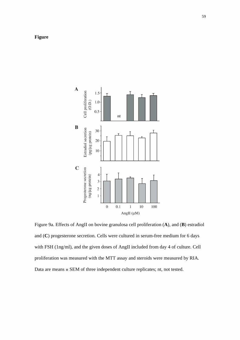
59
Figure
Figure 9a. Effects of AngII on bovine granulosa cell proliferation (A), and (B) estradiol
and (C) progesterone secretion. Cells were cultured in serum-free medium for 6 days
with FSH (1ng/ml), and the given doses of AngII included from day 4 of culture. Cell
proliferation was measured with the MTT assay and steroids were measured by RIA.
Data are means ± SEM of three independent culture replicates; nt, not tested.

60
Figure 9b. Effects of AngII on protease activity secreted from bovine granulosa cells.
Cells were cultured in serum-free medium for 6 days with FSH (1ng/ml), and the given
doses of AngII included from day 4 of culture. Protease activity was measured by
gelatin zymography. Proteolytic bands of Mr 83k and 62k are identified as MMP9 and
MMP2, respectively (representative gel shown above graph). Data are means ± SEM of
three independent culture replicates.

61
Fig 9c. AngII inhibits (A) SERPINE2 secretion and (B) gene expression in bovine
granulosa cells. Cells were cultured in serum-free medium for 6 days with FSH
(1ng/ml), and the AngII (10 μM) added from day 4 of culture. Data are means ± SEM
of three independent culture replicates. Asterisks denote means significantly different
from control cultures without AngII. A representative Western is shown for one
replicate, with samples in the same order as the graph in (A). A representative agarose
gel is shown to illustrate mRNA abundance in 2 replicates (B).

62
References
1. Paul M, Poyan Mehr A, Kreutz R. Physiology of local renin-angiotensin systems. Physiological Reviews 2006; 86: 747-803.
2. Mukhopadhyay AK, Holstein K, Szkudlinski M, Brunswig-Spickenheier B, Leidenberger FA. The relationship between prorenin levels in follicular fluid and follicular atresia in bovine ovaries. Endocrinology 1991; 129: 2367-2375.
3. Daud AI, Bumpus FM, Husain A. Evidence for selective expression of angiotensin II receptors on atretic follicles in the rat ovary: an autoradiographic study. Endocrinology 1988; 122: 2727-2734.
4. Pucell AG, Hodges JC, Sen I, Bumpus FM, Husain A. Biochemical properties of the ovarian granulosa cell type 2-angiotensin II receptor. Endocrinology 1991; 128: 1947-1959.
5. Kuji N, Sueoka K, Miyazaki T, Tanaka M, Oda T, Kobayashi T, Yoshimura Y. Involvement of angiotensin II in the process of gonadotropin-induced ovulation in rabbits. Biology of Reproduction 1996; 55: 984-991.
6. Pellicer A, Palumbo A, DeCherney AH, Naftolin F. Blockage of ovulation by an angiotensin antagonist. Science 1988; 240: 1660-1661.
7. Yoshimura Y, Karube M, Aoki H, Oda T, Koyama N, Nagai A, Akimoto Y, Hirano H, Nakamura Y. Angiotensin II induces ovulation and oocyte maturation in rabbit ovaries via the AT2 receptor subtype. Endocrinology 1996; 137: 1204-1211.
8. Yoshimura Y, Karube M, Oda T, Koyama N, Shiokawa S, Akiba M, Yoshinaga A, Nakamura Y. Locally produced angiotensin II induces ovulation by stimulating prostaglandin production in in vitro perfused rabbit ovaries. Endocrinology 1993; 133: 1609-1616.
9. Acosta TJ, Berisha B, Ozawa T, Sato K, Schams D, Miyamoto A. Evidence for a local endothelin-angiotensin-atrial natriuretic peptide systemin bovine mature follicles in vitro: effects on steroid hormones and prostaglandin secretion. Biology of Reproduction 1999; 61: 1419-1425.
10. Davis BJ, Lennard DE, Lee CA, Tiano HF, Morham SG, Wetsel WC, Langenbach R. Anovulation in cyclooxygenase-2-deficient mice Is restored by prostaglandin E2 and interleukin-1 . Endocrinology 1999; 140: 2685-2695.
11. Stirling D, Magness RR, Stone R, Waterman MR, Simpson ER. Angiotensin II inhibits luteinizing hormone-stimulated cholesterol side chain cleavage expression and stimulates basic fibroblast growth factor expression in bovine luteal cells in primary culture. Journal of Biological Chemistry 1990; 265: 5-8.
12. Li XM, Juorio AV, Murphy BD. Angiotensin II interferes with steroidogenesis in porcine granulosa cells. Biology of Reproduction 1995; 53: 791-799.
13. Johnson MC, Vega M, Vantman D, Troncoso JL, Devoto L. Regulatory role of angiotensin II on progesterone production by cultured human granulosa cells. Expression of angiotensin II type-2 receptor. Molecular Human Reproduction 1997; 3: 663-668.
14. Morris RS, Francis MM, Do YS, Hsueh WA, Lobo RA, Paulson RJ. Angiotensin II (AII) modulation of steroidogenesis by luteinized granulosa cells in vitro. Journal of Assisted Reproduction and Genetics 1994; 11: 117-122.

63
15. Costa APR, Fagundes-Moura CR, Pereira VM, Silva LF, Vieira MAR, Santos RAS, Dos Reis AM. Angiotensin-(1-7): a novel peptide in the ovary. Endocrinology 2003; 144: 1942-1948.
16. Cannon CM, Keeble SC, Curry TE, Jr. Effect of A23187 or angiotensin II on ovarian metalloproteinase inhibitors and steroidogenesis in rats. Journal of Reproduction and Fertility 1997; 111: 71-79.
17. Féral C, Le Gall S, Leymarie P. Angiotensin II modulates steroidogenesis in granulosa and theca in the rabbit ovary: its possible involvement in atresia. European Journal of Endocrinology 1995; 133: 747-753.
18. Kotani E, Sugimoto M, Kamata H, Fujii N, Saitoh M, Usuki S, Kubo T, Song K, Miyazaki M, Murakami K, Miyazaki H. Biological roles of angiotensin II via its type 2 receptor during rat follicle atresia. American Journal of Physiology - Endocrinology and Metabolism 1999; 276: E25-33.
19. Tanaka M, Ohnishi J, Ozawa Y, Sugimoto M, Usuki S, Naruse M, Murakami K, Miyazaki H. Characterization of angiotensin II receptor type 2 during differentiation and apoptosis of rat ovarian cultured granulosa cells. Biochemical and Biophysical Research Communications 1995; 207: 593-598.
20. de Gooyer TE, Skinner SL, Wlodek ME, Kelly DJ, Wilkinson-Berka JL. Angiotensin II influences ovarian follicle development in the transgenic (mRen-2)27 and Sprague-Dawley rat. Journal of Endocrinology 2004; 180: 311-324.
21. Gutiérrez CG, Campbell BK, Webb R. Development of a long-term bovine granulosa cell culture system: induction and maintenance of estradiol production, response to follicle-stimulating hormone, and morphological characteristics. Biology of Reproduction 1997; 56: 608-616.
22. Silva JM, Price CA. Effect of follicle-stimulating hormone on steroid secretion and messenger ribonucleic acids encoding cytochromes P450 aromatase and cholesterol side-chain cleavage in bovine granulosa cells in vitro. Biology of Reproduction 2000; 62: 186-191.
23. Cao M, Sahmi M, Lussier JG, Price CA. Plasminogen activator and serine protease inhibitor-E2 (protease nexin-1) expression by bovine granulosa cells in vitro. Biology of Reproduction 2004; 71: 887-893.
24. Bédard J, Brûlé S, Price CA, Silversides DW, Lussier JG. Serine protease inhibitor-E2 (SERPINE2) is differentially expressed in granulosa cells of dominant follicle in cattle. Molecular Reproduction & Development 2003; 64: 152-165.
25. Robert C, McGraw S, Massicotte L, Pravetoni M, Gandolfi F, Sirard MA. Quantification of housekeeping transcript levels during the development of bovine preimplantation embryos. Biology of Reproduction 2002; 67: 1465-1472.
26. Bélanger A, Couture J, Caron S, Roy R. Determination of nonconjugated and conjugated steroid levels in plasma and prostate after separation on C-18 columns. Annals of the New York Academy of Sciences 1990; 595: 251-259.
27. Lafrance M, Goff AK. Effect of pregnancy on oxytocin-induced release of prostaglandin F2 alpha in heifers. Biology of Reproduction 1985; 33: 1113-1119.
28. Portela VM, Gonçalves PBD, Jr B. A novel role for angiotensin II in the regulation of protease-nexin-1 expression and secretion in bovine follicles. 39th Annual Meeting of Socity for Study of Reproduction 2006.

64
29. Pucell AG, Bumpus FM, Husain A. Regulation of angiotensin II receptors in cultured rat ovarian granulosa cells by follicle-stimulating hormone and angiotensin II. Journal of Biological Chemistry 1988; 263: 11954-11961.
30. Pucell AG, Bumpus FM, Husain A. Rat ovarian angiotensin II receptors. Characterization and coupling to estrogen secretion. Journal of Biological Chemistry 1987; 262: 7076-7080.
31. Nogueira MF, Buratini J, Jr., Price CA, Castilho AC, Pinto MG, Barros CM. Expression of LH receptor mRNA splice variants in bovine granulosa cells: changes with follicle size and regulation by FSH in vitro. Molecular Reproduction and Development 2007; 74: 680-686.
32. Sahmi M, Nicola ES, Silva JM, Price CA. Expression of 17 - and 3 -hydroxysteroid dehydrogenases and steroidogenic acute regulatory protein in non-luteinizing bovine granulosa cells in vitro. Molecular and Cellular Endocrinology 2004; 223: 43-54.
33. Arenas IA, Xu Y, Lopez-Jaramillo P, Davidge ST. Angiotensin II-induced MMP-2 release from endothelial cells is mediated by TNF- . American Journal of Physiology. Cell Physiology 2004; 286: C779-C784.
34. Browatzki M, Larsen D, Pfeiffer CAH, Gehrke SG, Schmidt J, Kranzhöfer A, Katus HA, Kranzhöfer R. Angiotensin II Stimulates Matrix Metalloproteinase Secretion in Human Vascular Smooth Muscle Cells via Nuclear Factor-kB and Activator Protein 1 in a Redox-Sensitive Manner. Journal of Vascular Research 2005; 42: 415-423.
35. Rouet-Benzineb P, Gontero B, Dreyfus P, Lafuma C. Angiotensin II Induces Nuclear Factor- B Activation in Cultured Neonatal Rat Cardiomyocytes Through Protein Kinase C Signaling Pathway. Journal of Molecular and Cellular Cardiology 2000; 32: 1767-1778
. 36. van Leeuwen RT, Kol A, Andreotti F, Kluft C, Maseri A, Sperti G. Angiotensin
II increases plasminogen activator inhibitor type 1 and tissue-type plasminogen activator messenger RNA in cultured rat aortic smooth muscle cells. Circulation 1994; 90: 362-368.
37. Vaughan DE, Lazos SA, Tong K. Angiotensin II regulates the expression of plasminogen activator inhibitor-1 in cultured endothelial cells. A potential link between the renin-angiotensin system and thrombosis. Journal of Clinical Investigation 1995; 95: 995-1001.
38. Nakamura S, Nakamura I, Ma L, Vaughan DE, Fogo AB. Plasminogen activator inhibitor-1 expression is regulated by the angiotensin type 1 receptor in vivo. Kidney International 2000; 58: 251-259.
39. Cao M, Buratini Jr J, Lussier JG, Carriere PD, Price CA. Expression of protease nexin-1 and plasminogen activators during follicle growth and the periovulatory period in cattle. Reproduction 2006; 131: 125-137.
40. Dow MP, Bakke LJ, Cassar CA, Peters MW, Pursley JR, Smith GW. Gonadotrophin surge-induced upregulation of mRNA for plasminogen activator inhibitors 1 and 2 within bovine periovulatory follicular and luteal tissue. Reproduction 2002; 123: 711-719.
41. Smith MF, Gutierrez CG, Ricke WA, Armstrong DG, Webb R. Production of matrix metalloproteinases by cultured bovine theca and granulosa cells. Reproduction 2005; 129: 75-87.

65
42. Bleuel A, de Gasparo M, Whitebread S, Püttner I, Monard D. Regulation of protease nexin-1 expression in cultured Schwann cells is mediated by angiotensin II receptors. Journal of Neuroscience 1995; 15: 750-761.
43. Cao M, Nicola E, Portela VM, Price CA. Regulation of serine protease inhibitor-E2 and plasminogen activator expression and secretion by follicle stimulating hormone and growth factors in non-luteinizing bovine granulosa cells in vitro. Matrix Biology 2006; 25: 342-354.
44. Macchione E, Epifano O, Stefanini M, Belin D, Canipari R. Urokinase redistribution from the secreted to the cell-bound fraction in granulosa cells of rat preovulatory follicles. Biology of Reproduction 2000; 62: 895-903.

66
5. CAPÍTULO 3
THE EXPRESSION OF GENES INVOLVED IN OVULATION ARE
REGULATED BY ANGIOTENSIN II IN GRANULOSA CELLS IN VITRO.
Valério M Portela, Paulo BD Gonçalves, João FC de Oliveira, Paulo A Lovato,
Christopher A Price.

67
THE EXPRESSION OF GENES INVOLVED IN OVULATION ARE
REGULATED BY ANGIOTENSIN II IN GRANULOSA CELLS IN VITRO.
Valério M Portela 1,2, Paulo BD Gonçalves 1, João FC de Oliveira 1 , Paulo A Lovato 1,
Christopher A Price2,3.
Laboratório de Biotecnologia e Reprodução Animal1, Universidade Federal de Santa
Maria, Santa Maria, RS, Brazil; Centre de Recherche en Reproduction Animale2,
Faculty of Veterinary Medicine, University of Montreal, St-Hyacinthe, QC, Canada.
Key words: ovulation, cox2, serpine2, granulosa cell, follicle
Running head: Angiotensin and ovulation in bovine follicles
3 Correspondance: C.A.Price, CRRA, Faculte de médecine vétérinaire, C.P. 5000, St-
Hyacinthe QC, J2S 7C6 Canada. Email: Error! Contact not [email protected]

68
Abstract
Angiotensin II (AngII) induces follicular prostaglandin release in a number of
species, and induces ovulation in rabbits. The objective of the present study was to test
the hypotheses that AngII, alone or in combination with LH, induces the expression of
the enzyme responsible for prostaglandin synthesis, Ptgs2, and the prostaglandin-
dependent proteolytic cascade necessary for follicle rupture. Bovine granulosa cells
from large (>10 mm) follicles were cultured for 6h, 12h and 24h with LH (100ng/ml) or
AngII. These cells expressed Ptgs2 under basal culture conditions, which was not
upregulated by either LH or AngII alone. However, LH and AngII in combination
significantly enhanced Ptgs2 (P<0.05) mRNA and protein accumulation. Expression of
the proteolytic enzymes uPA and tPA, and their inhibitor, Serpine2 was also measured
by RT-PCR and, as for Ptgs2, the combination of LH and AngII but neither factor alone
upregulated expression of all three genes (P<0.05). These data suggest that AngII is a
physiological co-factor necessary for the expression of genes in granulosa cells that are
critical for ovulation.
Introduction
The preovulatory surge of LH initiates complex changes in gene expression in
preovulatory follicles, which culminate in ovulation and formation of the corpus
luteum. Granulosa cells of preovulatory follicles express LH receptors, and respond in
dramatic ways to the LH surge. A considerable body of work in rodents has suggested a
probable sequence of events triggered by the LH surge in granulosa cells. One of the
early genes upregulated by LH is the progesterone receptor (PR), and expression of this
gene is essential for ovulation as demonstrated by studies with PR knock-out mice

69
(PRKO) [1]. LH also rapidly induced the nuclear factor RIP140, which in turn induces
the expression of the proteases cathepsin L and a disintegrin and metalloproteinase with
thrombospondin repeats (ADAMTS1), epiregulin, amphiregulin, betacellulin,
hyaluronan synthase2 and prostaglandin-endoperoxide synthase2 (Ptgs2, also known as
Cox2) [2-4]. Ptgs2 catalyses the synthesis of prostaglandins [5], which are essential for
cumulus-oocyte-expansion and ovulation as Ptgs2 knock-out mice fail to ovulate [6].
Down-stream from the above genes is the regulation of proteins involved in the
physical rupture of the follicle wall. These include members of the matrix
metalloproteinase (MMP) family and their inhibitors (TIMP) [7], and the plasminogen
activators (PA) and their inhibitors (PAI). In cattle, hCG transiently upregulated tissue-
type PA (tPA), urokinase (uPA) and the inhibitors PAI-1 and protease nexin-1
(Serpine2, also known as PN1) until the time of ovulation when all except uPA
decreased [8-10]. Periovulatory changes in several MMPs and TIMPs have also been
described in cattle [11, 12]. Owing to the large numbers of proteases and inhibitors and
considerable potential for redundancy, the exact role of each protein in ovulation
remains unclear. However, injection of the prostaglandin inhibitor indomethacin, which
blocks ovulation in cattle [13], into bovine preovulatory follicles resulted in few
changes in follicular MMP or TIMP expression but significantly inhibited follicular
tPA, uPA and PAI-1 expression [14], suggesting that the PA system is part of the LH-
induced mechanism of follicle rupture.
Ovulation can be induced or inhibited by a number of factors other than LH.
One example is angiotensin II (AngII), which induced ovulation in perifused rabbit
ovaries [15, 16]. Conversely, AngII antagonists inhibit ovulation in rodents, rabbits and
cattle [17-19]. The mechanism of action of AngII on ovulation is not clear, although is
mediated through the type 2 receptor (AGTR2, also known as AT2) [15, 17]. AngII

70
stimulated prostaglandin secretion from rabbit ovaries [15, 17] and bovine preovulatory
follicles [20]. The objective of the present study was to determine whether AngII
stimulates the expression of Ptgs2 in cultured bovine granulosa cells independently or
in concert with LH, and to test the hypothesis that AngII stimulates genes involved in
follicle rupture.
Materials and methods
Cell culture
All materials were obtained from Invitrogen Life Technologies (Burlington, ON,
Canada) except where otherwise stated. Briefly, bovine ovaries were collected from
adult cows, irrespective of stage of the estrous cycle, at a local abattoir, and were
transported to the laboratory in PBS at 35°C containing penicillin (100 IU/ml),
streptomycin (100 μg/ml) and fungizone (1 μg/ml). Cells were collected from large
follicles (>8mm) by aspiration, and were washed twice by centrifugation at 219 g for
20 min each, and suspended in DMEM-F12, sodium bicarbonate (10 mM), sodium
selenite (4 ng/ml), BSA (0.1%; Sigma-Aldrich Canada, Oakville, ON, Canada),
penicillin (100 IU/ml), streptomycin (100 μg/ml), transferrin (2.5 μg/ml), non-essential
amino acid mix (1.1 mM), androstenedione (10-7 M at start of culture, and 10-6 M at
each medium change), FSH 1ng/ml, insulin (10 ng/ml) and 2% FCS. Cell viability was
estimated with 0.4% Trypan Blue Stain. Cells were seeded into 24-well tissue culture
plates (Sarstedt, Montreal, QC) at a density of 1 x 10⁶ viable cells per well in 1 ml
medium. Cultures were maintained at 37°C in 5% CO2. After 24h, medium was
changed to DMEM-F12 without supplementation for 10 h before adding insulin
(10ng/ml) and hormone treatments. Treatments were LH (100ng/ml; NIH) or LH +
AngII (100 μM) for the time periods described in Results.

71
Immunofluorescence
Protein localization in bovine granulosa cells in vitro was performed by
immunofluorescence. After cell culture the cells were fixed 4% paraformaldehyde for
20 min, washed 5 times for 5 min in PBS and were blocked in 5% BSA for 1 h and then
1:200 Ptgs2 antibody (cat. PG26, Oxford Biomedical Research, Oxford, MI) was
applied for 18 h at 4 C. After the primary antibody, the cells were washed in PBS and
then incubated with Cy3-conjugated second antibody (Jackson ImmunoResearch,West
Grove, PA) and counterstained with DAPI. Negative controls were performed in the
absence of primary antibody. Digital images were captured and mean fluorescence
intensity in each field was quantified with ImageJ (NIH). The number of cells in the
field was quantified by counting DAPI-stained nuclei, and the staining was expressed as
units/cell.
Nucleic acid extraction & semi-quantitative RT-PCR
Total RNA and DNA were extracted using Trizol according to the
manufacturer’s instructions. Total RNA was quantified by absorbance at 260 nm. Total
RNA (1 μg) was first treated with 1 U DNase (Promega, Madison, WI) at 37°C for 30
min to digest any contaminating DNA, followed by adding 1 μl of EDTA stop buffer at
65°C for 10 min. The RNA was reverse transcribed in the presence of 1 mM oligo(dT)
primer and 4 U Omniscript RTase (Omniscript RT Kit, Qiagen, Mississauga, ON), 0.25
mM dideoxy-nucleotide triphosphate (dNTP) mix, and 19.33 U RNase Inhibitor
(Amersham Biosciences, Baie D’Urfé, QC) in a volume of 20 μl at 42°C for 2 h. The
reaction was terminated by incubation at 93°C for 5 min.

72
Bovine-specific primers were used for amplifying uPA , tPA and Serpine2
exactly as described [21]. Primer for bovine Ptgs2 were as published [22]. Variability in
mRNA amounts was assessed by amplifying histone H2AFZ (H2AFZ; sense: 5’-
AGCGTATTACCCCTGGTCAC -3’, antisense: 5’-CCAGGCATCCTTTAGACAGT -
3’). An aliquot (1 μl) of the cDNA template was amplified by PCR using 0.25 μl (2.5
U) Taq Polymerase (Amersham Pharmacia Biotech Inc., Oakville, ON, Canada) in a 25-
μl PCR buffer (Amersham Pharmacia Biotech Inc.) containing 0.1 mM dNTP mix, and
0.2 μM specific primers. After an initial denaturation step for 3 min at 94°C, target
cDNA was amplified with a denaturation step at 94°C for 60 sec (or 45 for H2AFZ),
annealing for 45 sec at 55°C (tPA), 58°C (H2AFZ), 60°C (Ptgs2), 62°C (Serpine2) or
65°C (uPA for 30 sec, and elongation at 72°C for 1 min. All reactions were terminated
with a final elongation at 72°C for 5 min. Semiquantitative RT-PCR was validated for
each gene product. Reactions were performed for 26 cycles for tPA, 24 cycles for
Serpine2 and 30 cycles for uPA, H2AFZ and Ptgs2.
The PCR products were separated on 2 % agarose gels with 0.001% ethidium
bromide, and visualized under UV light. Quantification of band intensity was performed
with NIH Image software. Target gene mRNA abundance was expressed relative to
H2aFZ mRNA abundance.
Steroid assay
Estradiol was measured in conditioned medium in duplicate as described [23],
without solvent extraction. Intra- and inter-assay coefficients of variation were 8.5%
and 6.3%, respectively. Progesterone was measured in duplicate as described [24] with
mean intra- and inter-assay coefficients of variation were 7.2% and 18%, respectively.

73
The sensitivity of these assays were 10 pg and 4 pg per tube for estradiol and
progesterone, equivalent to 0.3 and 20 ng/ g protein, respectively. Steroid
concentrations in culture medium were corrected for cell number by expressing per unit
mass of total protein.
Statistical Analysis
Data that did not follow a normal distribution (Shapiro-Wilk test) were
transformed to logarithms. Homogeneity of variance was tested with O’Brien and
Brown-Forsythe tests. Analysis of data was performed with JMP software (SAS
Institute) with treatment or follicle group as main effect and culture replicate (where
appropriate) as a random variable in the F-test. For gene expression data, housekeeping
gene abundance was included as a covariate in the main effects model. Differences
between means were tested with the Tukey-Kramer HSD test. Data are presented as
means ± SEM.
Results
Steroid secretion from granulosa cells at 6, 12 and 24 h after the addition of LH
or LH plus AngII was assessed (Fig 1). Estradiol secretion was no higher at 12 or 24 h
than it was at 6 h, indicating no secretion after 6 h. In contrast, progesterone secretion
doubled from 6 to 12 h of culture, and then again at 24 h, demonstrating continued
secretion of progesterone in culture. AngII did not alter estradiol secretion, but
significantly enhanced progesterone secretion at 6 h post-treatment.
As AngII has been reported to increase prostaglandin secretion from bovine
follicles in vivo, we next assessed the effects of AngII on Ptgs2 mRNA and protein
abundance in granulosa cells in vitro. The addition of LH or AngII alone did not

74
significantly increase Ptgs2 expression over time, but the addition of AngII to LH-
stimulated cells significantly increased Ptgs2 mRNA at 6 and 24 h post-treatment,
although not at 12 h (Fig 2A). To assess which AngII receptor mediated the effect of
AngII, selective blockers of AGTR1 (Losartan) or of AGTR2 (PD123,319) were added
one hour before treatments. PD123,319 effectively inhibited the stimulatory effect of
AngII whereas Losartan did not (Fig 2B).
In concordance with the gene expression data, LH or AngII alone did not
stimulate Ptgs2 protein accumulation, whereas these two factors combined significantly
enhanced Ptgs2 protein accumulation at 6 h post-treatment (Fig 3).
Finally, we assessed the effects of AngII on the PA system. In comparison with
LH alone, AngII significantly increased uPA, tPA and Serpine2 expression after 6 h in
LH-stimulated cells (Fig 4).
Discussion
The present study clearly shows that AngII stimulates Ptgs2 gene expression and
protein accumulation, and expression of the proteases tPA, uPA and the protease
inhibitor PN1. AngII has been shown to induce ovulation in rabbit ovaries, and
antagonists of angiotensin receptors inhibit ovulation in rabbits, rats and cattle [15-19].
Direct evidence for the induction of ovulation by AngII comes from studies with
perfused rabbit ovaries [15, 16]. The rabbit is an induced-ovulator; therefore, the
mechanisms involved in ovulation may differ from those in other mammals. Infusion
of AngII into preovulatory rabbit and bovine follicles stimulated the release of
prostanglandin, suggesting a potential mechanism of AngII action on ovulation [20].
Recently, our group demonstrated that ovulation is blocked when an specific inhibitors

75
of AngII receptors (saralasin or PD123 319) is injected into ovulatory follicles in cattle
[24].
In the present study, we determined whether AngII is capable of inducing
ovulation alone or whether an interaction with LH is required. To do so, we used a cell
model in which cells from large follicles were cultured for 24 h with serum followed by
a serum-free period prior to testing the effects of LH and AngII. Under these conditions,
the granulosa cells secreted estradiol and progesterone, expressed Ptgs2, and did not
appear morphologically luteinized. Using a similar culture system with pig granulosa
cells, Li et al [25] showed that AngII inhibited LH-stimulated progesterone secretion. In
the present study, AngII increased progesterone secretion from bovine granulosa cells in
short-term (6 h) culture but not in longer-term (12 - 24 h) culture. A similar pattern of
stimulation of progesterone was observed in perfused rat ovaries [26].
LH alone had no effect on Ptgs2 protein accumulation in vitro in the present
study (Fig 3), which is not accord with the established induction of Ptgs2 by LH in
vivo. However, induction of Ptsg2 expression may be an effect of culture, as previously
noted in mice [27]. In this previous study, culture of granulosa cells from eCG-primed
mice for 4 h induced Ptgs2 expression, which was significantly enhanced by LH.
Comparison of Ptgs2 expression in granulosa cells of mice 4 h after hCG injection in
vivo with that 4 h after addition of LH in vitro demonstrated that Ptgs2 expression in
vivo was significantly higher than in vitro [27]. The authors concluded that a factor(s)
in addition to LH is required for optimal Ptgs2 expression. In agreement with this
hypothesis, the present data from bovine granulosa cells show that LH alone was not
capable of increasing Ptgs2 expression above levels induced by culture, as was also the
case for AngII alone, but that the combination of LH and AngII resulted in significantly

76
increased Ptgs2 expression. Thus, these data suggest that AngII may be one of the
previously unknown factors necessary for the chain of events leading to ovulation.
To explore further this hypothesis, we examined the effect of AngII on members
of the PA system, prostaglandin-dependent proteases that are likely important for the
tissue remodeling involved in follicle rupture. The addition of AngII to LH-treated cells
significantly increased expression of tPA, uPA and the inhibitor Serpine2 within 6 h.
This is in agreement with the increase in the expression of these genes observed in
bovine follicles in vivo following induction of ovulation with hCG or GnRH [8, 10]. As
AngII antagonists inhibit prostaglandin release and ovulation in cattle [19, 20] as well
as rodents, the likely mechanism is inhibition of prostaglandin-dependent PA activity
and the resulting proteolytic degradation of the follicle wall.
In conclusion, the present data strongly suggest that AngII is a mediator of the
LH to establish induction of Ptgs2 and to regulate the remodeling of extracellular matrix
in bovine granulosa cells. Moreover, there is evidence that AngII is an obligatory co-
factor for the LH-induced cascade of events leading to rupture of the follicle wall and
ovulation.

77
Figures
Fig 1. Effect of time in culture on estradiol and progesterone secretion in luteinizing
bovine granulosa cells incubated with LH or LH+AngII. Cells from large follicles
(>10mm diameter) were cultured with serum for 24 h, and then in serum-free medium
for a further 12 h. LH (100ng/ml) and AngII (dose) was then added for the times shown.
Means ± SEM are shown for 3 replicate cultures. Asterisk denotes a significant effect of
AngII (P<0.05).

78
Fig 2. Effect of AngII on Ptgs2 mRNA expression in luteinizing granulosa cells in vitro.
Cells were cultured as described in Fig 1, and Ptgs2 mRNA measured by RT-PCR. (A)
AngII increased Ptgs2 expression in cells cultured with LH. Asterisks denote
differences between LH alone and LH+AngII treatments. (B) Pretreatment with
selective AGTR1 (Losartan) and AGTR2 (PD) blockers reveal which AngII receptor
mediates the effect of AngII on Ptgs2 expression. Letters above bars denote differences
between means (P<0.05). Means ± SEM are shown for 3 replicate cultures. A
representative gel of PCR products is shown for each gene.

Cox-2 DAPI
Ang
LH
LH+Ang
Negative control
Ptgs2
pro
tein
conte
nt/
cell
(
arbit
rary
unit
s)
AU/Cell
ANG LH
+ -
-+
++
aa
b
0
2
4
6
8
10
12
14
16
18
20
--
a a
Control
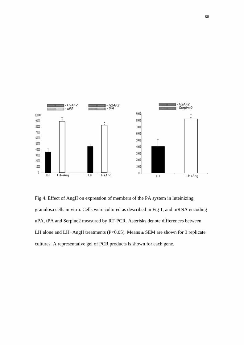
80
Fig 4. Effect of AngII on expression of members of the PA system in luteinizing
granulosa cells in vitro. Cells were cultured as described in Fig 1, and mRNA encoding
uPA, tPA and Serpine2 measured by RT-PCR. Asterisks denote differences between
LH alone and LH+AngII treatments (P<0.05). Means ± SEM are shown for 3 replicate
cultures. A representative gel of PCR products is shown for each gene.

81
References
1. Robker RL, Russell DL, Espey LL, Lydon JP, O'Malley BW, Richards JS. Progesterone-regulated genes in the ovulation process: ADAMTS-1 and cathepsin L proteases. Proceedings of the National Academy of Sciences of the United States of America 2000; 97: 4689-4694.
2. Russell DL, Robker RL. Molecular mechanisms of ovulation: co-ordination through the cumulus complex. Human Reproduction Update 2007; 13: 289-312.
3. Shimada M, Hernandez-Gonzalez I, Gonzalez-Robayna I, Richards JS. Paracrine and autocrine regulation of epidermal growth factor-like factors in cumulus oocyte complexes and granulosa cells: key roles for prostaglandin synthase 2 and progesterone receptor. Molecular Endocrinology 2006; 20: 1352-1365.
4. Tullet JM, Pocock V, Steel JH, White R, Milligan S, Parker MG. Multiple signaling defects in the absence of RIP140 impair both cumulus expansion and follicle rupture. Endocrinology 2005; 146: 4127-4137.
5. Sirois J, Sayasith K, Brown KA, Stock AE, Bouchard N, Dore M. Cyclooxygenase-2 and its role in ovulation: a 2004 account. Human Reproduction Update 2004; 10: 373-385.
6. Davis BJ, Lennard DE, Lee CA, Tiano HF, Morham SG, Wetsel WC, Langenbach R. Anovulation in cyclooxygenase-2-deficient mice Is restored by prostaglandin E2 and interleukin-1 . Endocrinology 1999; 140: 2685-2695.
7. Curry TE, Jr., Osteen KG. The Matrix Metalloproteinase System: Changes, Regulation, and Impact throughout the Ovarian and Uterine Reproductive Cycle. Endocrine Reviews 2003; 24: 428-465.
8. Cao M, Buratini Jr J, Lussier JG, Carriere PD, Price CA. Expression of protease nexin-1 and plasminogen activators during follicle growth and the periovulatory period in cattle. Reproduction 2006; 131: 125-137.
9. Dow MP, Bakke LJ, Cassar CA, Peters MW, Pursley JR, Smith GW. Gonadotrophin surge-induced upregulation of mRNA for plasminogen activator inhibitors 1 and 2 within bovine periovulatory follicular and luteal tissue. Reproduction 2002; 123: 711-719.
10. Dow MP, Bakke LJ, Cassar CA, Peters MW, Pursley JR, Smith GW. Gonadotropin surge-induced up-regulation of the plasminogen activators (tissue plasminogen activator and urokinase plasminogen activator) and the urokinase plasminogen activator receptor within bovine periovulatory follicular and luteal tissue. Biology of Reproduction 2002; 66: 1413-1421.
11. Bakke LJ, Dow MPD, Cassar CA, Peters MW, Pursley JR, Smith GW. Effect of the Preovulatory Gonadotropin Surge on Matrix Metalloproteinase (MMP)-14, MMP-2, and Tissue Inhibitor of Metalloproteinases-2 Expression Within Bovine Periovulatory Follicular and Luteal Tissue. Biology of Reproduction 2002; 66: 1627-1634.
12. Bakke LJ, Li Q, Cassar CA, Dow MPD, Pursley JR, Smith GW. Gonadotropin Surge-Induced Differential Upregulation of Collagenase-1 (MMP-1) and Collagenase-3 (MMP-13) mRNA and Protein in Bovine Preovulatory Follicles. Biology of Reproduction 2004; 71: 605-612.
13. De Silva M, Reeves JJ. Indomethacin inhibition of ovulation in the cow. Journal of Reproduction and Fertility 1985; 75: 547-549.

82
14. Li Q, Jimenez-Krassel F, Kobayashi Y, Ireland JJ, Smith GW. Effect of intrafollicular indomethacin injection on gonadotropin surge-induced expression of select extracellular matrix degrading enzymes and their inhibitors in bovine preovulatory follicles. Reproduction 2006; 131: 533-543.
15. Yoshimura Y, Karube M, Aoki H, Oda T, Koyama N, Nagai A, Akimoto Y, Hirano H, Nakamura Y. Angiotensin II induces ovulation and oocyte maturation in rabbit ovaries via the AT2 receptor subtype. Endocrinology 1996; 137: 1204-1211.
16. Yoshimura Y, Karube M, Oda T, Koyama N, Shiokawa S, Akiba M, Yoshinaga A, Nakamura Y. Locally produced angiotensin II induces ovulation by stimulating prostaglandin production in in vitro perfused rabbit ovaries. Endocrinology 1993; 133: 1609-1616.
17. Kuji N, Sueoka K, Miyazaki T, Tanaka M, Oda T, Kobayashi T, Yoshimura Y. Involvement of angiotensin II in the process of gonadotropin-induced ovulation in rabbits. Biology of Reproduction 1996; 55: 984-991.
18. Pellicer A, Palumbo A, DeCherney AH, Naftolin F. Blockage of ovulation by an angiotensin antagonist. Science 1988; 240: 1660-1661.
19. Ferreira R, Oliveira JFC, Fernandes R, Moraes JCF, Gonáalves PBD. The role of angiotensin II in the early stages of bovine ovulation. Reproduction 2007.
20. Acosta TJ, Berisha B, Ozawa T, Sato K, Schams D, Miyamoto A. Evidence for a local endothelin-angiotensin-atrial natriuretic peptide systemin bovine mature follicles in vitro: effects on steroid hormones and prostaglandin secretion. Biology of Reproduction 1999; 61: 1419-1425.
21. Cao M, Sahmi M, Lussier JG, Price CA. Plasminogen activator and serine protease inhibitor-E2 (protease nexin-1) expression by bovine granulosa cells in vitro. Biology of Reproduction 2004; 71: 887-893.
22. Hayashi K, Acosta TJ, Berisha B, Kobayashi Si, Ohtani M, Schams D, Miyamoto A. Changes in prostaglandin secretion by the regressing bovine corpus luteum. Prostaglandins and Other Lipid Mediators 2003; 70: 339-349.
23. Bélanger A, Couture J, Caron S, Roy R. Determination of nonconjugated and conjugated steroid levels in plasma and prostate after separation on C-18 columns. Annals of the New York Academy of Sciences 1990; 595: 251-259.
24. Lafrance M, Goff AK. Effect of pregnancy on oxytocin-induced release of prostaglandin F2 alpha in heifers. Biology of Reproduction 1985; 33: 1113-1119.
25. Li XM, Juorio AV, Murphy BD. Angiotensin II interferes with steroidogenesis in porcine granulosa cells. Biology of Reproduction 1995; 53: 791-799.
26. Costa APR, Fagundes-Moura CR, Pereira VM, Silva LF, Vieira MAR, Santos RAS, Dos Reis AM. Angiotensin-(1-7): a novel peptide in the ovary. Endocrinology 2003; 144: 1942-1948.
27. Joyce IM, Pendola FL, O'Brien M, Eppig JJ. Regulation of prostaglandin-endoperoxide synthase 2 messenger ribonucleic acid expression in mouse granulosa cells during ovulation. Endocrinology 2001; 142: 3187-3197.

83
6. DISCUSSÃO
No presente estudo foi demonstrado que o AGTR2 está presente nas células da
granulosa de bovinos, que o mRNA para esse gene está aumentado em folículos
dominantes e que a expressão na células da granulosa é estimulada por gonadotrofinas.
Esses dados evidenciam que a sinalização via AGTR2 está envolvida no crescimento e
não na atresia folicular em bovinos. Além disso, foi demonstrado que mRNA AGTR1 e
AGTR2 estão presentes em células da teca e granulosa de bovinos.
Em estudos anteriores havia sido demonstrado que a AngII está envolvida com a
atresia folicular através da redução na secreção de E2 e da expressão do AGTR2 em
células da granulosa de folículos atrésicos (KOTANI et al., 1999; DE GOOYER et al.,
2004). No entanto, a expressão do AGTR2 tinha sido detectada somente nas células da
teca e não em células da granulosa em folículos bovinos (SCHAUSER et al., 2001a),
sugerindo uma via alternativa de ação da AngII em bovinos. As técnicas utilizadas
anteriormente não foram sensíveis para detectar o AGTR2 em células da granulosa,
vasos e tecidos ricos em receptores de AngII (BRUNSWIG-SPICKENHEIER &
MUKHOPADHYAY, 1992a; TOUYZ & SCHIFFRIN, 2000; SCHAUSER et al.,
2001b). Neste experimento, foi utilizada a técnica de RT-PCR para investigar a
presença de AGTR2, e imunohistoquimica para detectar a proteína tanto nas células da
teca quanto nas células da granulosa. A expressão de AGTR1 nas células da teca e nas
células da granulosa não diferiu em folículos dominantes e atrésicos, concordando com
dados achados em células da teca em bovinos (ACOSTA et al., 1999). Em contraste
com estudos em ratos que apresentaram um aumento significativo de AGTR2 nas
células da granulosa de folículos atrésicos (DAUD et al., 1988; PUCELL et al., 1991;
DE GOOYER et al., 2004), a expressão de AGTR2 foi menor em células da granulosa
de folículos atrésicos do que em folículos dominantes.. Esta diferença demonstra uma
modificação no sistema fisiológico durante a evolução das espécies.
Células da granulosa quando cultivadas com FSH e fatores de crescimento como
BMP-7 e IGF-1 apresentaram maior expressão de mRNA e proteína para AGTR2. Essa
relação entre mRNA e proteína sugeri que os níveis de expressão dos receptores sejam
regulados nos mesmos níveis de transcrição e tradução. No entanto, a expressão total de
receptores pode não corresponder a quantidade de proteína funcional na superfície
celular, provavelmente por sofrer uma regulação endócrina ou parácrina que afete níveis
de mRNA de AGTR2 e tenha resposta na sinalização via AGTR2. Para avaliar o papel

84
de fatores de crescimento sobre a expressão de AGTR2, foi testada a ação de 3 tipos de
FGFs (FGF-2 (VERNON & SPICER, 1994), FGF-7 (PARROTT & SKINNER, 1998),
FGF-10 (BURATINI et al., 2007), que inibiram E2 em células da granulosa cultivadas
in vitro. Embora todos os FGFs tenham reduzido a secreção de E2, somente o FGF-7 e -
10 inibiram os níveis de expressão de proteína AGTR2. Os FGFs possuem efeitos
diversos nos diferentes tecidos e atuam através de diferentes receptores. O FGF-2 ativa
o receptor FGFR2c, FGFR3c e FGFR4, enquanto, o FGF-7 e -10 ativam
predominantemente o receptor FGFR2b (ORNITZ et al., 1996; IGARASHI et al.,
1998). Porém, se desconhece os receptores que são utilizados pelos FGFs para a
regulação da expressão de AGTR2.
Após estabelecer que os receptores de AngII estejam presentes nas células da
granulosa e que são regulados por hormônios e fatores de crescimento envolvidos no
desenvolvimento folicular, uma segunda etapa foi iniciada para estabelecer a ação da
AngII através da ativação de seus receptores. Células da granulosa cultivadas com FSH
na presença ou ausência de AngII não apresentaram diferença na secreção de E2 e P4,
bem como na proliferação celular, sugerindo que a AngII não induz a atresia folicular
em ruminantes. Para este estudo, foi desenvolvido um modelo de cultivo de células da
granulosa não luteinizada e indiferenciada, representando o folículo em crescimento,
antes de aquisição de receptores funcionais de LH.
A AngII está associada a alteração da expressão de membros ativos do RME,
tais com ativador plasminogênio e metaloproteínases de matriz, aumentando a
expressão desses genes em tecidos vasculares (NAKAMURA et al., 2000; ARENAS et
al., 2004; BROWATZKI et al., 2005). As células da granulosa de folículos em
desenvolvimento não expressam o inibidor do ativador do plasminogênio, mas
expressam e secretam MMP2, MMP9 e um inibidor específico das células da granulosa,
conhecido como SERPINE2 (DOW et al., 2002a; SMITH et al., 2005; CAO et al.,
2006a). Nós demonstramos que a AngII não altera a atividade de MMPs, mas inibe a
expressão de mRNA e secreção de SERPINE2, como anteriormente descrito em células
de Schawann de ratos (BLEUEL et al., 1995).
A regulação de SERPINE2 não foi acompanhada por mudança na secreção de
E2, demonstrando uma ação específica da AngII no controle do RME em células da
granulosa de bovinos.
Em uma terceira etapa, após ter sido mostrado que a ativação de receptores de
AngII ocorre através deste peptídeo em células da granulosa provenientes de folículos

85
pequenos (2-5mm), buscou-se estabelecer os possíveis efeito da AngII em um sistema
de cultivo de células da granulosa de folículos grandes (>8mm), mimetizando a
ovulação. Esse etapa mostrou que a AngII estimula a expressão de mRNA e proteína
para Ptgs2, bem com a expressão para APt, APu e SERPINE2.
Existem evidências de que a AngII está envolvida na ovulação em coelhos e
vacas (MUKHOPADHYAY et al., 1991; SCHAUSER et al., 2001a; FERREIRA et al.,
2007). No presente experimento, nós utilizamos um sistema de cultivo de 24 h sem a
presença de soro para demonstrar a ação da AngII associada ou não ao LH, resultando
na secreção de E2, P4 e expressão de Ptgs2. Em um sistema de cultivo similar, a AngII
inibiu a secreção de P4 estimulada por LH (BÉLANGER et al., 1990). Em contraste, foi
demonstrado que AngII aumenta a secreção de P4 e células da granulosa as 6 h porém
não as 12 e 24 h após o tratamento. Resultados similares foram observados em ovários
de ratos perfundidos com AngII.
A expressão de Ptgs2 não foi alterada pelos tratamentos com LH, não
concordando com o fato dessa gonadotrofina aumentar a proteína Ptgs2 in vivo
(BRIDGES et al., 2006). Em estudos prévios com células da granulosa de camundongos
sensibilizados com PMSG in vitro, foi encontrado um aumento na expressão de Ptgs2
induzido por LH. No entanto, a indução de Ptgs2 pode ser um efeito do cultivo pela
oresença do oócito (JOYCE et al., 2001). A hipótese é que a AngII seja um fator
necessário, presente no folículo in vivo, que associada ao LH aumenta a expressão de
Ptgs2. Essa hipótese é baseada nos resultados que mostram a necessidade dos dois
hormônios, LH e AngII, para aumentar a Ptgs2, não havendo resposta positiva quando
utilizados isoladamente em cultivo de células da granulosa. Isso sugeri que a AngII
possa ser um co-fator necessário na cascata de eventos que desencadeiam a ovulação.
Associado a esses achados, foi verificado o efeito da AngII em genes
relacionados ao sistema de RME durante a ovulação. A combinação de LH e AngII
aumenta a expressão de ativador do plasminogênio e SERPINE2 6 h após o tratamento.
Isto concorda com resultados in vivo onde a indução da ovulação com hCG ou GnRH
também induziu a expressão desses genes (PUCELL et al., 1991). Assim como
antagonistas da AngII inibiram a secreção de prostaglandina e ovulação em bovinos
(GUTIERREZ et al., 1997; FERREIRA et al., 2007). O provável mecanismo de ação da
AngII deve ser a secreção de prostaglandina e o aumento da atividade do ativador do
plasminogênio, resultando na degradação da parede folicular e ovulação.

86
7. CONCLUSÃO
O receptor AGTR2 está presente nas células da granulosa de bovinos e o mRNA
para o receptor AGTR2 está aumentado em folículos dominantes. Além disso, a
expressão do mRNA e a tradução da proteína para o AGTR2 são reguladas por FSH,
IGF1, BMP7, FGF-7 e FGF-10 em células da granulosa de bovinos cultivadas in vitro.
AngII também, regula a proteína PN-1, Ptgs2 e ativadores do plasminogênio em células
da granulosa, agindo como um co-fator fisiológico necessário para a ovulação.

87
8. REFERÊNCIAS BIBLIOGRÁFICAS
ACOSTA, T.J. et al. Evidence for a local endothelin-angiotensin-atrial natriuretic peptide systemin bovine mature follicles in vitro: effects on steroid hormones and prostaglandin secretion. Biology of Reproduction, v.61, n.6, p.1419-1425. 1999. ACOSTA, T.J. et al. Local Release of Steroid Hormones, Prostaglandin E2, and Endothelin-1 from Bovine Mature Follicles In Vitro: Effects of Luteinizing Hormone, Endothelin-1, and Cytokines. Biology of Reproduction, v.59, n.2, p.437-443. 1998. ACOSTA, T.J. et al. Periovulatory Changes in the Local Release of Vasoactive Peptides, Prostaglandin F2alpha, and Steroid Hormones from Bovine Mature Follicles In Vivo. Biology of Reproduction, v.63, n.5, p.1253-1261. 2000a. ACOSTA, T.J. et al. Periovulatory Changes in the Local Release of Vasoactive Peptides, Prostaglandin F2alpha, and Steroid Hormones from Bovine Mature Follicles In Vivo. Biology of Reproduction, v.63, n.5, p.1253-1261. 2000b. AGUILERA, G. et al. Angiotensin II receptors in the gonads. American Journal of Hypertension, v.2, n.5, p.395-402. 1989. ALWACHI, S.N. et al. Additional pathway of transfer of uterine prostaglandin F-2 alpha to the ovary in sheep. Journal of Reproduction and Fertility, v.61, n.1, p.197-200. 1981. ARENAS, I.A. et al. Angiotensin II-induced MMP-2 release from endothelial cells is mediated by TNF- . American Journal of Physiology. Cell Physiology, v.286, n.4, p.C779-C784. 2004. BAKKE, L.J. et al. Effect of the Preovulatory Gonadotropin Surge on Matrix Metalloproteinase (MMP)-14, MMP-2, and Tissue Inhibitor of Metalloproteinases-2 Expression Within Bovine Periovulatory Follicular and Luteal Tissue. Biology of Reproduction, v.66, n.6, p.1627-1634. 2002. BAKKE, L.J. et al. Gonadotropin Surge-Induced Differential Upregulation of Collagenase-1 (MMP-1) and Collagenase-3 (MMP-13) mRNA and Protein in Bovine Preovulatory Follicles. Biology of Reproduction, v.71, n.2, p.605-612. 2004. BÉDARD, J. et al. Serine protease inhibitor-E2 (SERPINE2) is differentially expressed in granulosa cells of dominant follicle in cattle. Molecular Reproduction & Development, v.64, n.2, p.152-65. 2003a. BÉDARD, J. et al. Serine protease inhibitor-E2 (SERPINE2) is differentially expressed in granulosa cells of dominant follicle in cattle. Molecular Reproduction and Development, v.64, n.2, p.152-65. 2003b.

88
BÉLANGER, A. et al. Determination of nonconjugated and conjugated steroid levels in plasma and prostate after separation on C-18 columns. Annals of the New York Academy of Sciences, v.595, p.251-9. 1990. BLASI, F. et al. Urokinase-type plasminogen activator: proenzyme, receptor, and inhibitors. Journal of Cell Biology , v.104, n.4, p.801-4. 1987. BLEUEL, A. et al. Regulation of protease nexin-1 expression in cultured Schwann cells is mediated by angiotensin II receptors. Journal of Neuroscience., v.15, n.1, p.750-761. 1995. BRIDGES, P.J. et al. Gonadotropin-Induced Expression of Messenger Ribonucleic Acid for Cyclooxygenase-2 and Production of Prostaglandins E and F2{alpha} in Bovine Preovulatory Follicles Are Regulated by the Progesterone Receptor. Endocrinology, v.147, n.10, p.4713-4722. 2006. BROWATZKI, M. et al. Angiotensin II Stimulates Matrix Metalloproteinase Secretion in Human Vascular Smooth Muscle Cells via Nuclear Factor-kB and Activator Protein 1 in a Redox-Sensitive Manner. Journal of Vascular Research, v.42, p.415-423. 2005. BROWN, N.J. et al. Synergistic Effect of Adrenal Steroids and Angiotensin II on Plasminogen Activator Inhibitor-1 Production. Journal of Clinical Endocrinology & Metabolism, v.85, n.1, p.336-344. 2000. BRUNSWIG-SPICKENHEIER, B.; MUKHOPADHYAY, A.K. Characterization of angiotensin-II receptor subtype on bovine thecal cells and its regulation by luteinizing hormone. Endocrinology, v.131, n.3, p.1445-52. 1992a. BURATINI, JR. et al. Expression and Function of Fibroblast Growth Factor 10 and Its Receptor, Fibroblast Growth Factor Receptor 2B, in Bovine Follicles. Biology of Reproduction, p.biolreprod. 2007a. BURATINI, J.JR. et al. Expression of fibroblast growth factor-8 and regulation of cognate receptors, fibroblast growth factor receptor (FGFR)-3c and -4, in bovine antral follicles. Reproduction, v.130, n.3, p.343-350. 2005a. BURATINI, J.JR. et al. Expression and Function of Fibroblast Growth Factor 10 and Its Receptor, Fibroblast Growth Factor Receptor 2B, in Bovine Follicles. Biology of Reproduction p.DOI 10.1095/biolreprod.107.062273. 2007b. CAMPBELL, B.K. et al. Induction and maintenance of oestradiol and immunoreactive inhibin production with FSH by ovine granulosa cells cultured in serum-free media. Journal of Reproduction and Fertility, v.106, n.1, p.7-16. 1996. CAMPBELL, E.J. et al. Extracellular matrix injury during lung inflammation. Chest, v.92, n.1, p.161-167. 1987. CANIPARI, R. et al. Mouse ovarian granulosa cells produce urokinase-type plasminogen activator, whereas the corresponding rat cells produce tissue-type plasminogen activator. Journal of Cell Biology , v.105, n.2, p.977-81. 1987.

89
CANNON, C.M. et al. Effect of A23187 or angiotensin II on ovarian metalloproteinase inhibitors and steroidogenesis in rats. Journal of Reproduction and Fertility, v.111, n.1, p.71-79. 1997. CAO, M. et al. Expression of protease nexin-1 and plasminogen activators during follicular growth and the periovulatory period in cattle. Reproduction, v.131, n.1, p.125-137. 2006. CAO, M. et al. Regulation of serine protease inhibitor-E2 and plasminogen activator expression and secretion by follicle stimulating hormone and growth factors in non-luteinizing bovine granulosa cells in vitro. Matrix Biology, v.25, n.6, p.342-54. 2006c. CAO, M. et al. Plasminogen activator and serine protease inhibitor-E2 (protease nexin-1) expression by bovine granulosa cells in vitro. Biology of Reproduction, v.71, n.3, p.887-93. 2004a. CHEN, H.C.; FEENER, E.P. MEK1,2 response element mediates angiotensin II-stimulated plasminogen activator inhibitor-1 promoter activation. Blood, v.103, n.7, p.2636-2644. 2004. CHIU, A.T. et al. Identification of angiotensin II receptor subtypes. Biochemical and Biophysical Research Communications, v.165, n.1, p.196-203. 1989. COSTA, A.P.R. et al. Angiotensin-(1-7): a novel peptide in the ovary. Endocrinology, v.144, n.5, p.1942-8. 2003. CURRY, T.E.JR.; OSTEEN, K.G. The Matrix Metalloproteinase System: Changes, Regulation, and Impact throughout the Ovarian and Uterine Reproductive Cycle. Endocrine Reviews, v.24, n.4, p.428-465. 2003. DAUD, A.I. et al. Evidence for selective expression of angiotensin II receptors on atretic follicles in the rat ovary: an autoradiographic study. Endocrinology, v.122, n.6, p.2727-2734. 1988. DAVIS, B.J. et al. Anovulation in cyclooxygenase-2-deficient mice Is restored by prostaglandin E2 and interleukin-1 . Endocrinology v.140, n.6, p.2685-2695. 1999. DE GOOYER, T.E. et al. Angiotensin II influences ovarian follicle development in the transgenic (mRen-2)27 and Sprague-Dawley rat. Journal of Endocrinology, v.180, n.2, p.311-324. 2004. DE SILVA, M.; REEVES, J.J. Indomethacin inhibition of ovulation in the cow. Journal of Reproduction and Fertility, v.75, n.2, p.547-549. 1985. DOW, M.P. et al. Gonadotrophin surge-induced upregulation of mRNA for plasminogen activator inhibitors 1 and 2 within bovine periovulatory follicular and luteal tissue. Reproduction, v.123, n.5, p.711-9. 2002a.

90
DOW, M.P. et al. Gonadotropin surge-induced up-regulation of the plasminogen activators (tissue plasminogen activator and urokinase plasminogen activator) and the urokinase plasminogen activator receptor within bovine periovulatory follicular and luteal tissue. Biology of Reproduction, v.66, n.5, p.1413-21. 2002b. ELVIN, J.A. et al. Paracrine Actions Of Growth Differentiation Factor-9 in the Mammalian Ovary. Molecular Endocrinology, v.13, n.6, p.1035-1048. 1999. ELVIN, J.A. et al. Growth differentiation factor-9 stimulates progesterone synthesis in granulosa cells via a prostaglandin E2/EP2 receptor pathway. Proceedings of the National Academy of Sciences, v.97, n.18, p.10288-10293. 2000. EVANS, A.C.O.; FORTUNE, J.E. Selection of the Dominant Follicle in Cattle Occurs in the Absence of Differences in the Expression of Messenger Ribonucleic Acid for Gonadotropin Receptors. Endocrinology, v.138, n.7, p.2963-2971. 1997. FÉRAL, C. et al. Angiotensin II modulates steroidogenesis in granulosa and theca in the rabbit ovary: its possible involvement in atresia. European Journal of Endocrinology, v.133, n.6, p.747-753. 1995. FERREIRA, R. et al. The role of angiotensin II in the early stages of bovine ovulation. Reproduction. 2007. FORTUNE, J.E. et al. The primordial to primary follicle transition. Molecular and Cellular Endocrinology, v.163, n.1-2, p.53-60. 2000. FORTUNE, J.E. et al. Differentiation of dominant versus subordinate follicles in cattle. Biology of Reproduction, v.65, n.3, p.648-54. 2001. GALWAY, A.B. et al. Epidermal growth factor stimulates tissue plasminogen activator activity and messenger ribonucleic acid levels in cultured rat granulosa cells: mediation by pathways independent of protein kinases-A and -C. Endocrinology, v.125, n.1, p.126-35. 1989. GINTHER, O.J. et al. Composition and characteristics of follicular waves during the bovine estrous cycle. Animal Reproduction Science, v.20, n.3, p.187-200. 1989. GINTHER, O.J. et al. Selection of the dominant follicle in cattle. Biology of Reproduction, v.55, n.6, p.1187-94. 1996. GIOMETTI, I.C. et al. Angiotensin II reverses the inhibitory action produced by theca cells on bovine oocyte nuclear maturation. Theriogenology, v.63, n.4, p.1014-1025. 2005a. GLISTER, C. et al. Bone morphogenetic protein (BMP) ligands and receptors in bovine ovarian follicle cells: actions of BMP-4, -6 and -7 on granulosa cells and differential modulation of Smad-1 phosphorylation by follistatin. Reproduction, v.127, n.2, p.239-254. 2004.

91
GUTIERREZ, C.G. et al. Development of a long-term bovine granulosa cell culture system: induction and maintenance of estradiol production, response to follicle- stimulating hormone, and morphological characteristics. Biology of Reproduction, v.56, n.3, p.608-616. 1997. HAGGLUND, A.C. et al. Coordinated and cell-specific induction of both physiological plasminogen activators creates functionally redundant mechanisms for plasmin formation during ovulation. Endocrinology, v.137, n.12, p.5671-5677. 1996. HASAN, S. et al. Coordinate expression of anticoagulant heparan sulfate proteoglycans and serine protease inhibitors in the rat ovary: a potent system of proteolysis control. Biology of Reproduction, v.66, n.1, p.144-58. 2002. HAYASHI, K. et al. Changes in prostaglandin secretion by the regressing bovine corpus luteum. Prostaglandins and Other Lipid Mediators, v.70, p.339-349. 2003. HAYASHI, K. et al. Regulation of Angiotensin II Production and Angiotensin Receptors in Microvascular Endothelial Cells from Bovine Corpus Luteum. Biology of Reproduction, v.62, n.1, p.162-167. 2000. HOSHI, H. et al. Fibroblast growth factor stimulates the gene expression and production of tissue inhibitor of metalloproteinase-1 in bovine granulosa cells. In Vitro Cell Dev Biol Anim, v.31, n.7, p.559-63. 1995. HUSAIN, A. et al. Localization of Angiotensin II Receptors in Ovarian Follicles and the Identification of Angiotensin II in Rat Ovaries. Proceedings of the National Academy of Sciences, v.84, n.8, p.2489-2493. 1987. IGARASHI, M. et al. Characterization of recombinant human fibroblast growth factor (FGF)-10 reveals functional similarities with keratinocyte growth factor (FGF-7). Journal of Biological Chemistry, v.273, n.21, p.13230-13235. 1998. IRELAND, J.J.; ROCHE, J.F. Development of nonovulatory antral follicles in heifers: changes in steroids in follicular fluid and receptors for gonadotropins. Endocrinology, v.112, n.1, p.150-6. 1983. JOHNSON, M.C. et al. Regulatory role of angiotensin II on progesterone production by cultured human granulosa cells. Expression of angiotensin II type-2 receptor. Molecular Human Reproduction, v.3, n.8, p.663-668. 1997. JOYCE, I. M. et al. Regulation of Prostaglandin-Endoperoxide Synthase 2 Messenger Ribonucleic Acid Expression in Mouse Granulosa Cells during Ovulation. Endocrinology, v.142, n.7, p.3187-3197. 2001. KAMBAYASHI, Y. et al. Insulin and insulin-like growth factors induce expression of angiotensin type-2 receptor in vascular-smooth-muscle cells. European Journal of Biochemistry, v.239, n.3, p.558-565. 1996.

92
KARAKJI, E.G.; TSANG, B.K. Follicular stage-dependent regulation of rat granulosa cell plasminogen activator system by transforming growth factor-alpha in vitro. Biology of Reproduction, v.52, n.2, p.411-8. 1995. KONISHI, H. et al. Novel Subtype of Human Angiotensin II Type 1 Receptor: cDNA Cloning and Expression. Biochemical and Biophysical Research Communications, v.199, n.2, p.467-474. 1994. KOTANI, E. et al. Biological roles of angiotensin II via its type 2 receptor during rat follicle atresia. American Journal of Physiology - Endocrinology and Metabolism, v.276, n.1, p.E25-33. 1999. KRUITHOF, E.K. Plasminogen activator inhibitors--a review. Enzyme, v.40, n.2-3, p.113-21. 1988. KUJI, N. et al. Involvement of angiotensin II in the process of gonadotropin-induced ovulation in rabbits. Biology of Reproduction, v.55, n.5, p.984-991. 1996. KUO, T.C. et al. Direct effect of angiotensin II on in-vitro perfused rabbit ovary. Journal of Reproduction and Fertility, v.92, n.2, p.469-474. 1991. LAFRANCE, M.; GOFF, A.K. Effect of pregnancy on oxytocin-induced release of prostaglandin F2 alpha in heifers. Biology of Reproduction, v.33, n.5, p.1113-9. 1985. LAPOLT, P.S. et al. Basic fibroblast growth factor induction of granulosa cell tissue-type plasminogen activator expression and oocyte maturation: potential role as a paracrine ovarian hormone. Endocrinology, v.127, n.5, p.2357-63. 1990. LEDOUX, S. et al. Adiponectin Induces Periovulatory Changes in Ovarian Follicular Cells. Endocrinology, v.147, n.11, p.5178-5186. 2006. LEONARDSSON, G. et al. Ovulation efficiency is reduced in mice that lack plasminogen activator gene function: functional redundancy among physiological plasminogen activators. Proceedings of the National Academy of Sciences, v.92, n.26, p.12446-50. 1995. LI, J. et al. Up-regulation of urokinase plasminogen activator messenger ribonucleic acid and protein in hen granulosa cells by transfoming growth factor alpha in vitro during follicular development. Biology of Reproduction, v.56, p.1317-1322. 1997a. LI, J.Y. et al. Effects of growth factors on cell proliferation and angiotensin II type 2 receptor number and mRNA in PC12W and R3T3 cells. Molecular and Cellular Endocrinology, v.139, n.1-2, p.61-69. 1998. LI, J.Y. et al. Transcriptional and Translational Regulation of Angiotensin II Type 2 Receptor by Angiotensin II and Growth Factors. Endocrinology, v.140, n.11, p.4988-4994. 1999. LI, M. et al. Expression of urokinase-type plasminogen activator and its receptor during ovarian follicular development. Endocrinology, v.138, n.7, p.2790-9. 1997b.

93
LI, Q. et al. Effect of intrafollicular indomethacin injection on gonadotropin surge-induced expression of select extracellular matrix degrading enzymes and their inhibitors in bovine preovulatory follicles. Reproduction, v.131, n.3, p.533-543. 2006. LI, X.M. et al. Angiotensin II interferes with steroidogenesis in porcine granulosa cells. Biology of Reproduction, v.53, n.4, p.791-799. 1995. LI, Y.H. et al. Localization of angiotensin II in pig ovary and its effects on oocyte maturation in vitro. Theriogenology, v.61, n.2-3, p.447-459. 2004. LIU, K. et al. Temporal expression of urokinase type plasminogen activator, tissue type plasminogen activator, plasminogen activator inhibitor type 1 in rhesus monkey corpus luteum during the luteal maintenance and regression. Molecular and Cellular Endocrinology, v.133, n.2, p.109-16. 1997. LIU, Y.X. Plasminogen activator/plasminogen activator inhibitors in ovarian physiology. Frontiers of Bioscience, v.9, p.3356-73. 2004. LUSSIER, J.G. et al. Growth rates of follicles in the ovary of the cow. Journal of Reproduction and Fertility, v.81, n.2, p.301-7. 1987. MACCHIONE, E. et al. Urokinase redistribution from the secreted to the cell-bound fraction in granulosa cells of rat preovulatory follicles. Biology of Reproduction, v.62, n.4, p.895-903. 2000. MARKSTRÖM, E. et al. Survival factors regulating ovarian apoptosis - dependence on follicle differentiation. Reproduction, v.123, n.1, p.23-30. 2002. MARTIN, G.B. et al. Interactions between inhibin, oestradiol and progesterone in the control of gonadotrophin secretion in the ewe. Journal of Reproduction and Fertility, v.82, n.1, p.319-328. 1988. MAZERBOURG, S. et al. Pregnancy-Associated Plasma Protein-A (PAPP-A) in Ovine, Bovine, Porcine, and Equine Ovarian Follicles: Involvement in IGF Binding Protein-4 Proteolytic Degradation and mRNA Expression During Follicular Development. Endocrinology, v.142, n.12, p.5243-5253. 2001. MORRIS, R.S. et al. Angiotensin II (AII) modulation of steroidogenesis by luteinized granulosa cells in vitro. Journal of Assisted Reproduction and Genetics, v.11, n.3, p.117-122. 1994. MUKHOPADHYAY, A.K. et al. The relationship between prorenin levels in follicular fluid and follicular atresia in bovine ovaries. Endocrinology, v.129, n.5, p.2367-2375. 1991. MURPHY, B.D. Luteinization. In: Leung P. C. K. e Adashi E. Y. (Ed.). The Ovary: Elsevier Academic Press, 2004. Luteinization, p.185-199

94
MURPHY, G. et al. Mechanisms for pro matrix metalloproteinase activation. Apmis, v.107, n.1, p.38-44. 1999. NAKAMURA, S. et al. Plasminogen activator inhibitor-1 expression is regulated by the angiotensin type 1 receptor in vivo. Kidney International, v.58, n.1, p.251-259. 2000. NIELSEN, A.H. et al. Angiotensin-II Receptor Density in Bovine Ovarian Follicles Relates to Tissue Renin and Follicular Size. Clinical and Experimental Pharmacology and Physiology, v.21, n.6, p.463-469. 1994. NOGUEIRA, M.F. et al. Expression of LH receptor mRNA splice variants in bovine granulosa cells: changes with follicle size and regulation by FSH in vitro. Molecular Reproduction and Development, v.74, n.6, p.680-686. 2007. ORNITZ, D.M. et al. Receptor specificity of the fibroblast growth factor family. Journal of Biological Chemistry, v.271, n.25, p.15292-7. 1996. PARROTT, J.A.; SKINNER, M.K. Developmental and hormonal regulation of hepatocyte growth factor expression and action in the bovine ovarian follicle. Endocrinology, v.139, p.228-235. 1998. PAUL, M. et al. Physiology of local renin-angiotensin systems. Physiological Reviews, v.86, n.3, p.747-803. 2006. PELLICER, A. et al. Blockage of ovulation by an angiotensin antagonist. Science, v.240, n.4859, p.1660-1661. 1988. PENG, X.R. et al. Transient and cell-specific expression of tissue-type plasminogen activator and plasminogen-activator-inhibitor type 1 results in controlled and directed proteolysis during gonadotropin-induced ovulation. European Journal of Biochemistry, v.214, n.1, p.147-56. 1993. PETERSON, C.M. et al. The angiotensin II antagonist saralasin inhibits ovulation in the perfused rat ovary. American Journal of Obstetrics and Gynecology, v.168, n.1 Pt 1, p.242-245. 1993. POLITIS, I. et al. Changes in and partial identification of the plasminogen activator and plasminogen activator inhibitor systems during ovarian follicular maturation in the pig. Biol Reprod, v.43, n.4, p.636-42. 1990. PORTELA, V.M. et al. A novel role for angiotensin II in the regulation of protease-nexin-1 expression and secretion in bovine follicles. In: ANNUAL MEETING OF SOCITY FOR STUDY OF REPRODUCTION, 39., 2006, Omaha. Proceedings… Omaha, 2006. 250p. PORTELA, V.M. et al. Regulation of angiotensin type 2 receptor in bovine granulosa cells in vitro. In: ANNUAL MEETING OF SOCITY FOR STUDY OF REPRODUCTION, 40., 2007, San Antonio. Proceedings… San Antonio, 2007. 262p.

95
PUCELL, A.G. et al. Rat ovarian angiotensin II receptors. Characterization and coupling to estrogen secretion. Journal of Biological Chemistry, v.262, n.15, p.7076-7080. 1987. PUCELL, A.G. et al. Regulation of angiotensin II receptors in cultured rat ovarian granulosa cells by follicle-stimulating hormone and angiotensin II. Journal of Biological Chemistry, v.263, n.24, p.11954-11961. 1988. PUCELL, A.G. et al. Biochemical properties of the ovarian granulosa cell type 2-angiotensin II receptor. Endocrinology, v.128, n.4, p.1947-1959. 1991. RICHARD, F.J.; SIRARD, M.A. Effects of follicular cells on oocyte maturation. II: Theca cell inhibition of bovine oocyte maturation in vitro. Biology of Reproduction, v.54, n.1, p.22-28. 1996. RIVERA, G.M.; FORTUNE, J.E. Selection of the dominant follicle and insulin-like growth factor (IGF)-binding proteins: evidence that pregnancy-associated plasma protein A contributes to proteolysis of IGF-binding protein 5 in bovine follicular fluid. Endocrinology, v.144, n.2, p.437-446. 2003. ROBERT, C. et al. Quantification of housekeeping transcript levels during the development of bovine preimplantation embryos. Biology of Reproduction, v.67, n.5, p.1465-72. 2002. ROBERTS, R.M. et al. Regulation and regulatory role of proteinase inhibitors. Critic Review in Eukaryotic Gene Expression, v.5, n.3-4, p.385-436. 1995. ROBKER, R.L. et al. Progesterone-regulated genes in the ovulation process: ADAMTS-1 and cathepsin L proteases. Proceedings of the National Academy of Sciences, v.97, n.9, p.4689-4694. 2000. RODGERS, R.J. et al. Roles of extracellular matrix in follicular development. Journal of Reproduction and Fertility Supplement, v.54, p.343-52. 1999. ROUET-BENZINEB, P. et al. Angiotensin II Induces Nuclear Factor- B Activation in Cultured Neonatal Rat Cardiomyocytes Through Protein Kinase C Signaling Pathway. Journal of Molecular and Cellular Cardiology, v.32, n.10, p.1767-1778. 2000. RUSSELL, D.L.; ROBKER, R. L. Molecular mechanisms of ovulation: co-ordination through the cumulus complex. Human Reproduction Update, v.13, n.3, p.289-312. 2007. SAHMI, M. et al. Expression of 17alpha- and 3beta-hydroxysteroid dehydrogenases and steroidogenic acute regulatory protein in non-luteinizing bovine granulosa cells in vitro. Molecular and Cellular Endocrinology, v.223, n.1-2, p.43-54. 2004. SCHAUSER, K.H. et al. Localization of the Renin-Angiotensin System in the Bovine Ovary: Cyclic Variation of the Angiotensin II Receptor Expression. Biology of Reproduction, v.65, n.6, p.1672-1680. 2001.

96
SHIMADA, M. et al. Paracrine and autocrine regulation of epidermal growth factor-like factors in cumulus oocyte complexes and granulosa cells: key roles for prostaglandin synthase 2 and progesterone receptor. Molecular Endocrinology, v.20, n.6, p.1352-1365. 2006. SHIMASAKI, S. et al. A functional bone morphogenetic protein system in the ovary. Proceedings of the National Academy of Sciences, v.96, n.13, p.7282-7287. 1999. SHUTTLEWORTH, G. et al. In vitro development of pig preantral follicles cultured in a serum-free medium and the effect of angiotensin II. Reproduction, v.123, n.6, p.807-818. 2002. SILVA, J. M.; PRICE, C.A. Effect of follicle-stimulating hormone on steroid secretion and messenger ribonucleic acids encoding cytochromes P450 aromatase and cholesterol side-chain cleavage in bovine granulosa cells in vitro. Biology of Reproduction, v.62, n.1, p.186-91. 2000. SILVERMAN, G.A. et al. The serpins are an expanding superfamily of structurally similar but functionally diverse proteins. Evolution, mechanism of inhibition, novel functions, and a revised nomenclature. J Biol Chem, v.276, n.36, p.33293-6. 2001. SIROIS, J. et al. Cyclooxygenase-2 and its role in ovulation: a 2004 account. Human Reproduction Update, v.10, n.5, p.373-85. 2004. SKURK, T. et al. Angiotensin II and Its Metabolites Stimulate PAI-1 Protein Release From Human Adipocytes in Primary Culture. Hypertension, v.37, n.5, p.1336-1340. 2001. SMITH, M.F. et al. Production of matrix metalloproteinases by cultured bovine theca and granulosa cells. Reproduction, v.129, n.1, p.75-87. 2005. SMITH, M.F. et al. Regulation of ovarian extracellular matrix remodelling by metalloproteinases and their tissue inhibitors: effects on follicular development, ovulation and luteal function. Journal of Reproduction and Fertility Supplement, v.54, p.367-81. 1999. STIRLING, D. et al. Angiotensin II inhibits luteinizing hormone-stimulated cholesterol side chain cleavage expression and stimulates basic fibroblast growth factor expression in bovine luteal cells in primary culture. Journal of Biological Chemistry, v.265, n.1, p.5-8. 1990. SULLIVAN, J.A. et al. Pregnancy and Ovarian Steroid Regulation of Angiotensin II Type 1 and Type 2 Receptor Expression in Ovine Uterine Artery Endothelium and Vascular Smooth Muscle. Endothelium, v.12, n.1, p.41-56. 2005. TANAKA, M. et al. Characterization of angiotensin II receptor type 2 during differentiation and apoptosis of rat ovarian cultured granulosa cells. Biochemical and Biophysical Research Communications, v.207, n.2, p.593-598. 1995.

97
TOUYZ, R.M.; SCHIFFRIN, E.L. Signal Transduction Mechanisms Mediating the Physiological and Pathophysiological Actions of Angiotensin II in Vascular Smooth Muscle Cells. Pharmacological Reviews, v.52, n.4, p.639-672. 2000. TULLET, J.M. et al. Multiple signaling defects in the absence of RIP140 impair both cumulus expansion and follicle rupture. Endocrinology, v.146, n.9, p.4127-37. 2005. VAN LEEUWEN, R.T. et al. Angiotensin II increases plasminogen activator inhibitor type 1 and tissue-type plasminogen activator messenger RNA in cultured rat aortic smooth muscle cells. Circulation, v.90, n.1, p.362-8. 1994. VAUGHAN, D.E. et al. Angiotensin II regulates the expression of plasminogen activator inhibitor-1 in cultured endothelial cells. A potential link between the renin-angiotensin system and thrombosis. Journal of Clinical Investigation, v.95, n.3, p.995-1001. 1995. VERNON, R.K.; SPICER, L.J. Effects of basic fibroblast growth factor and heparin on follicle-stimulating hormone-induced steroidogenesis by bovine granulosa cells. Journal of Animal Science, v.72, n.10, p.2696-702. 1994. WHITEBREAD, S. et al. Preliminary biochemical characterization of two angiotensin II receptor subtypes. Biochemical and Biophysical Research Communications, v.163, n.1, p.284-291. 1989. YOSHIMURA, Y. et al. Prolactin inhibits ovulation by reducing ovarian plasmin generation 10. Biology of Reproduction, v.50, n.6, p.1223-1230. 1994a. YOSHIMURA, Y. et al. Angiotensin II induces ovulation and oocyte maturation in rabbit ovaries via the AT2 receptor subtype. Endocrinology, v.137, n.4, p.1204-1211. 1996. YOSHIMURA, Y. et al. Angiotensin II directly induces follicle rupture and oocyte maturation in the rabbit. FEBS Letters, v.307, n.3, p.305-308. 1992. YOSHIMURA, Y. et al. Locally produced angiotensin II induces ovulation by stimulating prostaglandin production in in vitro perfused rabbit ovaries. Endocrinology, v.133, n.4, p.1609-1616. 1993. YOSHIMURA, Y. et al. Gonadotropin stimulates ovarian renin-angiotensin system in the rabbit. Journal of Clinical Investigation, v.93, p.180-187. 1994b.

98
Top Related