


Languages
Pages
Legal


H I G H - T E C H D I S P O S A B L E S
Reconstruction of full thickness skin equivalents usingBRANDplates® Insert System
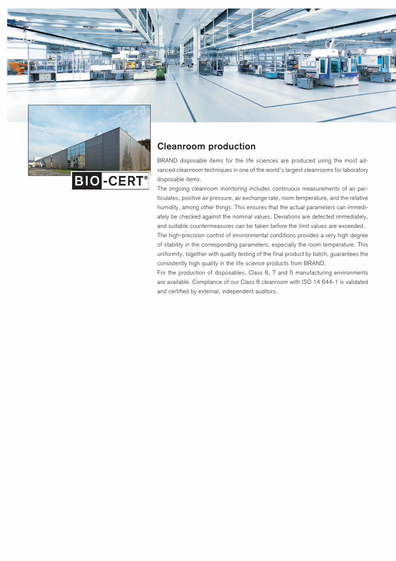
Cleanroom productionBRAND disposable items for the life sciences are produced using the most ad-
vanced cleanroom techniques in one of the world’s largest cleanrooms for laboratory
disposable items.
The ongoing cleanroom monitoring includes continuous measurements of air par-
ticulates, positive air pressure, air exchange rate, room temperature, and the relative
humidity, among other things. This ensures that the actual parameters can immedi-
ately be checked against the nominal values. Deviations are detected immediately,
and suitable countermeasures can be taken before the limit values are exceeded.
The high-precision control of environmental conditions provides a very high degree
of stability in the corresponding parameters, especially the room temperature. This
uniformity, together with quality testing of the final product by batch, guarantees the
consistently high quality in the life science products from BRAND.
For the production of disposables, Class 8, 7 and 5 manufacturing environments
are available. Compliance of our Class 8 cleanroom with ISO 14 644-1 is validated
and certified by external, independent auditors.
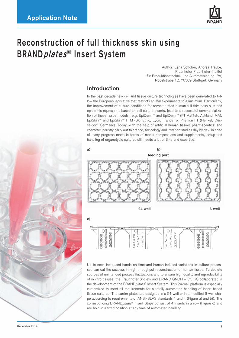
a) b)
c)
24-well 6-well
feeding port
Application Note
December 2014 3
Reconst ruct ion o f fu l l th ickness sk in us ing BRANDpla tes® Inser t System
Author: Lena Schober, Andrea Traube; Fraunhofer Fraunhofer-Institut
für Produktionstechnik und Automatisierung IPA, Nobelstraße 12, 70569 Stuttgart, Germany
IntroductionIn the past decade new cell and tissue culture technologies have been generated to fol-low the European legislative that restricts animal experiments to a minimum. Particularly, the improvement of culture conditions for reconstructed human full thickness skin and epidermis equivalents based on cell culture inserts, lead to a successful commercializa-tion of these tissue models , e.g. EpiDerm™ and EpiDerm™ (FT MatTek, Ashland, MA), EpiSkin™ and EpiSkin™ FTM (SkinEthic, Lyon, France) or Phenion FT (Henkel, Düs-seldorf, Germany). Today, with the help of artificial human tissues pharmaceutical and cosmetic industry carry out tolerance, toxicology and irritation studies day by day. In spite of every progress made in terms of media compositions and supplements, setup and handling of organotypic cultures still needs a lot of time and expertise.
Up to now, increased hands-on time and human-induced variations in culture proces-ses can cut the success in high throughput reconstruction of human tissue. To deplete sources of unintended process fluctuations and to ensure high quality and reproducibility of in vitro tissues, the Fraunhofer Society and BRAND GMBH + CO KG collaborated in the development of the BRANDplates® Insert System. This 24-well platform is especially customized to meet all requirements for a totally automated handling of insert-based tissue cultures. The carrier plates are designed in a 24-well or in a modified 6-well sha-pe according to requirements of ANSI/SLAS standards 1 and 4 (Figure a) and b)). The corresponding BRANDplates® Insert Strips consist of 4 inserts in a row (Figure c) and are hold in a fixed position at any time of automated handling.

Application Note
4 December 2014
6-well plate:
- Use just one or two inserts per well to extend medium change interval.
- For up to 4 inserts, medium in the well can be changed in one step (Figure b, pag. 3).
Inlet Opening System (IOS)
- No leaking during cell seeding or initial coa-ting.
- Simultaneous change of medium in the well and insert.
- Setup of air-liquid-interface in one step.
- Compatible with 24- und 6-well BRANDplates®.
Insert:
- Divided BRANDplates® Insert Strips for sub-sequent analysis.
BRANDplates® Inserts are available with the Inlet Opening System (patent pending, Figure c, pag. 3) which is dedicated to support the automated in vitro reconstruction of human skin. This peerless feature interconnects the medium of wells and inserts, giving the opportunity to establish the air-liquid interface without entering the inserts with pipet-te tips. In addition to this increase in safety for cultures, the IOS reduces the number of pipetting steps needed to change medium within the two compartments.
This user manual describes in short the reconstruction of full thickness skin equivalents and provides tips for the handling of BRANDplates® Insert System.
Volumes needed for different culture phases
24-well 6-well
Insert(e.g., coating, cell seeding)
50 - 400 µl 50 - 400 µl
Well: submerged culture 1.6 - 2 ml 8 - 10 ml
Well: air-liquid interface (wetted membrane)
0.8 ml 3.5 ml
1) Coating, cell seeding2) Medium application for
submerge culture
3) Medium change 4) Establish air-liquid interface
air-liquid interface
feeding port
InsertIOS
well
membrane
Application Note
December 2014 5
I . P reparat ion o f dermal components fo r fu l l th ickness sk in equ iva lents
5 min, 400 x g
5 min, 400 x g
transfer 4x10⁴ cells/equivalent into a new cen-
trifuge tube
sub cultivation
resuspend fibroblasts in required volume of collagen gel neutralizing medium
harvest cells
Splitting of fibroblasts:
1. Aspirate culture medium.2. Wash culture with prewarmed PBS.3. Add 0.25 % trypsin/EDTA, incubate for 5 min at 37°C, 5 % CO2.
- T 25 flask: 2 ml - T 75 flask: 4 ml - T175 flask: 8 ml
4. Stop trypsin digestion by DMEM + 10 % FCS.5. Transfer fibroblasts into a centrifuge tube.
2. Preparation of Collagen type I fibroblast mixture
on ice
collagen type I 266 µl/insert
fibroblasts 4x10⁴ in 133 µl/insert
3. Establishing fibroblast – collagen submerse culture
400 µl/insert
gel polymerization:
10-20 min at37°C, 5 % CO2
submers culture:2 ml of medium/well + 0.2 ml medium on
top of each insert take advantage of IOS:
2.2 ml medium/well and insert in one
step Medium:- DMEM, high glucose+ Glutamine+ Pyruvate + 10 % FCS+ 1 % Gentamycin
> 400 µl collagen - fibroblasts mix is needed per demal equivalent.
Advice: calculate an extra volume of 30 % to compensate for pipetting errors!> 4.0 x 10⁴ fibroblasts/400 µl> 5.2 x 10⁴ fibroblasts/520 µl
520 µl total
347 µl collagen
173 µl gel neutralization
medium
Gel neutralizing medium contains:1. 25 µl/ml Chondroitin-4-sulfate
(gel cross linker)2. 25 µl/ml Chondroitin-6-sulfate
(gel cross linker)3. 2 x DMEM4. 90 mM HEPES5. 3 % FCS
> Recommended collagen type I concentrati-on: 6 mg/ml.
> Preparation of collagen mixture on ice impe-de risk of premature gelation.
> Use chilled pipets while mixing collagen.> Avoid air bubbles!
> Collagen type I is usually dissolved in acetic acid (0.1 M, pH 3) and needs to be neu-tralized by titration of NaOH before adding cells.
> Alternatively use medium containing HEPES and/or sodium dicarbonate.
> Use inserts with membranes of 8 µm pore size!
Use of BRANDplates® Inserts with Inlet Opening System (IOS)> Prevents culture damaging during medium
application/changes.> Reduced number of pipetting step is needed
for medium application/changes.> Provides lateral nutrient supply for collagen
embedded fibroblasts.
Volume for submers culture in BRANDplates®:> 24 well: 2 ml> 6 well: 10 ml
homogenously suspend fibroblasts in collagen
on ice
ready to use col-lagen – fibroblasts
mixture
Protocol according to Promocell
1. Passaging of primary fibroblasts
Application Note
6 December 2014
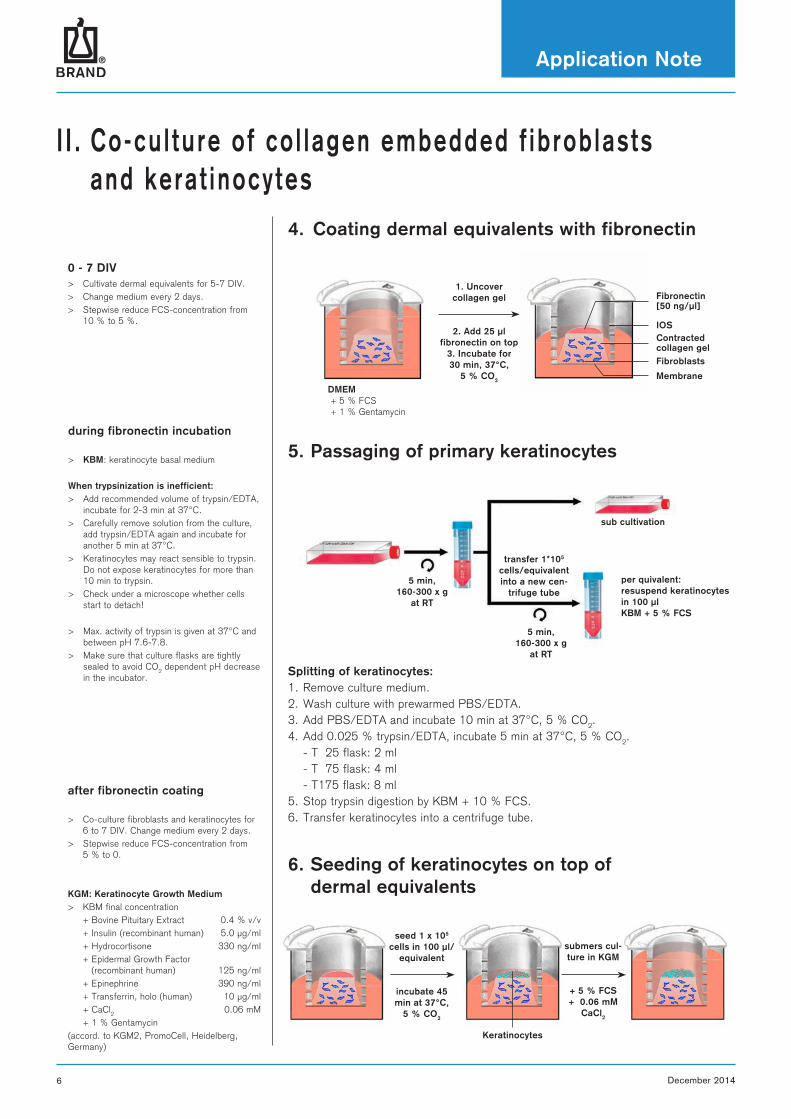
I I . Co-cu l ture o f co l lagen embedded f ib rob las ts and kera t inocytes
5. Passaging of primary keratinocytes
5 min, 160-300 x g
at RT
5 min, 160-300 x g
at RT
transfer 1*105 cells/equivalent into a new cen-
trifuge tube
sub cultivation
per quivalent: resuspend keratinocytes in 100 µl KBM + 5 % FCS
1. Uncover collagen gel
2. Add 25 µl fibronectin on top
3. Incubate for 30 min, 37°C,
5 % CO2
DMEM + 5 % FCS + 1 % Gentamycin
Fibronectin [50 ng/µl]
IOSContracted collagen gelFibroblasts
Membrane
Splitting of keratinocytes:1. Remove culture medium.2. Wash culture with prewarmed PBS/EDTA.3. Add PBS/EDTA and incubate 10 min at 37°C, 5 % CO2.4. Add 0.025 % trypsin/EDTA, incubate 5 min at 37°C, 5 % CO2. - T 25 flask: 2 ml - T 75 flask: 4 ml - T175 flask: 8 ml5. Stop trypsin digestion by KBM + 10 % FCS.6. Transfer keratinocytes into a centrifuge tube.
6. Seeding of keratinocytes on top of dermal equivalents
seed 1 x 10⁵ cells in 100 µl/
equivalent
incubate 45 min at 37°C,
5 % CO2
submers cul-ture in KGM
+ 5 % FCS+ 0.06 mM
CaCl2
Keratinocytes
0 - 7 DIV > Cultivate dermal equivalents for 5-7 DIV.> Change medium every 2 days.> Stepwise reduce FCS-concentration from
10 % to 5 %.
during fibronectin incubation
> KBM: keratinocyte basal medium
When trypsinization is inefficient:> Add recommended volume of trypsin/EDTA,
incubate for 2-3 min at 37°C. > Carefully remove solution from the culture,
add trypsin/EDTA again and incubate for another 5 min at 37°C.
> Keratinocytes may react sensible to trypsin. Do not expose keratinocytes for more than 10 min to trypsin.
> Check under a microscope whether cells start to detach!
> Max. activity of trypsin is given at 37°C and between pH 7.6-7.8.
> Make sure that culture flasks are tightly sealed to avoid CO2 dependent pH decrease in the incubator.
after fibronectin coating
> Co-culture fibroblasts and keratinocytes for 6 to 7 DIV. Change medium every 2 days.
> Stepwise reduce FCS-concentration from 5 % to 0.
KGM: Keratinocyte Growth Medium > KBM final concentration + Bovine Pituitary Extract 0.4 % v/v + Insulin (recombinant human) 5.0 µg/ml + Hydrocortisone 330 ng/ml + Epidermal Growth Factor
(recombinant human) 125 ng/ml + Epinephrine 390 ng/ml + Transferrin, holo (human) 10 µg/ml + CaCl2 0.06 mM + 1 % Gentamycin(accord. to KGM2, PromoCell, Heidelberg, Germany)
4. Coating dermal equivalents with fibronectin
stratum corneumstratum granulosum
stratum spinosum
stratum basale
dermis
picture © Frauenhofer IGB
suprabasal layer
Application Note
December 2014 7
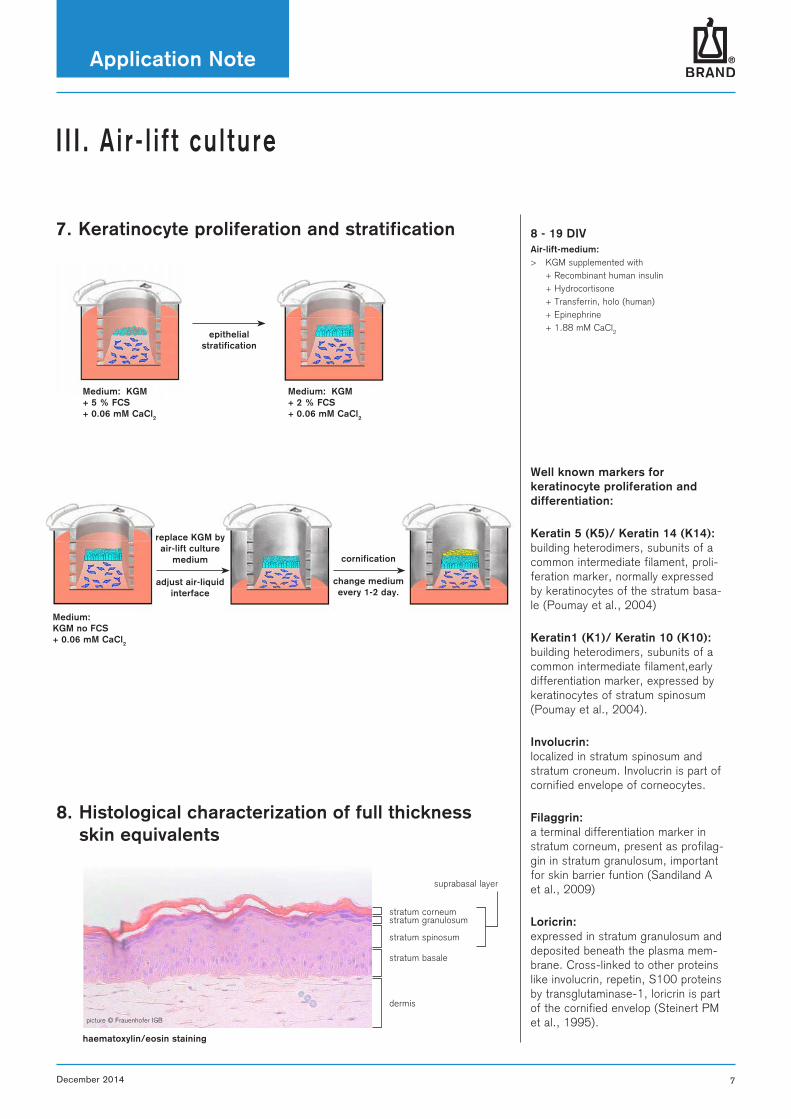
I I I . A i r - l i f t cu l ture
epithelial stratification
Medium: KGM + 5 % FCS + 0.06 mM CaCl2
Medium: KGM + 2 % FCS + 0.06 mM CaCl2
Medium: KGM no FCS+ 0.06 mM CaCl2
replace KGM by air-lift culture
medium
adjust air-liquid interface
cornification
change medium every 1-2 day.
8. Histological characterization of full thickness skin equivalents
haematoxylin/eosin staining
8 - 19 DIVAir-lift-medium:> KGM supplemented with + Recombinant human insulin + Hydrocortisone + Transferrin, holo (human) + Epinephrine + 1.88 mM CaCl2
Well known markers for keratinocyte proliferation and differentiation:
Keratin 5 (K5)/ Keratin 14 (K14): building heterodimers, subunits of a common intermediate filament, proli-feration marker, normally expressed by keratinocytes of the stratum basa-le (Poumay et al., 2004)
Keratin1 (K1)/ Keratin 10 (K10): building heterodimers, subunits of a common intermediate filament,early differentiation marker, expressed by keratinocytes of stratum spinosum(Poumay et al., 2004).
Involucrin: localized in stratum spinosum and stratum croneum. Involucrin is part of cornified envelope of corneocytes.
Filaggrin: a terminal differentiation marker in stratum corneum, present as profilag-gin in stratum granulosum, important for skin barrier funtion (Sandiland A et al., 2009)
Loricrin: expressed in stratum granulosum and deposited beneath the plasma mem-brane. Cross-linked to other proteins like involucrin, repetin, S100 proteins by transglutaminase-1, loricrin is part of the cornified envelop (Steinert PM et al., 1995).
7. Keratinocyte proliferation and stratification
BRAND GMBH + CO KG · P.O. Box 11 55 · 97861 Wertheim · GermanyTel.: +49 9342 808-0 · Fax: +49 9342 808-98000 · E-Mail: [email protected] · Internet: www.brand.deP
rinte
d in
Ger
man
y ·
WA
/121
4
BRANDplates®, BIO-CERT® and BRAND® are trademarks of BRAND GMBH + CO KG, Germany.
Other reproduced brands are the property of the respective owner.
Our technical literature is intended to inform and advise our customers. However, the validity of general empirical values, and of
results obtained under test conditions, for specific applications depends on many factors beyond our control. Please appreciate,
therefore, that no claims can be derived from our advice. The user is responsible for checking the appropriateness of the product
for any particular application.
Subject to technical modification without notice. Errors excepted.
Ordering Data
BRANDplates® Insert StripsInsert Strips, smooth-walled or with inlet channels (Inlet Opening System*)
PS. cellGrade™ plus surface, sterile. Strips of 4 inserts (divisible).
Description Pore size µm
Pack of PC membraneCat. No.
PET membraneCat. No.
smooth-walled 8.0 12 (individually wrapped) 7828 60 7828 70
with Inlet Opening System 8.0 12 (individually wrapped) 7828 61 7828 71
* patent pending
BRANDplates® Insert System6-well plates filled with 6 insert strips. PS. cellGrade™ plus surface, sterile. Insert strips, smooth-walled or with inlet channels (Inlet Opening System*). With lid with condensation rings.
Description Pore size µm
Pack ofplates with lid
PC membraneCat. No.
PET membraneCat. No.
smooth-walled 8.0 5 (30 insert strips) 7828 62 7828 72
with Inlet Opening System 8.0 5 (30 insert strips) 7828 63 7828 73
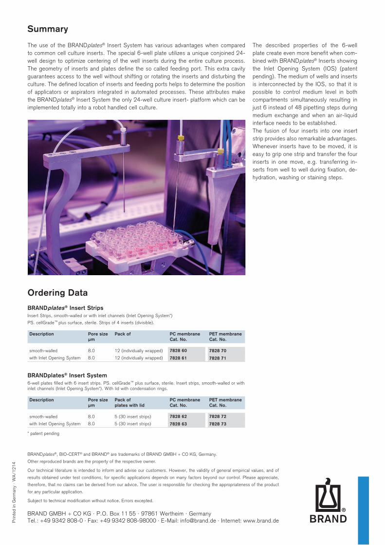
The use of the BRANDplates® Insert System has various advantages when compared to common cell culture inserts. The special 6-well plate utilizes a unique conjoined 24-well design to optimize centering of the well inserts during the entire culture process. The geometry of inserts and plates define the so called feeding port. This extra cavity guarantees access to the well without shifting or rotating the inserts and disturbing the culture. The defined location of inserts and feeding ports helps to determine the position of applicators or aspirators integrated in automated processes. These attributes make the BRANDplates® Insert System the only 24-well culture insert- platform which can be implemented totally into a robot handled cell culture.
The described properties of the 6-well plate create even more benefit when com-bined with BRANDplates® Inserts showing the Inlet Opening System (IOS) (patent pending). The medium of wells and inserts is interconnected by the IOS, so that it is possible to control medium level in both compartments simultaneously resulting in just 6 instead of 48 pipetting steps during medium exchange and when an air-liquid interface needs to be established.The fusion of four inserts into one insert strip provides also remarkable advantages. Whenever inserts have to be moved, it is easy to grip one strip and transfer the four inserts in one move, e.g. transferring in-serts from well to well during fixation, de-hydration, washing or staining steps.
Summary
Top Related