


Languages
Pages
Legal

台大農藝系 遺傳學 601
20000
Chapter 1 slide 1
Cytogenetics
MGL-3
Feb 17th 2013
Mohammed El-Khateeb
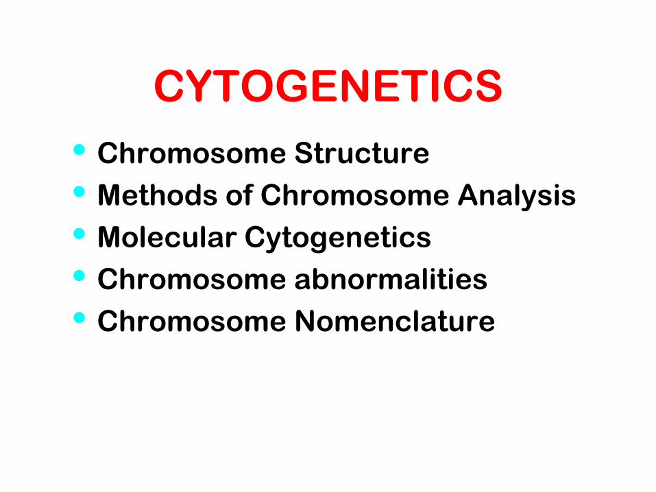
CYTOGENETICS
• Chromosome Structure
• Methods of Chromosome Analysis
• Molecular Cytogenetics
• Chromosome abnormalities
• Chromosome Nomenclature
Cytogenetics
The study of chromosome number, structure, function, and behavior in
relation to gene inheritance, organization and expression
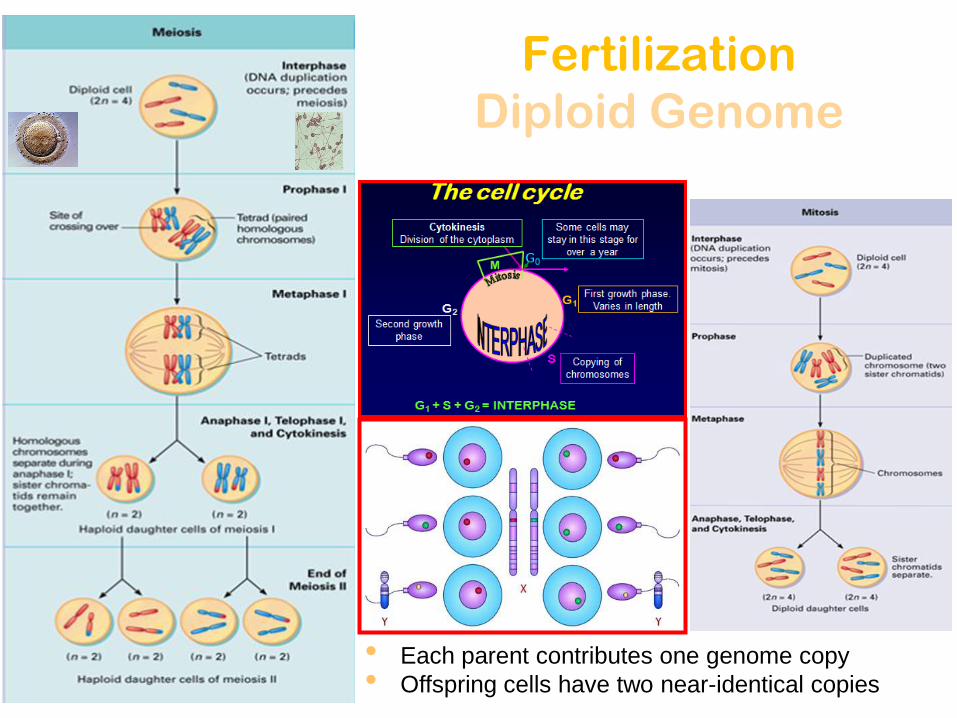
Fertilization
Diploid Genome
• Each parent contributes one genome copy
• Offspring cells have two near-identical copies

DNA Coiling Leading to the Visible
Structure of Chromosomes
DNA Nucleosomes Chromatin Fiber Loop Chromosome
Primary coiling of DNA double helix
Secondary coiling of DNA double helix
around the histone proteins to form
nucleosomes
Tetiary coiling of nucleosomes to form
chromatin fibres
Loops of chromatin fiber forming the
chromosome
2nm 10 nm
300 nm 700 nm

Sister Chromatides
Chromosome Chromo = colored in response to dye Some = body
Chromosome of Eukaryotes have been the traditional subject for cytogenetic analysis because they are large enough to be examined using light microscope
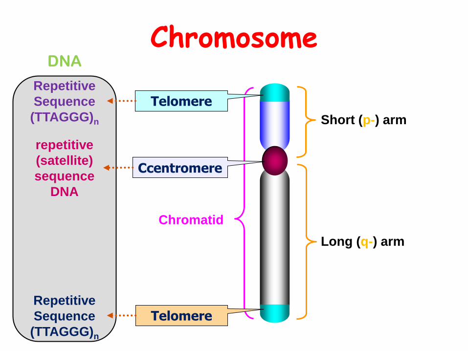
Chromosome
Short (p-) arm
Long (q-) arm
Ccentromere
Chromatid
Telomere
Telomere
repetitive
(satellite)
sequence
DNA
Repetitive
Sequence
(TTAGGG)n
Repetitive
Sequence
(TTAGGG)n
DNA
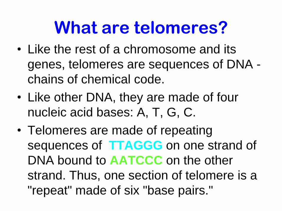
What are telomeres? • Like the rest of a chromosome and its
genes, telomeres are sequences of DNA -
chains of chemical code.
• Like other DNA, they are made of four
nucleic acid bases: A, T, G, C.
• Telomeres are made of repeating
sequences of TTAGGG on one strand of
DNA bound to AATCCC on the other
strand. Thus, one section of telomere is a
"repeat" made of six "base pairs."

DNA DNA Sequence for
Telomeres:
ttagggttagggttaggg…
||||||||||||||||||
aatcccaatcccaatccc…
Head
Telomere
Centromere
Tail
Telomere
NOTICE:
Tandem Repeats in Telomeres:
ttagggttagggttaggg…
||||||||||||||||||
aatcccaatcccaatccc…
Repeated 800-1600 times in each Telomere
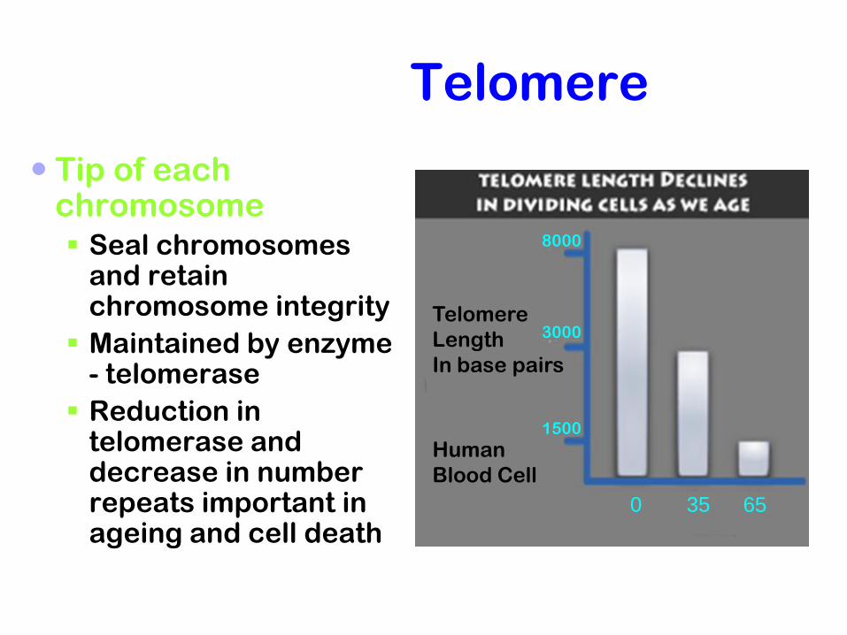
Telomere
Tip of each chromosome Seal chromosomes
and retain chromosome integrity
Maintained by enzyme - telomerase
Reduction in telomerase and decrease in number repeats important in ageing and cell death
Telomere
Length
In base pairs
Human
Blood Cell
0 35 65
1500
3000
8000
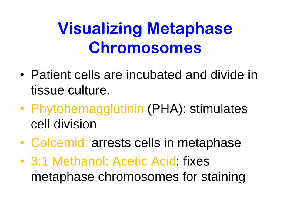
Visualizing Metaphase
Chromosomes
• Patient cells are incubated and divide in
tissue culture.
• Phytohemagglutinin (PHA): stimulates
cell division
• Colcemid: arrests cells in metaphase
• 3:1 Methanol: Acetic Acid: fixes
metaphase chromosomes for staining

Add a few drops of blood.
Add phytohemagglutinin to stimulate mitosis.
Draw 10 to 20 ml of blood.
Incubate at 37°C for 2 to 3 days.
Transfer to tube containing fixative.
Transfer cells to tube.
Add Colcemid to culture for 1 to 2 hours to stop mitosis in metaphase.
Centrifuge to concentrate cells. Add low-salt solution to eliminate red blood cells and swell lymphocytes.
Drop cells onto microscope slide.
Examine with microscope.
Digitized chromosome images processed to make karyotype.
Stain slide with Giemsa.
The steps in the process of creating a
karyotype for chromosome analysis. Preparation
of G banded
karyotype
•Peripheral blood
•Fibroblasts from skin bx
•Epithelial cells from
buccal smear
•Bone marrow
•Solid tumor biobsies
Specimens

Chromosome Number in
different animals and plants
• Human 46 • Chimpanzee 48 • Dog 78 • Horse 64 • Chicken 78 • Goldfish 94 • Fruit fly 8 • Mosquito 6 • Nematode 11(m), 12(f) • Horsetail 216 • Sequoia 22 • Round worm 2
• Onion 16 • Mold 16 • Carrot 20 • Tomato 24 • Tobacco 48 • Rice 24 • Maize 20 • Haploppus gracilis 4 • Crepis capillaris 6
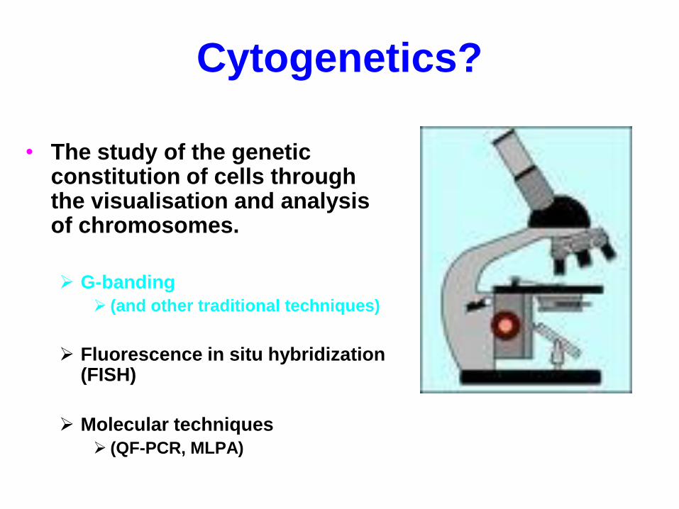
Cytogenetics?
• The study of the genetic constitution of cells through the visualisation and analysis of chromosomes.
G-banding
(and other traditional techniques)
Fluorescence in situ hybridization (FISH)
Molecular techniques
(QF-PCR, MLPA)
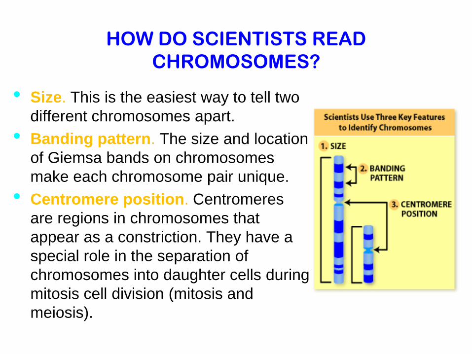
HOW DO SCIENTISTS READ
CHROMOSOMES?
• Size. This is the easiest way to tell two
different chromosomes apart.
• Banding pattern. The size and location
of Giemsa bands on chromosomes
make each chromosome pair unique.
• Centromere position. Centromeres
are regions in chromosomes that
appear as a constriction. They have a
special role in the separation of
chromosomes into daughter cells during
mitosis cell division (mitosis and
meiosis).
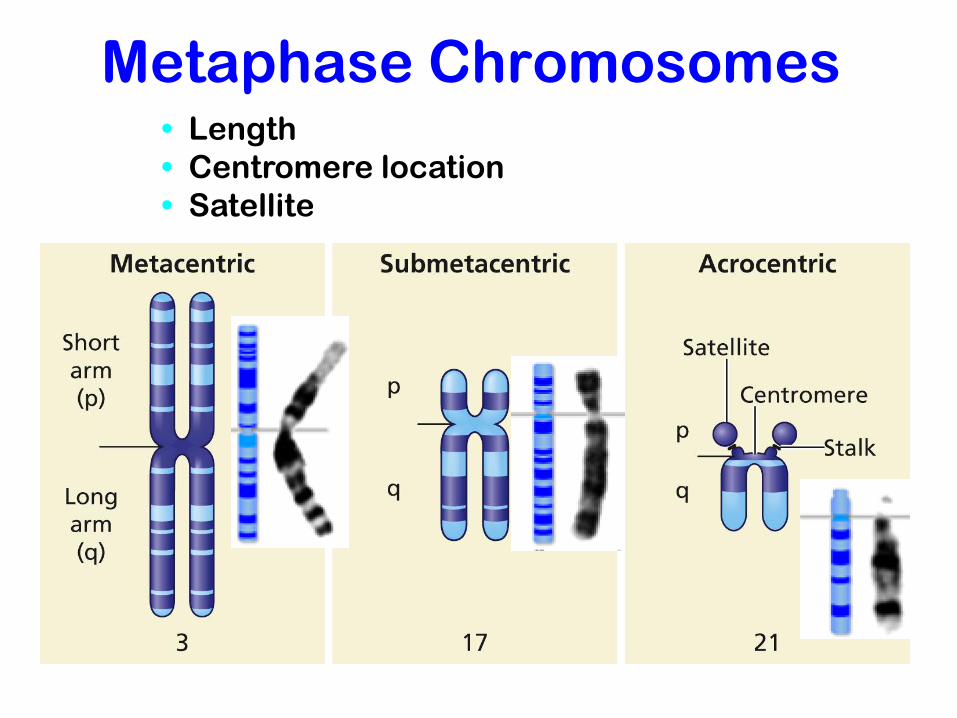
Metaphase Chromosomes
• Length
• Centromere location
• Satellite

Chromosome in general
(size, shape and number)
Two sister chromatids per chromosome
DNA replication chromatids Two sister chromatids joined
together at centromeres chromosomes differ in
size and appearance with staining
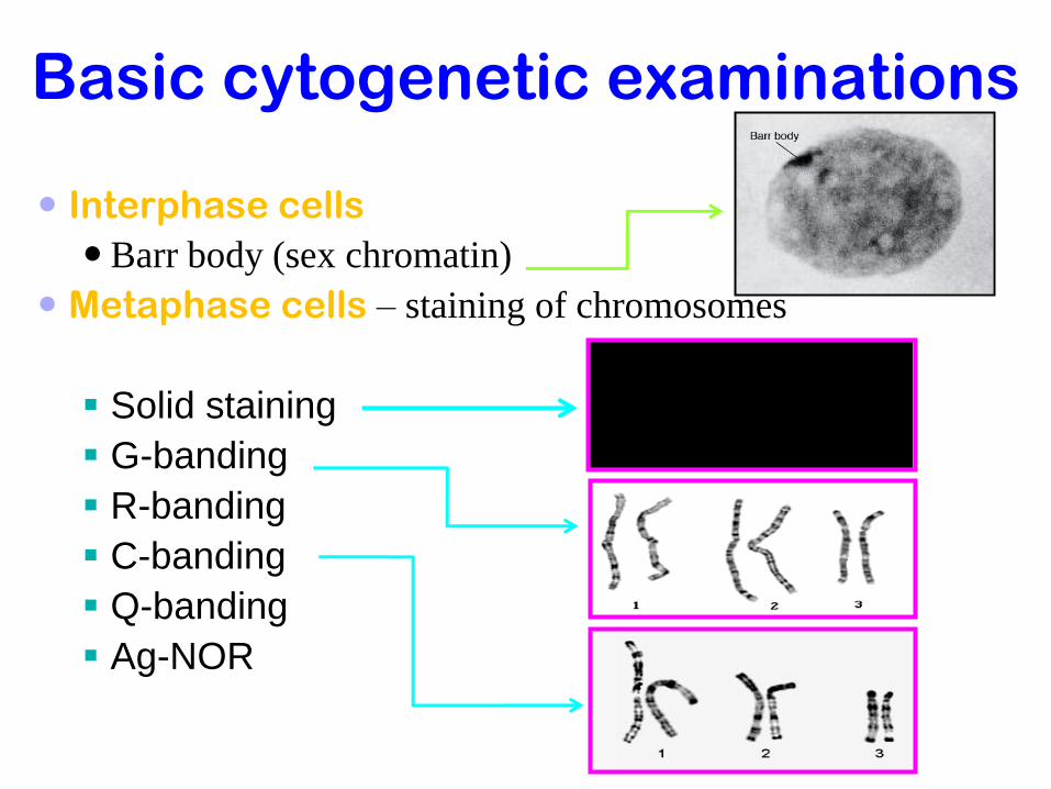
Basic cytogenetic examinations
Interphase cells
Barr body (sex chromatin)
Metaphase cells – staining of chromosomes
Solid staining
G-banding
R-banding
C-banding
Q-banding
Ag-NOR

Centromere
Joins sister chromatids
Essential for chromosome segregation at cell division
100s of kb of repetitive DNA: some non-specific, some chromosome specific
Dark (G) bands
Replicate late
Contain condensed chromatin
AT rich
Short arm
p (petit)
Long arm
q
q
Light bands
Replicate early in S phase
Less condensed chromatin
Transcriptionally active
Gene and GC rich
Telomere
DNA and protein cap
Ensures replication to tip
Tether to nuclear membrane
provide terminal stability to the
chromosome and ensure its survival
Telomere
Chromosomes as seen at metaphase
during cell division

Chromosomes Banding
Effect Area Stained Stain Type
Under UV light, distinct
fluorescent banded pattern
for each chromosome.
Chromosome arms; mostly
repetitive AT-rich DNA
Quinacrine Q-banding
Distinct banded pattern for
each chromosome; same as
Q-banding pattern except
single additional band near
centromere of chromosomes
1 and 16
Chromosome arms; mostly
repetitive AT-rich DNA
Giemsa G-banding
Reverse banding pattern of
that observed with Q- or G-
banding
Chromosome arms; mostly
unique GC-rich DNA
Variety of
techniques
R-banding
Largest bands usually on
chromosomes 1, 9, 16, and Y;
chromosomes 7, 10, and 15
have medium-sized bands;
size of C-bands highly
variable from person to
person
Centromere region of each
chromosome and distal portion
of Y chromosome; highly
repetitive, mostly AT-rich DNA
Variety of
techniques
C-banding
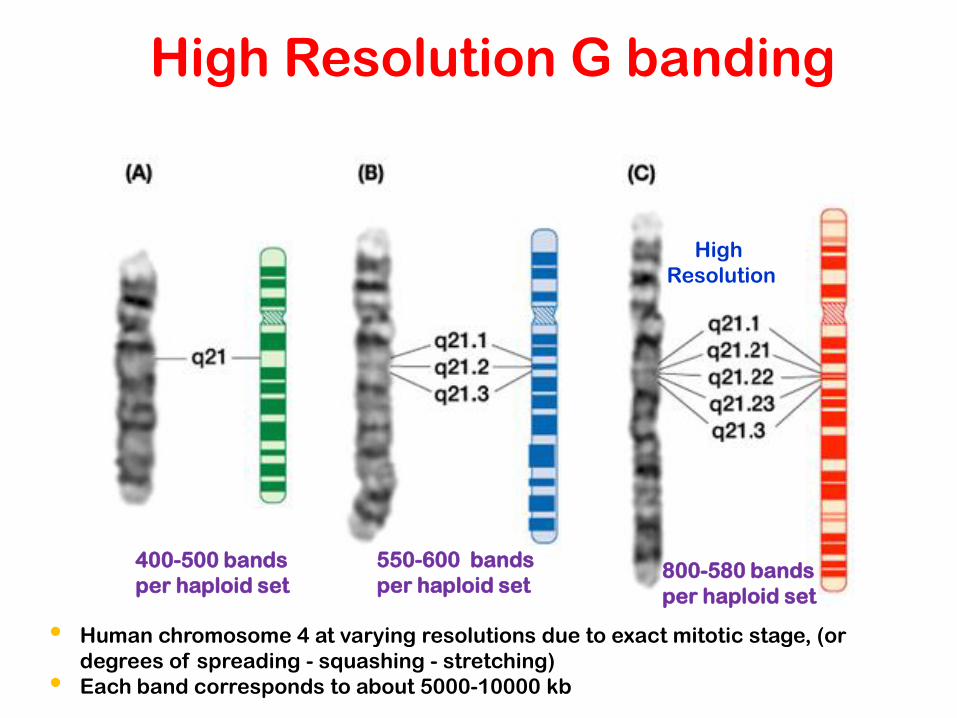
• Human chromosome 4 at varying resolutions due to exact mitotic stage, (or
degrees of spreading - squashing - stretching)
• Each band corresponds to about 5000-10000 kb
400-500 bands
per haploid set
550-600 bands
per haploid set 800-580 bands
per haploid set
High Resolution G banding
High
Resolution
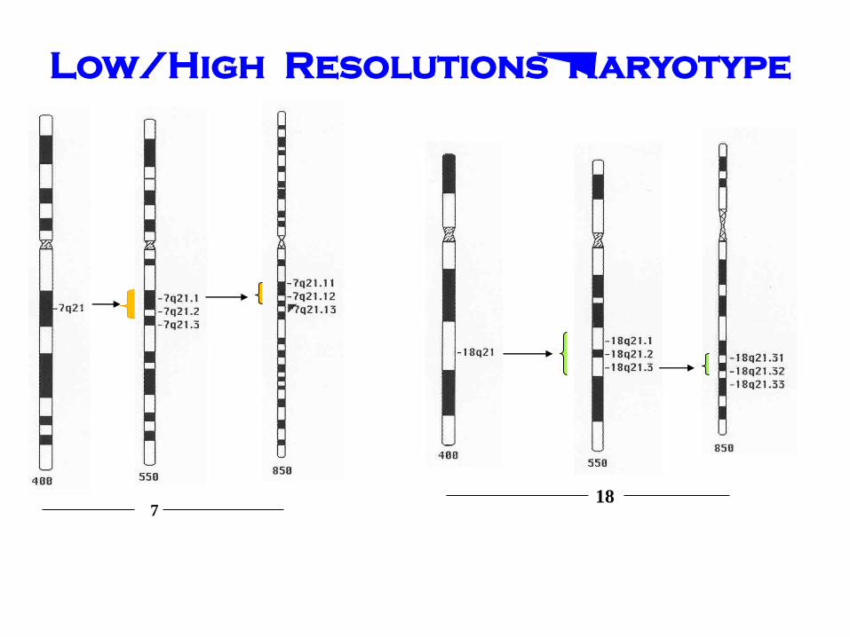
7 18
Low/High Resolutions Karyotype
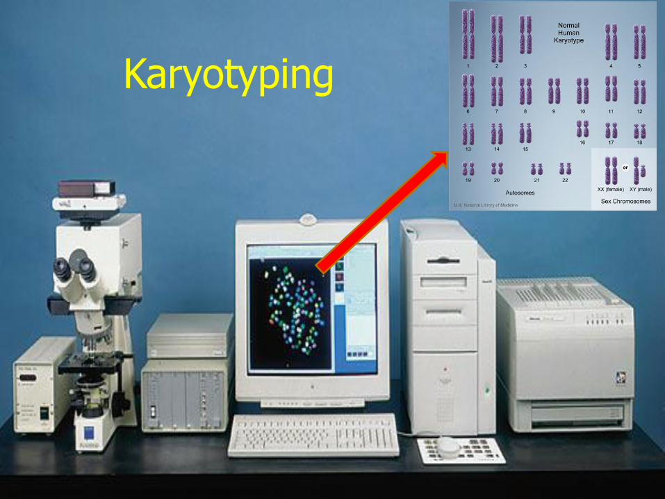
Karyotyping
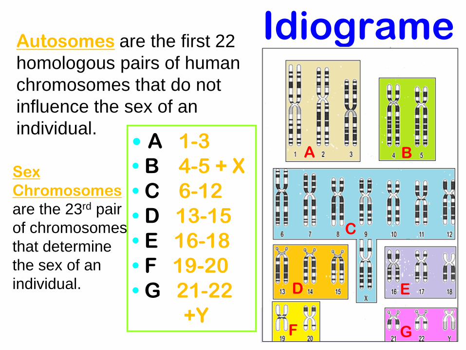
Idiograme
• A 1-3
• B 4-5 + X
• C 6-12
• D 13-15
• E 16-18
• F 19-20
• G 21-22
+Y
Autosomes are the first 22
homologous pairs of human
chromosomes that do not
influence the sex of an
individual.
Sex
Chromosomes
are the 23rd pair
of chromosomes
that determine
the sex of an
individual.
A B
C
D E
F G
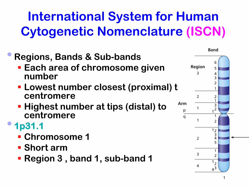
International System for Human
Cytogenetic Nomenclature (ISCN)
• Regions, Bands & Sub-bands
Each area of chromosome given number
Lowest number closest (proximal) to centromere
Highest number at tips (distal) to centromere
• 1p31.1
Chromosome 1
Short arm
Region 3 , band 1, sub-band 1
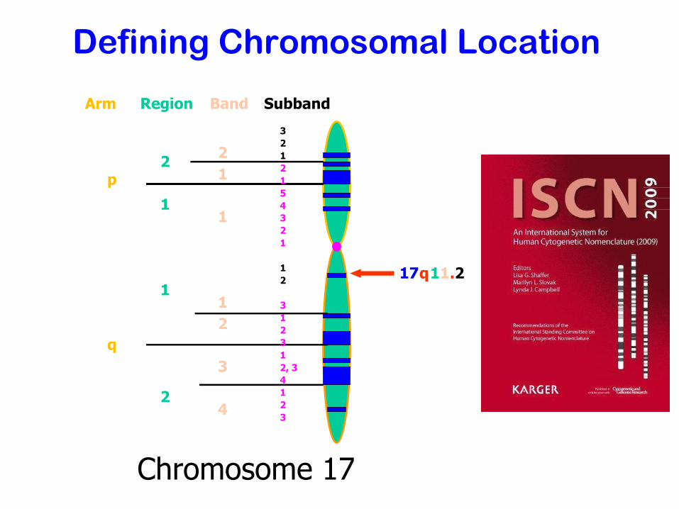
p
q
Arm Region Band Subband
2
1
1
2
2
1
1
1
2
3
4
3
2
1
2
1
5
4
3
2
1
1
2
3
1
2
3
1
2, 3
4
1
2
3
17 q 1 1 . 2
Chromosome 17
Defining Chromosomal Location
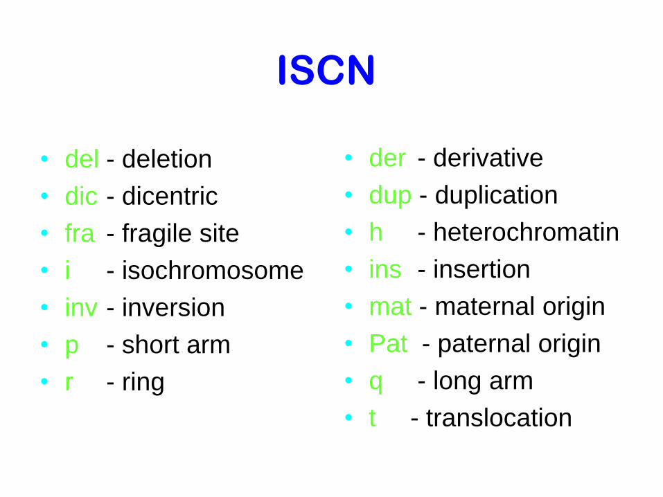
ISCN
• del - deletion
• dic - dicentric
• fra - fragile site
• i - isochromosome
• inv - inversion
• p - short arm
• r - ring
• der - derivative
• dup - duplication
• h - heterochromatin
• ins - insertion
• mat - maternal origin
• Pat - paternal origin
• q - long arm
• t - translocation
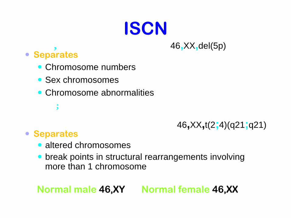
ISCN , 46,XX,del(5p) Separates
Chromosome numbers
Sex chromosomes
Chromosome abnormalities
;
46,XX,t(2;4)(q21;q21) Separates
altered chromosomes
break points in structural rearrangements involving more than 1 chromosome
Normal male 46,XY Normal female 46,XX

Cytogenetics?
• The study of the genetic constitution of cells through the visualisation and analysis of chromosomes.
– G-banding
(and other traditional techniques)
Fluorescence in situ hybridization (FISH)
– Molecular techniques
(QF-PCR, MLPA)
Molecular Cytogenetics
Fluorescent Inistu Hypridization (FISH)
Different Fish Probes Centromeric Probe
Chromosome specific unique sequence probe
Whole chromosome point probe
Reverse painting
Multicolor spectral karyotyping
Comparative Genomic Hypridization (CGH)
Flowcytometry

A G G C T
A
T
T C C G
A T
A COVALENT
BOND
F
F
Specimen DNA
FISH Probe DNA
DIRECT FLUORESCENT -
LABELED PROBE

FISH technique is based on the unique ability of a
single stranded piece of DNA (probe) to anneal or
hybridize with its complementary target sequence on
the chromosome
Advantages of Interphase
FISH
Interphase cells for FISH do not require culturing
of the cells and stimulating division to get
metaphase spreads
200–500 cells can be analyzed microscopically
using FISH
Monitor recurrent or residual disease in BMT pt.
33

Metaphase FISH
Uses fluorescent probes that bind
to metaphase chromosomal
regions or to whole chromosomes.
Whole chromosome paints:
Probes that cover the entire
chromosome, are valuable for
detecting small rearrangements
that are not apparent by regular
chromosome banding.
Telomeric and centromeric probes
are also applied to metaphase
chromosomes to detect
aneuploidy and structural
abnormalities 34
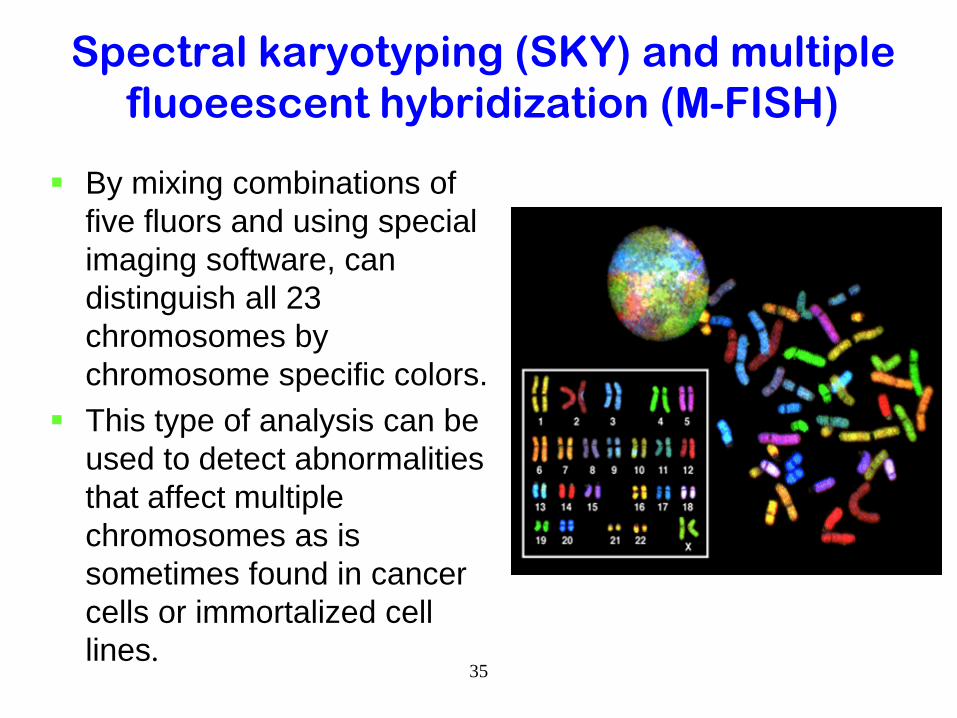
Spectral karyotyping (SKY) and multiple
fluoeescent hybridization (M-FISH)
By mixing combinations of
five fluors and using special
imaging software, can
distinguish all 23
chromosomes by
chromosome specific colors.
This type of analysis can be
used to detect abnormalities
that affect multiple
chromosomes as is
sometimes found in cancer
cells or immortalized cell
lines. 35
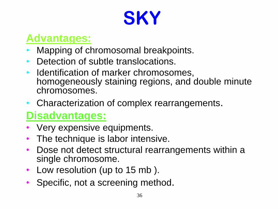
SKY Advantages: • Mapping of chromosomal breakpoints.
• Detection of subtle translocations.
• Identification of marker chromosomes, homogeneously staining regions, and double minute chromosomes.
• Characterization of complex rearrangements.
Disadvantages: • Very expensive equipments.
• The technique is labor intensive.
• Dose not detect structural rearrangements within a single chromosome.
• Low resolution (up to 15 mb ).
• Specific, not a screening method. 36
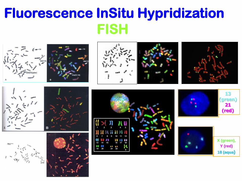
Fluorescence InSitu Hypridization
FISH
X (green),
Y (red)
18 (aqua)
13 (green)
21 (red)
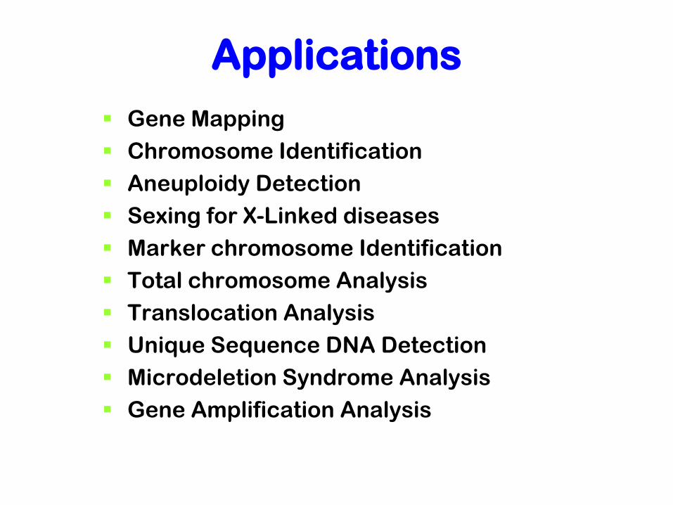
Applications
Gene Mapping
Chromosome Identification
Aneuploidy Detection
Sexing for X-Linked diseases
Marker chromosome Identification
Total chromosome Analysis
Translocation Analysis
Unique Sequence DNA Detection
Microdeletion Syndrome Analysis
Gene Amplification Analysis

Cytogenetics?
• The study of the genetic constitution of cells through the visualisation and analysis of chromosomes.
– G-banding
(and other traditional techniques)
– Fluorescence in situ hybridization (FISH)
Molecular techniques
(CGH, QF-PCR, MLPA, Microarray)

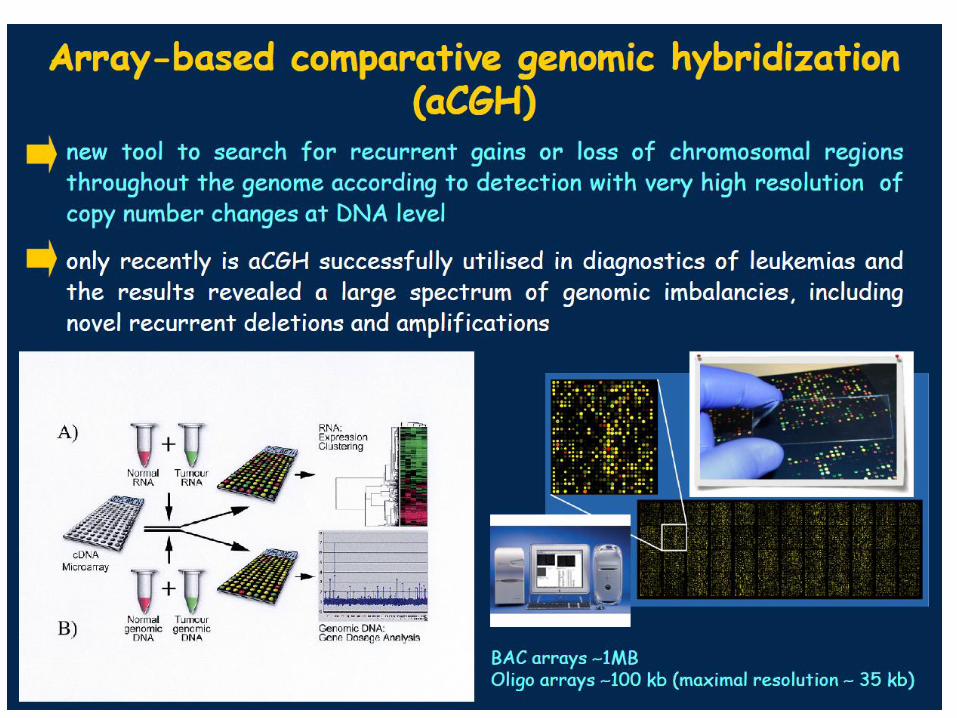
Methods:
• Isolate Genomic DNA from samples
• DNA digestion
• Label patient and control samples
• Hybridize to microarray
• Post hybridization washing
• Assay scanning and data analysis
Comparative Ggenomic
Hypridization (CGH)
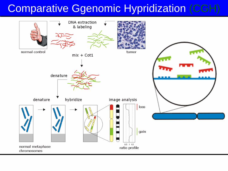
Comparative Ggenomic Hypridization (CGH)

Comparative Genomic Hybridisation
(CGH)
Amplified gene = Green Reduction of gene = Red
Flourochrom ratio = o.5 – 1.5
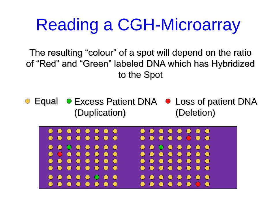
Reading a CGH-Microarray
The resulting “colour” of a spot will depend on the ratio
of “Red” and “Green” labeled DNA which has Hybridized
to the Spot
Equal Excess Patient DNA
(Duplication)
Loss of patient DNA
(Deletion)
Indications - Postnatal
• Multiple congenital anomalies
• Developmental delay/ mental retardation
of unknown origin
• Autism
• Any individual suspected of a
chromosomal imbalance, even with
normal karyotype
• High resolution mapping to identify
specific genes
Current Uses of Array CGH
• Define congenital genetic defects
• Define acquired genetic changes (in cancer)
• Molecular fingerprints of specific tumors and subtypes
• Identification of novel chromosomal regions for drug targets and new treatments
CGH
Advantages
whole genome in 1 experiment
no need to culture tumor cells
sensitive detection of gene amplification
Disadvantages
limited resolution (~10 Mb del/dup)
laborious
only gains and losses / no balanced rearrangements
no information on the nature of the aberrations
retrospective analysis
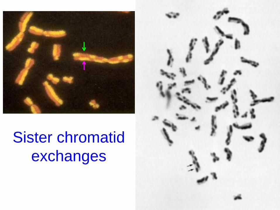
Sister chromatid
exchanges

CHROMOSOMAL
ABNORMALITIES
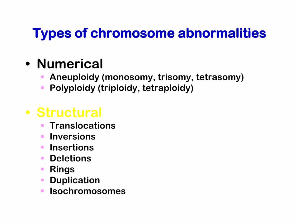
Types of chromosome abnormalities
• Numerical Aneuploidy (monosomy, trisomy, tetrasomy)
Polyploidy (triploidy, tetraploidy)
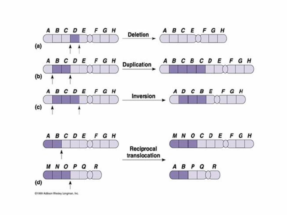
• Structural Translocations
Inversions
Insertions
Deletions
Rings
Duplication
Isochromosomes
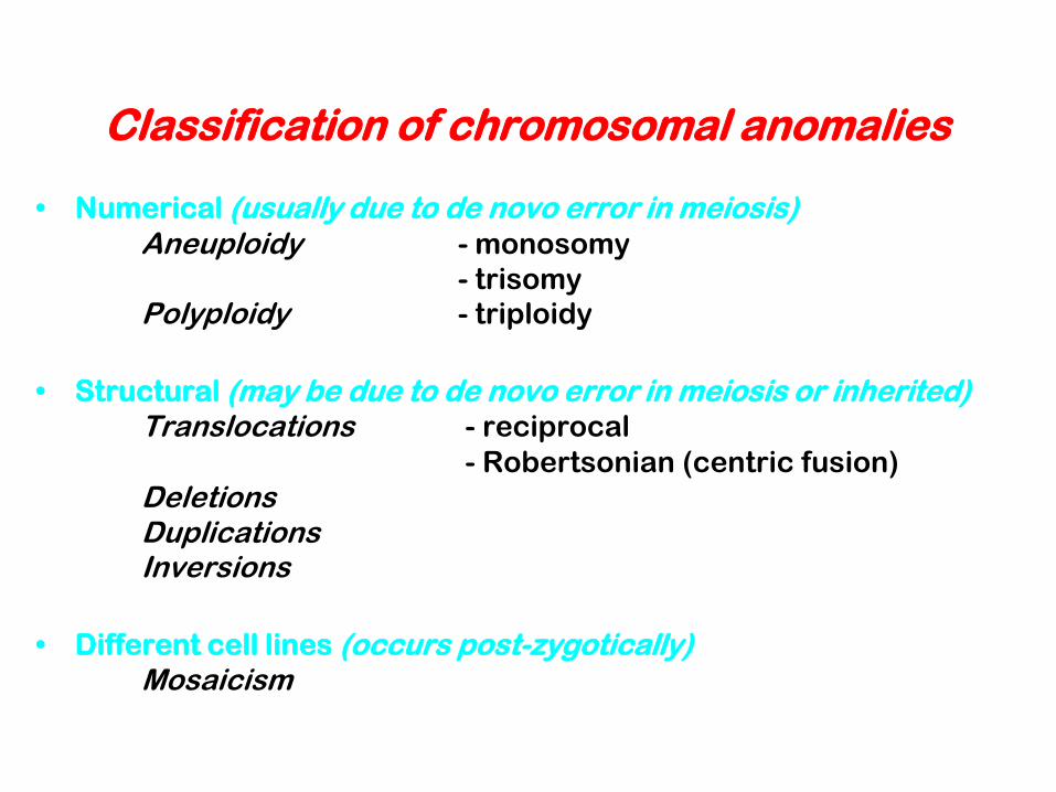
Classification of chromosomal anomalies
• Numerical (usually due to de novo error in meiosis)
Aneuploidy - monosomy
- trisomy
Polyploidy - triploidy
• Structural (may be due to de novo error in meiosis or inherited)
Translocations - reciprocal
- Robertsonian (centric fusion)
Deletions Duplications Inversions
• Different cell lines (occurs post-zygotically) Mosaicism
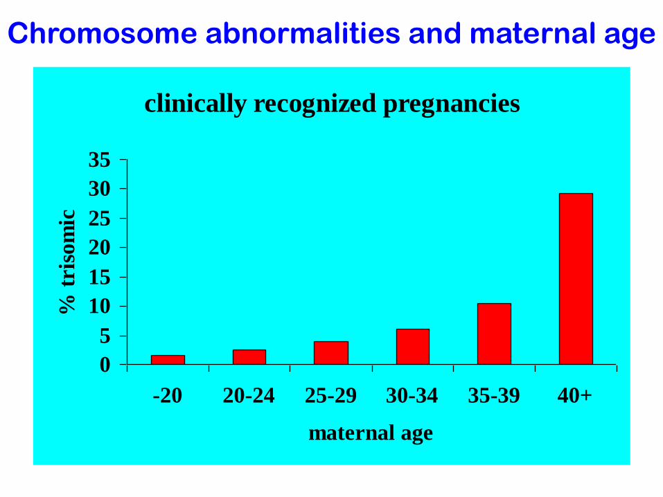
clinically recognized pregnancies
0
5
10
15
20
25
30
35
-20 20-24 25-29 30-34 35-39 40+
maternal age
% t
ris
om
ic
Chromosome abnormalities and maternal age
Top Related