


Languages
Pages
Legal

Journal of Korean Association of Pediatric Surgeons 7
pISSN 2383-5036 eISSN 2383-5508J Korean Assoc Pediatr Surg Vol. 21, No. 1, June 2015http://dx.doi.org/10.13029/jkaps.2015.21.1.7 Case Report
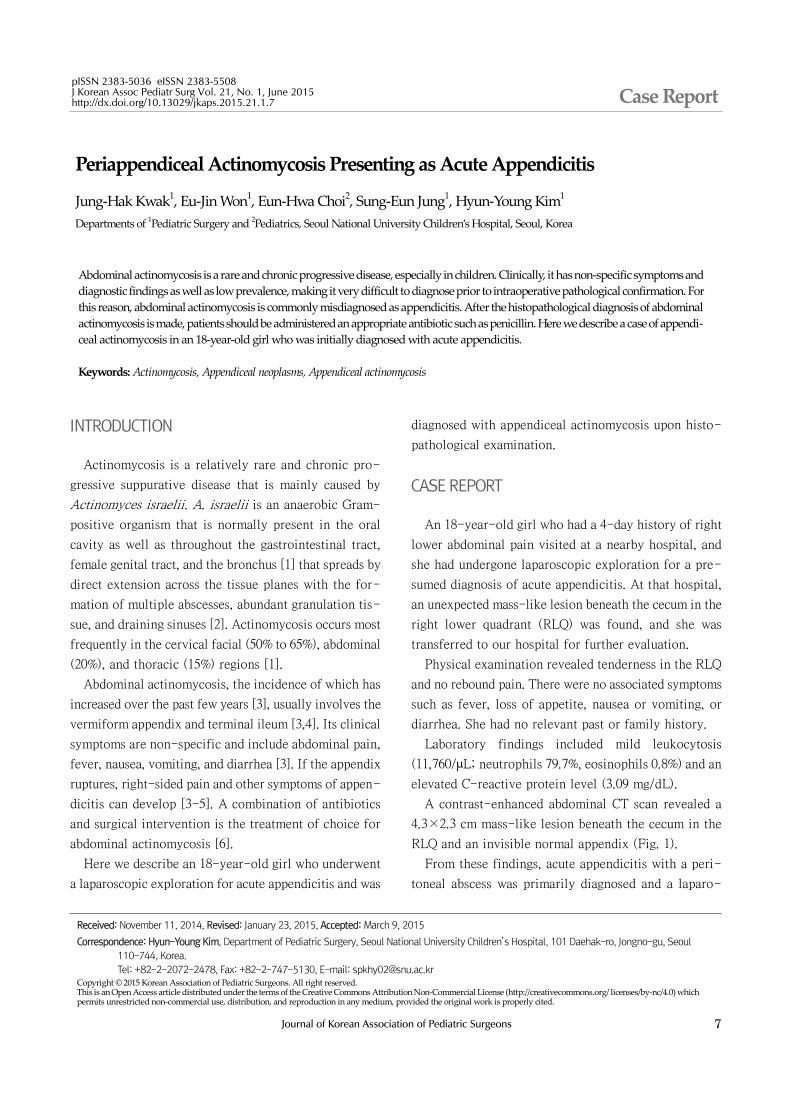
Periappendiceal Actinomycosis Presenting as Acute Appendicitis
Jung-Hak Kwak1, Eu-Jin Won1, Eun-Hwa Choi2, Sung-Eun Jung1, Hyun-Young Kim1
Departments of 1Pediatric Surgery and 2Pediatrics, Seoul National University Children’s Hospital, Seoul, Korea
Abdominal actinomycosis is a rare and chronic progressive disease, especially in children. Clinically, it has non-specific symptoms and diagnostic findings as well as low prevalence, making it very difficult to diagnose prior to intraoperative pathological confirmation. For this reason, abdominal actinomycosis is commonly misdiagnosed as appendicitis. After the histopathological diagnosis of abdominal actinomycosis is made, patients should be administered an appropriate antibiotic such as penicillin. Here we describe a case of appendi-ceal actinomycosis in an 18-year-old girl who was initially diagnosed with acute appendicitis.
Keywords: Actinomycosis, Appendiceal neoplasms, Appendiceal actinomycosis
Received: November 11, 2014, Revised: January 23, 2015, Accepted: March 9, 2015
Correspondence: Hyun-Young Kim, Department of Pediatric Surgery, Seoul National University Children’s Hospital, 101 Daehak-ro, Jongno-gu, Seoul 110-744, Korea.Tel: +82-2-2072-2478, Fax: +82-2-747-5130, E-mail: [email protected]
Copyright © 2015 Korean Association of Pediatric Surgeons. All right reserved.This is an Open Access article distributed under the terms of the Creative Commons Attribution Non-Commercial License (http://creativecommons.org/ licenses/by-nc/4.0) which permits unrestricted non-commercial use, distribution, and reproduction in any medium, provided the original work is properly cited.
INTRODUCTION
Actinomycosis is a relatively rare and chronic pro-
gressive suppurative disease that is mainly caused by
Actinomyces israelii. A. israelii is an anaerobic Gram-
positive organism that is normally present in the oral
cavity as well as throughout the gastrointestinal tract,
female genital tract, and the bronchus [1] that spreads by
direct extension across the tissue planes with the for-
mation of multiple abscesses, abundant granulation tis-
sue, and draining sinuses [2]. Actinomycosis occurs most
frequently in the cervical facial (50% to 65%), abdominal
(20%), and thoracic (15%) regions [1].
Abdominal actinomycosis, the incidence of which has
increased over the past few years [3], usually involves the
vermiform appendix and terminal ileum [3,4]. Its clinical
symptoms are non-specific and include abdominal pain,
fever, nausea, vomiting, and diarrhea [3]. If the appendix
ruptures, right-sided pain and other symptoms of appen-
dicitis can develop [3-5]. A combination of antibiotics
and surgical intervention is the treatment of choice for
abdominal actinomycosis [6].
Here we describe an 18-year-old girl who underwent
a laparoscopic exploration for acute appendicitis and was
diagnosed with appendiceal actinomycosis upon histo-
pathological examination.
CASE REPORT
An 18-year-old girl who had a 4-day history of right
lower abdominal pain visited at a nearby hospital, and
she had undergone laparoscopic exploration for a pre-
sumed diagnosis of acute appendicitis. At that hospital,
an unexpected mass-like lesion beneath the cecum in the
right lower quadrant (RLQ) was found, and she was
transferred to our hospital for further evaluation.
Physical examination revealed tenderness in the RLQ
and no rebound pain. There were no associated symptoms
such as fever, loss of appetite, nausea or vomiting, or
diarrhea. She had no relevant past or family history.
Laboratory findings included mild leukocytosis
(11,760/μL; neutrophils 79.7%, eosinophils 0.8%) and an
elevated C-reactive protein level (3.09 mg/dL).
A contrast-enhanced abdominal CT scan revealed a
4.3×2.3 cm mass-like lesion beneath the cecum in the
RLQ and an invisible normal appendix (Fig. 1).
From these findings, acute appendicitis with a peri-
toneal abscess was primarily diagnosed and a laparo-

8 Journal of Korean Association of Pediatric Surgeons
J Korean Assoc Pediatr Surg 2015;21(1):7-10
Fig. 1. CT image shows a 4.3×2.3 cm mass-like lesion beneath the
cecum within the right lower quadrant (arrow). This finding suggested
differential diagnosis of advanced appendicitis with phlegmon or a
tumorous condition.
Fig. 2. Intraoperative findings. (A) Lapar-
oscopic appearance of appendiceal ac-
tinomycosis. (B) Gross appearance of
appendiceal actinomycosis; a 7.0×4.0cm appendiceal mass. Severe inflam-
mation and adhesion between the ap-
pendix and the adjacent appendiceal serosal surface is visible.
Fig. 3. (A) Gross appearance of the excised specimen. (B) Pathological examination: Presence of Actinomyces israelii. Acute suppurative appendicitis
with a periappendiceal abscess and actinomycotic colonies characterized by sulfur granules (H&E stain, ×40). (C) Pathological examination: Presence of A. israelii. Acute suppurative appendicitis with a periappendiceal abscess and actinomycotic colonies characterized by sulfur granules (H&E stain,
×200).
scopic exploration was planned.
Operative findings showed a 3×3 cm appendiceal mass
with dense severe adhesions (Fig. 2A, B) for which we
performed an ileocecectomy.
Gross pathology revealed severe inflammation and
adhesion between the appendix and the adjacent appen-
diceal serosal surface. Irregular nodularity was seen in
the distal portion of the ileal mucosal area, while intra-
luminal obliteration was found due to the severe in-
flammation (Fig. 3A).
Light microscopic examination revealed acute suppu-
rative appendicitis with a periappendiceal abscess and
actinomycotic colonies. At higher magnification, a typi-
cal sulfur granule surrounded by neutrophils was found
that was confirmed as appendiceal actinomycosis (Fig.
3B, C).
After the operation, she was administered intravenous

Journal of Korean Association of Pediatric Surgeons 9
Kwak JH, et al: Periappendiceal Actinomycosis
antibiotics (cefotaxime, metronidazole, piperacillin/ta-
zobactam) for 20 days and then oral amoxicillin 1 g three
times a day for at least 3 months.
DISCUSSION
Abdominal actinomycosis is a rare and insidious dis-
ease [7,8]. The overall incidence of actinomycosis is vir-
tually impossible to ascertain since failure to consider it
as a diagnosis and difficulty confirming it lead to un-
der-reporting [9]. Approximately 65% of abdominal ac-
tinomycosis cases occur in association with acute appen-
dicitis [10].
The symptoms, signs, and laboratory findings of ab-
dominal actinomycosis are nonspecific, while its radio-
logic findings often show a poorly defined mass with ir-
regular margins [11]. As such, its diagnosis is often
delayed. Only 10% of cases are diagnosed preoperatively,
and it is difficult to differentiate abdominal actino-
mycosis from a malignant tumor or appendicitis [7].
The differential diagnosis of abdominal actinomycosis
should include Crohn’s disease, ulcerative colitis, diver-ticulitis, neoplasms, and intestinal tuberculosis [8,11].
The definitive diagnosis of actinomycosis requires mi-
croscopic proof of either the pathogen itself or the pres-
ence of specific “sulfur granules” [8].Tissue sampling and a histologic examination are im-
portant to ensuring a correct diagnosis, but the former is
not always possible for two reasons. First, the clinical
presentation of the disease may be acute or the sample
may be unobtainable using percutaneous methods. Second,
Actinomyces is a Gram-positive, slow-growing, non-
acid-fast, anaerobic, and filamentous bacterium that re-
quires specific incubation in special culture medium for
at least 7 days [1]. Accordingly, culture results are neg-
ative in 76% of cases [12] and excision biopsy for histo-
pathological examination is frequently needed [1].
The combined use of surgery and penicillin therapy
has a reported cure rate of 88.8% [13]. Antibiotic therapy
is useful for preventing the local recurrence of actino-
mycosis. Penicillin in particular has been shown to be ef-
fective in treating abdominal actinomycosis. Other com-
monly used antibiotics include tetracycline, erythromycin,
doxycycline, clindamycin, imipenem, ceftriaxone, and
ciprofloxacin [7].
A combination of long-term antibiotic therapy and
adequate surgery is necessary to ensure the complete
eradication of actinomycosis because of the large degree
of reactive fibrosis formed by the infection [14]. Howev-
er, recent studies have shown that a combination of com-
plete surgical resection followed by short-term anti-
biotic treatment is effective [15].
Similarly, in this case, as the disease does not present
with any typical features, either clinically or radio-
logically, the patient was initially diagnosed with appen-
dicitis, and received laparoscopic exploration for a pre-
sumed diagnosis of acute appendicitis. The diagnosis was
made after pathologic examination of the specimen, and
then antibiotics were changed according to the patho-
logic results.
In summary, abdominal actinomycosis is difficult to
diagnose preoperatively due to its atypical symptoms and
imaging findings. However, since its accurate diagnosis
is important to the prevention of unnecessary surgical
treatment or to adequate antibiotic therapy, abdominal
actinomycosis should be included as a differential diag-
nosis when an unusual abdominal mass or abscess pres-
ents on abdominal CT.
CONFLICTS OF INTEREST
No potential conflict of interest relevant to this article
was reported.
ACKNOWLEDGMENTS
Dr. Eun-Hwa Choi, Sung-Eun Jung, and Hyun-Young
Kim were made substantial contribution to this report.
REFERENCES
1. Acquaro P, Tagliabue F, Confalonieri G, Faccioli P, Costa M. Abdominal wall actinomycosis simulating a malignant neoplasm: Case report and review of the literature. World J Gastrointest Surg 2010;2:247-50.
2. Sung HY, Lee IS, Kim SI, Jung SE, Kim SW, Kim SY, et al. Clinical features of abdominal actinomycosis: a 15-year experience of a sin-gle institute. J Korean Med Sci 2011;26:932-7.
3. Bittencourt JA, Andreis EL, Lima EL, Dorn DE, Muller V. Actinomycosis simulating malignant large bowel obstruction.

10 Journal of Korean Association of Pediatric Surgeons
J Korean Assoc Pediatr Surg 2015;21(1):7-10
Braz J Infect Dis 2004;8:186-9. 4. Luncă S, Romedea N. Actinomycosis of the appendix. Case report.
Rev Med Chir Soc Med Nat Iasi 2004;108:640-3. 5. Davies M, Keddie NC. Abdominal actinomycosis. Br J Surg
1973;60:18-22.6. Hayashi M, Asakuma M, Tsunemi S, Inoue Y, Shimizu T, Komeda
K, et al. Surgical treatment for abdominal actinomycosis: a report of two cases. World J Gastrointest Surg 2010;2:405-8.
7. Russo T. Agents of actinomycosis. In: Mandell GL, Bennett JE, Dolin R, eds. Principles and practice of infectious diseases. 7th ed. New York: Churchill Livingstone; 2005. p.2924-34.
8. Huang CJ, Huang TJ, Hsieh JS. Pseudo-colonic carcinoma caused by abdominal actinomycosis: report of two cases. Int J Colorectal Dis 2004;19:283-6.
9. Garner JP, Macdonald M, Kumar PK. Abdominal actinomycosis. Int J Surg 2007;5:441-8.
10. Liu V, Val S, Kang K, Velcek F. Case report: actinomycosis of the ap-pendix--an unusual cause of acute appendicitis in children. J Pediatr Surg 2010;45:2050-2.
11. Abela J, Sciberras J, Meilak M, Felice AG, Degaetano J. Omental ac-tinomycosis presenting with right lower quadrant abdominal pain. J Clin Pathol 2004;57:671.
12. Sergent F, Marpeau L. Abdominopelvic actinomycosis: a tumoral syndrome due to bacterial infection. J Chir (Paris) 2004;141:150-6.
13. Spickett GP, Kipping RA. Pelvic actinomycosis presenting with rectal stricture. J R Soc Med 1985;78:674-6.
14. Atad J, Hallak M, Sharon A, Kitzes R, Kelner Y, Abramovici H. Pelvic actinomycosis. Is long-term antibiotic therapy necessary? J Reprod Med 1999;44:939-44.
15. Koren R, Dekel Y, Ramadan E, Veltman V, Dreznik Z. Periappendi-ceal actinomycosis mimicking malignancy report of a case. Pathol Res Pract 2002;198:441-3.
Top Related