


Languages
Pages
Legal

1
Evidence-Based
Evaluation & Treatment
of the Sacroiliac Joint
James R. Scifers, DScPT, PT, SCS, LAT, ATC
Western Carolina University
Athletic Training Education Program
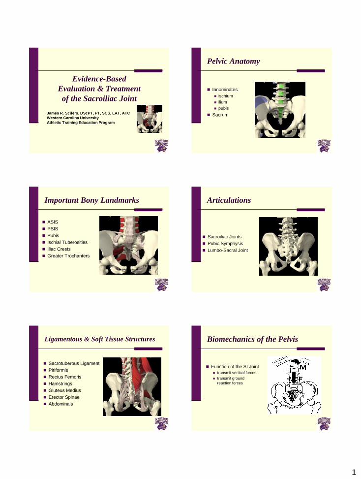
Pelvic Anatomy
Innominates
ischium
ilium
pubis
Sacrum
Important Bony Landmarks
ASIS
PSIS
Pubis
Ischial Tuberosities
Iliac Crests
Greater Trochanters
Articulations
Sacroiliac Joints
Pubic Symphysis
Lumbo-Sacral Joint
Ligamentous & Soft Tissue Structures
Sacrotuberous Ligament
Piriformis
Rectus Femoris
Hamstrings
Gluteus Medius
Erector Spinae
Abdominals
Biomechanics of the Pelvis
Function of the SI Joint
transmit vertical forces
transmit ground
reaction forces
2
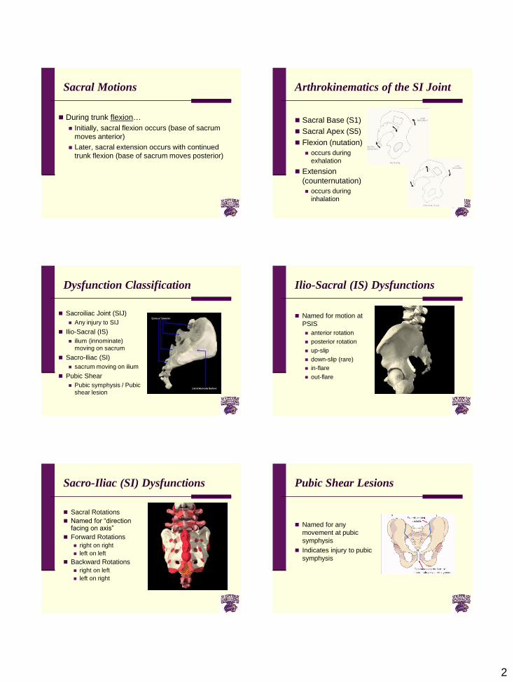
Sacral Motions
During trunk flexion…
Initially, sacral flexion occurs (base of sacrum
moves anterior)
Later, sacral extension occurs with continued
trunk flexion (base of sacrum moves posterior)
Arthrokinematics of the SI Joint
Sacral Base (S1)
Sacral Apex (S5)
Flexion (nutation)
occurs during
exhalation
Extension
(counternutation)
occurs during
inhalation
Dysfunction Classification
Sacroiliac Joint (SIJ)
Any injury to SIJ
Ilio-Sacral (IS)
ilium (innominate)
moving on sacrum
Sacro-Iliac (SI)
sacrum moving on ilium
Pubic Shear
Pubic symphysis / Pubic
shear lesion
Ilio-Sacral (IS) Dysfunctions
Named for motion at
PSIS
anterior rotation
posterior rotation
up-slip
down-slip (rare)
in-flare
out-flare
Sacro-Iliac (SI) Dysfunctions
Sacral Rotations
Named for “direction facing on axis”
Forward Rotations
right on right
left on left
Backward Rotations
right on left
left on right
Pubic Shear Lesions
Named for any
movement at pubic
symphysis
Indicates injury to pubic
symphysis
3
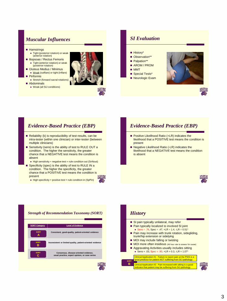
Muscular Influences
Hamstrings Tight (posterior rotation) or weak
(anterior rotation)
Iliopsoas / Rectus Femoris Tight (anterior rotation) or weak
(posterior rotation)
Gluteus Medius / Minimus Weak (outflare) or tight (inflare)
Piriformis Stretch (forward sacral rotations)
Abdominals Weak (all SIJ conditions)
SI Evaluation
History*
Observation**
Palpation**
AROM / PROM
MMT
Special Tests*
Neurologic Exam
Evidence-Based Practice (EBP)
Reliability (k) is reproducibility of test results, can be
intra-tester (within one clinician) or inter-tester (between
multiple clinicians)
Sensitivity (sens) is the ability of test to RULE OUT a
condition. The higher the sensitivity, the greater
chance that a NEGATIVE test means the condition is
absent
High sensitivity + negative test = rule condition out (SnNout)
Specificity (spec) is the ability of test to RULE IN a
condition. The higher the specificity, the greater
chance that a POSITIVE test means the condition is
present
High specificity + positive test = rule condition in (SpPin)
Evidence-Based Practice (EBP)
Positive Likelihood Ratio (+LR) indicates the
likelihood that a POSITIVE test means the condition is
present
Negative Likelihood Ratio (-LR) indicates the
likelihood that a NEGATIVE test means the condition
is absent
Strength of Recommendation Taxonomy (SORT)
SORT Category Level of Evidence
Consistent, good-quality, patient-oriented evidence
Inconsistent or limited-quality, patient-oriented evidence
Consensus, disease-oriented evidence,
usual practice, expert opinion, or case series
SORT
A
SORT
B
SORT
C
History
SI pain typically unilateral, may refer
Pain typically localized to involved SI joint Sens = .76, Spec = .47, +LR = 1.4, -LR = 0.511
Pain may increase with trunk rotation, sidegliding, trunk/hip extension or sidelying
MOI may include falling or twisting
MOI more often insidious (48 hour rule to assess for cause)
Aggravating Activities usually includes sitting Sens = .03, Spec = .90, +LR = 0.3, -LR = 1.071
SORT
B
Clinical Application #1: Failure to report pain at the PSIS is a
good predictor for patient NOT suffering from SIJ pathology
Clinical Application #2: Pain increased with sitting is a good
indicator that patient may be suffering from SIJ pathology
4
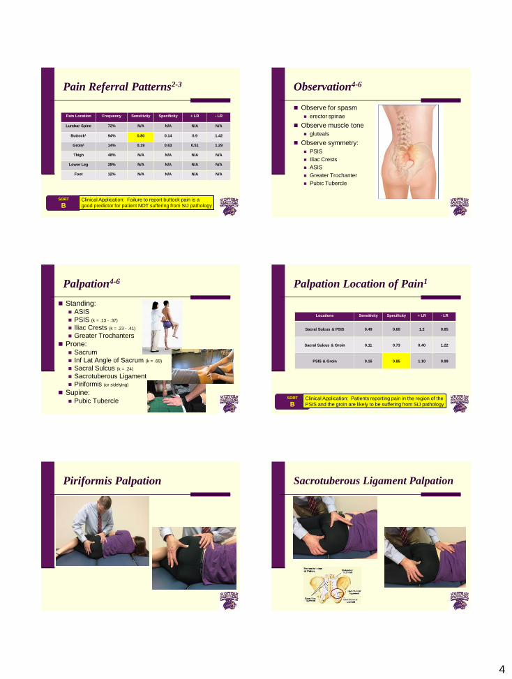
Pain Referral Patterns2-3
Pain Location Frequency Sensitivity Specificity + LR - LR
Lumbar Spine 72% N/A N/A N/A N/A
Buttock1 94% 0.80 0.14 0.9 1.42
Groin1 14% 0.19 0.63 0.51 1.29
Thigh 48% N/A N/A N/A N/A
Lower Leg 28% N/A N/A N/A N/A
Foot 12% N/A N/A N/A N/A
SORT
BClinical Application: Failure to report buttock pain is a
good predictor for patient NOT suffering from SIJ pathology
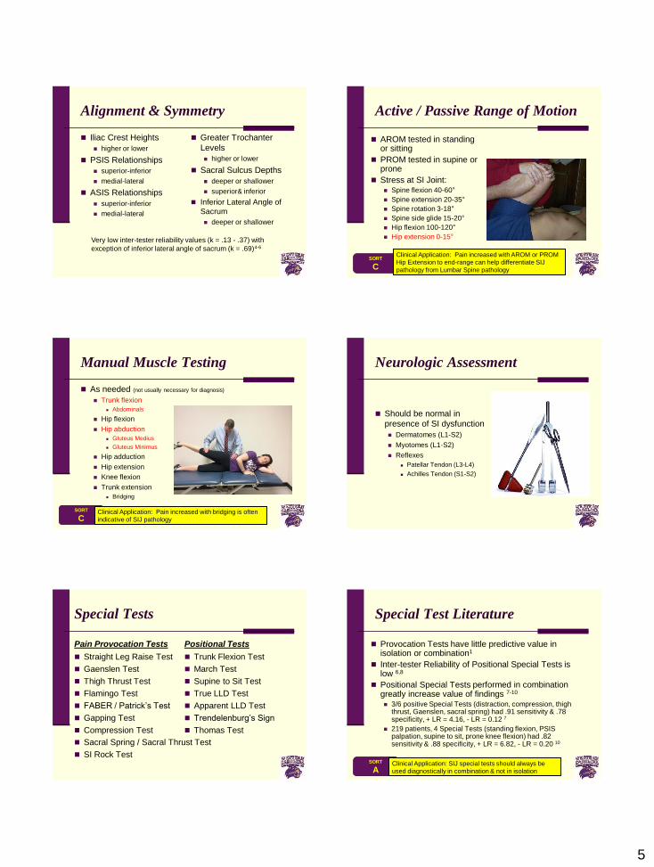
Observation4-6
Observe for spasm
erector spinae
Observe muscle tone
gluteals
Observe symmetry:
PSIS
Iliac Crests
ASIS
Greater Trochanter
Pubic Tubercle
Palpation4-6
Standing: ASIS
PSIS (k = .13 - .37)
Iliac Crests (k = .23 - .41)
Greater Trochanters
Prone: Sacrum
Inf Lat Angle of Sacrum (k = .69)
Sacral Sulcus (k = .24)
Sacrotuberous Ligament
Piriformis (or sidelying)
Supine: Pubic Tubercle
Palpation Location of Pain1
Locations Sensitivity Specificity + LR - LR
Sacral Sulcus & PSIS 0.49 0.60 1.2 0.85
Sacral Sulcus & Groin 0.11 0.73 0.40 1.22
PSIS & Groin 0.16 0.85 1.10 0.99
SORT
BClinical Application: Patients reporting pain in the region of the
PSIS and the groin are likely to be suffering from SIJ pathology
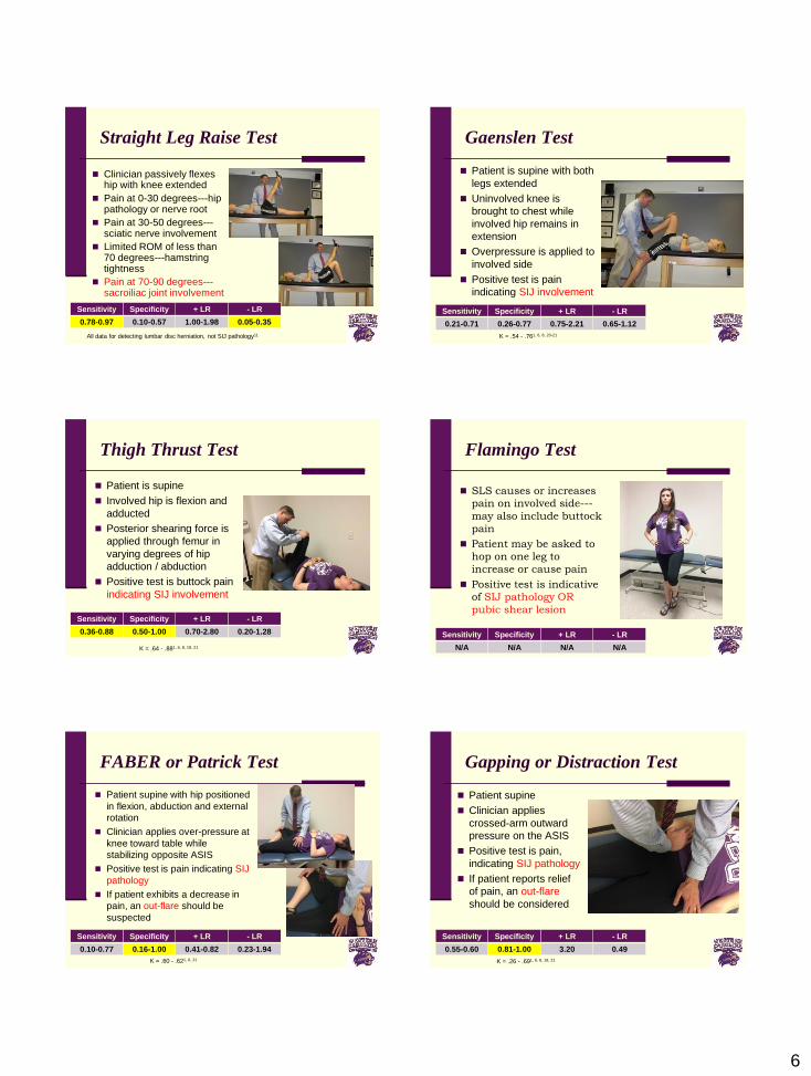
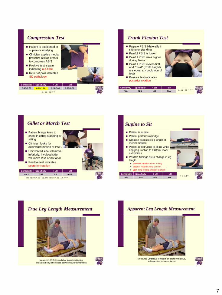
Piriformis Palpation Sacrotuberous Ligament Palpation
5
Alignment & Symmetry
Iliac Crest Heights
higher or lower
PSIS Relationships
superior-inferior
medial-lateral
ASIS Relationships
superior-inferior
medial-lateral
Greater Trochanter
Levels
higher or lower
Sacral Sulcus Depths
deeper or shallower
superior& inferior
Inferior Lateral Angle of
Sacrum
deeper or shallower
Very low inter-tester reliability values (k = .13 - .37) with
exception of inferior lateral angle of sacrum (k = .69)4-6
Active / Passive Range of Motion
AROM tested in standing or sitting
PROM tested in supine or prone
Stress at SI Joint: Spine flexion 40-60°
Spine extension 20-35°
Spine rotation 3-18°
Spine side glide 15-20°
Hip flexion 100-120°
Hip extension 0-15°
SORT
C
Clinical Application: Pain increased with AROM or PROM
Hip Extension to end-range can help differentiate SIJ
pathology from Lumbar Spine pathology
Manual Muscle Testing
As needed (not usually necessary for diagnosis)
Trunk flexion
Abdominals
Hip flexion
Hip abduction
Gluteus Medius
Gluteus Minimus
Hip adduction
Hip extension
Knee flexion
Trunk extension
Bridging
SORT
CClinical Application: Pain increased with bridging is often
indicative of SIJ pathology
Neurologic Assessment
Should be normal in
presence of SI dysfunction
Dermatomes (L1-S2)
Myotomes (L1-S2)
Reflexes
Patellar Tendon (L3-L4)
Achilles Tendon (S1-S2)
Special Tests
Pain Provocation Tests
Straight Leg Raise Test
Gaenslen Test
Thigh Thrust Test
Flamingo Test
FABER / Patrick’s Test
Gapping Test
Compression Test
Sacral Spring / Sacral Thrust Test
SI Rock Test
Positional Tests
Trunk Flexion Test
March Test
Supine to Sit Test
True LLD Test
Apparent LLD Test
Trendelenburg’s Sign
Thomas Test
Special Test Literature
Provocation Tests have little predictive value in isolation or combination1
Inter-tester Reliability of Positional Special Tests is low 6,8
Positional Special Tests performed in combination greatly increase value of findings 7-10
3/6 positive Special Tests (distraction, compression, thigh thrust, Gaenslen, sacral spring) had .91 sensitivity & .78 specificity, + LR = 4.16, - LR = 0.12 7
219 patients, 4 Special Tests (standing flexion, PSIS palpation, supine to sit, prone knee flexion) had .82 sensitivity & .88 specificity, + LR = 6.82, - LR = 0.20 10
SORT
AClinical Application: SIJ special tests should always be
used diagnostically in combination & not in isolation
6
Straight Leg Raise Test
Clinician passively flexes hip with knee extended
Pain at 0-30 degrees---hip pathology or nerve root
Pain at 30-50 degrees---sciatic nerve involvement
Limited ROM of less than 70 degrees---hamstring tightness
Pain at 70-90 degrees---sacroiliac joint involvement
All data for detecting lumbar disc herniation, not SIJ pathology11
Sensitivity Specificity + LR - LR
0.78-0.97 0.10-0.57 1.00-1.98 0.05-0.35
Gaenslen Test
Patient is supine with both
legs extended
Uninvolved knee is
brought to chest while
involved hip remains in
extension
Overpressure is applied to
involved side
Positive test is pain
indicating SIJ involvement
K = .54 - .761, 6, 8, 20-21
Sensitivity Specificity + LR - LR
0.21-0.71 0.26-0.77 0.75-2.21 0.65-1.12
Thigh Thrust Test
Patient is supine
Involved hip is flexion and
adducted
Posterior shearing force is
applied through femur in
varying degrees of hip
adduction / abduction
Positive test is buttock pain
indicating SIJ involvement
K = .64 - .881, 6, 8, 18, 21
Sensitivity Specificity + LR - LR
0.36-0.88 0.50-1.00 0.70-2.80 0.20-1.28
Flamingo Test
SLS causes or increases
pain on involved side---
may also include buttock
pain
Patient may be asked to
hop on one leg to
increase or cause pain
Positive test is indicative
of SIJ pathology OR
pubic shear lesion
Sensitivity Specificity + LR - LR
N/A N/A N/A N/A
FABER or Patrick Test
Patient supine with hip positioned
in flexion, abduction and external
rotation
Clinician applies over-pressure at
knee toward table while
stabilizing opposite ASIS
Positive test is pain indicating SIJ
pathology
If patient exhibits a decrease in
pain, an out-flare should be
suspected
K = .60 - .621, 6, 21
Sensitivity Specificity + LR - LR
0.10-0.77 0.16-1.00 0.41-0.82 0.23-1.94
Gapping or Distraction Test
Patient supine
Clinician applies
crossed-arm outward
pressure on the ASIS
Positive test is pain,
indicating SIJ pathology
If patient reports relief
of pain, an out-flare
should be considered
K = .26 - .691, 6, 8, 18, 21
Sensitivity Specificity + LR - LR
0.55-0.60 0.81-1.00 3.20 0.49
7
Compression Test
Patient is positioned in
supine or sidelying
Clinician applies medial
pressure at iliac crests
to compress ASIS
Positive test is pain
indicating out-flare
Relief of pain indicates
SIJ pathology
K = .26 - .736, 17-20
Sensitivity Specificity + LR - LR
0.60-0.70 0.69-1.00 2.20-7.00 0.33-1.00
Trunk Flexion Test
Palpate PSIS bilaterally in sitting or standing
Painful PSIS is lower
Painful PSIS rises higher during flexion
Painful PSIS moves first and “most” (PSIS heights are equal at conclusion of test)
Positive test indicates posterior rotation
K = .08 - .68 4-6, 12-14
Sensitivity Specificity + LR - LR
N/A N/A N/A N/A
Gillet or March Test
Patient brings knee to
chest in either standing or
sitting
Clinician looks for
downward motion of PSIS
Uninvolved side will move
inferiorly, involved side
will move less or not at all
Positive test indicates
posterior rotation
Intra-tester K = .02 – .31, Inter-tester K = .02 - .591, 5-6, 15-17
Sensitivity Specificity + LR - LR
0.43 0.68 1.3 0.84
Supine to Sit
Patient is supine
Patient performs a bridge
Clinician assesses leg length at
medial malleoli
Patient is instructed to sit up while
applying traction to bilateral lower
extremities
Positive findings are a change in leg
length
posterior rotation: short to long
anterior rotation: long to short
LLD: long to long or short to short
Sensitivity Specificity + LR - LR
N/A N/A N/A N/AK = .19 10
True Leg Length Measurement
Measured ASIS to medial or lateral malleolus,
indicates bony differences between lower extremities
Apparent Leg Length Measurement
Measured Umbilicus to medial or lateral malleolus,
indicates innominate rotation
8
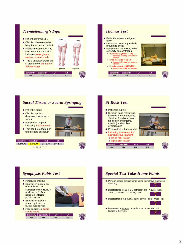
Trendelenburg’s Sign
Patient performs SLS
Clinician observes pelvic
height from behind patient
Inferior movement of iliac
crest on non-stance side
indicates weak gluteus
medius on stance side
This is an associated sign
in presence of out-flare or
SIJ pathologypositive negative
Sensitivity Specificity + LR - LR
N/A N/A N/A N/A
Thomas Test
Patient is supine at edge of table
Uninvolved knee is passively brought to chest
Positive test is involved lower extremity demonstrating: Hip flexion (tight iliopsoas)
Associated finding with anterior rotation
Knee extension (tight RF) Associated finding with anterior
rotation
Hip abduction (tight ITB/TFL) Associated finding with in-flare
Sensitivity Specificity + LR - LR
N/A N/A N/A N/A
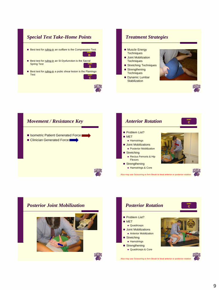
Sacral Thrust or Sacral Springing
Patient is prone
Clinician applies
downward pressure to
sacrum
Positive test is pain,
indicating sacral rotation
Test can be repeated on
four corners of sacrum
K = .30 - .561, 6, 17-18, 20
Sensitivity Specificity + LR - LR
0.27-0.75 0.29-1.00 0.75-3.00 0.50-1.62
SI Rock Test
Patient is supine
Clinician passively brings involved knee to opposite shoulder (combination of hip flexion and internal rotation) and applies overpressure
Positive test is buttock pain
Indicating involvement of sacrotuberous ligament
left on right rotation
right on left rotation
Sensitivity Specificity + LR - LR
N/A N/A N/A N/A
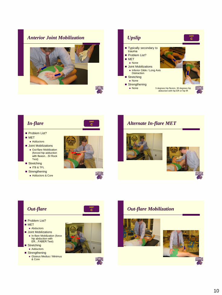
Symphysis Pubis Test
Patient is supine
Examiner places heel of one hand on
superior pubic ramus and heel of other hand on inferior pubic ramus
Examiner applies shearing force to pubic symphysis
Pain indicates pubic shear lesion
Sensitivity Specificity + LR - LR
N/A N/A N/A N/A
Special Test Take-Home Points
Perform special tests in combination to improve diagnostic
accuracy
Best tests for ruling-in SIJ pathology are FABER, Thigh
Thrust, Gaenslen & Gapping Tests
Best test for ruling-out SIJ pathology is Thigh Thrust Test
Best tests for ruling-in posterior rotation are March &
Supine to Sit Tests
SORT
A
SORT
B
SORT
C
SORT
B
9
Special Test Take-Home Points
Best test for ruling-in an outflare is the Compression Test
Best test for ruling-in an SI Dysfunction is the Sacral
Spring Test
Best test for ruling-in a pubic shear lesion is the Flamingo
Test
SORT
B
SORT
C
SORT
B
Treatment Strategies
Muscle Energy
Techniques
Joint Mobilization
Techniques
Stretching Techniques
Strengthening
Techniques
Dynamic Lumbar
Stabilization
Movement / Resistance Key
Isometric Patient Generated Force
Clinician Generated Force
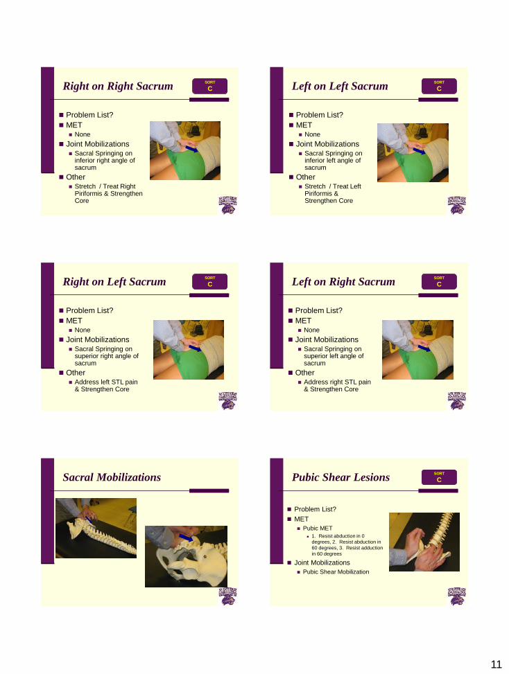
Anterior Rotation
Problem List?
MET
Hamstrings
Joint Mobilizations
Posterior Mobilization
Stretching
Rectus Femoris & Hip
Flexors
Strengthening
Hamstrings & Core
Also may use Scissoring or Arm Break to treat anterior or posterior rotation
SORT
C
Posterior Joint Mobilization Posterior Rotation
Problem List?
MET
Quadriceps
Joint Mobilizations
Anterior Mobilization
Stretching
Hamstrings
Strengthening
Quadriceps & Core
Also may use Scissoring or Arm Break to treat anterior or posterior rotation
SORT
C
10
Anterior Joint Mobilization Upslip
Typically secondary to trauma
Problem List?
MET
None
Joint Mobilizations
Inferior Glide / Long Axis Distraction
Stretching
None
Strengthening
None 5 degrees hip flexion, 30 degrees hip
abduction with hip ER or hip IR
SORT
C
In-flare
Problem List?
MET
Adductors
Joint Mobilizations
Out-flare Mobilization
(forced hip adduction
with flexion…SI Rock
Test)
Stretching
ITB & TFL
Strengthening
Adductors & Core
SORT
C Alternate In-flare MET
Out-flare
Problem List?
MET
Abductors
Joint Mobilizations
In-flare Mobilization (force hip abduction with ER…FABER Test)
Stretching
Adductors
Strengthening
Gluteus Medius / Minimus & Core
SORT
C Out-flare Mobilization
11
Right on Right Sacrum
Problem List?
MET None
Joint Mobilizations Sacral Springing on
inferior right angle of sacrum
Other Stretch / Treat Right
Piriformis & Strengthen Core
SORT
C Left on Left Sacrum
Problem List?
MET None
Joint Mobilizations Sacral Springing on
inferior left angle of sacrum
Other Stretch / Treat Left
Piriformis & Strengthen Core
SORT
C
Right on Left Sacrum
Problem List?
MET None
Joint Mobilizations Sacral Springing on
superior right angle of sacrum
Other Address left STL pain
& Strengthen Core
SORT
C Left on Right Sacrum
Problem List?
MET None
Joint Mobilizations Sacral Springing on
superior left angle of sacrum
Other Address right STL pain
& Strengthen Core
SORT
C
Sacral Mobilizations Pubic Shear Lesions
Problem List?
MET
Pubic MET
1. Resist abduction in 0
degrees, 2. Resist abduction in
60 degrees, 3. Resist adduction
in 60 degrees
Joint Mobilizations
Pubic Shear Mobilization
SORT
C
12
Order of Treatment Procedures
Pubic Lesions
Sacral Lesions (SI)
Innominate Lesions (IS)
Dynamic Lumbar Stabilization
Function Strengthening / Progression
SORT
C
References
1 Dreyfuss, et al (1996). The value of medical history and physical
examination in diagnosing sacroiliac joint pain. Spine, 21: 2594-2602.
2 Slipman, et al (2000). Sacroiliac joint pain referral zones. Archives of
Physical Medicine & Rehabilitation, 81: 334-338.
3 Schwartzer, et al (1996). The sacroiliac joint in chronic low back pain:
Joint double block and value of sacroiliac provocative tests. Spine, 21:
1889-1892.
4 Riddle & Freburger (2002). Evaluation of the presence of sacroiliac
joint dysfunction using a combination of tests: A multicenter intertester
reliability study. Physical Therapy, 82: 772-781.
5 Potter & Rothstein (1985). Intertester reliability of selected clinical
tests of the sacroiliac joint. Physical Therapy, 65: 1671-1675.
6 Flynn, et al (2002). A clinical prediction rule for classifying patients
with low back pain who demonstrate short-term improvement with
spinal manipulation. Spine, 27: 2835-2843.
References
7 Laslett, et al (2003). Diagnosing painful sacroiliac joints: A validity study of McKenzie evaluation and sacroiliac provocation tests. Australian Journal of Physiotherapy, 49: 89-97.
8 Laslett & Williams (1994). The reliability of selected pain provocation tests for sacroiliac joint pathology. Spine, 19: 1243-1249.
9 Cibulka & Koldehoff (1999). The clinical usefulness of a cluster of tests for sacroiliac joint dysfunction in patients with and without low back pain. JOSPT, 29: 83-89.
10 Levangie (1999). Four clinical tests of sacroiliac joint dysfunction: The association of test results with innominate torsion among patients with and without low back pain. Physical Therapy, 79: 1043-1057.
11 Scifers (2008). Special Tests for Neurologic Examination. SLACK Inc., Thorofare, NJ.
12 Vincent-Smith & Gibbons (1999). Inter-examiner and intra-examiner reliability of the standing flexion test. Manual Therapy, 4: 87-93.
13 Toussaint, et al (1999). Sacroiliac dysfunction in construction workers. Journal of Manipulative Physical Therapy, 22: 134-139.
14 Toussaint, et al (1999). Sacroiliac joint diagnosis in the Hamburg construction workers study. Journal of Manipulative Physical Therapy, 22: 139-143.
References
15 Carmichael (1987). Inter- and intra-examiner reliability of palpation for sacroiliac joint dysfunction. Journal of Manipulative Physical Therapy, 10: 164-171.
16 Meijne, et al (1999). Intraexaminer and interexaminer reliability of the Gillet test. Journal of Manipulative Physical Therapy, 22: 4-9.
17 Herzog, et al (1989). Reliability of motion palpation procedures to detect sacroiliac joint fixations. Journal of Manipulative Physical Therapy, 12: 86-92.
18 Broadhurst & Bond (1998). Pain provocation tests for the assessment of sacroiliac joint dysfunction. Journal of Spinal Disorders, 11: 341-345.
19 Blower & Griffin (1984). Clinical sacroiliac tests in ankylosing spondylitis and other causes of low back pain – 2 studies. Annuals of Rheumatoid Disorders, 43: 192-195.
20 Russell, et al (1981). Clinical examination of the sacroiliac joints: A prospective study. Arthritis Rheumatology, 24: 1575-1577.
21 Kokmeyer, et al (2002). The reliability of multi-test regimens with sacroiliac pain provocation tests. Journal of Manipulative Physical Therapy, 25: 42-48.
Questions?
Top Related