


Languages
Pages
Legal

Overview of Ophthalmic Equipmentand Support Systems for
OphthalmologyHealth Care Technology UnitORBIS DC-10 Flying Eye Hospital
Introduction
• Eye anatomy and common diseases
• Diagnostic instruments
• Therapeutic instruments
• Additional ophthalmic instruments
• Support systems for Ophthalmology
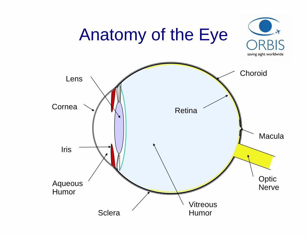
Macula
Aqueous Humor
Iris
Cornea
Lens
Vitreous Humor
Optic Nerve
Retina
Choroid
Sclera
Anatomy of the Eye
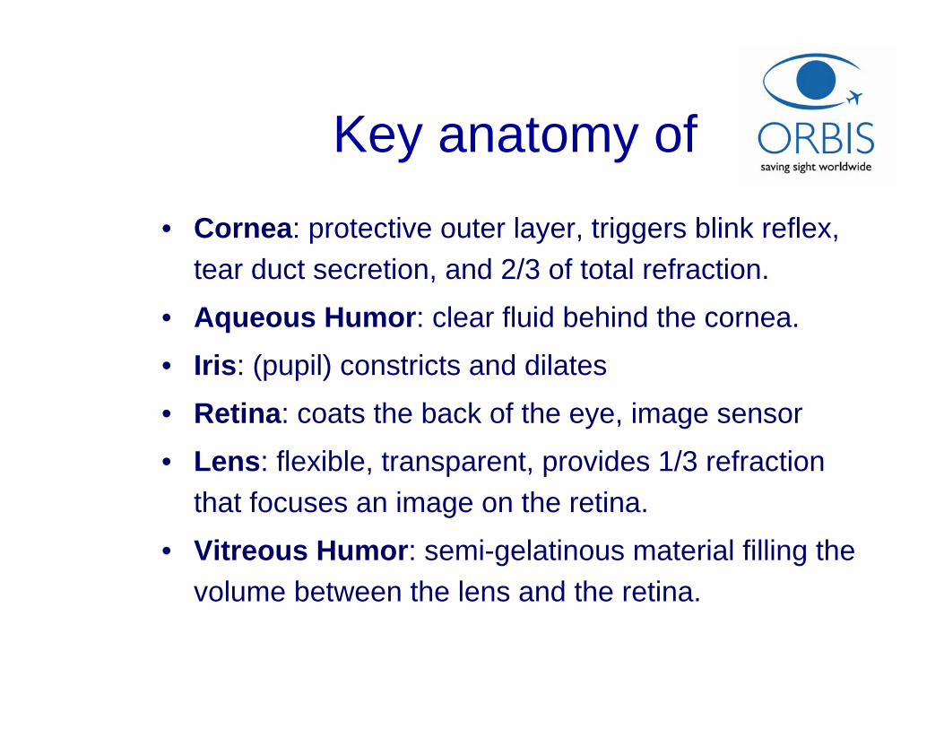
Key anatomy of• Cornea: protective outer layer, triggers blink reflex,
tear duct secretion, and 2/3 of total refraction.
• Aqueous Humor: clear fluid behind the cornea.
• Iris: (pupil) constricts and dilates
• Retina: coats the back of the eye, image sensor
• Lens: flexible, transparent, provides 1/3 refraction that focuses an image on the retina.
• Vitreous Humor: semi-gelatinous material filling the volume between the lens and the retina.
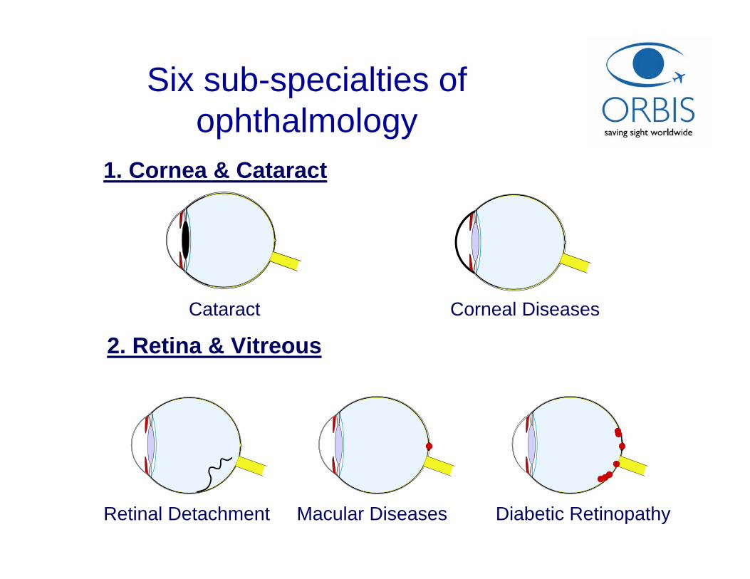
1. Cornea & Cataract
2. Retina & Vitreous
Six sub-specialties of ophthalmology
Cataract Corneal Diseases
Retinal Detachment Macular Diseases Diabetic Retinopathy
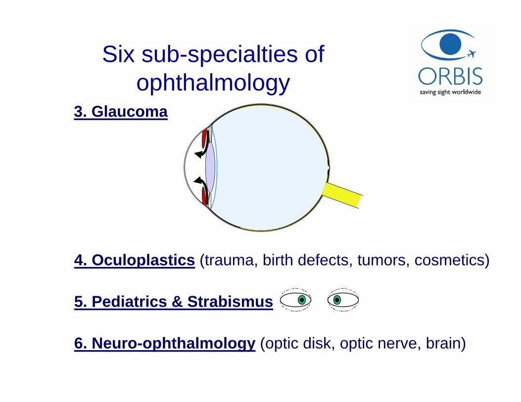
4. Oculoplastics (trauma, birth defects, tumors, cosmetics)
5. Pediatrics & Strabismus
6. Neuro-ophthalmology (optic disk, optic nerve, brain)
3. Glaucoma
Six sub-specialties of ophthalmology
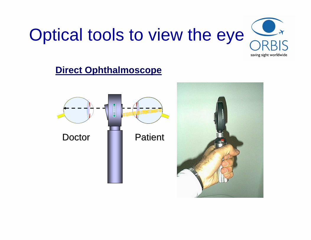
Direct Ophthalmoscope
DoctorDoctor PatientPatient
Optical tools to view the eye
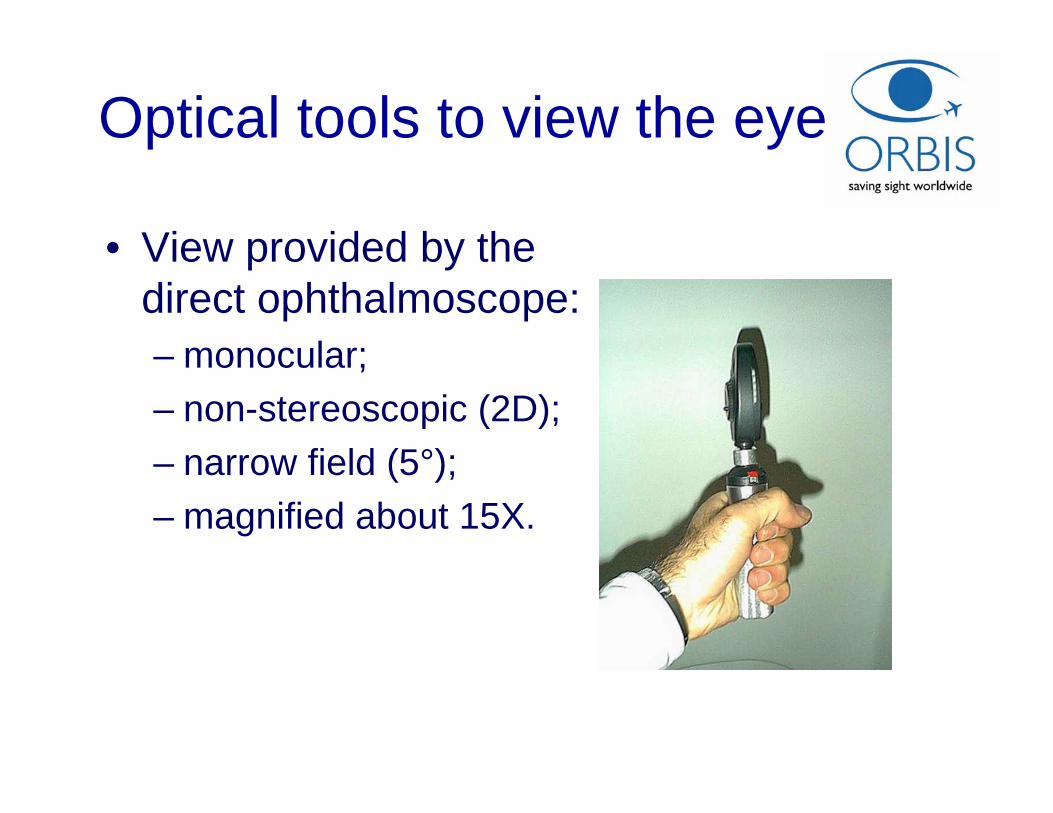
Optical tools to view the eye
• View provided by the direct ophthalmoscope:– monocular; – non-stereoscopic (2D);– narrow field (5°);– magnified about 15X.
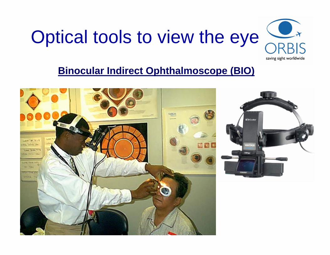
Binocular Indirect Ophthalmoscope (BIO)
Optical tools to view the eye
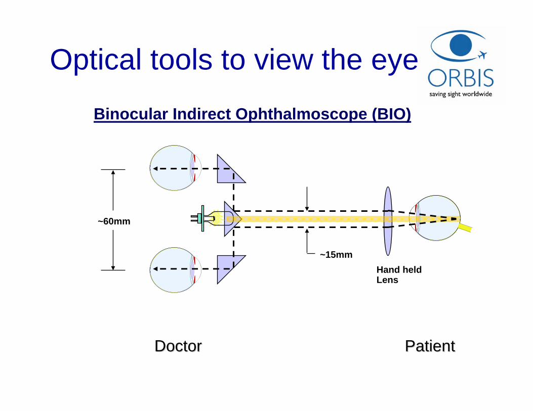
Binocular Indirect Ophthalmoscope (BIO)
~15mm
~60mm
Hand held Lens
DoctorDoctor PatientPatient
Optical tools to view the eye
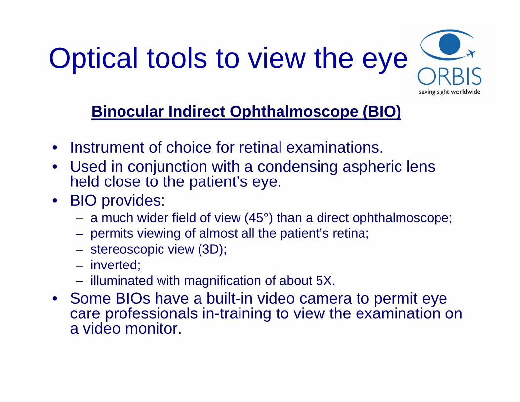
Binocular Indirect Ophthalmoscope (BIO)
Optical tools to view the eye
• Instrument of choice for retinal examinations.• Used in conjunction with a condensing aspheric lens
held close to the patient’s eye. • BIO provides:
– a much wider field of view (45°) than a direct ophthalmoscope;– permits viewing of almost all the patient’s retina; – stereoscopic view (3D);– inverted;– illuminated with magnification of about 5X.
• Some BIOs have a built-in video camera to permit eye care professionals in-training to view the examination on a video monitor.
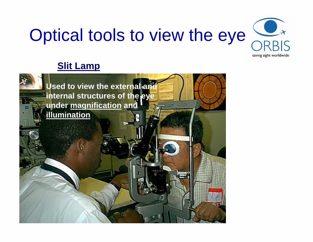
Slit Lamp
Optical tools to view the eye
• Used to view the external and internal structures of the eye under magnification and illumination
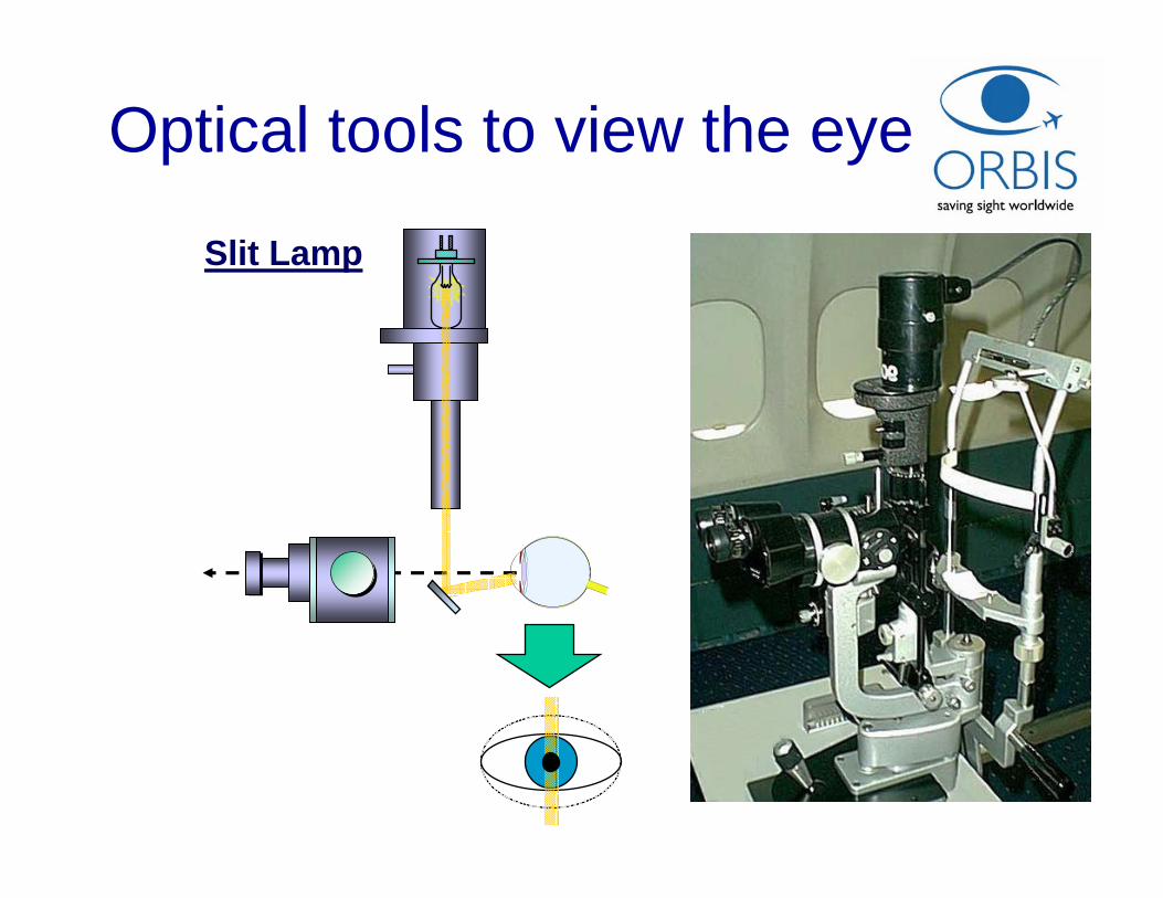
Slit Lamp
Optical tools to view the eye
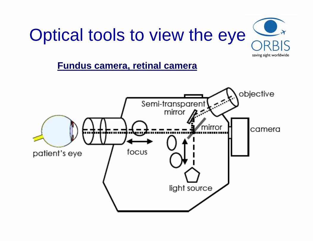
Fundus camera, retinal camera
Optical tools to view the eye

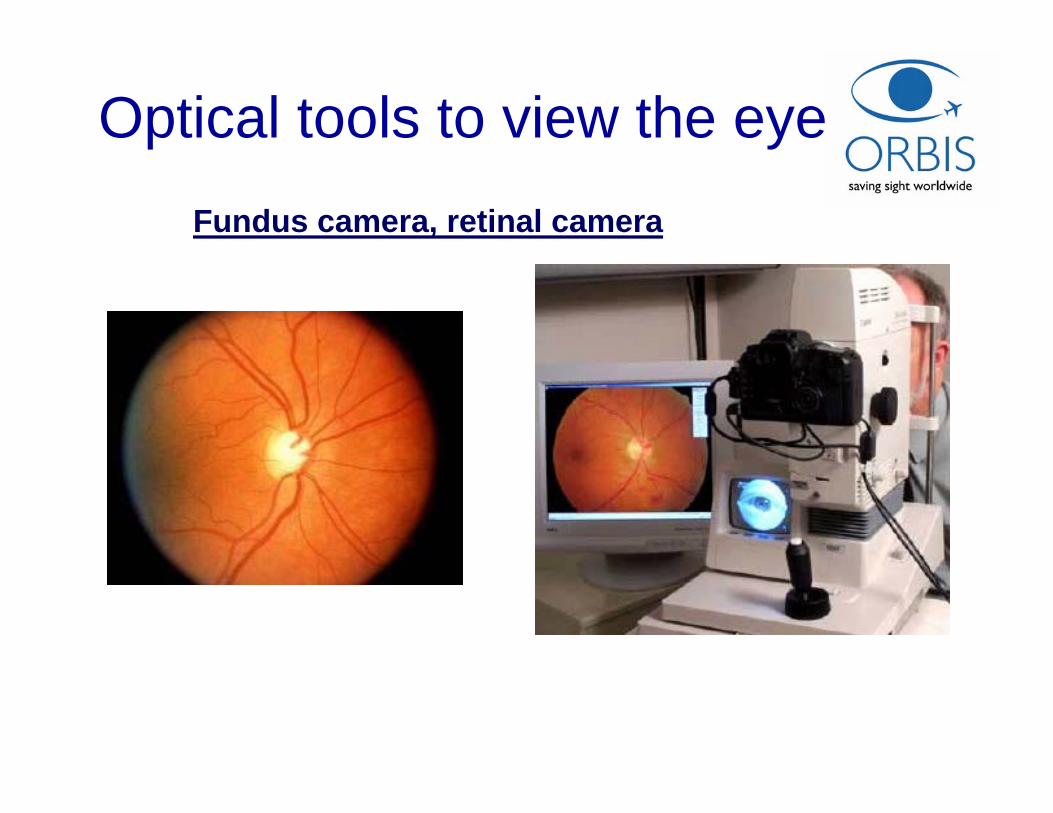
Fundus camera, retinal camera
Optical tools to view the eye
• Specialized low power microscope with an attached camera, designed for taking pictures of the back of the eye, or fundus.
• Often used in fluorescein angiography:– fluorescein dye is injected into a patient to reveal retinal
circulation.• Digital fundus cameras can be interfaced with a
computer for storage of the retinal images as graphic files:– files can be archived, edited, printed or sent to other eye care
specialists through a local area network or over the World Wide Web.
Fundus camera, retinal camera
Optical tools to view the eye
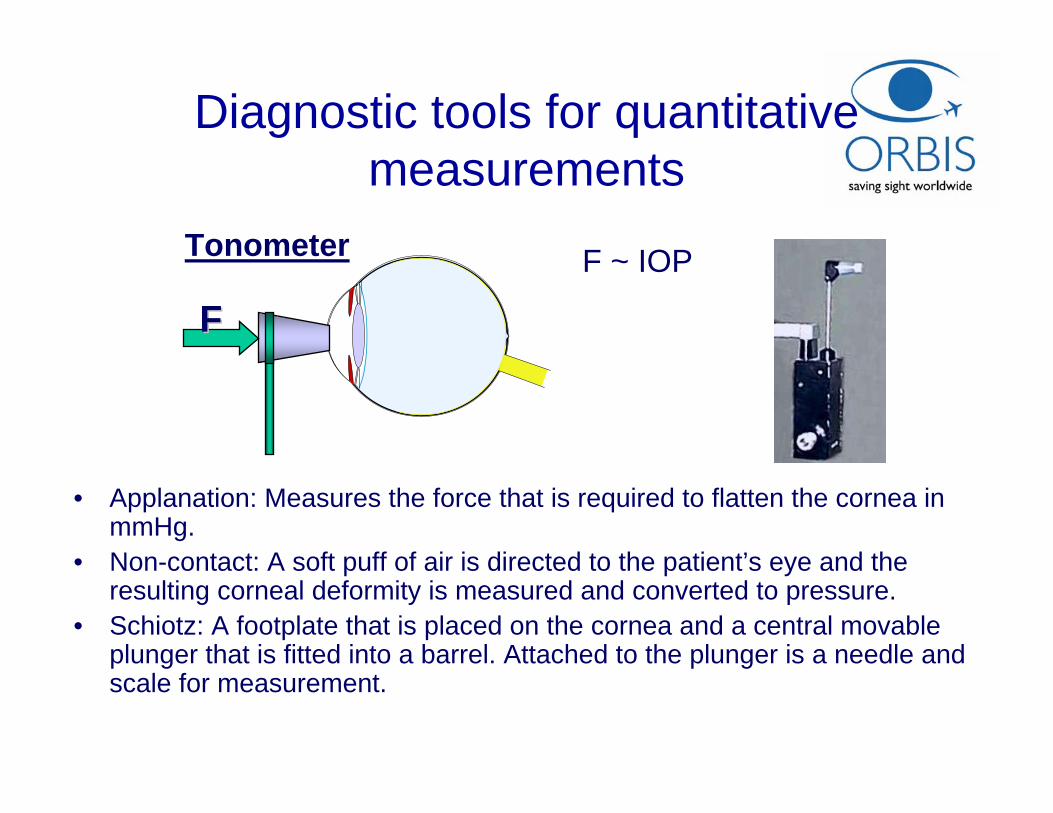
Tonometer F ~ IOP
Diagnostic tools for quantitative measurements
FF
• Applanation: Measures the force that is required to flatten the cornea in mmHg.
• Non-contact: A soft puff of air is directed to the patient’s eye and the resulting corneal deformity is measured and converted to pressure.
• Schiotz: A footplate that is placed on the cornea and a central movableplunger that is fitted into a barrel. Attached to the plunger is a needle and scale for measurement.
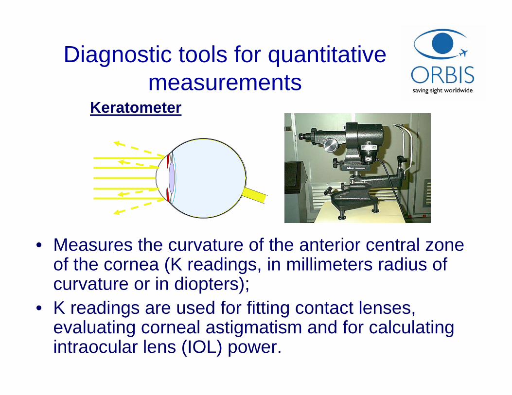
Keratometer
Diagnostic tools for quantitative measurements
• Measures the curvature of the anterior central zone of the cornea (K readings, in millimeters radius of curvature or in diopters);
• K readings are used for fitting contact lenses, evaluating corneal astigmatism and for calculating intraocular lens (IOL) power.
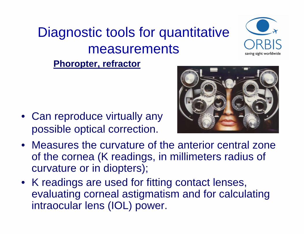
Phoropter, refractor
Diagnostic tools for quantitative measurements
• Measures the curvature of the anterior central zone of the cornea (K readings, in millimeters radius of curvature or in diopters);
• K readings are used for fitting contact lenses, evaluating corneal astigmatism and for calculating intraocular lens (IOL) power.
• Can reproduce virtually any possible optical correction.
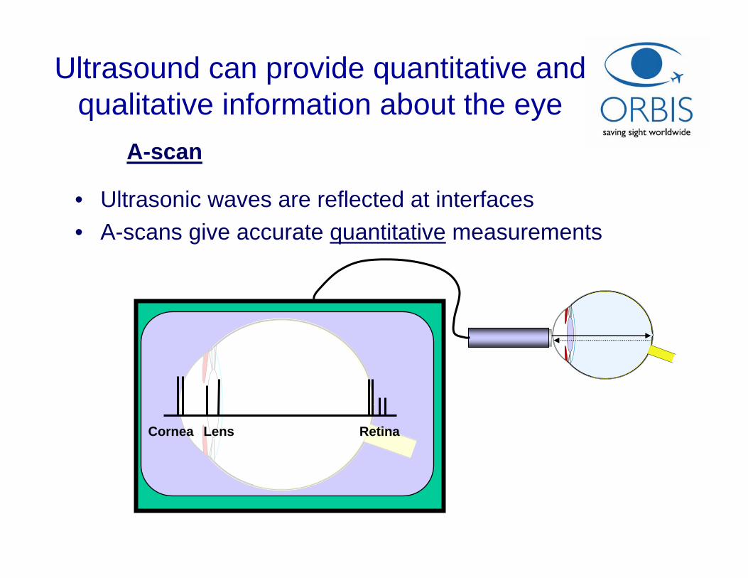
A-scan
Ultrasound can provide quantitative and qualitative information about the eye
Cornea Lens Retina
• Ultrasonic waves are reflected at interfaces• A-scans give accurate quantitative measurements
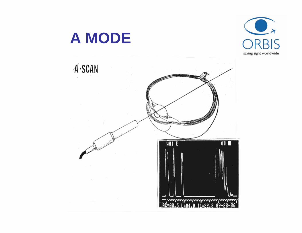
A MODE
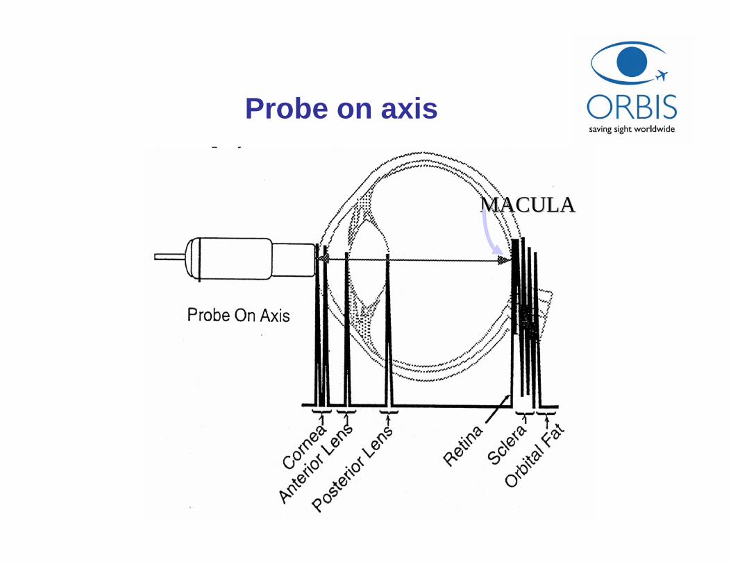
Probe on axis
MACULAMACULA
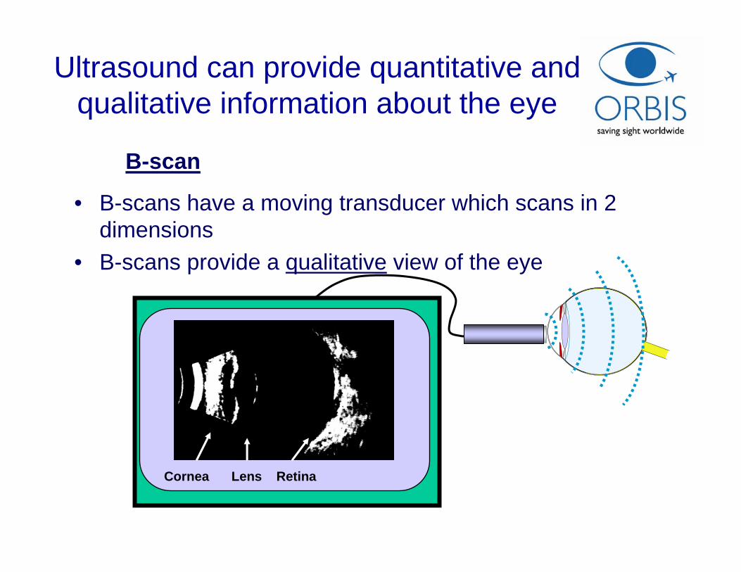
Ultrasound can provide quantitative and qualitative information about the eye
Cornea Lens Retina
• B-scans have a moving transducer which scans in 2 dimensions
• B-scans provide a qualitative view of the eye
B-scan
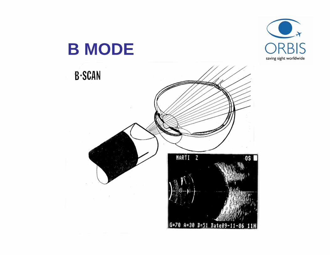
B MODE
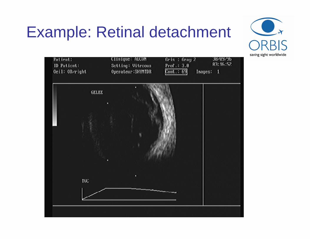
Example: Retinal detachment
THE ULTRASONIC EXAMINATIONOF THE EYE
•• Topographic analysis: B mode (Brightness)
• Quantitative analysis: A mode (Amplitude)Biometry
• Kinetic analysis: B Mode and/or Doppler
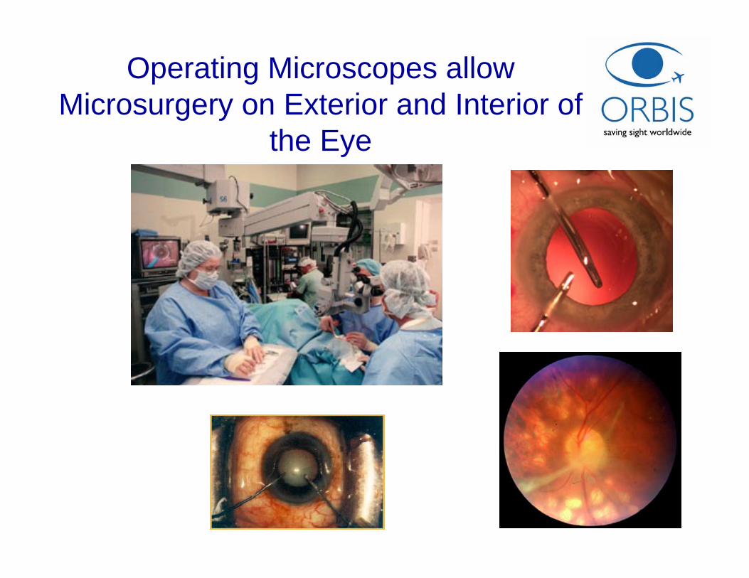
Operating Microscopes allow Microsurgery on Exterior and Interior of
the Eye
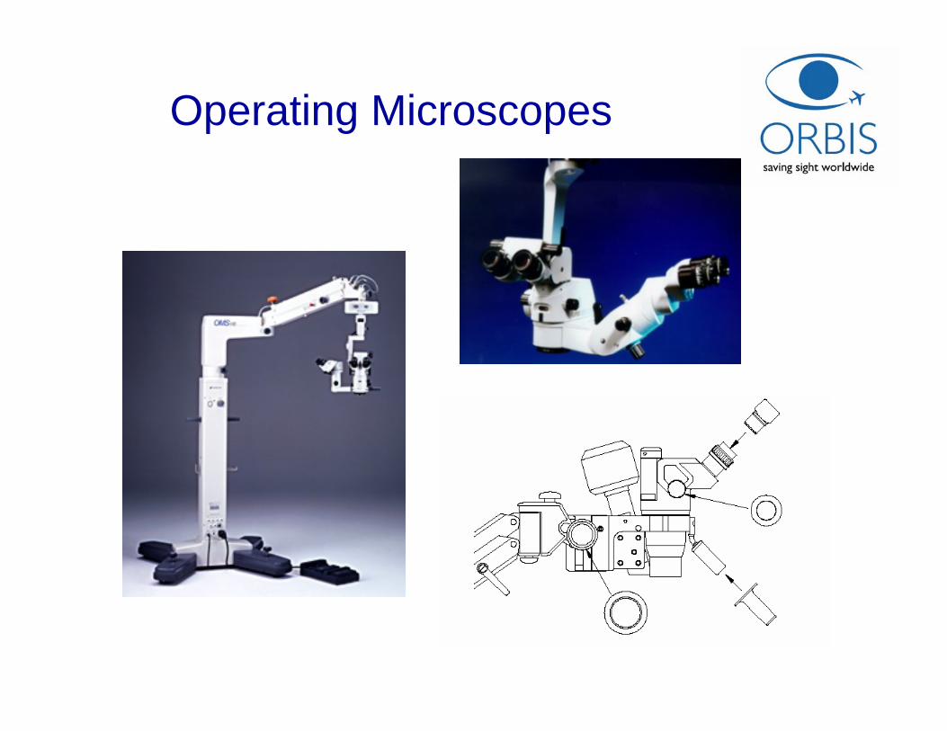
Operating Microscopes
Operating Microscopes
• Utilized for procedures that require high magnification and variable focusing.
• Light from a halogen light source is directed into the tube through prisms or fiber optic cables and shines through the objective lens onto the operating field.
• Magnification of the eyepieces is typically 8X to 20X.
• The typical focal length (working distance) of objective lenses for eye surgery using a 12.5X eyepiece is 175 to 200 mm
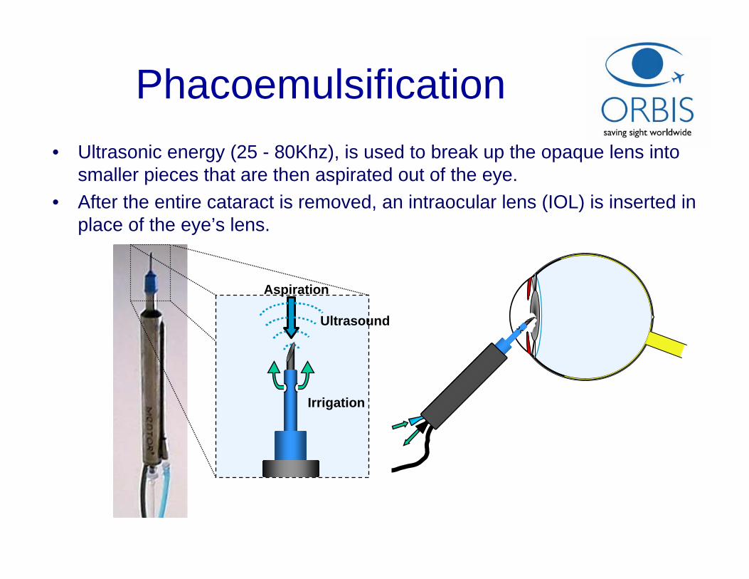
Phacoemulsification• Ultrasonic energy (25 - 80Khz), is used to break up the opaque lens into
smaller pieces that are then aspirated out of the eye.• After the entire cataract is removed, an intraocular lens (IOL) is inserted in
place of the eye’s lens.
Irrigation
Aspiration
Ultrasound
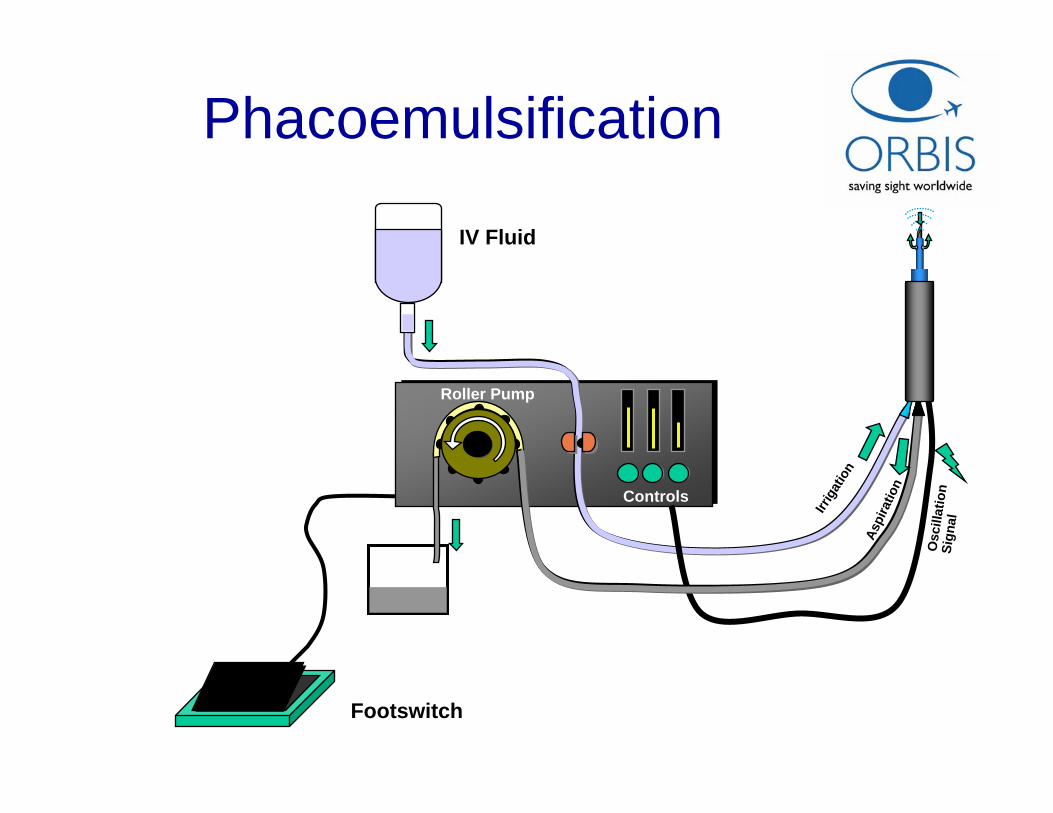
Phacoemulsification
IV Fluid
Irrig
atio
n
Aspi
ratio
n
Osc
illat
ion
Sign
al
Controls
Roller Pump
Footswitch
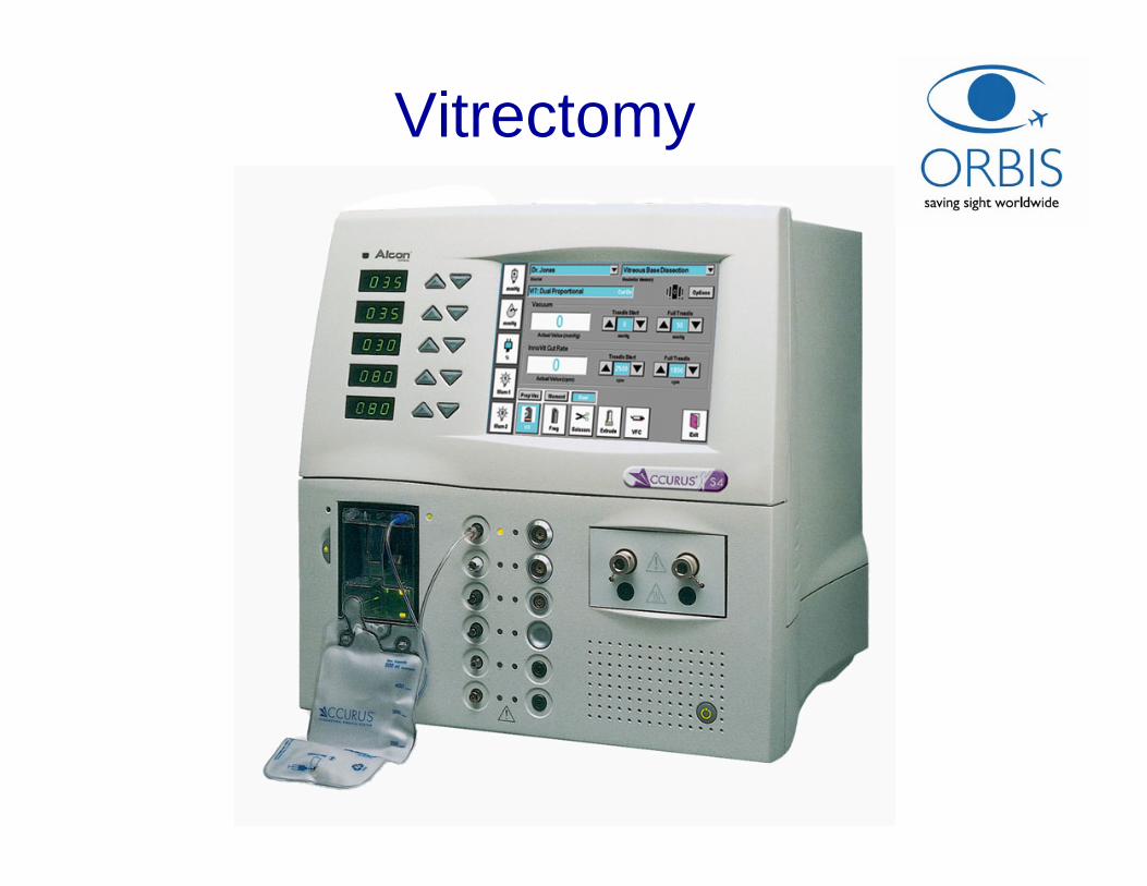
Vitrectomy
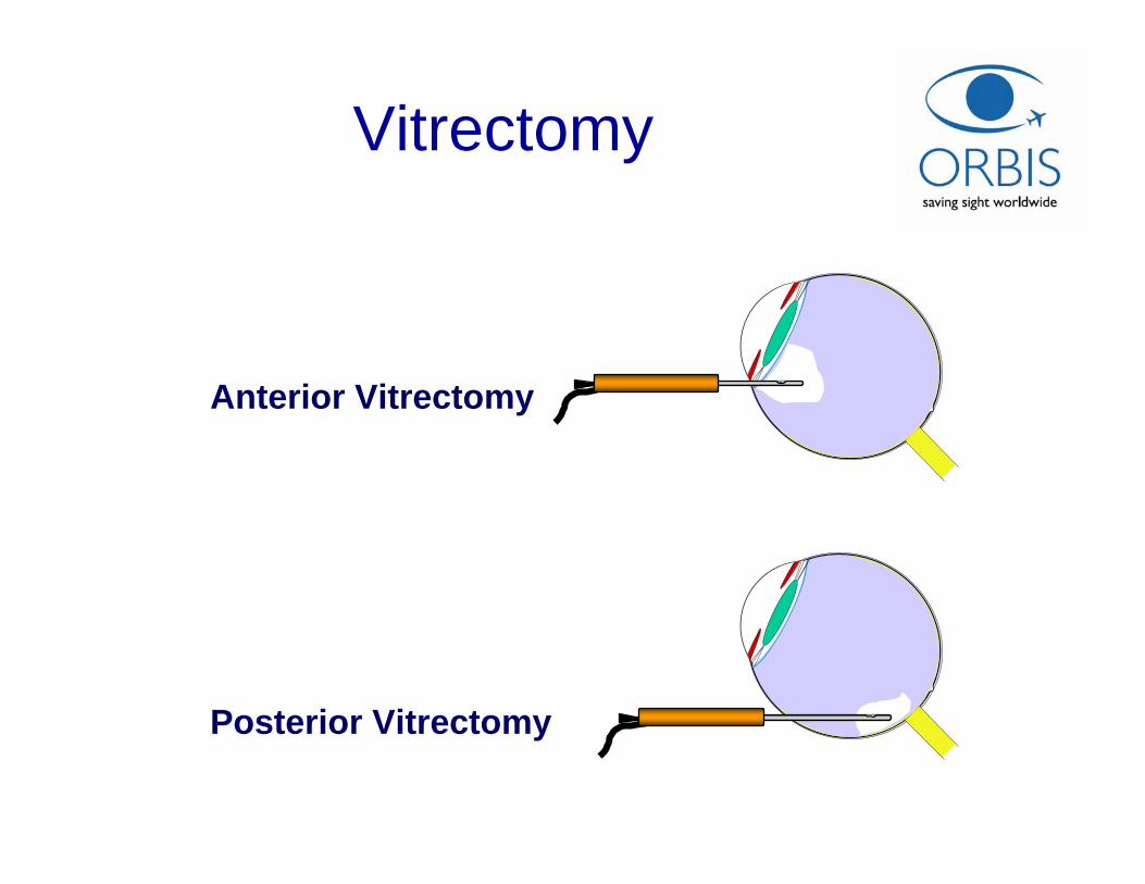
Vitrectomy
Posterior Vitrectomy
Anterior Vitrectomy
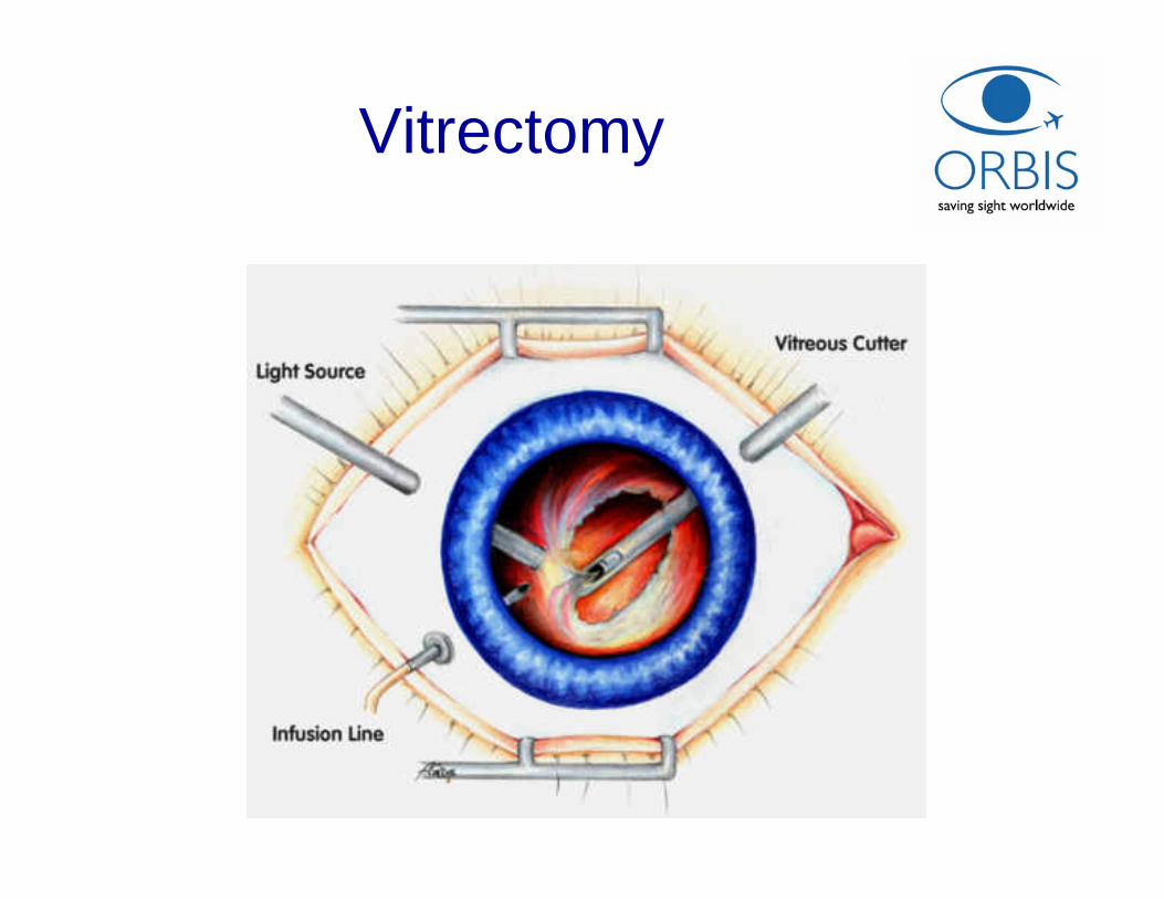
Vitrectomy
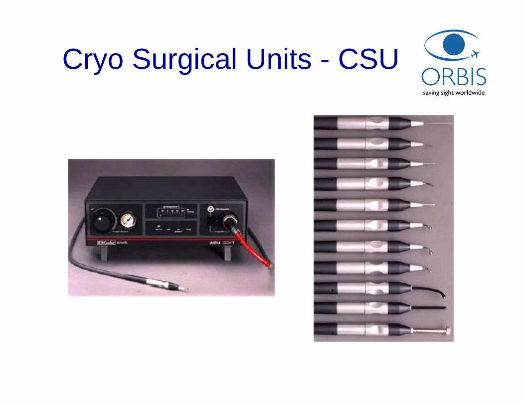
Cryo Surgical Units - CSU
Cryo Surgical Units - CSU
• CSU apply a refrigerant (cryogen) to withdraw heat from target tissue through contact with a cryogen-cooled probe.
• The effect is to freeze the surrounding tissue so that it dies.
• In the tissue immediately beyond the killed zone a degree of coagulation occurs thus limiting the resulting bleeding.
• Different types of interchangeable cryo probes are available for different applications.
• Cryogens in ophthalmology: Compressed nitrous oxide (N2O) and carbon dioxide (CO2).
Ophthalmic LASERs

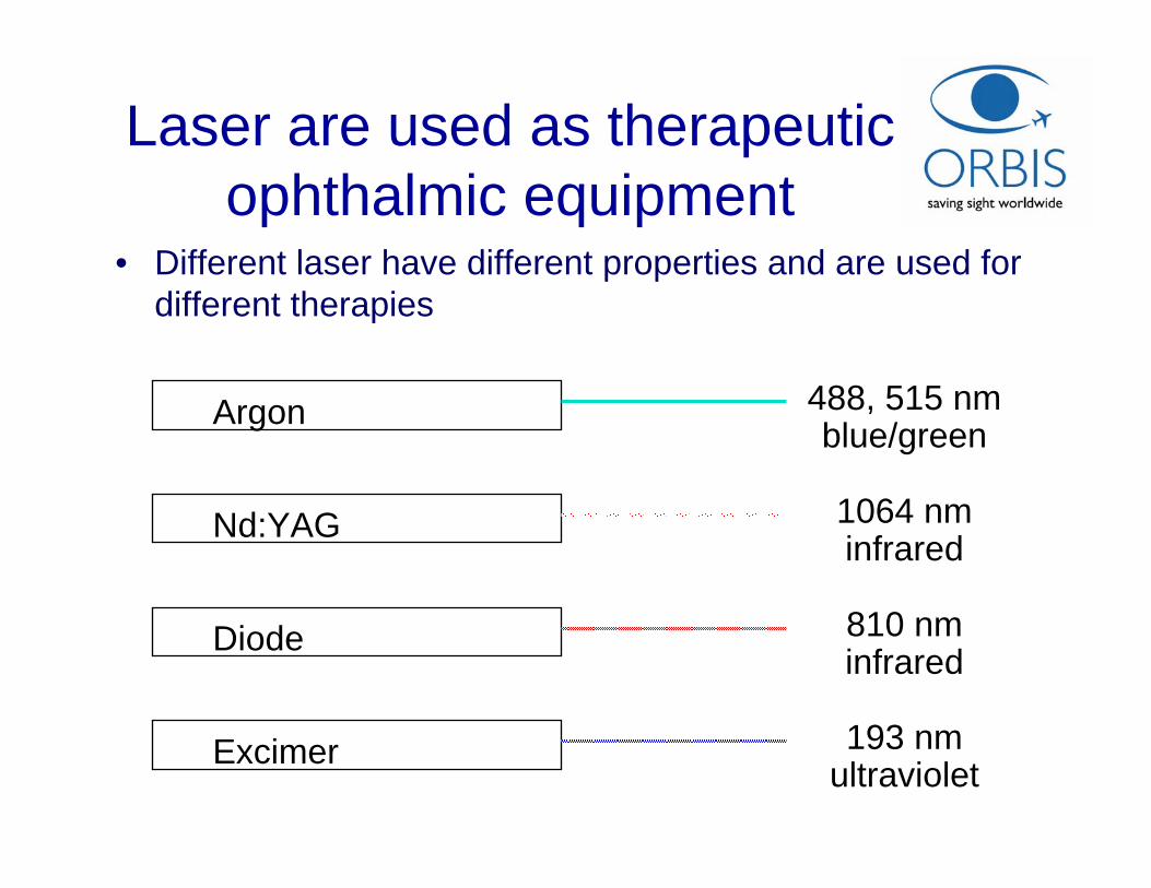
Laser are used as therapeutic ophthalmic equipment
Argon
Nd:YAG
Diode
Excimer
488, 515 nmblue/green
193 nmultraviolet
810 nminfrared
1064 nminfrared
• Different laser have different properties and are used for different therapies
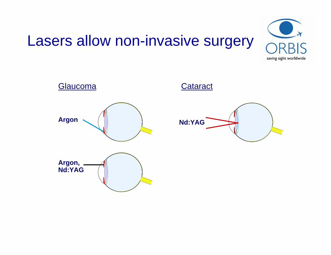
Lasers allow non-invasive surgery
Argon
Glaucoma
Nd:YAG
Cataract
Argon, Nd:YAG
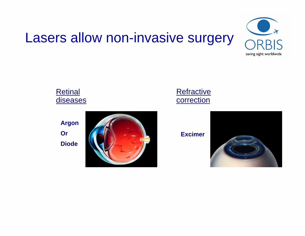
Lasers allow non-invasive surgery
ArgonOr Diode
Retinal diseases
Excimer
Refractive correction
Laser Delivery systems
• Slit lamp
• Endoprobe
• Indirect ophthalmoscope
• Operating microscope
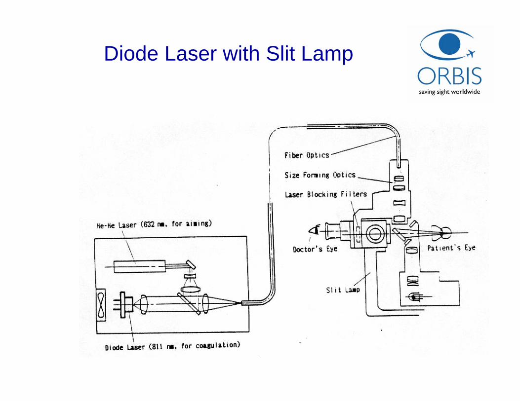
Diode Laser with Slit Lamp
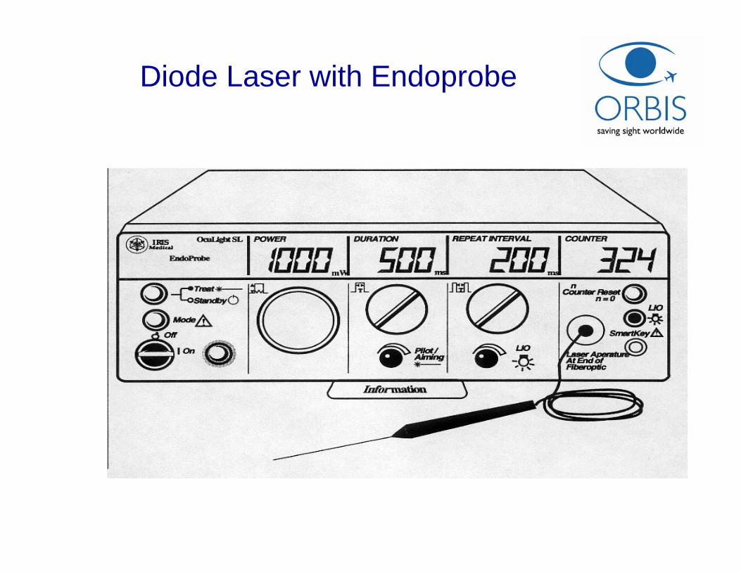
Diode Laser with Endoprobe
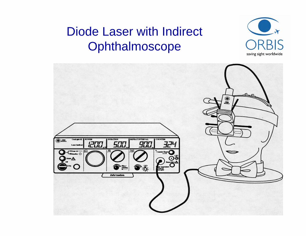
Diode Laser with Indirect Ophthalmoscope
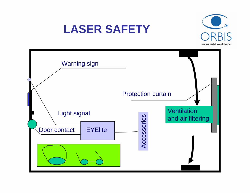
Door contact EYElite
Light signal
Warning sign
Protection curtain
Acc
esso
ries
Ventilation and air filtering
LASER SAFETY
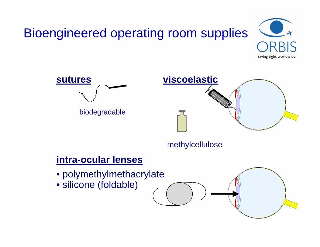
sutures viscoelastic
intra-ocular lenses
biodegradable
• polymethylmethacrylate• silicone (foldable)
methylcellulose
Bioengineered operating room supplies
Support systems for ophthalmology
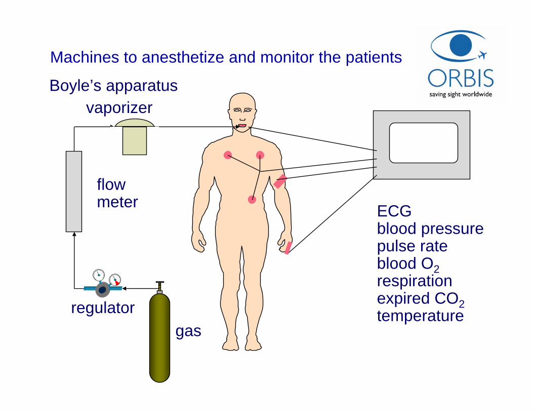
gasregulator
flowmeter
vaporizer
ECGblood pressurepulse rateblood O2respirationexpired CO2temperature
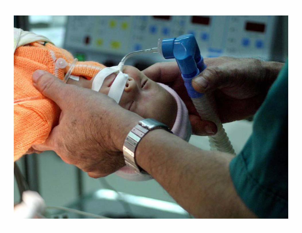
Machines to anesthetize and monitor the patients
Boyle’s apparatus
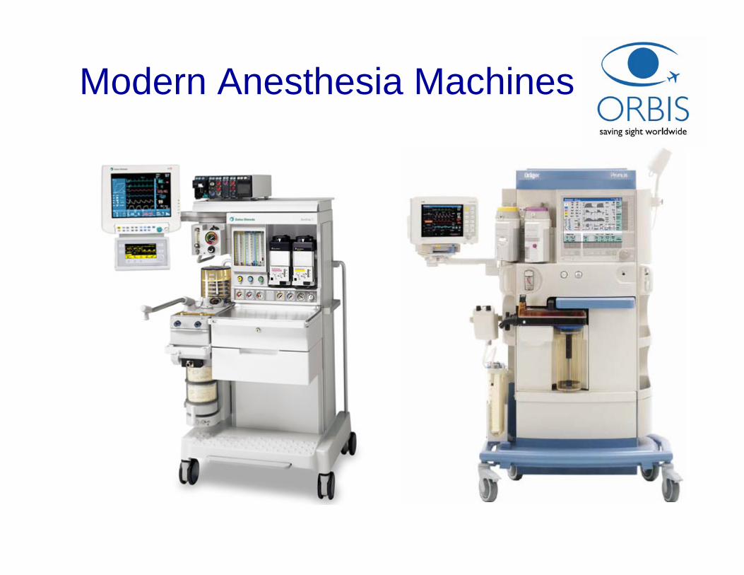
Modern Anesthesia Machines
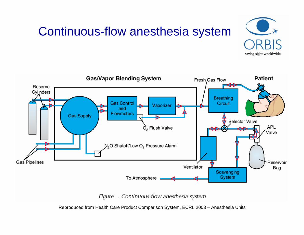
Continuous-flow anesthesia system
Reproduced from Health Care Product Comparison System, ECRI. 2003 – Anesthesia Units
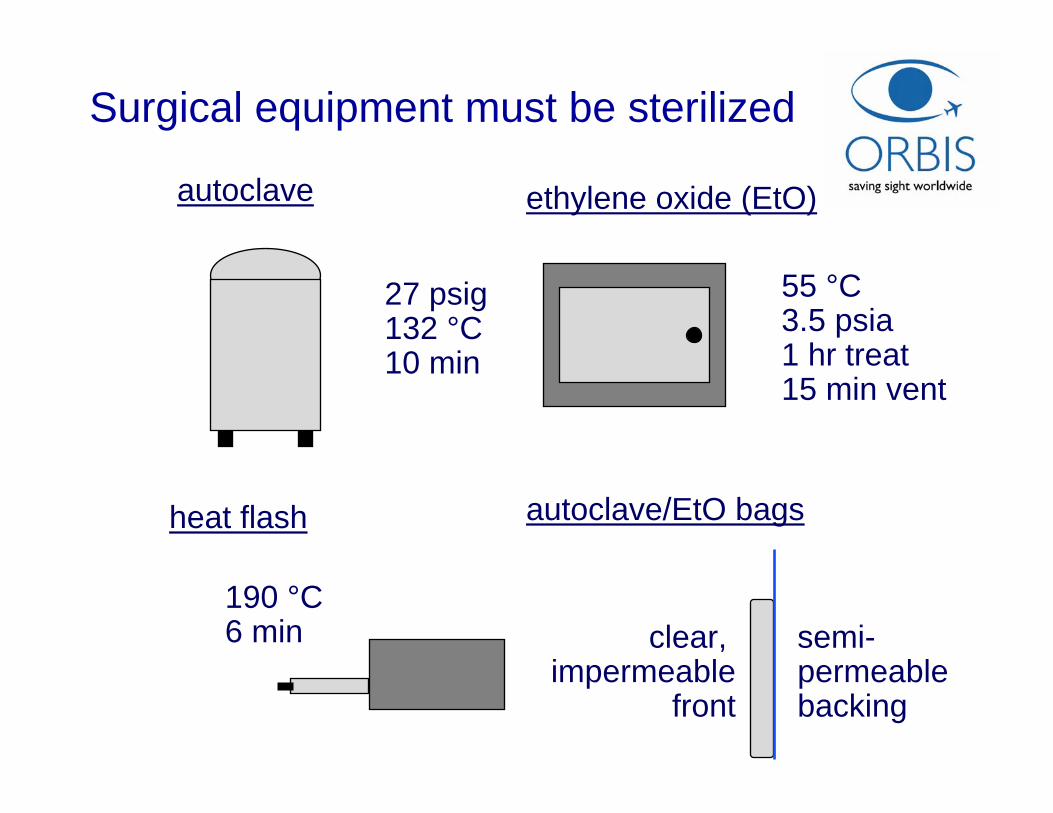
Surgical equipment must be sterilized
autoclave
27 psig132 °C10 min
heat flash
190 °C6 min
ethylene oxide (EtO)
55 °C3.5 psia1 hr treat15 min vent
autoclave/EtO bags
clear, impermeable
front
semi-permeablebacking
Acknowledgements
• ALCON Laboratories


Top Related