


Languages
Pages
Legal

Med & Health 2016; 11(2): 232-244
ORIGINAL ARTICLE
232
https://doi.org/10.17576/MH.2016.1102.12
Address for correspondence and reprint requests: Azian Abd Latiff. Anatomy Discipline, Faculty of Medicine, UniversitiTeknologi MARA, Sungai Buloh Campus, Jalan Hospital, 47000 Sungai Buloh, Selangor, Malaysia. Tel: 006-03-61267568 Fax: 006-03-61267483 E-mail: [email protected]
Oral Administration of Tocotrienol Ameliorates Lead-Induced Toxicity in the Rat Brain
NOOR AZLIZA WANI AA1, ZAR CHI T2, MOHAMAD FAIRUZ Y3, TEOH SL3, TATY ANNA K3, AZIAN AL2
1Department of Preclinical Science Studies, Faculty of Dentistry, 2Anatomy Discipline, Faculty of Medicine, Universiti Teknologi MARA, Sungai Buloh Campus, Jalan Hospital,
47000 Sungai Buloh, Selangor, Malaysia.3Department of Anatomy, Faculty of Medicine, Universiti Kebangsaan Malaysia Medical Centre, Jalan Yaacob Latif, Bandar Tun Razak, 56000 Cheras, Kuala Lumpur, Malaysia.
ABSTRAK
Kes keracunan plumbum (Pb) yang membimbangkan telah meningkat di beberapa negara tertentu. Semakin banyak bukti kajian menunjukkan pendedahan kronik terhadap plumbum memainkan peranan besar yang menyebabkan ketidakseimbangan prooksidan: antioksidan di dalam tisu otak dan perubahan histologi otak. Kajian ini dilakukan untuk mengenal pasti kesan agen antioksidan daripada kelapa sawit Malaysia, fraksi kaya tokotrienol (TRF) pada tisu otak tikus eksperimen berikutan keracunan plumbum. Lapan belas (n=18) tikus Sprague-Dawley jantan, berusia 6 minggu, dibahagikan secara rawak kepada kumpulan kawalan (CTRL) dan kumpulan eksperimen; didedahkan kepada 0.2% w / v plumbum asetat, sebagai kumpulan PB2; dan yang didedahkan kepada 0.2% w / v plumbum asetat bersama pengambilan suplemen TRF (200 mg / kg berat badan) sebagai kumpulan PB2T. Eksperimen ini dijalankan selama 30 hari. Pada akhir kajian, tisu otak telah diambil dan perubahan histopatologi di kawasan hippocampus diperhatikan. Keputusan biokimia seperti aras plumbum, TRF dan malondialdehid (MDA) dalam tisu otak serta aras aktiviti superoksida dismutase (SOD) dalam eritrosit telah ditentukan. Terdapat neuron atipikal berciri apoptosis dan tidak teratur diperhatikan di kawasan hippocampus bagi kumpulan Pb2 yang didedahkan kepada plumbum berbanding kumpulan Pb2T. Parameter- parameter biokimia tisu otak menunjukkan penurunan aras plumbum yang siginifikan (p<0.05) di dalam kumpulan Pb2T berbanding Pb2. Walaupun tiada perbezaan yang signifikan diperolehi (p>0.05) bagi aras MDA, terdapat peningkatan yang signifikan (p<0.05) bagi aktiviti SOD eritrosit dalam PB2T berbanding PB2 dan CTRL. Pengambilan suplemen TRF dilihat dapat membaiki perubahan histopatologi
233
Tocotrienol on Lead-Induced Brain Toxicity Med & Health 2016;11(2): 232-244
dalam tisu otak yang disebabkan oleh pendedahan plumbum melalui air minuman dengan mengurangkan pengumpulan plumbum dalam tisu otak tikus eksperimen.
Kata kunci: plumbum, neurotoksisiti, tekanan oksidatif, tokotrienol
ABSTRACT
The occurrence of severe lead (Pb) poisoning has risen in certain countries. There is increasing evidence that chronic lead exposure disturbs the prooxidant: antioxidant balance in the brain tissue and alters brain histology. The present study observed the antioxidant effect of tocotrienol-rich fraction (TRF) on brain tissues of the experimental rats following lead poisoning. Eighteen (n=18) male Sprague-Dawley rats, 6-weeks old, were randomly divided into control (CTRL) group and experimental groups; fed with 0.2% w/v lead acetate, as PB2 group; and fed with 0.2% w/v lead acetate and daily TRF supplementation (200 mg/kg body weight) as PB2T group. The experiment was conducted for 30 days. At the end of the study, the brain tissues were harvested and histopathological changes of the hippocampal region were observed. Biochemical findings such as brain lead, TRF and malondialdehyde (MDA) levels, and erythrocyte superoxide dismutase (SOD) activity were determined. It was observed that atypical apoptotic-like and disorganized neurons were present in the hippocampal region of the untreated PB2 group compared to PB2T group. Biochemical parameters showed a significant decrease (p < 0.05) in brain lead level in PB2T compared to PB. Even though no significant difference (p > 0.05) was obtained for MDA level, there was a significant increase (p < 0.05) in the erythrocyte SOD activity in PB2T compared to PB2 and CTRL. Supplementation with TRF improved histopathological changes in the brain tissues caused by lead exposure in drinking water by reducing lead accumulation in the brain of experimental rats.
Keywords: lead, neurotoxicity, oxidative stress, rat, tocotrienol
INTRODUCTION
Lead (Pb) poisoning is one of the major public health concerns worldwide due to its adverse effects to the human body systems (Iyer et al. 2015). Lead poisoning can occur as a result of exposure to lead through air, household dust, sand, water and commercial productssuch as paint (Cao et al. 2014). Risk factors of lead poisoning include the use of leaded gasoline,
occupational exposure to heavy metals and battery recycling, water harvesting techniques using old pipes as well as the level of education among parents, guardian or child’s primary caregiver (Brown et al. 2005; Albalak et al. 2003). Lead poisoning in human is indicated when blood lead levels reach ≥≥ 10 μg/dL whole blood (Taylor et al. 2013). The fatal dose is estimated at 500 mg of absorbed lead (Noji & Kelen 1989). The threshold for toxic effects of lead is yet
234
Med & Health 2016;11(2): 232-244 Noor Azliza Wani A.A. et al.
to be identified. However, blood lead levels of ≥≥ 80 μg/dL in children were reported to cause encephalopathy (Cao et al. 2014), whereas a lower blood lead level caused adverse effects on the central nervous system, though symptomless (Iyer et al. 2015). High blood lead levels were reported to affect physical development, auditory system, kidneys, bones and the most feared was the effect on the nervous system. Lead was reported to be highly neurotoxic and particularly affected the developing central nervous system (CNS) (Antonio-García & Massó-Gonzalez 2008). Previous studies of lead toxicity on the nervous system of mice found disruption in cognitive function due to changes in the cell morphology in certain regions of the brain (Huang & Schneider 2004; Yang et al. 2003). The present study hypothesized that lead-induced brain toxicity is highly attributable to oxidative stress. There is increase in evidence that chronic lead exposure plays an important role in the disruption of prooxidant: antioxidant balance in the brain tissue and alters brain histology which leads to physiological dysfunction (Bokara et al. 2008; Hamed et al. 2010). Oxidative stress occurs in the plasma and brain tissue in lead-exposed rats as evidenced by decreased important intracellular antioxidant enzymes and increased lipid peroxidation products (Xia et al. 2010). To the best of our knowledge, past studies have observed the effect of lead consumption along with chelating agent or other supplementary elements. Other researchers have observed the histopathological changes in the lead
exposed group (without treatment).Our interest in the present study was to evaluate the brain histopathological and biochemical effects after the consumption of lead, along with chelating agent from Malaysian palm, tocotrienol-rich fraction (TRF) treatment.TRF is a well known therapeutic agent as it possesses several medicinal properties including antioxidant, anti-inflmmatory and effective free radical scavenging agent (Abd Aziz et al. 2012). Results obtained from the pilot experiment of the present study found that thirty days of lead ingestion even in a small dose of 0.2% v/w was capable of causing histopathological changes in the hippocampus region of rats (Abd Aziz et al. 2012). The present study was therefore aimed to determine the possible effects of TRF on rat brain exposed to 0.2% v/w lead. Determination of lead, TRF, superoxide dismutase (SOD) activity and malondialdehyde (MDA) levels as well as microscopic observation of brain tissue in the hippocampus were performed in order to better understand the involvement of oxidative stress as a mechanism of neurotoxicity oflead.
MATERIALS AND METHODS
ANIMALS
Ethical approval was obtained from the Universiti Kebangsaan Malaysia Animal Ethics Committee (UKMAEC), with approval number of PP/ANAT/2011/FAIRUZ/19-MAY/377-MAY-2011-MAY-2012 prior to the commencement of study. A total of eighteen male Sprague Dawley rats (6-weeks old,
235
Tocotrienol on Lead-Induced Brain Toxicity Med & Health 2016;11(2): 232-244
weighing 150-200 gm) were obtained from the Laboratory Animal Resource Unit (LARU), Universiti Kebangsaan Malaysia. The rats were housed one animal per cage, acclimatized for one week, fed with commercial rat chow (Gold Coin, Malaysia) with distilled water ad libitum and maintained under controlled environment of 12 hrs light and dark cycle.
EXPERIMENTAL DESIGN
Rats were randomly divided into three groups, with each group comprising of six animals (n=6). The groups were i) control group which received distilled water ii) group which received lead acetate (0.2% w/v) in drinking water, and iii) group which received lead acetate (0.2% w/v) in drinking water and daily force-fed with tocotrienol-rich fraction, TRF (Golden Hope Bioganic Sdn. Bhd., Malaysia) using oral gavage at 200 mg/kg body weight. TRF used in the study composed of 235.8 mg/g (31.16%) of γ-tocotrienol (GTT), 111.5 mg/g (14.74%) of δδ-tocotrienol (DTT), 208.5 mg/g (27.56%) of α-tocotrienol (ATT), 168.9 mg/g (22.33%) of αα-tocoferol (ATF) and 31.8 mg/g (4.21%) of β-tocotrienol (BTT). In all groups, rats were maintained for thirty days and were sacrificed at the end of exposure. Histopathological study of the hippocampus was done using light microscope following hematoxylin and eosin (H&E) staining (Merck, Germany). In addition, another brain hemisphere were homogenized with ice-cold buffer (Tris-HCL, pH 7.5) (Sigma, United States of America) and centrifuged at 10,000 x g at 4˚C for 10 mins. Brain lead levels were determined using
atomic absorption spectroscopy (Perkin Elmer, United States of America), brain TRF levels by high-performance liquid chromatography (Shimadzu Corporation, Japan), erythrocyte superoxide dismutase (SOD) enzyme activity by antioxidant enzyme assay (Cayman, United States of America) and brain malondialdehyde (MDA) levels by lipid peroxidation product assay (Cayman, United States of America).
BRAIN HISTOLOGICAL STUDY
A hemisection of brain was analyzed for histopathological changes within the hippocampal subregions following lead exposure. Brain tissues were processed according to the standard protocol for light microscopy (Olympus, Japan). Tissues were fixed with 10% formalin (Ajax, Australia), dehydrated through graded alcohol series (25%-100%) (Hayman Ltd., England), cleared in toluene (Systerm Chemicals, Malaysia) and embedded in paraffin wax (Fisher Scientific, United States of America). Serial sections of 3 μm thickness were made using a microtome (Leica, Germany) and stained with H&E staining (Merck, Germany). Quantitative estimation of the number of apoptotic-like cells was done using an image analyzer (Leica, Germany) at x 20 objective based on a described method (Haynes et al. 2001).
ATOMIC ABSORPTION SPECTROSCOPY
The brain lead concentration was analyzed by atomic absorption spectrophotometer (Perkin-Elmer, United States of America) at 283.3 nm
236
Med & Health 2016;11(2): 232-244 Noor Azliza Wani A.A. et al.
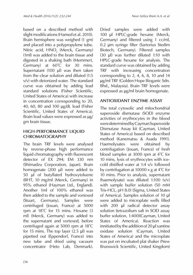
based on a described method with slight modifications (Hamed et al. 2010). Brain hemisphere was weighed (1 gm) and placed into a polypropylene tube. Nitric acid, HNO3 (Merck, Germany) (1ml) was added to the brain tissue and digested in a shaking bath (Memmert, Germany) at 60˚C for 30 mins. Supernatant (100 µl) was then taken from the clear solution and diluted (1:5 v/v) with deionized water. The standard curve was obtained by adding lead standard solutions (Fisher Scientific, United States of America) with increase in concentration corresponding to 20, 40, 60, 80 and 100 µg/dL lead (Fisher Scientific, United States of America). Brain lead values were expressed as µg/gm brain tissue.
HIGH-PERFORMANCE LIQUID CHROMATOGRAPHY
The brain TRF levels were analysed by reverse-phase high performance liquid chromatography with fluorescent detector of EX 294; EM 330 nm (Shimadzu Corporation, Japan). Brain homogenate (200 µl) were added to 50 µl of butyllated hydroxytoluene (BHT, 10 mg/ml (Merck, Germany) in 95% ethanol (Hayman Ltd., England). Another 1ml of 100% ethanol was then added to the sample and vortexed (Stuart, Germany). Samples were centrifuged (Jouan, France) at 5000 rpm at 18˚C for 15 mins. Hexane (3 ml) (Merck, Germany) was added to the supernatant and vortexed, before centrifuged again at 5000 rpm at 18˚C for 15 mins. The top layer (2.5 μl) was pipetted out (Eppendorf, France) into new tube and dried using vacuum concentrator (Heto Lab, Denmark).
Dried samples were added with 100µμl HPLC-grade hexane (Merck, Germany) and filtered using 1 cc/ml, 0.2 μm syringe filter (Sartorius Stedim Biotech, Germany). Filtered samples (30 μl) was further diluted 1:10 with HPLC-grade hexane for analysis. The standard curve was obtained by adding TRF with increasing concentration corresponding to 2, 4, 6, 8, 10 and 14 μg/ml TRF (Golden Hope Bioganic Sdn. Bhd., Malaysia). Brain TRF levels were expressed as μg/ml brain homogenate.
ANTIOXIDANT ENZYME ASSAY
The total cytosolic and mitochondrial superoxide dismutase (SOD) enzyme activities of erythrocytes in the blood were determined by Cayman Superoxide Dismutase Assay kit (Cayman, United States of America) based on described method (Kanematsu & Asada 1991). Haemolysates were obtained by centrifugation (Jouan, France) of fresh blood samples at 3000 rpm at 4˚C for 10 mins, lysis of erythrocytes with ice-cold distilled water at 1:4 v/v followed by centrifugation at 10000 x g at 4˚C for 10 mins. Prior to analysis, supernatant (haemolysate) was diluted 1:100 (v/v) with sample buffer solution (50 mM Tris-HCL, pH 8.0) (Sigma, United States of America). Samples solution of 10 μl were added to microplate wells filled with 200 μl radical detector assay solution (tetrazolium salt in SOD assay buffer solution, 1:400)(Cayman, United States of America). Reaction was innitiated by the addition of 20 μl xantine oxidase solution (Cayman, United States of America) and the microplate was put on incubated plat shaker (New Brunswick Scientific, United Kingdom)
237
Tocotrienol on Lead-Induced Brain Toxicity Med & Health 2016;11(2): 232-244
mins. Samples (150µμl) were used for each microplate well (Cayman, United States of America). The standard curve was obtained by adding MDA standard solutions (Cayman, United States of America) with increasing concentration corresponding to 0.625, 1.25, 2.5, 5.0, 10.0, 25.0 and 50.0 μM MDA (Cayman, United States of America). Brain MDA levels were determined using ELISA reader (Biotech, United States of America) at 530-540 nm. Results were expressed as μM/ml brain homogenate.
STATISTICAL ANALYSIS
SPSS software version 19.0 (SPSS Incorporation, United States of America) was used in all analyses. ANOVA, with post-hoc Tukey HSD were performed to compare variables. All results were expressed as mean ± SD and P value <0.05 was accepted as statistically significant.
RESULTS
BRAIN HISTOLOGICAL STUDY
Effect of 30 days lead exposure with TRF supplementation on the hippocampus
A formal quantitative analysis by one-way ANOVA demonstrated that lead caused an increase in the number of apoptotic-like cells in the hippocampal subregion scornusammonis CA1 (F= 92.264, p<0.05) and cornusammonis CA3 (F= 78.704, p<0.05) (Table 1). Lead exposure was shown to cause histopathological changes in the hippocampus area cornusammonis CA1 (Figure 1) and area cornusammonis
for 20 mins. The standard curve was obtained by adding SOD standard solutions (Cayman, United States of America) with increasing concentration corresponding to 0.025, 0.05, 0.1, 0.15, 0.2 and 0.25 U/ml SOD (Cayman, United States of America). SOD activities were read using ELISA reader (Biotech, United States of America) at 440-460 nm. Erythrocytes SOD activities obtained through the SOD enzyme assay kit (Cayman, United States of America) were compared to the levels of haemoglobin (Hb) concentrations in each haemolysate sample using Cyanomethaemoglobin assay (Sigma, United States of America) based on a described method (Lewis & Shinton 1980). Results were expressed as U/mg Hb.
LIPID PEROXIDATION PRODUCT ASSAY
The brain malondialdehyde (MDA) levels were determined by Cayman Malondialdehyde Assay kit (Cayman, United States of America) based on described method (Yagi 1998). Sodium dodecyl sulphate (SDS) solution (100 μl)(Cayman, United States of America) were added to the brain homogenate (100 μl)in a 5 ml tube and shaken thoroughly.Colour reagent (thiobarbituric acid solution) (Cayman, United States of America) (4 ml) were added to the bottom of the tube. Tubes were capped and put vertically in the rack. After all tubes were brought to boil on a heated plate (Leica, Germany) for 1 hr, the chemical reaction was stopped by immediately putting the samples on ice for 10 mins. Samples were centrifuged (Jouan, France) at 1600 x g at 4˚C for 10
238
Med & Health 2016;11(2): 232-244 Noor Azliza Wani A.A. et al.
CA3 (Figure 2). Compared to the control (CTRL group), the group fed with 0.2% w/v lead acetate (PB2) showed neurons with apoptotic-like appearance with shrunken and condensed cytoplasm in addition to disorganized arrangement of neurons. TRF supplementation during lead exposure (PB2T group) was found to reduce the number of neuronal cells with apoptotic-like appearance at hippocampal subregion cornusammonis CA1 (Figure 1) and cornusammonis CA3 (Figure 2) when compared to PB2 group (P<0.05). However, disorganized arrangement of neurons was still markedly observed
in PB2T group ascompared to CTRL group.
BIOCHEMICAL STUDY
Effect of 30 days lead exposure with TRF supplementation on brain lead level
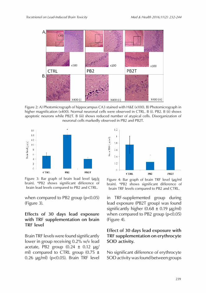
Brain lead levels were found significantly higher in group receiving 0.2% w/v lead acetate, PB2 group (14.20 ± 1.01 μg/gm) compared to CTRL group (5.36 ± 1.27 μg/gm) (p<0.05). Brain lead level in TRF-supplemented group during lead exposure (PB2T group) was found significantly lower (4.07 ± 0.78 μg/m)
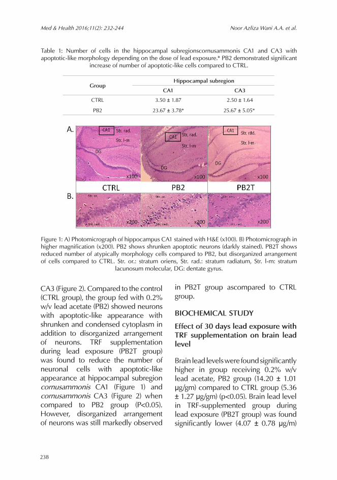
Table 1: Number of cells in the hippocampal subregionscornusammonis CA1 and CA3 with apoptotic-like morphology depending on the dose of lead exposure.* PB2 demonstrated significant
increase of number of apoptotic-like cells compared to CTRL.
Figure 1: A) Photomicrograph of hippocampus CA1 stained with H&E (x100). B) Photomicrograph in higher magnification (x200). PB2 shows shrunken apoptotic neurons (darkly stained). PB2T shows reduced number of atypically morphology cells compared to PB2, but disorganized arrangement of cells compared to CTRL. Str. or.: stratum oriens, Str. rad.: stratum radiatum, Str. l-m: stratum
lacunosum molecular, DG: dentate gyrus.
Group Hippocampal subregion
CA1 CA3
CTRL 3.50 ± 1.87 2.50 ± 1.64
PB2 23.67 ± 3.78* 25.67 ± 5.05*
239
Tocotrienol on Lead-Induced Brain Toxicity Med & Health 2016;11(2): 232-244
when compared to PB2 group (p<0.05) (Figure 3).
Effects of 30 days lead exposure with TRF supplementation on brain TRF level
Brain TRF levels were found significantly lower in group receiving 0.2% w/v lead acetate, PB2 group (0.24 ± 0.12 μg/ml) compared to CTRL group (0.75 ± 0.26 μg/ml) (p<0.05). Brain TRF level
in TRF-supplemented group during lead exposure (PB2T group) was found significantly higher (0.68 ± 0.19 μg/ml) when compared to PB2 group (p<0.05) (Figure 4).
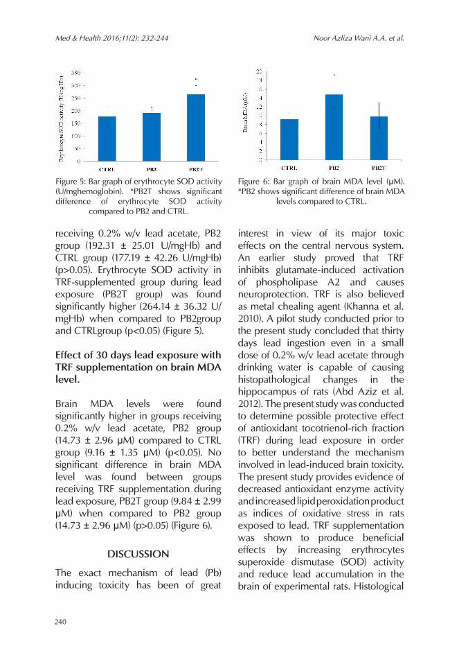
Effect of 30 days lead exposure with TRF supplementation on erythrocyte SOD activity.
No significant difference of erythrocyte SOD activity was found between groups
Figure 2: A) Photomicrograph of hippocampus CA3 stained with H&E (x100). B) Photomicrograph in higher magnification (x400). Normal neuronal cells were observed in CTRL. B (i). PB2. B (ii) shows apoptotic neurons while PB2T. B (iii) shows reduced number of atypical cells. Disorganization of
neuronal cells markedly observed in PB2 and PB2T.
Figure 3: Bar graph of brain lead level (μg/g brain). *PB2 shows significant difference of brain lead levels compared to PB2 and CTRL.
Figure 4: Bar graph of brain TRF level (μg/ml brain). *PB2 shows significant difference of brain TRF levels compared to PB2 and CTRL.
240
Med & Health 2016;11(2): 232-244 Noor Azliza Wani A.A. et al.
receiving 0.2% w/v lead acetate, PB2 group (192.31 ± 25.01 U/mgHb) and CTRL group (177.19 ± 42.26 U/mgHb) (p>0.05). Erythrocyte SOD activity in TRF-supplemented group during lead exposure (PB2T group) was found significantly higher (264.14 ± 36.32 U/mgHb) when compared to PB2group and CTRLgroup (p<0.05) (Figure 5).
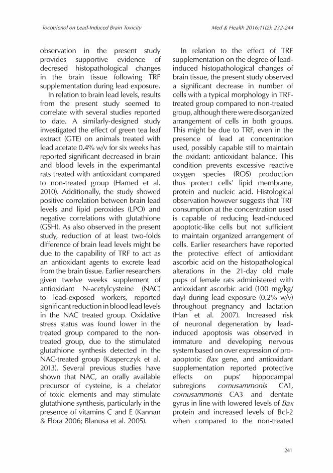
Effect of 30 days lead exposure with TRF supplementation on brain MDA level.
Brain MDA levels were found significantly higher in groups receiving 0.2% w/v lead acetate, PB2 group (14.73 ± 2.96 μM) compared to CTRL group (9.16 ± 1.35 μM) (p<0.05). No significant difference in brain MDA level was found between groups receiving TRF supplementation during lead exposure, PB2T group (9.84 ± 2.99 μM) when compared to PB2 group (14.73 ± 2.96 μM) (p>0.05) (Figure 6).
DISCUSSION
The exact mechanism of lead (Pb) inducing toxicity has been of great
interest in view of its major toxic effects on the central nervous system. An earlier study proved that TRF inhibits glutamate-induced activation of phospholipase A2 and causes neuroprotection. TRF is also believed as metal chealing agent (Khanna et al. 2010). A pilot study conducted prior to the present study concluded that thirty days lead ingestion even in a small dose of 0.2% w/v lead acetate through drinking water is capable of causing histopathological changes in the hippocampus of rats (Abd Aziz et al. 2012). The present study was conducted to determine possible protective effect of antioxidant tocotrienol-rich fraction (TRF) during lead exposure in order to better understand the mechanism involved in lead-induced brain toxicity. The present study provides evidence of decreased antioxidant enzyme activity and increased lipid peroxidation product as indices of oxidative stress in rats exposed to lead. TRF supplementation was shown to produce beneficial effects by increasing erythrocytes superoxide dismutase (SOD) activity and reduce lead accumulation in the brain of experimental rats. Histological
Figure 5: Bar graph of erythrocyte SOD activity (U/mghemoglobin). *PB2T shows significant difference of erythrocyte SOD activity
compared to PB2 and CTRL.
Figure 6: Bar graph of brain MDA level (μM). *PB2 shows significant difference of brain MDA
levels compared to CTRL.
241
Tocotrienol on Lead-Induced Brain Toxicity Med & Health 2016;11(2): 232-244
observation in the present study provides supportive evidence of decresed histopathological changes in the brain tissue following TRF supplementation during lead exposure. In relation to brain lead levels, results from the present study seemed to correlate with several studies reported to date. A similarly-designed study investigated the effect of green tea leaf extract (GTE) on animals treated with lead acetate 0.4% w/v for six weeks has reported significant decreased in brain and blood levels in the experimantal rats treated with antioxidant compared to non-treated group (Hamed et al. 2010). Additionally, the study showed positive correlation between brain lead levels and lipid peroxides (LPO) and negative correlations with glutathione (GSH). As also observed in the present study, reduction of at least two-folds difference of brain lead levels might be due to the capability of TRF to act as an antioxidant agents to excrete lead from the brain tissue. Earlier researchers given twelve weeks supplement of antioxidant N-acetylcysteine (NAC) to lead-exposed workers, reported significant reduction in blood lead levels in the NAC treated group. Oxidative stress status was found lower in the treated group compared to the non-treated group, due to the stimulated glutathione synthesis detected in the NAC-treated group (Kasperczyk et al. 2013). Several previous studies have shown that NAC, an orally available precursor of cysteine, is a chelator of toxic elements and may stimulate glutathione synthesis, particularly in the presence of vitamins C and E (Kannan & Flora 2006; Blanusa et al. 2005).
In relation to the effect of TRF supplementation on the degree of lead-induced histopathological changes of brain tissue, the present study observed a significant decrease in number of cells with a typical morphology in TRF-treated group compared to non-treated group, although there were disorganized arrangement of cells in both groups. This might be due to TRF, even in the presence of lead at concentration used, possibly capable still to maintain the oxidant: antioxidant balance. This condition prevents excessive reactive oxygen species (ROS) production thus protect cells’ lipid membrane, protein and nucleic acid. Histological observation however suggests that TRF consumption at the concentration used is capable of reducing lead-induced apoptotic-like cells but not sufficient to maintain organized arrangement of cells. Earlier researchers have reported the protective effect of antioxidant ascorbic acid on the histopathological alterations in the 21-day old male pups of female rats administered with antioxidant ascorbic acid (100 mg/kg/day) during lead exposure (0.2% w/v) throughout pregnancy and lactation (Han et al. 2007). Increased risk of neuronal degeneration by lead-induced apoptosis was observed in immature and developing nervous system based on over expression of pro-apoptotic Bax gene, and antioxidant supplementation reported protective effects on pups’ hippocampal subregions cornusammonis CA1, cornusammonis CA3 and dentate gyrus in line with lowered levels of Bax protein and increased levels of Bcl-2 when compared to the non-treated
242
Med & Health 2016;11(2): 232-244 Noor Azliza Wani A.A. et al.
group (Han et al. 2007). Additionally, a study on lead chloride (4 mM) interaction with selenium (Se) (60µgm/kg body weight/day) reported marked reversing effect of both lead-induced alterations in neural cell adhesion molecules (NCAM), also concentration of lead in rat blood and hippocampus (Wang et al. 2013). The present study showed significant high levels of TRF in the brain of TRF-supplemented group close to the levels of the control group, when compared to the non-treated group.TRF detected in the non-treated group therefore might have resulted from the amount of TRF that already exist in the rats’ body or obtained through diet given, however not sufficient to prevent the over production of free radicals which had lowered the brain TRF levels. Additionally, TRF levels in brain homogenate were also found to be negatively related to the brain lead levels. This is in accordance to a report by National Health and Nutrition Examination Survey III (NHANES III) on the possitive relationship between blood lead levels and oxidative stress markers in 10,098 adult subjects, in addition to negative correlations between blood lead levels and serum vitamin E, carotenoids and vitamin C (Lee et al. 2006). A study on the effect of vitamin E and vitamin C on lead exposure (500 mg/L) for 12 weeks to the liver and kidney of rats has reported a significant decrease of about thirty percents of initial TRF level, but not vitamin C when compared to the control group (Jurczuk et al. 2007). Beneficial effect of vitamin E (50 IU/kg body weight) intraperitoneally
administered during lead exposure (12 mg/kg body weight) for 24 days has also been reported to prevent lead-induced testicular toxicity due to its antioxidant properties (Al-Masri 2015). The present study evaluated the effect of TRF supplementation on the indices of oxidative stress in order to better understand the mechanism involved in lead toxicity. SOD enzyme activity per mg haemoglobin was determined in haemolysates of erythrocytes instead of brain homogenates. This is because SOD is present much greater in erythrocytes than in most tissues. Futuremore, previous literatures have shown that lead is capable in reducing SOD activity and other reactive oxygen species in erythrocytes (Mousa et al. 2002; Rendon-Ramirez et al. 2007). Erythrocyte SOD enzyme activitiy in TRF-treated group were found significantly higher compared to the control and non-treated group. This might be due to highly significant existing SOD enzymes in rats and in the presence of lead at the concentration used, able to neutralize excessive superoxide radicals produced by lead at the concentration used in the exposure. Besides, brain malondialdehyde (MDA) levels were also detected lower in the TRF-treated group compared to non-treated group. This might be due to TRF supplementation, at the concentration given, capable to protect cells’ lipid membrane from excessive superoxide radicals produced by lead, thus maintaining MDA levels similar to the control group. Effects of antioxidants like tocopherol, ascorbic acid and L-methionine following lead-induced toxicity to the liver, kidney
243
Tocotrienol on Lead-Induced Brain Toxicity Med & Health 2016;11(2): 232-244
and the brain of mice have shown that tocopherol (100 mg/kg body weight) significantly reduced lipid peroxidation products in both liver and brain tissues (Patra et al. 2001). A study on the effect of L-ascorbic acid and α-tocopherol on male rats exposed to lead has reported a decrease in nitric oxide and lipid peroxide levels to the normal level, in addition to a significant increase in SOD, glutathione peroxidase (GSHPx) and catalase (CAT) in both antioxidant-treated groups (Das & Saha 2010). Indices of oxidative stress determined in the present study thus provide evidence of lead ability to produce oxidative stress in cells.
CONCLUSION
The present study provided evidence of beneficial effect of tocotrienol-rich fraction (TRF) supplementation during lead exposure. TRF supplementation during lead exposure is capable of maintaining vitamin E at normal levels, reduces indices of oxidative stress, prevents histopathological alterations as well as reduces brain lead levels. Based on the limited parameters of antioxidant enzyme and product of lipid peroxidation determined in the present study, therefore further studies are needed to ascertain the exact action of lead in producing brain toxicity and other indices of oxidative stress following lead consumption. Due to limited duration of the study and limited technique used, a few histomorphometric analyses along with quantitative analysis on apoptosis or necrosis of the brain cells were unable to confirm the event of apoptosis in the
present study. Future studies should confirm the event of apoptosis using specific apoptotic assay.
ACKNOWLEDGEMENTS
The authors wish to thank the departments involved in the study, especially the Department of Anatomy, Faculty of Medicine, Universiti Kebangsaan Malaysia for providing the research grant UKM-GGPM-TKP-155-2010.
REFERENCESAbd Aziz, N.A.W., Yahaya, M.F., Teoh, S.L., Latiff,
A.A., Das, S., Kamarudin, T.A. 2012. Histological observation of the hippocampus and frontal cortex of experimental Sprague-Dawley rats fed with lead in a dose-related manner. Br J Med Health Sci 1(1): 99-106.
Albalak, R., Noonan, G., Buchanan, S., Flanders, W.D., Gotway-Crawford, C., Kim, D., Jones, R.L., Sulaiman, R., Blumenthal, W., Tan, R., Curtis, G., McGeehin, M.A. 2003. Blood lead levels and risk factors for lead poisoning among children in Jakarta, Indonesia. Sci Total Environ 301(1-3): 75-85.
Al-Masri, S.A. 2015. Effect of pumpkin oil and vitamin E on lead-induced testicular toxicity in male rats. J Anim Plant Sci 25(1): 72-77.
Antonio-García, M.T., Massó-Gonzalez, E.L. 2008. Toxic effects of perinatal lead exposure on the brain of rats: involvement of oxidative stress and the beneficial role of antioxidants. Food Chem Toxicol 46(6): 2089-95.
Blanusa, M., Varnai, V.M., Piasek, M., Kostial, K. 2005. Chelators as antidotes of metal toxicity: therapeutic and experimental aspects. Curr Med Chem 12(23): 2771-94.
Bokara, K.K., Brown, E., McCormick, R., Yallapragada, P.R., Rajanna, S., Bettaiya, R. 2008. Lead-induced increase in antioxidant enzymes and lipid peroxidation products in developing rat brain. Biometals 21(1): 9-16.
Brown, L.M., Kim, D., Yomai, A., Meyer, P.A., Noonan, G.P., Huff, D., Flanders, W.D. 2005. Blood lead levels and risk factors for lead poisoning in children and caregivers in Chuuk State, Micronesia. Int J Hyg Environ Health 208(4): 231-6.
Cao, J., Li, M., Wang, Y., Yu, G., Yan, C. 2014. Environmental lead exposure among preschool
244
Med & Health 2016;11(2): 232-244 Noor Azliza Wani A.A. et al.
children in Shanghai, China: blood lead levels and risk factors. PLoS One 9(12): e113297.
Das, K. K, Saha, S. 2010. L-ascorbic acid and alpha tocopherol supplementation and antioxidant status in nickel- or lead-exposed rat brain tissue. JBasic Clin Physiol Pharmacol 21(4): 325-46.
Hamed, E.A., Meki, A.R., Abd El-Mottaleb, N.A. 2010. Protective effect of green tea on lead-induced oxidative damage in rat’s blood and brain tissue homogenates. J Physiol Biochem 66(2): 143-51.
Han, J.M., Chang, B.J., Li, T.Z., Choe, N.H., Quan, F.S., Jang, B.J., Cho, I.H., Hong, H.N., Lee, J.H. 2007. Protective effects of ascorbic acid against lead-induced apoptotic neurodegeneration in the developing rat hippocampus in vivo. Brain Res 1185: 68-74.
Haynes, L.E., Griffiths, M.R., Hyde, R.E., Barber, D.J., Mitchell, I.J. 2001. Dexamethasone induces limited apoptosis and extensive sublethal damage to specific subregions of the striatum and hippocampus: Implications for mood disorders. Neuroscience 104(1): 57-69.
Huang, F., Schneider, J.S. 2004. Effects of lead exposure on proliferation and differentiation of neural stem cells derived from different regions of embryonic rat brain. Neurotoxicology 25(6): 1001-12.
Iyer, S., Sengupta, C., Velumani, A. 2015. Lead toxicity: An overview of prevalence in Indians. Clin Chim Acta 451(Pt B): 161-4.
Jurczuk, M., Brzóska, M.M., Moniuszko-Jakoniuk, J. 2007. Hepatic and renal concentrations of vitamins E and C in lead- and ethanol-exposed rats. An assessment of their involvement in the mechanisms of peroxidative damage. Food Chem Toxicol 45(8): 1478-86.
Kasperczyk, S., Dobrakowski, M., Kasperczyk, A., Ostałowska, A., Birkner, E. 2013. The administration of N-acetylcysteine reduces oxidative oxidative stress and regulates glutathione metabolism in the blood cells of workers exposed to lead. ClinToxicol (Phila) 51(6): 480-6.
Kanematsu, S., Asada, K. 1991. Chloroplast and cytosol isozymes of CuZn-superoxide dismutase: their characteristic amino acid sequences. Free Radic Res Commun 12-13 Pt 1: 383-90.
Kannan, G.M., Flora, S.J. 2006. Combined administration of N-acetylcysteine and monoisoamyl DMSA on tissue oxidative stress during arsenic chelation therapy. Biol Trace Elem Res 110(1): 43-59.
Khanna, S., Parinandi, N.L., Kotha, S.R., Roy, S., Rink, C., Bibus, D., Sen, C.K. 2010. Nanomolar
vitamin E alpha-tocotrienol inhibits glutamate-induced activation of phospholipase A2 and causes neuroprotection. J Neurochem 112(5): 1249-60.
Lee, D.H., Lim, J.S., Song, K., Boo, Y., Jacobs, D.R. 2006. Graded Associations of Blood Lead and Urinary Cadmium Concentrations with Oxidative-Stress–Related Markers in the U.S. Population: Results from the Third National Health and Nutrition Examination Survey. Environmental Health Perspectives 114(3): 350–4.
Lewis, S.M., Shinton, N.K. 1980. Cyanmethaemoglobin reference preparations. J Clin Pathol 33(7): 700-1.
Mousa, H.M., Al-Qarawi, A.A., Ali, B.H., Abdel-Rahman, H.A., ElMougy, S.A. 2002. Effect of lead exposure on the erythrocytic antioxidant levels in goats. J Vet Med A Physiol Pathol Clin Med 49(10): 531-4.
Noji, E.K., Kelen, G.D. 1989. Manual of Toxicologic Emergencies. Chicago: Year Book Medical Publishers; 231-76.
Patra, R.C., Swarup, D., Dwivedi. S.K. 2001. Antioxidant effects of a-tocopherol, ascorbic acid and L-methionine on lead induced oxidative stress to the liver, kidney and brain in rats. Toxicology 162(2): 81-8.
Rendon-Ramirez, A., Cerbon-Solorzano, J., Maldonado-Vega, M., Quintanar-Escorza, M.A., Calderon-Salinas, J.V. 2007. Vitamin-E reduces the oxidative damage on delta-aminolevulinicdehydratase induced by lead intoxication in rat erythrocytes. Toxicol In Vitro 21(6): 1121-6.
Taylor, M.P., Lanphear, B.P., Winder, C. 2013. Eliminating childhood lead toxicity in Australia: a call to lower the intervention level. In reply. Med J Aust 199(5): 323-4.
Wang, M., Fu, H., Xiao, Y., Ai, B., Wei, Q., Wang, S., Liu, T., Ye, L., Hu, Q. 2013. Effects of low-level organic selenium on lead-induced alterations in neural cell adhesion molecules. Brain Res 1530: 76-81.
Xia, D., Yu, X., Liao, S., Shao, Q., Mou, H., Ma, W. 2010. Protective effect of Smilax glabra extract against lead-induced oxidative stress in rats. J Ethnopharmacol 130(2): 414-20.
Yagi, K. 1998. Simple assay for the level of total lipid peroxides in serum or plasma. Methods Mol Biol 108: 101-6.
Yang, Y., Ma, Y., Ni, L., Zhao, S., Li, L., Zhang, J., Fan, M., Liang, C., Cao, J., Xu, L. 2003. Lead exposure through gestation-only caused long-term learning/memory deficits in young adult offspring. Exp Neurol 184(1): 489-95.
Top Related