


Languages
Pages
Legal

iVue® with Corneal Epithelial Thickness Mapping (ETM) Software
Optovue Traditional 510(k) Premarket Notification
510(k) SUMMARY
(per 21 CFR §807.92)
iVue Model 100 with Corneal Epithelial Thickness Mapping
I. GENERAL INFORMATION
Submitter: Optovue, Inc.
2800 Bayview Drive
Fremont, CA 94538
Est. Registration No.: 3005950902
Contact Person: Thomas Navarro, RAC
Advisor, Regulatory and Quality Affairs
Direct: (510) 897-1728
Fax: (510) 651-2516
Email: [email protected]
Date Summary Prepared: November 23, 2016
II. SUBJECT DEVICE
Trade Name: iVue® Model 100
Common Name: Optical Coherence Tomography (OCT)
Classification Name: Ophthalmoscope, AC-powered
Device Classification: Class II (21 CFR§ 886.1570)
Product Code: HLI (ophthalmoscope, ac-powered)
Subsequent Product Code: OBO (tomography, optical coherence)
III. PREDICATE DEVICE
Company: Optovue, Inc.
Device: iVue® with Normative Database (K121739)
Cleared: 510(k) K121739 on 18-January-2013
iVue® with Corneal Epithelial Thickness Mapping (ETM) Software
Optovue Traditional 510(k) Premarket Notification
IV. DEVICE DESCRIPTION
Introduction
The iVue is used to capture, store, display and print spectral domain-optical coherence
tomography (SD-OCT) images of the posterior and anterior structure of the eye. The device
software includes a Normative Database (NDB), consisting of OCT data from a range of known
normal subjects that can be used to compare a new patient’s measurements in relation to the
normal distribution.
iVue is a computer controlled ophthalmic imaging system using either a laptop computer or “all-
in-one” computer (where the computer is integrated into the monitor). For laptop systems there
are two control box options 110 vs 230 volts. They interface between the motor column and the
medical power supply for the computer.
iVue System Key Functional Components
The iVue system contains the following hardware components:
Scanner Head: the scanner is the main component of the iVue system. It is used to view
and scan the patient’s eye, collect the OCT signal, and send it to the computer for
processing.
Computer: the system computer, either a laptop or All-in-One (AIO, which includes the
computer and monitor in one unit), is approved for medical use. It supports scanner
operation and processes, stores and displays exam data through the application software.
The searchable iVue database stores and organizes patient and exam data.
Control Box: the control box supports operation of the scanner and contains the backup
hard disk.
Joystick and Chinrest Assembly: the joystick moves the scanner left and right, forward
and back, and align it with the patient’s eye to capture the scan.
Footswitch (optional): the footswitch provides another way to capture scans, including
auto-adjustment, capture and saving.
Motorized Table (optional): a motorized table is optional. Customers may order the table in
two input voltages of 120V or 230V.
Cornea Adapter Module: the cornea lens adapter is attached to the front of the instrument
to enable the iVue to image the cornea and anterior chamber of the eye.
iVue® with Corneal Epithelial Thickness Mapping (ETM) Software
Optovue Traditional 510(k) Premarket Notification
iVue Device Characteristics
The device scans a patient’s eye and uses a low coherence interferometer to measure the
reflectivity of the retinal tissue. The cross sectional retinal tissue structure is composed of
sequence of A-scans. It has a traditional patient and instrument interface like most ophthalmic
devices. The patient will rest their head on the forehead and chin rest while the operator uses
joystick to align the device to patient’s eye. The computer has a graphic user interface for
acquiring and analyzing the image.
iVue also has a cornea lens adapter, which can be attached to the front of the instrument to enable
the iVue to image the cornea and anterior chamber of the eye. This lens adapter is called the
Corneal Anterior Module, or “CAM.”
The iVue device uses two light sources for illumination. A near-infrared (but still visible) LED is
used for illumination during alignment to the patient’s eye with a central wavelength in the 735-
850nm range. A superluminescent diode (SLD) is used to illuminate the retina using a wavelength
of 840nm.
The subject iVue device with ETM software measures the epithelial cell layer. The device scans
the patient’s eye using a low coherence interferometer to measure the reflectivity of the corneal
tissue. The cross-sectional corneal image is composed of a sequence of A-scans. The computer
has a graphic user interface for acquiring and analyzing the image. The iVue ETM software
modification also expands the pachymetry scan pattern for corneal epithelial measurements,
automates segmentation for the posterior boundary, provides a thickness map for the central 6
mm diameter, and generates reports and associated data.
The iVue device has a chin rest and forehead rest that come into contact with the patient’s skin.
The chin rest and forehead rest can be cleaned with a disinfectant, such as a wipe with an isopropyl
alcohol pad or with another germicide using a clean cloth. The iVue is intended to be used in
clinical settings such as a hospital, eye clinic or doctor’s exam room.
V. PROPOSED INDICATIONS FOR USE
The iVue® is a non-contact, high resolution tomographic imaging device. It is intended for in-vivo
imaging, axial cross-sectional, and three-dimensional imaging and measurement of anterior and
posterior ocular structures, including retina, retinal nerve fiber layer, ganglion cell complex
(GCC), optic disc, cornea, corneal epithelia, corneal stroma and anterior chamber of the eye. With
the integrated normative database, the iVue with Normative Database is a quantitative tool for the
comparison of retina, retinal nerve fiber layer, ganglion cell complex, and optic disc
measurements to a database of known normal subjects. The iVue is indicated for use as a device
to aid in the diagnosis, documentation, and management of ocular health and diseases in the adult
population.
iVue® with Corneal Epithelial Thickness Mapping (ETM) Software
Optovue Traditional 510(k) Premarket Notification
VI. COMPARISON OF TECHNOLOGICAL CHARACTERISTICS WITH THE
PREDICATE DEVICE
Both the predicate iVue device and subject iVue with corneal ETM software are used to capture
spectral domain-optical coherence tomography (SD-OCT) images of the eye. The predicate iVue
device with Normative Database (NDB) was previously verified for performance and
functionality to assure conformance to the requirements for its intended use.
At a high level, both devices are based on the following same technological elements:
Scanner Head: used to view and capture scans of the patient’s eye
Computer: supports scanner operation and processes, stores and displays exam data
Normative Database (NDB): consisting of OCT data that can be used to compare a new
patient’s measurements in relation to the normal distribution
The following technological differences exist between the subject and predicate devices:
Corneal Epithelial Thickness Mapping (ETM): used to provide the pachymetry maps and
the corneal epithelial thickness maps
Automatic Segmentation: for the posterior boundary of the epithelial layer to generate
ETM thickness maps
Software-generated Reports: to include corneal epithelial thickness maps and associated
data
VII. PERFORMANCE DATA
The following performance data were provided in support of the substantial equivalence
determination.
Software Validation
The subject iVue device has the additional corneal ETM software module. Software
documentation complies with the determination of a “moderate” Level of Concern device. Device
software was verified and validated to support the proposed indications for use according to IEC
62304:2006 Medical device software – Software life cycle processes and FDA’s General
Principles of Software Validation; Final Guidance for Industry and FDA Staff.
The iVue software version 2016.2 with corneal ETM software is the final version incorporated
into this 510(k) premarket notification. The iVue with ETM software provides a corneal thickness
(pachymetry) map, and the corneal epithelial thickness map (ETM) for the central 6mm diameter.
The new software version consists of modifying the cornea pachymetry scan pattern for ETM
scan, adding automatic segmentation for the posterior boundary of the epithelial layer to generate
iVue® with Corneal Epithelial Thickness Mapping (ETM) Software
Optovue Traditional 510(k) Premarket Notification
ETM thickness maps, and updating software-generated reports to include corneal epithelial
thickness map and associated data.
Scan Reports
Corneal Pachymetry & Epithelium Report
The “Corneal Pachymetry & Epithelium Report” has been expanded to include an OCT image
and a map showing Pachymetry and Epithelial thickness; alternately, a map showing the thickness
of the Stroma is also available. A “Cornea Pachymetry & Epithelial Change Analysis” shows
thickness measurements from all patient visits.
Cornea Pachymetry/Lens Fitting Scan
The Cornea Pachymetry scan has improved functionality for “lens fitting”. The Lens Fitting
Report maps the clearance space between the posterior surface of the scleral lens and the anterior
surface of the cornea.
Cornea Angle Scan
The Cornea Angle scan has been improved to visualize the “landing zones” of lenses, nasal,
temporal, inferior and superior. The angle scan line is perpendicular to where the lens contacts
the sclera. For convenience, multiple (4) images can be selected to display on a single page.
3D Fundus En Face
The 3D Fundus En Face scan option improves usability by offering an NFL thickness map
window to visualize the nerve fiber over the superior and inferior arches.
Electrical Safety and Electromagnetic Compatibility (EMC)
Electrical safety and EMC testing were conducted on the iVue device and complies with the IEC
60601-1 standards for safety and the IEC 60601-1-2 standard for EMC.
Clinical Evaluation
Two (2) clinical studies were conducted to demonstrate substantial equivalence:
Clinical Study for Repeatability and Reproducibility of Corneal Epithelial Mapping with iVue®
SD-OCT
The objective of this clinical study was to evaluate the repeatability and reproducibility of the
iVue ETM scan for the corneal thickness (pachymetry), the epithelial thickness, and the stromal
thickness mapping in normal subjects and corneal patients with iVue SD-OCT ETM software
based on a crossed-study design and crossed random-effects ANOVA model.
A heterogeneous population of qualified study subjects was evaluated and included the Normal
Subjects group (12 subjects), and the Corneal Patients group further stratified to target 12 contact
lens patients, 12 post-refractive surgery patients, 12 dry eye patients, and 14 keratoconus patients.
iVue® with Corneal Epithelial Thickness Mapping (ETM) Software
Optovue Traditional 510(k) Premarket Notification
The study inclusion criteria required subject who were 18 years of age or older, able to provide
consent, and were willing to complete the required examinations. In addition, subjects were
qualified based on a history or clinical diagnosis of one or more of the following conditions:
Dry eye patients with no history of refractive surgery
Contact lens patients without complications, refractive surgery or dry eye
Post-laser refractive surgery patients with 1 month post-surgery without complications
Keratoconus patients with a clinical diagnosis of keratoconus in the study eye
The study exclusion criteria excluded those with the inability to complete the required SD-OCT
scans (e.g., unable to fixate due to poor vision).
Based on a crossed-study design, each study subject was imaged with all three iVue/operator
pairs, and within each iVue/operator pair, at least three ETM scans were acquired with the
operator realigning the instrument on the study eye for each scan acquisition. To ensure
realignment for each scan, the operator asked test subject to sit back after a scan acquisition and
then reposition for the next scan.
The repeatability and reproducibility of the ETM scan are assessed for all zonal thickness
parameters and the summary statistics parameters displayed on screen for the corneal thickness
(pachymetry), the epithelial thickness, and the stromal thickness.
For qualified scans, the operators reviewed the thickness maps for obvious segmentation error
and reviewed the individual corneal meridian images to verify segmentation for erroneous maps.
Segmentation edit tools were used to perform manual correction and then the epithelial map was
reprocessed. Noticeable segmentation errors were manually corrected by the operator and marked
for “Manual Correction” in the case report form. Scans with manual correction qualified for R&R
data analysis.
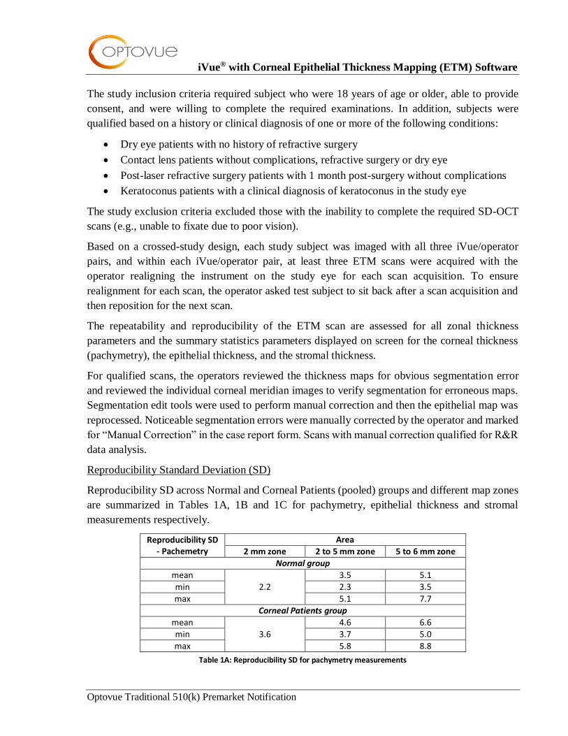
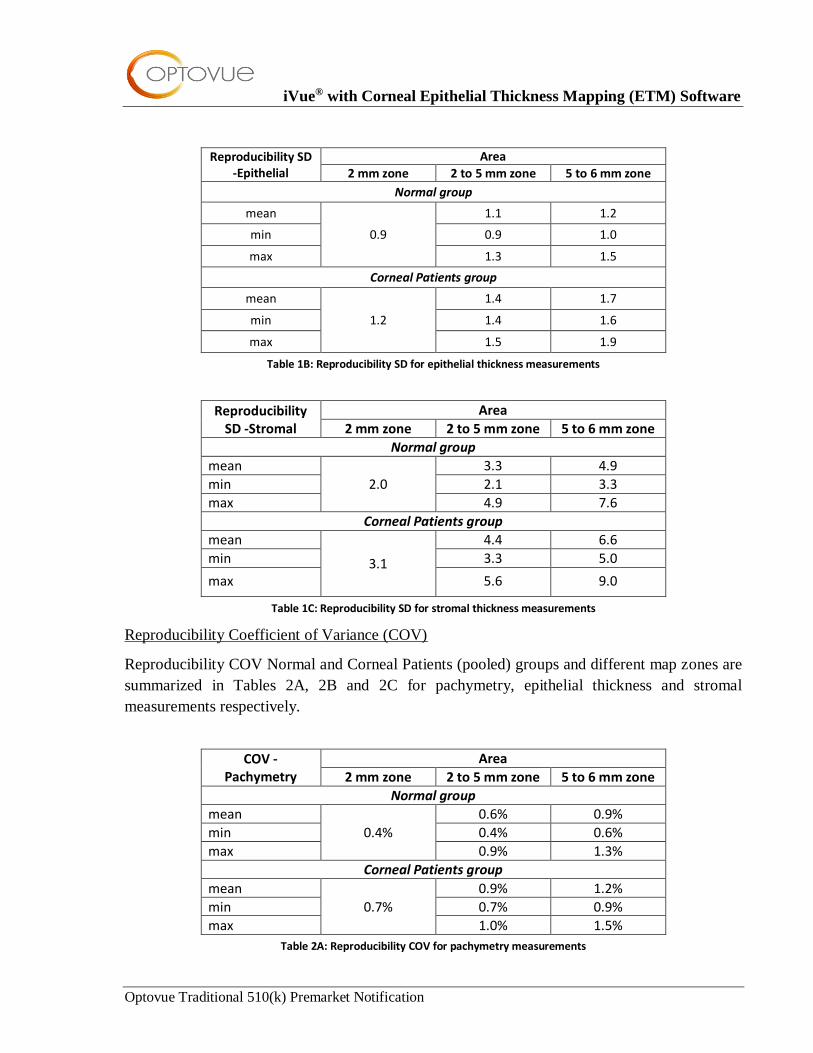
Reproducibility Standard Deviation (SD)
Reproducibility SD across Normal and Corneal Patients (pooled) groups and different map zones
are summarized in Tables 1A, 1B and 1C for pachymetry, epithelial thickness and stromal
measurements respectively.
Reproducibility SD - Pachemetry
Area
2 mm zone 2 to 5 mm zone 5 to 6 mm zone
Normal group
mean
2.2
3.5 5.1
min 2.3 3.5
max 5.1 7.7
Corneal Patients group
mean
3.6
4.6 6.6
min 3.7 5.0
max 5.8 8.8
Table 1A: Reproducibility SD for pachymetry measurements
iVue® with Corneal Epithelial Thickness Mapping (ETM) Software
Optovue Traditional 510(k) Premarket Notification
Reproducibility SD -Epithelial
Area
2 mm zone 2 to 5 mm zone 5 to 6 mm zone
Normal group
mean
0.9
1.1 1.2
min 0.9 1.0
max 1.3 1.5
Corneal Patients group
mean
1.2
1.4 1.7
min 1.4 1.6
max 1.5 1.9
Table 1B: Reproducibility SD for epithelial thickness measurements
Reproducibility SD -Stromal
Area
2 mm zone 2 to 5 mm zone 5 to 6 mm zone
Normal group
mean
2.0
3.3 4.9
min 2.1 3.3
max 4.9 7.6
Corneal Patients group
mean
3.1
4.4 6.6
min 3.3 5.0
max 5.6 9.0
Table 1C: Reproducibility SD for stromal thickness measurements
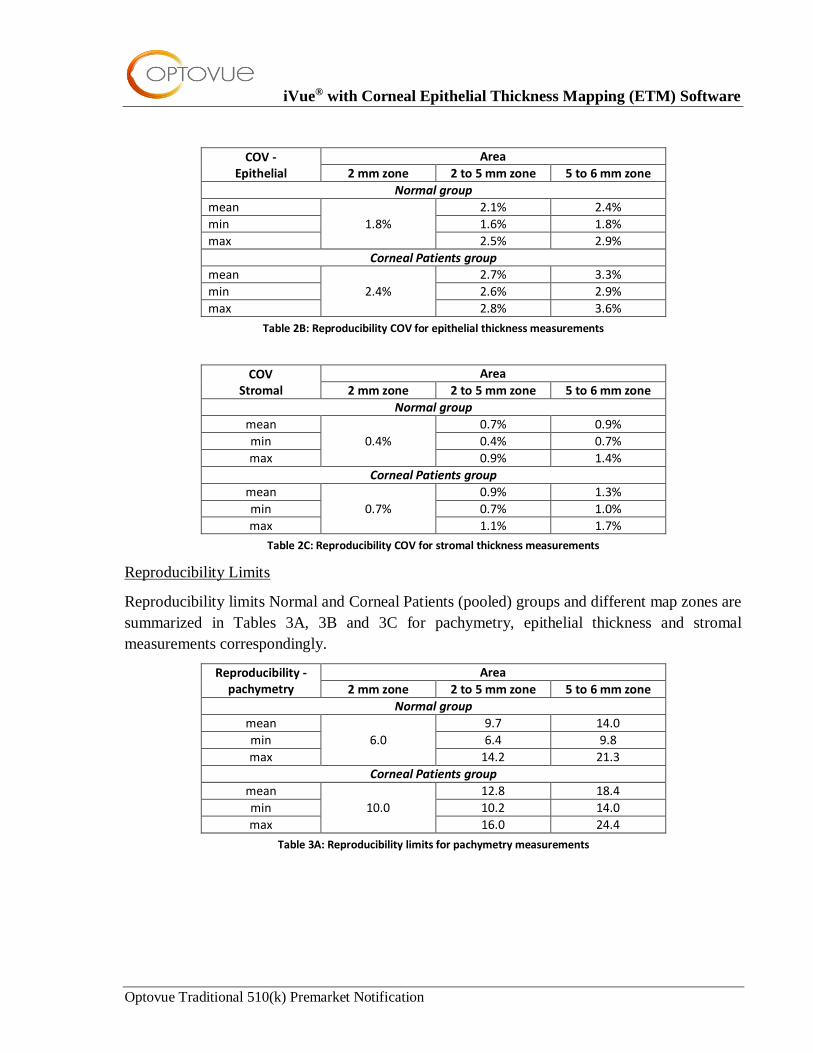
Reproducibility Coefficient of Variance (COV)
Reproducibility COV Normal and Corneal Patients (pooled) groups and different map zones are
summarized in Tables 2A, 2B and 2C for pachymetry, epithelial thickness and stromal
measurements respectively.
COV - Pachymetry
Area
2 mm zone 2 to 5 mm zone 5 to 6 mm zone
Normal group
mean
0.4%
0.6% 0.9%
min 0.4% 0.6%
max 0.9% 1.3%
Corneal Patients group
mean
0.7%
0.9% 1.2%
min 0.7% 0.9%
max 1.0% 1.5%
Table 2A: Reproducibility COV for pachymetry measurements
iVue® with Corneal Epithelial Thickness Mapping (ETM) Software
Optovue Traditional 510(k) Premarket Notification
COV - Epithelial
Area
2 mm zone 2 to 5 mm zone 5 to 6 mm zone
Normal group
mean
1.8%
2.1% 2.4%
min 1.6% 1.8%
max 2.5% 2.9%
Corneal Patients group mean
2.4%
2.7% 3.3%
min 2.6% 2.9%
max 2.8% 3.6%
Table 2B: Reproducibility COV for epithelial thickness measurements
COV Stromal
Area
2 mm zone 2 to 5 mm zone 5 to 6 mm zone
Normal group
mean
0.4%
0.7% 0.9%
min 0.4% 0.7%
max 0.9% 1.4%
Corneal Patients group
mean
0.7%
0.9% 1.3%
min 0.7% 1.0%
max 1.1% 1.7%
Table 2C: Reproducibility COV for stromal thickness measurements
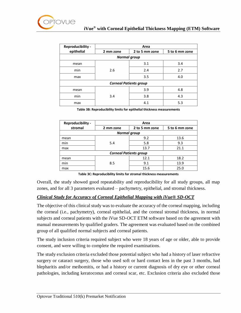
Reproducibility Limits
Reproducibility limits Normal and Corneal Patients (pooled) groups and different map zones are
summarized in Tables 3A, 3B and 3C for pachymetry, epithelial thickness and stromal
measurements correspondingly.
Reproducibility - pachymetry
Area
2 mm zone 2 to 5 mm zone 5 to 6 mm zone
Normal group
mean
6.0
9.7 14.0
min 6.4 9.8
max 14.2 21.3
Corneal Patients group
mean
10.0
12.8 18.4
min 10.2 14.0
max 16.0 24.4
Table 3A: Reproducibility limits for pachymetry measurements
iVue® with Corneal Epithelial Thickness Mapping (ETM) Software
Optovue Traditional 510(k) Premarket Notification
Reproducibility - epithelial
Area
2 mm zone 2 to 5 mm zone 5 to 6 mm zone
Normal group
mean
2.6
3.1 3.4
min 2.4 2.7
max 3.5 4.0
Corneal Patients group
mean
3.4
3.9 4.8
min 3.8 4.3
max 4.1 5.3
Table 3B: Reproducibility limits for epithelial thickness measurements
Reproducibility - stromal
Area
2 mm zone 2 to 5 mm zone 5 to 6 mm zone
Normal group
mean
5.4
9.2 13.6
min 5.8 9.3
max 13.7 21.1
Corneal Patients group
mean
8.5
12.1 18.2
min 9.1 13.9
max 15.6 25.0
Table 3C: Reproducibility limits for stromal thickness measurements
Overall, the study showed good repeatability and reproducibility for all study groups, all map
zones, and for all 3 parameters evaluated – pachymetry, epithelial, and stromal thickness.
Clinical Study for Accuracy of Corneal Epithelial Mapping with iVue® SD-OCT
The objective of this clinical study was to evaluate the accuracy of the corneal mapping, including
the corneal (i.e., pachymetry), corneal epithelial, and the corneal stromal thickness, in normal
subjects and corneal patients with the iVue SD-OCT ETM software based on the agreement with
manual measurements by qualified graders. The agreement was evaluated based on the combined
group of all qualified normal subjects and corneal patients.
The study inclusion criteria required subject who were 18 years of age or older, able to provide
consent, and were willing to complete the required examinations.
The study exclusion criteria excluded those potential subject who had a history of laser refractive
surgery or cataract surgery, those who used soft or hard contact lens in the past 3 months, had
blepharitis and/or meibomitis, or had a history or current diagnosis of dry eye or other corneal
pathologies, including keratoconus and corneal scar, etc. Exclusion criteria also excluded those
iVue® with Corneal Epithelial Thickness Mapping (ETM) Software
Optovue Traditional 510(k) Premarket Notification
with the inability to complete the required SD-OCT scans (e.g., unable to fixate due to poor
vision).
The study data were collected at three separate clinical study sites. One eye per study subject was
included in the study. All scans from a given study subject were acquired in one visit.
One scan of sufficient image quality was required per study eye. If scan quality issue was observed
during scan acquisition by the operator, he/she could take up to two additional scans in attempt to
obtain a scan of sufficient quality. Study site operators evaluated scan quality and determined
whether a scan met image quality criteria and documented scan quality observations in the case
report form.
Manual image grading was performed by 3 qualified graders from the Optovue clinical team.
Each grader performed manual measurements for all ETM scans included in the grading data set.
Three randomized grading orders were generated and each grader was assigned one randomized
grading order to follow. The graders were masked to each other’s results throughout the grading
process.
Distribution of Study Measurements
The distributions of the corneal epithelial thickness, stroma thickness, and corneal thickness
(pachymetry) are based on software output for the combined group using simple descriptive
statistics such as the mean, median, standard deviation (stdev), 1st and 3rd quartiles (Q1 and Q3),
and range (min, max).
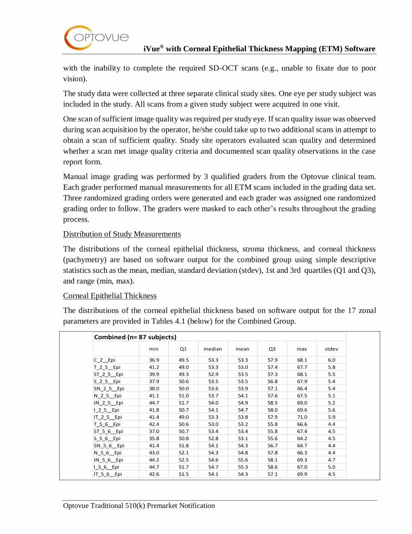
Corneal Epithelial Thickness
The distributions of the corneal epithelial thickness based on software output for the 17 zonal
parameters are provided in Tables 4.1 (below) for the Combined Group.
min Q1 median mean Q3 max stdev
C_2__Epi 36.9 49.5 53.3 53.3 57.9 68.1 6.0
T_2_5__Epi 41.2 49.0 53.3 53.0 57.4 67.7 5.8
ST_2_5__Epi 39.9 49.3 52.9 53.5 57.3 68.1 5.5
S_2_5__Epi 37.9 50.6 53.5 53.5 56.8 67.9 5.4
SN_2_5__Epi 38.0 50.0 53.6 53.9 57.1 66.4 5.4
N_2_5__Epi 41.1 51.0 53.7 54.1 57.6 67.5 5.1
IN_2_5__Epi 44.7 51.7 54.0 54.9 58.5 69.0 5.2
I_2_5__Epi 41.8 50.7 54.1 54.7 58.0 69.6 5.6
IT_2_5__Epi 41.4 49.0 53.3 53.8 57.9 71.0 5.9
T_5_6__Epi 42.4 50.6 53.0 53.2 55.8 66.6 4.4
ST_5_6__Epi 37.0 50.7 53.4 53.4 55.8 67.4 4.5
S_5_6__Epi 35.8 50.8 52.8 53.1 55.6 64.2 4.5
SN_5_6__Epi 41.4 51.8 54.1 54.3 56.7 64.7 4.4
N_5_6__Epi 43.0 52.1 54.3 54.8 57.8 66.3 4.4
IN_5_6__Epi 44.2 52.5 54.6 55.6 58.1 69.3 4.7
I_5_6__Epi 44.7 51.7 54.7 55.3 58.6 67.0 5.0
IT_5_6__Epi 42.6 51.5 54.1 54.3 57.1 69.9 4.5
Combined (n= 87 subjects)
iVue® with Corneal Epithelial Thickness Mapping (ETM) Software
Optovue Traditional 510(k) Premarket Notification
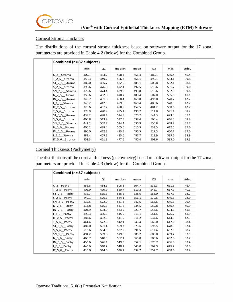
Corneal Stroma Thickness
The distributions of the corneal stroma thickness based on software output for the 17 zonal
parameters are provided in Table 4.2 (below) for the Combined Group.
min Q1 median mean Q3 max stdev
C_2__Stroma 309.1 433.2 458.3 451.4 480.1 556.4 46.4
T_2_5__Stroma 358.3 449.2 466.2 466.1 490.1 563.1 39.8
ST_2_5__Stroma 385.0 465.7 482.6 485.1 506.8 582.1 38.6
S_2_5__Stroma 390.6 476.6 492.4 497.5 518.6 591.7 39.0
SN_2_5__Stroma 379.6 474.4 489.0 493.8 516.6 592.0 39.6
N_2_5__Stroma 359.6 462.0 478.7 480.4 507.0 585.0 41.1
IN_2_5__Stroma 349.7 451.0 468.4 468.8 493.0 578.7 42.2
I_2_5__Stroma 345.2 442.3 459.6 460.4 488.6 570.3 42.7
IT_2_5__Stroma 328.6 437.2 458.5 457.5 484.2 558.6 42.7
T_5_6__Stroma 378.9 470.9 485.1 490.2 511.8 591.4 38.2
ST_5_6__Stroma 439.2 498.4 514.8 520.2 541.3 623.3 37.1
S_5_6__Stroma 460.8 513.9 537.5 538.4 560.4 646.3 38.8
SN_5_6__Stroma 442.2 507.7 524.4 530.9 550.8 648.7 37.7
N_5_6__Stroma 406.2 488.4 505.6 510.3 533.5 612.5 37.6
IN_5_6__Stroma 396.0 472.2 493.5 496.5 517.5 600.7 37.6
I_5_6__Stroma 383.4 463.3 483.6 487.7 511.9 589.6 38.9
IT_5_6__Stroma 352.3 461.3 477.6 480.4 502.6 583.0 39.3
Combined (n= 87 subjects)
Corneal Thickness (Pachymetry)
The distributions of the corneal thickness (pachymetry) based on software output for the 17 zonal
parameters are provided in Table 4.3 (below) for the Combined Group.
min Q1 median mean Q3 max stdev
C_2__Pachy 356.6 484.5 508.8 504.7 532.3 611.6 46.4
T_2_5__Pachy 402.9 499.9 520.7 519.2 542.7 617.9 40.1
ST_2_5__Pachy 432.7 515.5 536.6 538.6 558.0 637.3 38.7
S_2_5__Pachy 449.1 526.6 544.1 551.1 570.6 646.8 38.9
SN_2_5__Pachy 435.5 522.9 541.4 547.6 568.6 645.8 39.4
N_2_5__Pachy 414.8 515.5 531.8 534.5 559.8 640.4 40.9
IN_2_5__Pachy 404.9 503.9 523.9 523.7 547.6 634.8 41.5
I_2_5__Pachy 398.3 496.3 515.5 515.1 541.4 626.2 41.9
IT_2_5__Pachy 382.6 492.3 511.5 511.2 537.6 614.5 42.3
T_5_6__Pachy 441.4 522.6 542.1 543.4 565.0 647.0 38.4
ST_5_6__Pachy 482.0 551.4 569.3 573.6 593.5 678.3 37.4
S_5_6__Pachy 513.6 564.9 587.5 591.5 612.4 697.5 38.7
SN_5_6__Pachy 494.2 559.8 579.6 585.2 606.0 699.7 37.9
N_5_6__Pachy 460.7 540.9 562.1 565.0 586.6 667.6 37.7
IN_5_6__Pachy 453.6 526.1 549.8 552.1 570.7 656.0 37.4
I_5_6__Pachy 443.6 518.2 540.7 543.0 567.9 645.7 38.8
IT_5_6__Pachy 410.0 514.8 536.7 534.7 557.7 638.0 39.4
Combined (n= 87 subjects)
iVue® with Corneal Epithelial Thickness Mapping (ETM) Software
Optovue Traditional 510(k) Premarket Notification
Limits of Agreement (see Section: Performance Testing – Clinical)
The limits of agreement of the corneal thickness mapping between the software output and manual
measurements are provided for the Combined Group, and, for additional information, also for the
5 sub-groups (Normal, Contact Lens, Dry Eye, Post-LRS, and KCN) respectively.
The Bland-Altman plots for the agreement between software output and manual measurement are
provided for the Combined Group and the 5 sub-groups. The Bland-Altman plot is a scatter plot
of the difference between the software-based and manual methods, using manual measurements
as reference as represented by the horizontal axis. Reference lines were added to the plot including
that at mean differences and the upper and lower limits of agreement (LOA).
Deming regression analysis and Scatter plots for the corneal epithelial thickness between the
software output and the manual measurements are provided for the Combined Group and the 5
sub-groups. Deming regression analysis on corneal epithelial thickness for the Combined Group
is also provided, including intercept, confidence intervals (CI) of intercept, slope, and confidence
intervals (CI) of slope.
Summary
Overall, the clinical data and analyses demonstrate agreement of the automated corneal epithelial
thickness segmentation algorithm to qualified, independent, masked graders performing manual
corneal epithelial thickness segmentation.
VIII. CONCLUSIONS
The conclusions drawn from the non-clinical validation tests (software, electrical safety, and EMC)
demonstrate that the iVue subject device with corneal ETM software is as safe, as effective, and
performs as well as or better than the legally marketed iVue predicate device identified in this 510(k)
premarket notification.
We also believe that these study results achieved the clinical objectives and that the performance
of the iVue device is characterized sufficiently to help the Agency make a determination of
substantial equivalence. The cumulative performance testing data indicate the iVue as safe and
effective as applicable in the proposed labeling.
Top Related