


Languages
Pages
Legal


On Remodeling and Function of Autogenous
Bone Grafts in Maxillary Reconstruction
Amir Dasmah
Department of Oral & Maxillofacial Surgery
Institute of Odontology at Sahlgrenska Academy
University of Gothenburg
Gothenburg, Sweden
Göteborg 20131

On Remodeling and Function of Autogenous Bone Grafts in Maxillary
Reconstruction
© 2013 Amir Dasmah
email: [email protected]
http://hdl.handle.net/2077/33132
ISBN 978-91-628-8820-6
Printed in Sweden by Kompendiet
2

To
my father Parviz
my mother Jila
my brother Ali
with love
3

CONTENT
ABSTRACT 6
LIST OF PUBLICATIONS 8
INTRODUCTIONS 9
Background 9
Bone 11
Origin and function 11
Bone cells 11
The extra-cellular matrix 15
Bone structure 15
Bone formation and remodeling 16
Bone repair 18
Healing of autogenous bone grafts 21
Guided bone regeneration 22
Cardinal factors for predictable bone regeneration 23
Different donor sites in jaw bone reconstruction 24
Smoking 26
Surgical techniques 26
Osseointegration of titanium implants 30
Osseointegration of titanium implants in autogenous bone grafts 31
Titanium implant surface topography 32
AIMS 35
MATERIALS AND METHODS 36
Animal studies I and II 36
Animals and anaesthesia 36
Implants 36
Surgery protocols 37
4

Specimen preparation 39
Analysis and calculations 40
Resonance frequency analysis: implant stability measurements 41
Clinical and radiographic studies III and IV 42
Patients 42
Pre-surgical examination, inclusion and exclusion criteria 42
Pre- and post-surgical care 43
Bone harvesting and preparation 43
Bone augmentation of the anterior maxilla 44
Implants 45
Radiographic examination 45
Statistics 47
RESULTS 48
Integration of moderately rough fluoridated implants in 48
autogenous bone grafts
Marginal bone-level alterations and three-dimensional 49
analysis of volumetric change in autogenous bone grafts
DISCUSSION 52
Animal studies 52
Clinical and radiographic studies 56
CONCLUSIONS 67
FUTURE PERSPECTIVES 68
ACKNOWLEDGEMENTS 69
REFERENCES 70
5

ABSTRACT
Background Reconstruction of the jaws due to resorption of the alveolar crest may require bone augmentation for placement and integration of endosseous implants and future rehabilitation with a prosthetic supra-construction. Autogenous bone grafts from the iliac crest have frequently been used for this purpose in oral and maxillofacial surgery. Experimental studies have shown stronger bone tissue responses to surface-modified implants than to implants with machined surfaces and a delayed surgical protocol has been recommended. Whether surface modification of dental implants enhances osseointegration in grafted bone and how far the remodeling and resorption process of the grafted bone continue, has been a matter of debate.Aims The aim of the first two studies was to analyse the effect of surface modification of dental implants installed in grafted bone. In Study I, surface-modified (test) implants were compared with non-modified (control) implants in autogenous bone grafts with regard to osseointegration and stability in terms of bone-to-implant contact (BIC) and resonance frequency analysis (RFA). The aim of Study II was to evaluate osseointegration and stability of surface-modified implants in one-stage (test) vs. two-stage (control) surgery protocols using the same histomorphometric analysis and stability measurements as in the previous study. Study III focuses on differences in marginal bone-level alterations between autogenous particulate (test) and block (control) onlay grafts. Stability measurements were also studied using RFA. Finally, the objective of Study IV was to examine changes in volume reduction of grafted bone. Furthermore, we wanted to compare the amount of resorption between particulate bone (test) and block bone (control) grafts. Materials & Methods In Study I, we used eight rabbits. A bone graft from each side of the sagittal suture in the calvarial bone was harvested and fixed bicortically to each proximal tibial metaphysis through a dental implant with a blasted, fluoridated (test) surface and a machined (control) surface. Test and control sides were randomized. After 8 weeks, the rabbits were sacrificed for light microscopic analysis. Resonance frequency analysis was performed both at the time of surgery and at the end of the study. In Study II, six rabbits were subjected to the same bone grafting procedure; however, only implants with blasted, fluoridated surfaces were used in fresh (test) and healed (control) bone grafts. The healing time before stage two surgery was 8 weeks, with another 8 weeks between stage two surgery and sacrifice. The specimens were studied by light microscopic analysis and RFA was performed both at the time of surgery and at the end of the study. Study III included 15 patients who had undergone reconstruction of the maxillary alveolar bone with autogenous bone grafts from the iliac crest, particulate (test) grafts on one side and block (control) grafts on the contralateral side. Six months after the grafting procedure, surface-modified dental implants with titanium dioxide were installed. After an additional 6 months, abutments were placed in all cases. As a parallel intra-oral technique, radiographs were taken to measure the marginal bone level at baseline (after completion of the prosthetic treatment), after 1 year and again after 5 years of loading. Resonance frequency analysis was conducted after fixture installation, at abutment connection, and after 1 and 3 years.Study IV included eleven patients from the same group as included in Study III. Radiographic examinations using computed tomography (CT) were carried out within 1 month of the grafting procedure, and after 6 months and 24 months in function.
6
Results Study I shows that implants with blasted, fluoridated surface (test side) achieve greater osseointegration and stability in terms of BIC and RFA results. In Study II, no statistically significant difference could be observed in osseointegration between test and control sides. The RFA appeared to be higher at implant placement in favour of the two-stage surgery protocol, but the difference was levelled out by the time of sacrifice. Study III showed a tendency for more marginal bone resorption on the control side augmented by block bone grafting at baseline and after 1 and 5 years of loading, but the difference was not statistically significant. In addition, no significant difference in RFA could be observed between the test and control sides at any time. Study IV showed that the volume reduction on both the test and the control side was extensive after 6 months. Further volume reduction could be observed at the 2-year follow-up. At the particulate (test) side, 81.1% resorption could be observed, while on the control side augmented by block grafting, the resorption rate was 77.8%. The difference between test and control sides was not statistically significant. Despite major resorption of the augmented bone, no implant losses were occurred.Conclusion This thesis shows that greater osseointegration can be achieved when using fluoridated, moderately rough titanium implants in augmented bone during the healing period compared with non-modified implants. In our material, there was no difference in marginal bone loss whether implants were placed in block or particulate bone. Volume changes in autogenous block or particulate bone from the iliac crest showed no significant difference in resorption. Most of the resorption took place during the first 6 months of healing. Although the resorption continued after 6 months, implants remained imbedded and stable in the grafted bone. Key words autogenous bone graft, experimental study, radiographic study, surface-modified implants
7

LIST OF PUBLICATIONS
This thesis is based on the following papers:
I. Dasmah A, Kashani H, Thor A, Rasmusson L. Integration of fluoridated
implants in onlay autogenous bone grafts -an experimental study in the rabbit tibia.
Journal of Cranio-Maxillofacial Surgery 2013, Accepted.
II. Dasmah A, Rasmusson C, Thor A, Rasmusson L. Simultaneous or Delayed
Placement of Surface Modified and Fluoridated Dental Implants into Autogenous
Block Bone Grafts: A Histologic and Biomechanical Study in the Rabbit.
Clin Implant Dent Relat Res 2013, In press.
III. Dasmah A, Thor A, Ekestubbe A, Sennerby L, Rasmusson L. Marginal bone-
level alterations at implants installed in block versus particulate onlay bone grafts
mixed with platelet-rich plasma in atrophic maxilla. a prospective 5-year follow-up
study of 15 patients.
Clin Implant Dent Relat Res. 2013 Feb;15(1):7-14.
IV. Dasmah A, Thor A, Ekestubbe A, Sennerby L, Rasmusson L. Particulate vs.
block bone grafts: three-dimensional changes in graft volume after reconstruction of
the atrophic maxilla, a 2-year radiographic follow-up.
J Craniomaxillofac Surg. 2012 Dec;40(8):654-659.
8

INTRODUCTIONBackgroundAutogenous bone grafts are frequently used in cranio-maxillofacial and orthopaedic
surgery. Data in the scientific literature regarding maturation and resorption of
autogenous onlay bone grafts are sparse.
Edentulism is a matter of discomfort in terms of both aesthetics and loss of functional
ability. Although the rate of edentulism has declined in some European countries1, the
expectation of better masticatory ability has increased among patients, perhaps
because of the development of implant dentistry. Initial implant research was
performed by Brånemark and co-workers2. When the term “osseointegration” was
coined in 19773, osseointegration was more a concept than a precisely defined
biological term4. In 1985, Brånemark et al.5 provided a scientific definition of the
term.
Figure 1. Lateral view of a resorbed maxilla
Rehabilitation of edentulous jaws with
endosseous implants has been performed
for more than 3 decades. Although many
edentulous patients have been treated
with endosseous implants with fixed oral
prostheses, there is a patient group in
whom fixed restorations with endosseous
implants remain a challenge because of
inadequate residual bone volume both in
width and in vertical dimensions. To
achieve primary stability as well as
9

longevity of endosseous implants, bone grafting may be inevitable. Many grafting
materials and procedures have been tested and documented in the literature, with
varying clinical outcomes6–11, but in patients with large areas of resorption, especially
within the maxilla, autogenous bone grafts have been regarded as a treatment with
predictable and successful results12,13.
The disadvantages of using autogenous bone grafts have been discussed in the
literature, mostly being various donor site morbidities14. The most common reported
post-surgical sequels for bone grafts from the iliac crest are: gait disturbance15–19,
infection15, haematomas15, altered sensation along the course of the lateral femoral
cutaneous nerve15,17, stress fracture15 and even meralgia paraesthetica15, to name a
few. The advantages, on the other hand, are the graft’s ability to be both
osteoconductive and osteoinductive20,21. Besides functioning as space holders and
scaffolding for new bone formation in sinus floor augmentation, autogenous bone
grafts have proved to function as lateral onlays for increasing the width of a resorbed
alveolar crest22–24. Autogenous bone blocks have also been used as interpositional
bone grafts to correct large sagittal discrepancies after a LeFort I down fracture of the
maxilla25,26. Therefore, this augmentation procedure has been an issue for research
over many years. However, one of the greatest challenges that the surgeons are faced
with is the amount of resorption that takes place after the grafting procedure, at least
when the aim is to gain greater width of the alveolar crest for optimal implant
positioning. Johansson et al.27 have for example reported a decrease in bone volume
of 47% for buccal onlays after 6 months.
Since surface modification of dental implants was first attempted, higher implant
survival rates have been reported in clinical studies. Histological studies have also
reported greater bone-to-implant contact (BIC), with higher implant stability28.
However, most of these studies were conducted in patients with implants embedded
in their residual bone. Furthermore, in bone grafting procedures using autogenous
bone grafts from the iliac crest, a two-stage protocol has been recommended29,30. One
of the issues to be addressed is whether surface-modified dental implants present
10

greater BIC and higher implant stability in comparison with implants with machined
surfaces when placed in autogenous bone blocks? And if so, could surface-modified
implants achieve enough BIC and implant stability when placed simultaneously in a
grafted bone, as in a delayed approach? In addressing these issues, there is a need to
study osseointegration of implants with rough surfaces when placed in autogenous
bone grafts. Furthermore, since autogenous bone grafts are frequently used as lateral
onlays, a relatively long-term follow-up of grafted bone and its interaction with
surface-modified implants is needed.
BoneOrigin and function
Bone is a connective tissue that consists of cells and extracellular matrix. The
craniofacial skeleton is formed from the neural crest cells31. In regions of the
craniofacial skeleton, differentiation into osteoblasts produces intramembranous (IM)
bones directly, while differentiation into chondrocytes produces a framework of
cartilage models of the future bones in the remaining skeleton. These cartilage
models are subsequently replaced by bone and bone marrow through the process of
endochondral (EC) ossification31. The principal role of the skeleton is to provide
structural support for the body. It opposes muscular contraction resulting in motion,
withstands functional load and protects internal organs32. Furthermore, bone
functions as a site for haemopoiesis, and a reservoir for calcium storage and ion
homeostasis33.
Bone cells Bone cells constitute about 10% of total bone volume34. They arise from two different
cell lines: osteoprogenitor cells arise from mesenchymal stem cells that differentiate
into osteoblasts and osteocytes. Whereas osteoclasts are of hematopoietic origin35,36.
11

The osteoblastOsteoblasts line the surface of bone and pack tightly against each other. When active,
they have a rounded, oval polyhedral form and an osteoid seam separates them from
the mineralized matrix36. Osteoblasts are the only cells with capability of bone
formation37. They synthesize both the collagen and the ground substance that
constitutes the initial unmineralized bone or osteoid38. Type I collagen is the major
protein in the matrix. Its fibres provide the structure on which mineral is deposited39.
Non-collagenous proteins that constitute the ground substance are proteoglycans and
glycoproteins40.
Osteoblasts are also responsible for calcifying the matrix through secretion of small
membrane-limited matrix vesicles that accumulate calcium and phosphate38,41. In
addition, osteoblasts are responsible for regulating the differentiation of the bone-
resorbing osteoclasts39. Osteoblasts produce the receptor activator NF-ĸB ligand
(RANKL), a cell surface protein. It binds to the receptor (RANK) on the surface of
mononuclear osteoclast precursors which fuse to form multi-nucleate osteoclasts39,42.
Some factors that act on osteoblasts to increase RANKL expression are: parathyroid
hormone (PTH), PTH-related peptide (PTHrP), tumour necrosis factor alpha (TNF-α)
and interleukin (IL)-1 (IL-1)37,43–47. Four maturational stages have been identified in
osteoblast differentiation: pre-osteoblast, osteoblast, osteocyte and bone lining cells.
Once the appropriate stimulus is present, the mesenchymal stem cells turn into pre-
osteoblasts37. Histologically, these cells resemble osteoblasts; however, they lack
some of the characteristics of mature osteoblasts including the ability to produce
mineralized tissue48. Mature osteoblasts face one of three fates: they either undergo
apoptosis, or differentiate into osteocytes, or become quiescent lining cells37,49,50.
The osteocyteOsteocytes are cells which have been differentiated from osteoblasts and are
embedded in the bone matrix51. They are the most numerous specialized bone cell
type in mammalian bone and are found within individual lacunae in the mineralized
bone matrix52. Osteocytes are smaller than osteoblasts and have lost many of their
12

cytoplasmic organelles53. Once embedded in the osteoid, they start to extend dendritic
projections51. These dendritic projections extend through channels in the bone matrix,
called canaliculi54,55, and help the osteocyte to be in communication with already
imbedded cells and other bone cells on the bone surface51, such as bone lining cells
and osteoblasts52. The function of osteocytes is to maintain the bone matrix33 and to
function as mechanosensors56,57. Osteocytes do not normally express alkaline
phosphatase, but they express several matrix proteins that facilitate intercellular
communication and regulate the mineral exchange in the bone fluid within the
lacunae and canaliculi system35. It is through the intercellular communication
network between bone lining cells, osteoblasts and osteocytes that mechanical strains
can be translated into electric fields in the cells which can induce osteogenic
stimulus58.
Bone lining cells
Bone lining cells are cells that are closely apposed to the bone surface. They are thin,
and have a flat nuclear profile with a cytoplasm that is extended through the bone
surface. Gap junctions exist between bone lining cells and osteocytes. It has been
proposed that bone lining cells act as a functional membrane, separating bone fluids
from interstitial fluids59, and are responsible for the immediate release of calcium
from bone when the blood calcium level is low60. When exposed to PTH, bone lining
cells secrete enzymes that remove the osteoid layer covering the mineralized
matrix61.
The osteoclast
Osteoclasts are giant, multi-nucleated cells and are the only cell type that can resorb
bone62. According to Lerner62, when mononucleated osteoclast precursor cells that
are derived from stem cells in the hematopoietic tissues enter circulation, they
migrate to the fibrous part of the periosteal tissues. At the same time, osteoblasts that
are in the periosteum form a one-cell layer covering the mineralized bone.
Osteoblasts express receptors for hormones and cytokines. Activation of these
receptors by hormones such as PTH results in a new phenotype of the osteoblast,
13

causing osteolytic degradation of the osteoid layer which is a zone of unmineralized
osteoid separating osteoblasts from the mineralized bone. Next follows a paracrine
stimulation of the osteoclast precursor cells which further proliferate, differentiate
and fuse to latent osteoclasts. Finally, the osteoblasts withdraw from the non-osteoid,
covered mineralized bone and the latent osteoclasts that are activated by osteoblasts
migrate and attach to the mineralized bone surface and initiate the resorptive process.
The further differentiation from the osteoclast progenitor cell into the osteoclast is
also controlled by macrophage colony-stimulating factor (M-CSF), osteoclast
differentiation factor (ODF) and osteoprotegerin (OPG), to name a few. These factors
are expressed by cells in the hematopoietic tissues and act as activator/inhibitor of
osteoclast formation62,63.
Figure 2. Cells responsible for bone remodeling
14

The extra-cellular matrixThe extra-cellular matrix composes about 90% of the total bone volume34. It consists
of 50–70% inorganic or mineral matrix, about 20–40% organic matrix, 5–10% water
and <3% lipids35.
The mineral content of bone is mostly in the form of hydroxy-apatite (HA) crystals
[Ca10(PO4)6(OH)2] and because they are smaller and less perfect in structure than
naturally occurring apatites, they are more reactive and soluble32,. While the
inorganic matrix provides mechanical rigidity and load-bearing strength, the organic
matrix provides elasticity and flexibility to bone35.
The organic matrix of bone consists largely of type I collagen34,35 which is fibril-
forming. Fibril-associated collagens with interrupted triple helix (FACIT collagens)
are a group of non-fibrillar collagens that serve as molecular bridges, thus
establishing organization and stability of the extracellular matrix35. The molecular
conformation of the collagen triple helix confers strict amino acid sequence
constraints64. There are also non-collagenous proteins in the extracellular matrix,
such as osteocalcin, osteopontin and bone sialoprotein. It is believed that these
calcium- and phosphate-binding proteins help regulate the amount and size of the
HA crystals35.
Bone structure
Bone can adapt to functional loading conditions and has a great potential to heal.
Bone is composed of a cortical (compact) dense layer that forms the outside of the
bone tissue while centrally, a cancellous (trabecular or spongy) arrangement of thin,
inter-communicating spicules form a meshwork. Long bones consist also of bone
marrow, which consists of hematopoietic tissue and fat cells. Mature cortical bone
consists of cylindrical systems of bone structure, called “osteons” or “Haversian
systems”. The Haversian canals are surrounded by concentric lamellae that run
parallel to each other. There are also interstitial lamellae between every osteon.
Haversian canals are in contact with each other through Volkman’s canals, which are
15

channels in lamellar bone containing blood vessels and nerve fibres. Cortical bone is
highly mineralized and is more rigid than cancellous bone, which consists mostly of
bone marrow. Mineralized bone can be distinguished as woven or lamellar. Woven
bone is formed at an early stage of bone formation, and consists of irregularly packed
collagen fibres, large osteocyte lacunae, and minerals. As the mineralization process
proceeds, this softer bone is replaced by lamellar bone, which has an organized
structure.
Bone formation and remodeling As mentioned previously, bone develops via two different mechanisms: IM and EC
bone formation. In IM bone formation, mesenchymal stem cells differentiate directly
into osteoblasts and proceed to form bone by mineralization of an organic matrix.
This process forms the facial bones and the vault of the skull65. Endochondral bone
formation occurs when mesenchymal cells proceed via chondrocytes, which form
cartilaginous templates for the future bones. The long bones, pelvis, vertebrae and
base of the skull are formed via EC bone formation65. Throughout life, the bone is
continuously remodelled. This remodelling procedure involves replacement of woven
bone by lamellar bone and also a continuous remodeling process in which
replacement of mature lamellar bone takes place through osteoclastic and osteoblastic
activities66.
The regulation of bone remodelling is both systemic and local66. The major systemic
regulators are the two major calcium-regulating hormones PTH and 1,25-dihydroxy
vitamin D. Parathyroid hormone is a potent stimulator of bone resorption and has a
biphasic effect on bone formation67. It stimulates bone formation when given
intermittently and bone resorption when secreted continuously66,67. Furthermore,
PTH and vitamin D in high doses decrease collagen synthesis67. Calcitonin can
inhibit bone resorption but appears to play little role in the regulation of the
physiologic calcium level in adult humans. However, it is a potent inhibitor of bone
resorption and is used clinically in the treatment of osteoporosis67. Growth hormone
(GH), acting through both systemic and local insulin-like growth factor (IGF)
16

production, stimulates bone formation and resorption67. The GH/IGF-1 system and
IGF-2 are important for skeletal growth, especially at the cartilaginous templates and
plates and during EC bone formation. They are among the major determinants of the
bone mass through their effect on regulation of both bone formation and resorption66.
Glucocorticoids are necessary for bone cell differentiation during development, but
their post-natal effect is to inhibit bone formation67. Thyroid hormones stimulate both
bone resorption and formation66. Probably the most important systemic hormone in
maintaining normal bone turnover is estrogen67. Estrogen deficiency leads to an
increase in bone remodelling, in which resorption exceeds formation and bone mass
decreases. This can be observed, not only in post-menopausal women but also in men
with defects in either oestrogen receptor or the synthesis of oestrogen from
testosterone67.
Local regulators of bone remodelling are cytokines, prostaglandins and growth
factors. Cytokines that cause bone loss are IL-1, TNF and ODF. There are some
cytokines that prevent bone loss, such as IL-4 and OPG67. Bone remodelling also
involves proteins that are responsible for the interaction between cells of the
osteoblastic and the osteoclastic lineage67. These proteins belong to the family of
TNF receptors. Osteoblast precursors express a molecule called “TNF activation-
induced cytokine (TRANCE)”, also known as “RANKL”68. As described earlier,
RANKL, expressed on the surface of preosteoblastic cells, binds to RANK on the
preosteoclastic precursor cells and is critical for the differentiation, fusion into multi-
nucleated cells, activation, and survival of osteoclastic cells66.
Osteoclastic resorption produces irregular, scalloped cavities on the trabecular bone
surface, called “Howship lacunae”, and cylindrical Haversian canals in cortical bone.
These cavities are finally filled by new bone from osteoblasts67. Rasmusson69 refers
to the cells responsible for this osteoclastic/osteoblastic activity, as cutting and filling
cones in cortical bone, and as bone-metabolizing units (BMUs) in trabecular bone, a
term first coined by Frost70 in 1963. Terms such as “basic multicellular unit” and
“basic metabolizing unit” have also been used in the literature, referring to the same
specialized group of cells71. Bone resorption followed by bone formation was
17

referred to as a “creeping substitution” by Albrektsson72, a process which results in
secondary osteon formation in which a resorption canal is formed by osteoclasts. The
osteoblasts then refill these canals with concentric lamellae69. Primary osteon
formation appears during the appositional bone growth from the perimeter towards
the Haversian canals.
Bone repairThe mechanisms of IM and EC bone formation also apply to bone repair following
fractures or osteotomies73. The three stages of normal wound healing of soft tissue,
the inflammatory stage, fibroblastic stage and remodeling stage, are also present in
the normal wound healing of bone tissues, with a some modification due to the
presence of osteoblasts and osteoclasts74. Shapiro73 describes bone healing as
following one of four different patterns:
1. Endochondral bone repair (a repair by callus formation), mediated by the inner
periosteal layer and marrow tissue, synthesizing first cartilage and then woven
and lamellar bone. This form of bone repair takes place in an environment of
inter-fragmentary space and mobility.
2. Primary bone repair (direct contact repair) is mediated by osteoclasts and
osteoblasts from the intraosseous Haversian system without a cartilage phase.
Primary bone repair occurs strictly within the cortex in situations where
fractures or osteotomies are rigidly compressed with no inter-fragmentary gap,
causing repair to occur via initial lamellar bone deposition already parallel to the
longitudinal axis of the bone.
3. Direct bone repair is also mediated without a cartilage phase by marrow-derived
vessels and mesenchymal cells perpendicular to the long axis of bone in an
inter-fragmentary space with rigid stability. The gap is >0.1 mm; however, in
such dimensions, repair can occur without cartilage mediation. The bone
originates from the marrow cells and is aligned at right angles to the long axis of
the bone. Therefore, it must undergo remodelling to align the lamellar bone to
the longitudinal axis of the bone.
18

4. Distraction osteogenesis is the fourth pattern of bone healing and is mediated by
an inner periosteal layer and marrow tissue including endosteal tissue
synthesizing woven and lamellar bone in a slowly widening gap.
According to Hing75, any fractured bone heals through EC ossification in a five-step
process:
1. A haematoma is formed in response to an injury to the periosteum, which is a
fibrous membrane containing blood vessels.
2. Due to this disruption of the blood supply, the osteocytes nearest to the fracture
die, resulting in local necrosis of the bone tissue around the fracture.
3. Because of the necrotic tissue, macrophages and fibroblasts are recruited to the
damaged site, to remove tissue debris and express extracellular matrix,
respectively. In response to growth factors and cytokines released by
inflammatory cells, mesenchymal cells are recruited from the bone marrow and
the periosteum then proliferates and differentiates into osteoprogenitor cells.
4. This results in thickening of the periosteum and production of external callus
around the fracture site. Those osteoprogenitor cells that are close to undamaged
bone and lie within the reach of the oxygen supply differentiate into osteoblasts
and form osteoid, which is rapidly calcified into bone, while those farther away
turn into chondroblasts and form cartilage. Angiogenesis is induced and as soon
as the cartilage has been formed and the fracture site is stabilized, it is replaced
by woven cancellous bone via EC ossification in which osteoclasts and
osteoprogenitor cells invade the cartilaginous callus.
5. The woven bone is then remodeled to lamellar bone and the process is
completed by the return of normal bone marrow within cancellous regions,
while in repairing cortical bone, the spaces between trabeculae are gradually
filled with successive layers of bone, forming new Haversian canals.
19

According to Shapiro73, when a stable environment for repair is established by early
surgical fixation of the fragments, the need for a large external cartilage callus is
bypassed. With very rigid fixation, the entire EC sequence can be bypassed and new
bone can be formed without the interposition of cartilage tissue at all. Furthermore, it
has been noted by the same author that a slight opening between two bone fragments
leads to repair of bone without a cartilaginous stage as the slight inter-fragmentary
space allows for vascular invasion from the marrow cavity along the mesenchymal
cells, which synthesizes lamellar bone at right angles to the longitudinal axis of
bone73. Therefore, the presence of oxygen is crucial for direct bone repair.
The upper limit size of the gap for primary repair of bone has been estimated to be
about 0.5 mm by some authors and 0.1 mm by others76. In addition, the absence of
micro-movements is decisive for direct bone repair. According to Philips and Rahn77,
improved results with respect to graft resorption can be expected if onlay bone grafts
are stabilized. Hjorting-Hansen et al.78, claim that micro-movements during the early
healing phases influences cellular differentiation. The authors describe that if the
distance in healing site is increased by 100% during the very early stages of fracture
healing, the primitive mesenchymal cells tend to differentiate to fibroblasts rather
than osteoblasts.
In implant dentistry, bone healing is described as contact osteogenesis, which implies
bone formation in direct contact with the implant surface, and distance osteogenesis,
meaning new bone formation on the surfaces of the parent bone79. Using Labrador
dogs, Botticelli and co-workers80 studied the amount of new bone formation adjacent
to implants placed in recipient sites with a wide marginal defect. They also studied
the degree of BIC. In each dog, mandibular premolars and first molars were
extracted. After 3 months of healing, defect preparation and implant installation were
performed. Implants installed had sandblasted, large-grit, acid-etched (SLA) surface
treatment (ITI® system; Straumann, Waldenburg, Switzerland). The implants were 3.3
mm in diameter and 10 mm in length. The defects were 5.3 mm wide and 5 mm deep,
creating a distance of 1–1.25 mm between the implant and the bone walls. Traditional
20
implant installation was performed in one site as control. The results showed that
large marginal defects had been filled with newly formed bone after 4 months of
healing. The degree of BIC at all test sites was similar to that at control sites.
Furthermore, placement of a barrier membrane did not improve the outcome of
healing. The authors concluded that marginal defects >1 mm may heal, with new
bone and a high degree of osseointegration to an implant with an SLA surface80. In
another experimental study81, implants using SLA surface ITI® system were
compared with turned implants in defect areas of the same size as described above.
The results showed significantly greater distance between the implant margin and the
most coronal level of BIC for the turned implants. It was concluded that surface
characteristics influence osseointegration of implants placed with marginal defects.
Further experiments82 have shown significantly larger areas of osseointegration for
OsseoSpeedTM implants with a fluoride-modified surface (test side) compared with
MicroThreadTM implants with TiOblast surface (control side). Following implant
installation, a 1 mm wide gap occurred between the implant surface and the bone
wall. Moreover, specimens obtained after 2 weeks of healing showed that woven
bone had formed from the apical and lateral areas of the defect on both the test and
control sides. After 6 weeks of healing, bone formation had continued and bone
occupied a substantial part of the defect.
Therefore, it appears that in situations with marginal bone defects about 1–1.25 mm
wide, bone healing may occur and surface modification may play a crucial role in
osseointegration when placing the implants in defects.
Healing of autogenous bone grafts
Autogenous bone grafts are considered to be the gold standard because of the lack of
an immunologic rejection mechanism and the presence of stem cells and growth
factors, both with osteoinductive and osteoconductive properties83. Because the major
challenges of bone augmentation with an autogenous bone graft are the graft’s
21
incorporation in the recipient bone tissue and the resulting volume change, a
thorough understanding of the healing process of the grafted bone is important.
Cortical versus cancellous bone
There are some differences in the histologic events during incorporation of cortical
vs. cancellous bone84. Cancellous bone is revascularized more rapidly than cortical
bone, owing to its porous nature, therefore permitting more complete incorporation
and perhaps even total replacement. It is also believed that new bone formation on
transplanted trabecular surfaces precedes resorptive activity84,85. In addition, while
creeping substitution of cancellous bone initially involves an appositional bone
formation phase followed by a resorptive phase, cortical grafts undergo a reverse
creeping substitution process. Lastly, cancellous bone tends to repair completely with
time, while cortical grafts remain a blend of necrotic and viable bone21. However, the
initial events in the incorporation of a non-vascularized, fresh autogenous cortical
graft and a cancellous graft are suggested to be identical84. First, a haematoma is
formed around the grafted bone. Then, necrosis of the graft stimulates an
inflammatory response which causes the milieu to transform into a fibrovascular
stroma. This connective tissue conveys blood vessels from the recipient bed and
osteogenic precursor cells to the graft84. The major contributions from the bone graft
are space keeping, osteoconduction and osteoinduction84. Osteoconduction is
characterized by the graft acting as a scaffold on which new bone is deposited while
the graft itself functions in a passive mode. Osteoinduction occurs when graft-derived
factors actively stimulate the recipient bone to invade the structure with osteogenic
activity. The source of stimulation may partially reside with cells in the bone graft but
most certainly emanate from matrix in the form of bone morphogenic protein
(BMP)86.
Guided bone regeneration
Guided bone regeneration (GBR) is a surgical method by which the alveolar bone
volume in areas designated for future implant placement or around previously placed
implants is augmented87. By a mechanical hindrance, using a membrane technique,
22

fibroblasts and other soft connective tissue cells are prevented from entering the bone
defect so that the presumably slower migrating cells with osteogenic potential are
allowed to repopulate the defect88. Four major principles for GBR have been
described in the literature87: primary wound closure, angiogenesis, space
maintenance, and stability of the wound and implant.
Cardinal factors for predictable bone regeneration
The principles mentioned above may also be applied when discussing prerequisites
for healing of autogenous bone grafts without GBR technique. Cardinal factors for
predictable bone regeneration include:✦ The intention of primary wound closure is to place the edge of the wound in the
same position as prior to the incision. Passive closure of wound edges enables
the wound to heal with reduced re-epithelialization, collagen formation and
remodelling, and wound contraction87. Goldstein et al.89 describe some factors
that must be taken into consideration when managing soft tissues in the oral
cavity: complete and tension-free flap coverage of the wound, maintenance of
the vestibule depth and preservation of the keratinized tissue.
✦ Angiogenesis is a crucial factor for the initial healing process, providing
nutrient, gas and undifferentiated mesenchymal cells, which enhances bone
regeneration through newly formed blood vessels90. Several studies have shown
close correlation between angiogenesis and bone formation91–94. Angiogenesis is
a multi-step process leading to the formation of new vessels by sprouting from
pre-existing ones. It involves activation, adhesion, migration, proliferation and
transmission of endothelial cells across cell matrices to or from new capillaries
and from existing vessels95. Furthermore, angiogenesis is a process that is highly
dependent on coordinated production of angiogenesis- stimulatory and
inhibitory factors95. Schmid et al.94 elaborate on the effect of temporary removal
of the overlying periosteum during bone surgery, which will cause a tear in
some small blood vessels extending from the periosteum into the bone, and
thereby cause some vessel wounding. This wounding, in turn, may be sufficient
to cause a biological cascade that will end up with new bone formation. This
23
may explain successful bone regeneration without further bone wounding,
according to the authors. Some authors have described the role of cortical
perforation of the recipient bed90, proposing that cortical perforation of the
recipient bed and the autogenous bone block could enhance initial angiogenesis
and thereby the integration of the graft. ✦ Space maintenance in the bone grafting procedure relates to the autogenous
bone graft in the shape of either block or particulate bone, which functions as
space holder by its very nature while acting as scaffold for new bone formation,
and also initiating osteogenesis through its osteoinductive ability.✦ Fixation is another factor that needs to be taken into consideration when
performing augmentation of the alveolar ridge by means of an autogenous bone
block. Phillips and Rahn77 report that in their material, the volume of fixed bone
grafts was significantly higher compared with that of non-fixed grafts after 20
weeks. La Trenta et al.96 examined the role of rigid skeletal fixation in bone graft
augmentation of the craniofacial skeleton. Their results showed bony union of
bone grafts fixed with rigid skeletal fixation, while fibrous union predominated
in bone grafts fixed with wire technique.
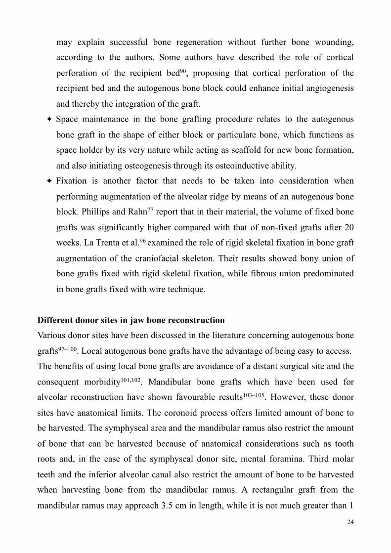
Different donor sites in jaw bone reconstructionVarious donor sites have been discussed in the literature concerning autogenous bone
grafts97–100. Local autogenous bone grafts have the advantage of being easy to access.
The benefits of using local bone grafts are avoidance of a distant surgical site and the
consequent morbidity101,102. Mandibular bone grafts which have been used for
alveolar reconstruction have shown favourable results103–105. However, these donor
sites have anatomical limits. The coronoid process offers limited amount of bone to
be harvested. The symphyseal area and the mandibular ramus also restrict the amount
of bone that can be harvested because of anatomical considerations such as tooth
roots and, in the case of the symphyseal donor site, mental foramina. Third molar
teeth and the inferior alveolar canal also restrict the amount of bone to be harvested
when harvesting bone from the mandibular ramus. A rectangular graft from the
mandibular ramus may approach 3.5 cm in length, while it is not much greater than 1
24

cm in height. These dimensions apply to a span of three to four tooth sites106.
Therefore, in cases with total tooth loss in the maxilla and severe resorption of the
alveolar ridge, using autogenous bone grafts from the iliac crest is usually necessary.
In some patients with severe maxillary atrophy (class V and VI),107 a reversed inter-
maxillary relation or increased vertical distance between the jaws may result108. The
indication for harvesting autogenous bone block from the ilium becomes more
evident in these cases, not only for the purpose of optimal implant positioning but
also for restoring the correct facial height and morphology.
Another aspect regarding the choice of donor site relates to its origin, namely,
whether the harvested bone has an EC or IM origin. Clinical studies have shown that
IM onlay bone grafts tend to resorb less compared with EC bone grafts in the
craniofacial skeleton109,110. Experimental studies likewise have shown more
favourable results, in terms of volume maintenance, for IM bone grafts111–113. Ozaki
and Buchman114 point out that in some previous studies115,116, IM bone, owing to its
ability to maintain volume, has been reported to have inherent embryogenic
advantage over EC bone. However, the authors then challenge this idea by suggesting
that the micro-architecture of the IM bone graft has more cortical bone compared
with EC grafts, and hence that IM bone is less prone to resorption. In that study114,
cortical bone grafts of membranous origin and cortical and cancellous bone grafts of
EC origin were compared by placing them onto cranium of rabbits. Volume analysis
showed a statistically greater resorption rate in the cancellous EC bone graft than in
either the EC or the membraneous cortical bone grafts. Furthermore, no statistical
difference was observed in the resorption rates between the two cortical onlay bone
grafts of different embryonic origin.
In an experimental study, Kusiak and co-workers117 relate the ability of greater
volume maintenance of IM bone grafts to more rapid vascularization compared with
EC bone.
25

SmokingCigarette smoking may have a negative influence on wound healing118. Bain and
Moy119 ascribe the negative effects of cigarette smoking on wound healing to the
direct cutaneous vasoconstrictive action of nicotine, increased platelet aggregation
and compromised polymorphonuclear (PMN) leucocyte function, to name a few
causes. Several studies report a correlation between smoking and higher risk of
implant failure120-124. Based on these findings, smoking could be regarded as a
contraindication also for bone augmentation. A systemic review of the orthopaedic
literature regarding the impact of smoking on bone healing has revealed that smoking
has a negative effect on bone healing in terms of delayed union and non-union125.
Nicotine decreases blood flow to the extremities owing to the increased peripheral
vasoconstriction, especially relating to digital and forearm haemodynamics126.
Furthermore, carbon monoxide has a high affinity for haemoglobin, reducing the
amount of oxygen carried by this molecule127. Smoking has been reported to be one
of the predictors of implant failure after maxillary sinus floor augmentation and
reconstruction128. It has also been reported in the literature that post-operative healing
complications occur significantly more often in smokers compared with non-
smokers129.
Surgical techniquesSurgical procedure of the reconstruction of the atrophic maxilla can be divided into:
inlay, onlay, and interpositional bone grafting. In a systemic review article Del
Fabbro et al.130 report the survival rates of implants in the grafted maxillary sinus as
follows: the overall implant survival rate in 39 studies was 91.49%. The loaded
follow-up time ranged from 12 to 75 months. Simultaneous vs. delayed procedure
displayed almost similar survival rates, of 92.17% vs. 92.93%. Furthermore, when
implants were installed in grafted maxillary sinus, the performance of rough implants
was shown to be superior to that of smooth surface implants. Bone substitute material
proved to be as successful as autogenous bone grafts. In another study131 the use of
cancellous block allografts for sinus floor augmentation with simultaneous implant
placement was evaluated, with a mean follow-up of 27 months. The inclusion
26

criterion was a residual alveolar ridge height of ≤4 mm. The success rate was
reported to be 94.4%. Olson and colleagues132 report a long-term assessment of
endosseous dental implants in the augmented maxillary sinus. The follow-up began at
stage two (abutment connection) and ranged from 5 to 71 months. Although the
amount of residual bone was not measured and recorded, the results showed high
implant survival rates in grafted sinuses (97.5%). Out of 120 implants placed in 45
grafted sinuses, 88 implants were placed simultaneously and 32 were placed 3–12
months after sinus augmentation. The sinus augmentation material did not appear to
affect the long-term success, from implant placement to loading, or function as
described by the authors. However, when comparing a one-stage surgery protocol
with a delayed placement of implants, it appeared that all the failed implants occurred
when using a one-stage surgery protocol. Based on these studies, it can be concluded
that when placing an endosseous implant into grafted bone, primary implant stability
is of utmost importance for osseointegration to take place. Consequently, when
primary implant stability cannot be achieved, a staged surgery protocol is
recommended. This approach is also valid when augmenting a resorbed maxillary
ridge with onlay buccal or vertical bone grafts. However, since this type of bone
augmentation is more susceptible to lateral and occlusal forces, using a one-stage
surgery protocol is not as straightforward and conclusive as is a maxillary sinus
augmentation procedure.
The onlay group can be divided into horizontal (buccal veneer) grafting and vertical
grafting. While buccal onlay grafting has been used to augment the width of a
resorbed maxilla, some clinicians have reported satisfying results also when
augmenting the height. Nyström et al.133 conducted a study to post-operatively
evaluate combined use of bone grafts and implants, using computed tomography
(CT). The harvested bone was from both the lateral and the medial aspect of the
ilium, forming a horseshoe shape. According to the authors, the graft was then
modelled to fit the residual maxillary alveolar crest. Using a one-stage surgery
protocol, six self-tapping fixtures were inserted, penetrating the bone graft and the
residual bone. Rigid fixation was established. Out of 120 fixtures inserted, 14 were
27

reported lost during the observation period of ≥24 months. Prior to this study, a 2-
year longitudinal study was initiated by Nyström and colleagues134 using the same
surgical procedure in 30 patients. The first ten patients were classified as the
development group and the remaining 20 patients as the routine group. The implant
survival rate after 2 years was reported to be 54.4% in the development group and
88.3% in the routine group. In a 10-year follow-up study of the same patient group,
the implant success rate was reported to be 83.1% in the routine group135. Van
Steenberghe et al.136 report a cumulative success rate of 85% after a 10-year follow-
up when placing implants simultaneously with autogenous onlay bone grafts. The
bone graft harvested was also in the shape of a horseshoe and was stabilized with
four to seven self-tapping, machined surface implants (using the Brånemark system).
It was concluded that the self-tapping, screw-shaped implants lead to an excellent
adaptation of the graft and even compression of the graft towards the residual bone.
In some circumstances, using residual bone is not suitable for one-stage surgery, for
instance when the residual alveolar ridge is too small for the fixture to be penetrated
in both the residual and the grafted bone, allowing compression of the bone graft, or
when bone grafts are used solely as lateral onlay. Sjöström and colleagues22 report a
90% survival rate in a total of 192 implants after a 3-year follow-up. Using a delayed
placement of titanium implants with a turned surface, 29 patients were reconstructed
with free iliac crest grafts using onlay/inlay or interpositional bone grafts; 25 patients
remained for the follow-up period. In the same study, a literature survey was also
conducted, indicating that while the one-stage technique is the most commonly used
procedure, delayed placement of implants results in a higher survival rate. Triplett
and Schow30 have shown that the success rate of implants placed in grafted areas 6–9
months after bone augmentation is higher than when implants are placed
simultaneously with the grafting procedure. The authors have suggested four
important and valid factors:
28
1. Rigid fixation and a tension-free primary closure of the soft-tissue flap
minimizes complications that lead to failure.
2. Most of the grafting failure is due to infections or exposure of the graft to the
oral cavity because of dehiscence. Early loading of grafts with a transitional
prosthesis is another potential cause of graft failure.
3. Success of the placement of endosseous implants in the grafted area is more
predictable using a delayed surgical procedure.
4. Failure of individual implants in the grafted bone does not imply failure of the
bone graft. In most cases there will be enough bone volume after 6–8 months for
successful implant placement.
Becktor and co-workers137 have indicated that the trauma caused by a provisional
maxillary denture opposed by a mandibular dentition, creating force concentration
rather than force distribution, could induce further trauma to the maxilla.
Furthermore, the authors have implied that there is an association between unilateral
mandibular dentition and an increase in implant failure in the maxilla.
Another method for reconstruction of cranio-maxillofacial defects is through tissue
engineering. A primary source of mesenchymal stem cells (MSCs) for bone
regeneration is from adipose tissue to provide adipose-derived stem cells (ASCs)138.
Sándor et al.139 used autogenous fat from the anterior abdominal wall of a patient
who had undergone resection of a 10 cm anterior mandibular ameloblastoma.
Adipose-derived stem cells were isolated and expanded ex vivo. The expanded cells
were seeded onto a mixture of β-tricalcium phosphate (β-TCP) granules and
recombinant human BMP-2 as a scaffold. Ten months after reconstruction, dental
implants were inserted into the grafted site. It was concluded that ASCs in
combination with β-TCP and BMP-2 offers a promising construct for the treatment of
large mandibular defects without the need for ectopic bone formation and allowing
rehabilitation with dental implants139.
29
Osseointegration of titanium implantsOsseointegration was defined by Brånemark5 as a direct structural and functional
connection between ordered, living bone and the surface of a load-carrying implant.
According to Albrektsson140, establishment of osseointegration is dependent on the
implant material, implant design, implant surface, status of the bone, surgical
technique, and implant-loading condition.
According to Rasmusson69, the most common methods of analysis of the interactions
between bone tissue and titanium are: 1. descriptive histology using light and/or
electron microscopy (scanning and transmission electron microscopy (SEM and
TEM, respectively); 2. quantitative histology using morphometry of ground sections
for light microscopic analysis; and 3. biomechanical tests such as the push/pull tests
or removal torque tests, as well as resonance frequency analysis (RFA).
Nygren et al.141 describe the effect of the titanium surface on different biological
components that come in contact with the surface as soon as the implant is placed
into the surgical prepared site, as a crucial factor in the healing process. The authors
describe that the surface influences protein adsorption, platelet adhesion and
haemostasis, inflammation and osteogenic cell response. Bone regeneration around
oral titanium implants resembles the healing phases of bone injury or fracture, i.e.
inflammation, regeneration, and remodelling142. In 1991, Sennerby and co-workers143
examined the bone-titanium interface in retrieved clinical oral implants. Using light
microscopy and TEM, the authors observed that the threads of the implant were filled
79–95% with dense lamellar bone, and that a large fraction of the implant surface,
56–85%, appeared to be in direct contact with the mineralized bone. In areas of direct
mineralized bone-titanium contact at the ultrastructural level, mineralized bone
reached close to the implant surface but was separated by an amorphous layer 100–
400 nm tick. Furthermore, Sennerby et al.144 have shown early bone tissue response
to titanium implants. Placing titanium implants in rabbit tibia, they observed a
cellular response after 3 days. Osteoblast-producing osteoids were observed at the
endosteal surface and elongated mesenchymal cells were present at the site of injury.
30

Some macrophages, but rather few other inflammatory cells, were identified. From
day 7, multi-nuclear giant cells were observed in direct contact with the implant and
forming a continuous layer along the surface. Bone formation was first identified at
day 7 as woven trabecular bone formed from the endosteal surface and extended
towards the implant surface as a solitary formation. This solitary bone matrix was
described as a base for surface osteoblasts which produced osteoid in a lamellar
arrangement. With time the two types of newly formed bone fused and more bone
filled the threads and became remodelled by bone remodelling units. The authors also
observed that bone-titanium contact and the bone area in the threads increased with
time until 6 months after implant placement.
Osseointegration of titanium implants in autogenous bone grafts
The healing of turned-surface titanium implants into grafted bone has been
previously studied. Nyström et al.145 performed a histological examination on one of
a series of patients who had undergone treatment with bone grafts from the iliac crest
in combination with self-tapping fixtures. The patient had died in an accident 4
months after the operation. Autopsy specimens from the patient were used to analyse
the amount of osseointegration after 4 months of healing. The graft from the maxilla,
including all six implants, was retrieved. A specimen from the donor site was also
removed post-mortem and prepared for histological examination. The results showed
no clear distinction between the grafted and the residual bone. Marginal aspects of
the implant showed signs of resorption while the apical portion of the implants
seemed to be imbedded in the original maxillary bone. The interface between bone
and implant was to some extent soft tissue, which reflected a delayed remodelling
process. In only a small section of the implant circumference was a direct BIC
observed. At the donor site, there was evidence of new bone formation but the gap
was not bridged. There was no inflammatory reaction in the soft tissue.
Lundgren and colleagues29 analysed the bone graft-titanium implant interface of
titanium micro-implants placed simultaneously or after primary healing of the grafts.
Histological analysis of micro-implants representing healing periods of 0–6 months,
31

0–12 months (simultaneous placement) and 6–12 months (delayed placement)
revealed that the delayed micro-implants had more bone within the threads and more
bone in direct contact with the implant surface compared with simultaneous micro-
implant specimens. Furthermore, histological findings of biopsies without micro-
implants at day 0, and 6 and 12 months post-grafting showed signs of ongoing
resorption, bone formation and remodelling at 6 and 12 months. Morphological
measurements of bone areas from these biopsies showed more areas of new bone
after 12 months of healing compared with 6 months post-healing. The authors could
show that although titanium micro-implants integrate in free autogenous iliac crest
bone grafts, when used as either onlays or as interpositional bone grafts, the micro-
implants placed in a delayed procedure showed more bone in the implant interface
compared with the simultaneous procedure.
Titanium implant surface topography
According to Albrektsson and Wennerberg146, surface quality of an implant can be
looked at in terms of mechanical, topographic and physiochemical properties.
Frandsen and colleagues147 found that the holding power of different screws in the
cancellous bone of femoral head increases with the length and the diameter of the
thread. According to Albrektsson148, look alike implants do not necessarily show
similar long-term clinical results. Moreover while, threaded implants have shown to
become osseointegrated, non-threaded implants result in patches of BIC interrupted
by areas with a fibrous tissue contact149.
A long-term follow-up study150 involving standard Brånemark System fixtures
revealed implant survival rates of 89% at 5 years, 81% at 10 years and 78% at 15
years for maxillary fixtures. For fixtures placed in the mandible, the survival rates
reported were 97% at 5 years, 95% at 10 years and 86% at 15 years. The topographic
surface of standard Brånemark System fixtures has been described in the literature151
as machined by turning.
32

A systemic review152 on implant surface roughness and bone healing has revealed
enhanced BIC with increasing surface roughness. In 2000, Cooper153 described the
role of surface topography on osseointegration. The author concluded that increasing
implant surface roughness does increase the surface area of the implant and that it
would be an advantage if osseointegration consisted of a cohesive bond between the
implant and the bone. The author also reported that the implant surface topography
affected the amount of bone formed at the implant-bone interface. The mechanism of
endosseous integration has been termed “contact osteogenesis” (bone growth on the
implant surface) by Davies154, a mechanism that can be divided into three phases: 1.
osteoconduction, where a migration of differentiating osteogenic cells takes place to
the implant surface through a connective tissue scaffold; 2. new bone formation,
which results in a mineralized matrix being laid down on the implant surface; and 3.
bone remodelling. Albrektsson & Wennerberg146 have defined different surface
roughnesses as follows:
✦ Smooth surfaces have an Sa value of <0.5 µm.✦ Minimally rough surfaces have an Sa value of 0.5–1 µm.
✦ Moderately rough surfaces have an Sa value of 1–2 µm.✦ Rough surfaces have an Sa value of >2 µm.
They concluded that moderately rough implant surfaces have some clinical
advantages over smoother or rougher surfaces by showing stronger bone responses.
While surface topography can be changed by either subtractive or additive processes,
as described by Albrektsson & Wennerberg146, surface treatment with fluoride has
been shown to enhance the retention of titanium implants fourfold compared with
implants with machined surfaces in rabbit ulna155. According to Ellingsen156, fluor
ions have documented activity in bone. This element is known to form fluoridated
HA or fluorapatite, the latter with improved crystallinity and better resistance to
dissolution compared with HA. In a study conducted by Ellingsen and co-workers157,
the fluoride modification of the titanium surface and its effect on bone response was
investigated by comparing titanium oxide (TiO2)-blasted titanium implants with and
33

without fluoride-modified surfaces. The results showed a significantly higher
removal torque value for the fluoride-modified implants after 3 months.
Histomorphometric analysis showed higher BIC for the fluoridated test implants.
Further discussion about different types of implant surface modification is beyond the
scope of the present thesis.
34

AIMSThe aims of the present thesis were:
To compare the bone tissue response for machined and fluoridated implants with
increased surface roughness installed in onlay bone grafts in one-stage surgery, using
histomorphometry and RFA.
To determine if there are any differences in stability and osseointegration of implants
with bioactive surface installed in autogenous bone grafts using a simultaneous and a
delayed approach in test and control groups, respectively.
To evaluate marginal bone-level alterations around moderately rough implants
installed in block vs. particulate autogenous bone grafts.
To evaluate and compare the extent of resorption of autogenous bone grafts between
block and particulate bone by three-dimensional (3D) radiographic examination after
2 years.
35

MATERIALS AND METHODSAnimal studies I and IIAnimals and anaesthesiaFor the experimental studies I and II, female New Zealand White rabbits were
chosen. The animals were kept in specially designed rooms and fed with a standard
diet and water ad libitum.
Surgery was initiated by sedation with an intraperitoneal (i.p.) injection of Stesolid® (Dumex, Copenhagen, Denmark) 1.5 mg/kg. General anaesthesia was administered
by intramuscular (i.m.) injection of Hypnorm® (Janssen Pharmaceutica, Brussels,
Belgium) 0.2 ml/kg. In addition, 0.8 ml of local anaesthesia (2% lidocain/epinephrine
1:80 000; Astra AB, Södertälje, Sweden) was given. Post-operatively, single i.m.
injections of antibiotics (Intencillin® 2 250 000 IE/5 ml, 0.1 ml/kg body weight;
LEO, Helsingborg, Sweden) and analgesic (Temgesic® 0.05 mg/kg body weight;
Reckitt & Coleman, Hull, UK) were given. The healing times for both studies I and II
were 8 weeks. After completion of healing, the animals were sacrificed by a mixture
overdose of Rompun® Vet. (Bayer A/S, Animal Health Division, Copenhagen,
Denmark) 20 mg/ml and Ketalar® (Pfizer AB, Sollentuna, Sweden) 50 mg/ml.
Implants
Screw-shaped titanium implants from commercially pure titanium 9 mm long and 3.5
mm in diameter were used in Study I. Control implants had a machined surface while
test implants had a fluoridated surface (OsseoSpeedTM; Astra Tech AB, Mölndal,
Sweden).
The same screw-shaped titanium implants with fluoridated surface (OsseoSpeedTM;
Astra Tech AB, Mölndal, Sweden) and similar dimensions were used in one-stage
and two-stage surgery protocols in Study II. Fixation of the bone graft in the two-
36

stage surgery protocol was achieved by using a titanium mini-screw 5 mm long and 2
mm in diameter.
Surgery protocols
Study I
Eight adult (8–9-month-old) female New Zealand White rabbits were used in this
study. The proximal tibial metaphysis on both sides was used as experimental sites.
The autogenous bone grafts were harvested from the calvarial bone. Surgery was
performed under aseptic conditions and was initiated by making an incision and
raising a sub-periosteal, full-thickness flap. A trephine bur was used to harvest a disc-
shaped bone graft 8 mm in diameter and 2 mm thick from the lateral aspect of the
sagittal suture of the calvarium, with care taken not to penetrate the dura. After bone
harvesting, the donor site was sutured in layers using Vicryl 5.0 (Ethicon®; Johnson
& Johnson, Livingston, Scotland) and the skin using Monocryl 3.0 (Ethicon®;
Johnson & Johnson, Livingston, Scotland). The proximal tibial methaphyses were
exposed bilaterally through a skin and fascia-periosteal flap. A 3.2 mm hole was
drilled in the centre of each bone graft, using a 3.2 mm twist drill. The hole and the
proximal tibial metaphysis were then prepared for a dental implant (Astra Tech AB,
Mölndal, Sweden) 9 mm long and 3.5 mm in diameter with a machined surface as
control and a fluoridated surface (OsseoSpeedTM; Astra Tech AB, Mölndal, Sweden)
as test. Test and control sides were randomized. All implants were installed with good
primary stability. Cover screws were placed and fascia-periosteal flaps were sutured
in the same way as the donor site. After 8 weeks of healing, the animals were
sacrificed by a mixture overdose of Rompun® Vet. (Bayer A/S, Animal Health
Division, Copenhagen, Denmark) 20 mg/ml and Ketalar® (Pfizer AB, Sollentuna,
Sweden) 50 mg/ml. The right and left tibia and excess tissue were removed.
37

Figure 3. An implant placed in a one-stage
surgery protocol
Study II
Six adult female New Zealand White
rabbits were used in this study. The
autogenous bone graft was harvested
from the calvarium and placed on the
tibial metaphysis as the recipient site, as
described in Study I. However, the bone
graft surgery was conducted on two
occasions to acquire specimens with
different healing times. During the initial bone harvesting procedure, the bone graft
was fixed on the tibial bone by means of a mini-screw 5 mm long and 2 mm in
diameter. The fascia-periosteal flaps were then sutured in layers using Vicryl 5.0
(Ethicon®; Johnson & Johnson, Livingston, Scotland) and the skin using Monocryl
3.0 (Ethicon®; Johnson & Johnson, Livingston, Scotland) representing the control
site.
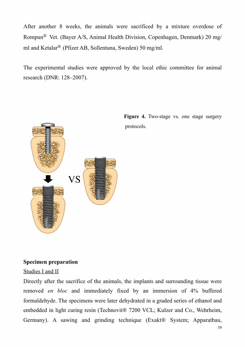
After 8 weeks, a second bone graft from the other side of the calvarium was
harvested and directly placed on and fixed to the tibial bone by a fluoridated implant
3.5 mm x 9 mm (Astra Tech AB, Mölndal, Sweden) on the other tibial metaphysis,
representing the test site.
During the same surgery, the fixation screw on the control side, which by this time
had undergone 8 weeks of healing, was removed. After preparation using the same
specifications as for the test site, a 3.5 mm x 9 mm OsseoSpeed® implant (Astra Tech
AB, Mölndal, Sweden) was installed (control site). Therefore, the implants placed in
healed bone grafts represented control sites and two-stage surgery, while the test sites
were operated using a one-stage surgery protocol. 38
After another 8 weeks, the animals were sacrificed by a mixture overdose of
Rompun® Vet. (Bayer A/S, Animal Health Division, Copenhagen, Denmark) 20 mg/
ml and Ketalar® (Pfizer AB, Sollentuna, Sweden) 50 mg/ml.
The experimental studies were approved by the local ethic committee for animal
research (DNR: 128–2007).
Figure 4. Two-stage vs. one stage surgery
protocols.
Specimen preparationStudies I and II
Directly after the sacrifice of the animals, the implants and surrounding tissue were
removed en bloc and immediately fixed by an immersion of 4% buffered
formaldehyde. The specimens were later dehydrated in a graded series of ethanol and
embedded in light curing resin (Technovit® 7200 VCL; Kulzer and Co., Wehrheim,
Germany). A sawing and grinding technique (Exakt® System; Apparatbau, 39

Norderstedt, Germany) was used to make approximately 10 µm thick ground sections
of each specimen. The sections were then stained with 1% toluidine blue and
pyronin G.
Analysis and calculationsStudies I and II
The sections were viewed and analysed in a light microscope (Nikon Eclipse 80i;
Tekno Optik AB, Göteborg, Sweden) using 1.8–100x magnification, connected to a
personal computer with software for morphometry (Easy Image Measurements 2000;
Tekno Optik AB, Göteborg, Sweden). Morphometrical measurements of the
following dimensions were made:
1. Bone-to-implant contact in the grafted bone.
2. Bone-to-implant contact in the total amount of bone (bone graft and residual
bone).
3. Total bone structure within a region of interest (ROI).
The above calculations were done by measuring the following parameters:
1. Implant length in the grafted bone.
2. Bone-to-implant contact of the grafted bone.
3. Implant length in the residual bone.
4. Bone-to-implant contact of the residual bone.
To calculate the total BIC, parameters (2) and (4) were added together. These
measurements were performed at both sides of the longitudinal axis of each implant
and a mean value was calculated for each implant. Bone area within an ROI was
measured by drawing a vertical line about 1.0 mm from the outer border of each
implant surface, parallel to the longitudinal axis of each implant including seven
implant threads within the grafted area.
40

Resonance frequency analysis: implant stability measurementsFor performing a non-invasive, in vivo assessment of implant stability, a resonance
frequency measurement method has been proposed by Meredith and co-
workers158,159. The principle of this method is to attach a transducer to an implant
fixture either directly or through a transmucosal abutment. Via a sinusoidal signal, the
transducer is vibrated and implant stability is assessed by measuring the resonance
frequency of the transducer. There is a clear correlation between resonance frequency
and the exposed height of the fixture. The transducer is also sensitive to the stiffness
of the surrounding tissue. According to Rasmusson69, the technique measures the first
bending resonance frequency of a small transducer that is attached to a fixture or
abutment. Rasmusson reports that the resonance frequency is dependent on two
factors: the stiffness of the implant-bone system, and the height of the transducer
above the marginal bone level.
Studies I and II
As a non-invasive implant stability measurement, RFA was used according to the
method described by Meredith et al.159. Resonance frequency analysis was performed
in all animals at the time of surgery and at the end of the experiment using OsstellTM
(Integration Diagnostics AB, Göteborg, Sweden). A transducer was attached and
registrations were made perpendicular and longitudinal to the long axis of each
implant. Mean values for the test and control implants were calculated.
41

Clinical and radiographic studies III and IVPatientsStudy III
Fifteen patients (two men and 13 women, mean age 58 years, range 35–75 years)
with severe resorption of the maxilla were reconstructed with autogenous bone grafts
from the anterior iliac crest and rehabilitated with oral implants with full fixed
bridges.
Study IV
Using the patient group from Study III, eleven edentulous patients (all female) with
severe resorption of the maxilla were followed up with CT scans taken pre-
operatively, directly after grafting surgery, and 6 and 24 months post-grafting surgery.
The clinical studies were approved by the local ethics committee for human trials
(DNR: 19199-11-01).
Pre-surgical examination, inclusion and exclusion criteriaAfter clinical examinations, the extent of horizontal and vertical bone deficiencies
was assessed by CT scans. Inclusion criteria were severely resorbed maxilla with an
anterior crestal width of <3 mm and/or height of <7 mm. Inclusion criteria for
vertical bone height at the posterior aspect of the maxilla were set at <5 mm. Patients
smoking fewer than ten cigarettes per day, with no alcohol abuse, and between 20 and
75 years old were selected.
Twelve out of 15 patients in Study III were smokers (<10 cigarettes/day) prior to
treatment. One patient smoked during the period from bone grafting to the abutment
connection. In Study IV, all eleven patients who were followed up were among those
who were smokers before the bone grafting procedure. Exclusion criteria were acute
illness, ongoing chemotherapy, ongoing or recent (within 3 years) radiotherapy and
i.v. bisphosphonate treatment. 42

Pre- and post-surgical careAt the start of the bone grafting surgery, benzylpenicillin (3 g x 3) or clindamycin
(600 mg x 3) was given peri-operatively and for the following 24 hours. Patients
received a prophylactic antibiotic regimen for 10 days after the grafting procedure
with either phenoxymethylpenicillin (1 g x 3) and metronidazole (400 mg x 3) or
clindamycin (300 mg x 3).
At the time of implant installation, patients received a single dose of antibiotic, either
phenoxymethylpenicillin (2 g) or clindamycin (300 mg). The antibiotic cure
continued for 5 days after implant surgery, using either phenoxymethylpenicillin (1 g
x 3) or clindamycin (300 mg x 3). As analgesic, acetaminophen with codeine or non-
steroidal anti-inflammatory drug was prescribed for a period of 1–2 weeks after
surgical procedures. Dentures were not used during the first month following the
grafting procedure and for 10 days after the implant placement.
Bone harvesting and preparation
Under general anaesthesia, an incision was made about 2 cm posterior of the anterior
superior iliac spine on either the right or the left side. A cortico-cancellous block of
bone from the medial aspect of the iliac crest was harvested by means of a reciprocal
saw, leaving the lateral part of the iliac crest intact. The incision was sutured in
layers. To achieve particulate bone (test side), a bone mill (Tessier Osseous
Microtome (TOM®); Stryker Leibinger GmbH, Freiburg, Germany) was used.
Harvested cortico-cancellous bone graft was milled down to pieces and mixed with
platelet-rich plasma (PRP) prepared from withdrawal of whole blood from a
peripheral vein of the patient.
43

Figure 5. A cortico-cancellous bone being
harvested from the medial aspect of the iliac
crest
Bone augmentation of the anterior maxilla
Bone blocks as onlay graft (control)Once a mucoperiosteal flap was raised on
the maxilla, the recipient site was
prepared using a small round bur until
small spots of bleeding were observed. A
cortico-cancellous bone block harvested
from the anterior iliac crest was placed on the right side (frontal sub-nasal area) of the
maxilla. The bone block was fixed to the residual bone by a minimum of two
titanium screws (6–13 mm in length) to achieve a rigid fixation of the bone graft.
Particulate bone as onlay graft (test)On the left side, the particulate bone mixed with PRP was placed onto the recipient
site.
No randomization was made for the test and control sides.
To gain full, tension-free soft tissue coverage of the grafted area, an incision was
made through the periosteum and the flap was elevated in order to cover the
augmented area. The incision was then sutured using resorbable sutures (Vicryl®;
Johnson & Johnson AB, Sollentuna, Sweden).
44
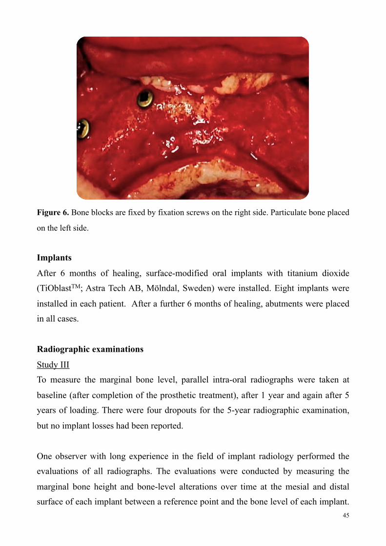
Figure 6. Bone blocks are fixed by fixation screws on the right side. Particulate bone placed
on the left side.
Implants
After 6 months of healing, surface-modified oral implants with titanium dioxide
(TiOblastTM; Astra Tech AB, Mölndal, Sweden) were installed. Eight implants were
installed in each patient. After a further 6 months of healing, abutments were placed
in all cases.
Radiographic examinations
Study III
To measure the marginal bone level, parallel intra-oral radiographs were taken at
baseline (after completion of the prosthetic treatment), after 1 year and again after 5
years of loading. There were four dropouts for the 5-year radiographic examination,
but no implant losses had been reported.
One observer with long experience in the field of implant radiology performed the
evaluations of all radiographs. The evaluations were conducted by measuring the
marginal bone height and bone-level alterations over time at the mesial and distal
surface of each implant between a reference point and the bone level of each implant. 45

The highest point of the vertical section of each implant at the mesial and distal
surfaces was chosen as reference point. Signs of radiographic changes at the BIC
zones, indicating loss of osseointegration, and signs of bone loss related to
mechanical components of the implants were registered. Bone-level measurements
were conducted to the nearest 0.1 mm by means of a magnifying lens x7. A mean
value of the mesial and distal measurements was calculated per implant. To determine
the error of radiographic measurements, double recordings of one randomly selected
implant per patient from the 5-year follow-up examination were performed and the
mean difference between the two readings was set as the degree of marginal bone
resorption.
Study IV
To establish 3D volumetric changes of the bone grafts, we performed CT
examinations using a CT Pace Plus (General Electric, Milwaukee, MI, USA) for 2
mm axial scans. Scans were taken within 1 month of the grafting procedure (the first
post-operative CT examination) and after 6 and 24 months. All CT scans were taken
at one hospital, following the same procedure and analysed by one person. The total
volume of the grafted areas was measured separately at the test and control sides
using axial and sagittal images. The two types of bone grafts were identified on the
lateral aspect of the anterior maxilla. Next, the volume of each bone graft was
calculated by measuring the following dimensions: thickness of the bone graft in a
bucco-palatal aspect, and vertical height and horizontal length in an anterior-posterior
direction of the grafted areas. To reduce the error in the radiographic measurements,
three different CT sections at each dimension were selected. In order to be as
consistent as possible, we used sections showing the largest amount of bone in all
dimensions. Furthermore, we used a drawing function for measurement of distance,
to manually plot the ranges in all three sections of each dimension. In this fashion,
the mean value for each dimension was calculated, and multiplied to obtain a value in
cubic mm.
46

Based on clinical experience, we suspected that the width of the bone grafts was
more prone to resorption, therefore the results of changes in the bucco-palatinal
aspect were presented separately.
StatisticsStudies I–IV
The Wilcoxon signed-rank test was used for statistical analysis and a difference
between the two groups was considered significant if p<0.05.
47

RESULTS
Integration of moderately rough fluoridated implants in
autogenous bone grafts Studies I and II
The aim of the first animal study (Study I) was to establish whether there would be
any differences in bone response between machined, and moderately rough and
fluoridated implants with increased surface roughness when placed in autogenous
bone blocks using a one-stage surgery protocol.
Study II aimed to answer if there are any differences in bone tissue response to
implants with increased surface roughness when simultaneously placing them with
block bone grafts or when using a delayed surgery protocol after 8 weeks of graft
healing.
Clinical observations
Because of unexpected death of one animal in the first study, the total number of
animals was reduced to seven. In Study II, all six animals could be analysed through
the planned time protocol.
At the time of sacrifice and specimen preparation, all bone grafts had healed and
integrated into the residual bone with smooth and mature texture. In Study I, the RFA
showed statistically higher stability for the implants with moderately rough
fluoridated surface (test surface), both at the time of implant placement and after 8
weeks of healing. In Study II, RFA results were higher for implants when using a
delayed approach at the time of implant installation. However, the difference between
control and test sides had levelled out by the time of sacrifice.
48

Histological observationCross-sections of experimental sites in each specimen showed implants well
integrated into both residual and grafted bone. Although grafted bone tissues were
well integrated into the residual bone, a demarcation could be observed between the
two. This demarcation was more distinct in Study II when placing implants at the
time of bone augmentation, hence with shorter healing time for the bone grafts prior
to specimen preparation. With higher magnification, the grafted area appeared to be
more mature bone when using a delayed surgical protocol (control).
Histomorphometry
In Study I, histomorphometry revealed that the BIC within the grafted area as well as
total bone (grafted bone + residual bone) was higher for the fluoridated, rough (test)
compared with machined (control) implants. The difference was statistically
significant, with p<0.05.
In Study II, results comparing the same type of moderately rough implants installed
in autogenous bone blocks in a simultaneous procedure (test implants) with same
type of implants installed in healed bone grafts (control implants) showed a tendency
for a higher BIC for the control side (n.s.). Results for total BIC were almost identical
for test and control sides.
Marginal bone-level alterations and three-dimensional
analysis of volumetric change in autogenous bone grafts Studies III and IV
The purpose of the third study in this thesis was to: 1. assess the marginal bone-level
alterations around moderately rough oral implants (TiOblastTM; Astra Tech AB,
Mölndal, Sweden); 2. evaluate whether there is any difference in marginal bone-level
alterations between implants placed in autogenous block (control implants) or
particulate bone (test implants); and 3. whether there is any difference in stability
between test and control sites as measured by RFA. 49

Study IV aimed to investigate volumetric changes of autogenous bone grafts placed
as lateral onlay in the anterior maxilla. The extent of resorption of the grafted bone
after 2 years was investigated. Furthermore, we wanted to establish whether there is
any difference in the amount of resorption between autogenous block and particulate
bone.
In Study III, radiographic findings revealed a tendency towards more marginal bone
resorption from the reference point at sites augmented with autogenous block bone
(control) at baseline, after 1 year and again after 5 years. However, the difference
between test and control sides was not statistically significant. Resonance frequency
analysis showed almost similar results at implant installation between test and control
sides and a tendency for higher values for the implants placed in particulate bone
(test) at the time of abutment surgery and after 1 year in function. After 3 years, the
difference in RFA value had levelled out between the test and control sides. No
statistically significant difference was observed at any time during the follow-up
period.
In Study IV, the volumetric change, measured by CT, showed extensive resorption of
both particulate (test) and block (control) bone grafts after 24 months. The amount of
volume reduction appeared to be higher on the test side (81.1±8.3%) augmented with
particulate bone, compared with the control side (77.8±5.2%) augmented with block
bone, but the difference was not statistically significant.
50

Table 1. This table shows the volume reduction of the grafted bone
0
1000
2000
3000
4000
5000
First post grafting 6 months 24 months
Kub
ic m
m
Time period
Test (Particulate bone) Control (Block bone)
51

DISCUSSIONAnimal studiesThe results showed significantly higher BIC and implant stability when using
fluoridated surface-modified implants installed in autogenous bone grafts compared
with implants with machined surface. Although factors such as stability of the grafted
bone, healing time, primary stability of the implant and surface dimensions of the
implant are crucial factors for successful integration and stability of both the grafted
bone and the implant, surface configuration and properties of the implant seem to
play a crucial role in osseointegration.
According to Sennerby and Meredith160, the main determinants of implant stability
are: 1. The mechanical properties of the bone tissue at the implant site; and 2. how
well the implant is engaged with that bone tissue. Furthermore, the authors state that
the mechanical properties of bone are determined by the composition of the bone at
the implant site and may increase during healing because soft trabecular bone tends
to undergo a transformation to dense cortical bone in the vicinity of the implant
surface. Therefore, stability of the grafted bone can be related to the mechanical
properties of the bone tissue at the implant site. In addition, it has been shown by
Philips and Rahn161 that bone grafts need to be stabilized for predictable graft
survival and that rigid fixation facilitates the preservation of the bone graft volume,
probably because this creates an appositional phase earlier and, consequently,
improved osteoconduction161.
In studies I and II, rigid fixation of the bone graft was gained by placing the implants
bicortically through the cortical layers of the graft and the thicker outer cortex of the
tibial bone. By doing so, good primary stability of the implants was achieved in all
animals. By the time of sacrifice, the bone grafts had fully integrated with the tibial
bone with smooth surface and rounded edges, displaying ongoing remodeling.
Consequently, we conclude that autogenous bone block can be rigidly fixed by means
of an implant in one-stage surgery. However, it must be noted that in this study, since
52

the bone grafts were placed and fixed on the tibial bone, the grafts could easily be
stabilized by one implant. This procedure does not fully reflect the clinical situation
where the alveolar crest has undergone extensive resorption resulting in inadequate
alveolar width and a need for bone augmentation with lateral onlay grafts. However,
when vertical bone augmentation is needed, as described by Nyström and
colleagues133, the results of studies I and II may be applicable to understanding the
healing process.
A disadvantage of using the animal model we used in studies I and II is that other
factors such as primary wound closure, loading due to masticatory forces, the
preservation of keratinized soft tissue and other factors that are relevant within the
oral cavity milieu, cannot be taken into account when the bone grafting procedure is
performed on the tibial bone. Another aspect that should be taken into consideration
in the animal models used in studies I and II, is use of IM bone as the bone graft.
Bone grafts from different donor sites and different origins have been debated by
some authors who relate the quality of IM bone graft for better volume maintenance,
to different factors as described earlier in this thesis.
The most important limitation of the animal studies must be the low number of
animals used. Even so, we were able to show statistically significant differences
between the test and control sides in Study I. The results in Study I are clearly in
favour of fluoridated implants and seem likely to be due to the fluoridated surface
configuration of the test implants. Previous studies investigating the influence of
fluoridated implant surface in non-grafted bone show favourable results for this
surface modification. Using the same animal model, Ellingsen and colleagues157
compared TiO2-blasted titanium implants with and without fluoride modification with
respect to BIC, bone area in threads, and removal torque resistance in rabbit tibia.
The authors showed results in which the mean BIC value for all threads after 1 and 3
months of healing was significantly higher for the fluoridated compared with control
implants. The authors also showed significantly higher bone area filling the threads in
the best three consecutive threads as well as bone area in all threads after a 3-month
53

healing period. Removal torque in the same study displayed a higher mean value after
1 and 3 months, the latter being statistically significant. Other experimental studies
have presented similar results with fluoride-modified implant surfaces. Abrahamsson
and co-workers82 showed a significantly higher degree of BIC within a defect area at
fluoride-modified implants than at implants with only titanium dioxide-blasted
surfaces. Therefore, our results from Study I are in accordance with previously
conducted studies performed in non-grafted bone.
In Study II, we sought to examine whether there are any differences in
osseointegration of oral implants placed in autogenous onlay bone grafts in one-stage
surgery vs. a two-stage surgery protocol. The focus was solely on implants with
moderately rough fluoridated surface and our intention was to investigate whether
these implants would osseointegrate to the same extent when installed simultaneously
with the graft as when installed using a delayed procedure. Previous studies using
implants with turned surface have shown results in favour of a delayed procedure.
Rasmusson and co-workers162 showed a significantly higher degree of BIC in the
grafted part of the bone, with the delayed procedure. Resonance frequency analysis
results were also significantly higher for implants placed after 8 weeks of healing. It
was concluded that a delayed placement of titanium implants in autogenous onlay
bone grafts results in greater implant stability because of a greater degree of bone
formation and more bone contact with the implant. Using rabbit mandible as the
recipient site, Shirota et al.163 studied HA implants in grafted bone harvested from
rabbit ilium. The authors observed that new bone formation is delayed when implants
are placed immediately at the grafting procedure. Moreover, they report that a
delayed placement (after 90 days) of HA-coated implants resulted in a greater amount
of bone bonding to the implant surface. Using RFA, Sjöström and colleagues164
compared implants placed in grafted and non-grafted normal bone in the maxilla.
They present results in which 8% of the implants were lost when placed in the grafted
bone vs. 1% in non-grafted bone. The bone grafting procedure was either
interpositional grafting following a LeFort I osteotomy if patients had a reversed
inter-maxillary relation (five patients), or onlay bone grafting because of thin alveolar
54

crest and loss of vertical bone height (24 patients). In grafted patients, the bone grafts
were harvested from the iliac crest and the implants were placed 6 months after the
grafting procedure. Another group consisting of ten patients with edentulous but non-
grafted maxillae were included in that study. Two-stage surgery for the implant
placement was chosen and the healing time for the implants before loading was 6–8
months depending of their primary stability. The results, using RFA, showed that
when applying a delayed surgical procedure in bone grafting, implants placed in
grafted bone after a minimum primary healing period of 6 months can be as stable as
implants placed in non-grafted maxillary bone164.
Our results from Animal study II showed a tendency of greater amount of BIC in the
grafted bone area with the two-stage surgery protocol. This is in line with previously
described studies. However, the difference in BIC was not statistically significant.
Moreover, the difference between test and control implants for total BIC and for bone
area within an ROI was nearly the same. The data in the present thesis could be
interpreted as either resulting from insufficient sample size or indicating that a higher
amount of BIC can actually be achieved with implants with a rough, fluoridated
surface. If the latter is true, this would be in line with Animal study I, in which we
showed that fluoridated titanium implants achieve greater BIC and higher implant
stability in terms of RFA when placed in an autogenous bone graft.
Although RFA results at implant placement were higher for the group undergoing
two-stage surgery (controls), the values were almost identical after 8 weeks of
healing. The deviation in RFA value at implant placement could be attributed to the
non-incorporated graft and consequently lower implant stability for the group
operated with one-stage surgery (test group); however, once the grafted bone was
healed and incorporated into the residual bone, RFA results proved to be as high as
for the control group.
The exact role of fluoridation cannot be explained regarding the healing process in
these animal studies; however, fluoridation of titanium implants could have an
influence on the healing process of the graft. Any superiority of fluoridated implants
55

is probably not entirely due to their topographic features but could also be related to
chemical composition. It has been documented that NaF can increase the proliferation
rate, and the alkaline phosphatase content of bone cells in vitro165. It has also been
reported in the literature that fluoride may act directly on osteoprogenitor cells and
undifferentiated osteoblasts which synthesize growth factors rather than stimulating
the proliferation of highly differentiated osteoblasts166–168. To my knowledge, there is
no evidence that there exists a true chemical bond between fluoride ions and bone
cells when using fluoridated implant surfaces. However, it may be true that
fluoridation of implants stimulates mesenchymal proliferation of stem cells and could
induce more rapid bone healing along the bone-titanium implant interface.
Clinical and radiographic studiesIn Study III, the intention was to investigate whether there are any differences in
marginal bone alterations between implant sites previously augmented with
autogenous block, and particulate bone. The radiographic examinations were
conducted at baseline (i.e. just after completion of the prosthetic supra-construction),
and after 1 and 5 years of loading. However, clinical evaluation of implant stability
by means of RFA was performed at the time of implant installation, at abutment
surgery, and after 1 year and 3 years in function. Consequently, there is no evaluation
of implant stability at 5 years that could be correlated to the radiographic findings.
This is clearly a limitation of this study. However, if we separate the results from our
radiographic findings and implant stability measurements, we can conclude that since
the difference in marginal bone alterations at the implant sites between particulate
bone (test) and block bone (control) is insignificant, no obvious guidance can be
given as to whether to use particulate or block bone as lateral onlay in cases of
severely resorbed and thin maxillae.
Furthermore, we noted in Study III that the marginal bone-level alteration for test and
control sites could not be distinguished in the period between 1 year and 5 years of
loading. Most of the marginal bone resorption took place during the first year of
56

loading and only a limited recession of the marginal bone occurred between 1 year
and 5 years in function. This is in line with the findings of Nyström et al.133 who
showed that most of the reduction in height of the grafted bone occurs between 3
months and 1 year. They found that the reduction of the bone height was insignificant
during the first 3 post-operative months, but it increased and became statistically
significant between 3 and 6 months. The reduction in height continued to be
significant during the following 6 months but levelled out and had become
insignificant by the second follow-up. Nyström et al.’s follow-up study133 lasted 24
months and was conducted using CT examinations rather than intra-oral radiographic
imaging as used in Study III in this thesis. Additionally, bone grafts were in the shape
of a horseshoe from the iliac crest and placed on the alveolar ridge as an onlay and
fixed using a self-tapping fixture. Despite the fact that our method in Study III differs
from that employed in the study by Nyström et al.133, convincing parallel conclusions
can be drawn to state that most of the reduction in graft height occurs during the first
year. A plausible cause for the marginal resorption is the remodelling of the bone
graft during the initial healing phase.
It can be argued that block bone grafts that are cortico-cancellous could be more
stable assuming that they are rigidly fixed. On the other hand, the revascularization
process takes longer compared with particulate bone. The particulate bone used in
this study was also cortico-cancellous. It can be assumed that because of its
particulate nature, the revascularization process could start earlier, hence more rapid
maturation and perhaps incorporation of the grafted bone into the residual bone could
occur. However, particulate bone grafts could be more susceptible to lateral and even
occlusal forces during masticatory function. Clinical circumstances should act as a
guide to the type of bone graft best suited for the individual case. For instance, if
there are extensive areas with ridge discontinuities and valleys on the lateral alveolar
bone, bone blocks may be difficult to fit and may not remain stable even with fixation
screws. In these cases a cortico-cancellous bone graft can be milled to pieces and
more easily be fit to the defected areas while at the same time augmenting the lateral
alveolar ridge for implant placement. However, whenever a more continuous and
57

even alveolar bone is present, a block bone graft can be chosen for lateral
augmentation.
Another issue that needs to be addressed in Study III is the survival and stability of
the implants in grafted bone. It was reported8 that only two fixtures in two patients
were found to be mobile at the abutment surgery and consequently removed.
Resonance frequency analysis revealed no significant differences between test and
control implants during the 3-year observation period. In another study, conducted by
Rasmusson and colleagues169, evaluation of TiO2-blasted implants integrated in
grafted and non-grafted maxillary bone was performed by RFA at implant placement
and abutment surgery 6 months later. The implants were placed in the following
groups: onlay block graft, onlay particulate graft, particulate sinus inlay,
interpositional block graft, and non-grafted maxilla. No significant differences were
observed between the first and second measurements; however, implants placed in
interpositional bone grafts exhibited a significantly lower RFA value compared with
other groups. The implants used in Study III were surface-modified titanium implants
with titanium dioxide (TiOblastTM; Astra Tech AB, Mölndal, Sweden). Our results are
to some extent in line with the results presented by Rasmusson et al.169. Whether or
not titanium dioxide surface modifications used in our study had any effect on the
results remains unknown because the same implant type was used on both test and
control sides. However, it should be added that the moderately rough TiOblastTM
implants used in our study have an Sa value of 1.1 µm170. Moderately rough surfaces
have previously been described by Albrektsson & Wennerberg146 to exhibit stronger
bone response. It has also been reported in the literature that TiOblastTM, like
OsseoSpeedTM, has micro-threads which have been clinically documented to maintain
bone level better compared with implants without micro-threads171. De Bruyn and
colleagues172 have reported a total survival rate of 100% during a 3-year follow-up of
immediate function of seven to nine Astra Tech TiOblastTM implants placed in 25
patients. The authors did not exclude patients with smoking habits. Based on
individual implants, a mean bone loss of 0.16 mm was reported between 3 months
and 1 year. Only 3.5% of the implants showed bone loss above 1 mm. According to
58

the report, abutments were placed in a one-stage procedure and fixtures were
functionally loaded on the day of surgery with a ten-unit provisional bridge. In
another study, by Steveling and co-workers173, the same type of TiO2-blasted
implants were used for treatment of single-tooth and partial edentulism in the
maxilla. The implants were loaded after 3 months of healing and were followed up
for 5 years. No implants were lost during the observation period. The average
marginal bone loss was reported to be 0.5±0.7 mm after 1 year, 0.6±0.7 mm after 3
years and 0.9±1.6 mm after 5 years.
It can be concluded from Study III and previously reported studies that marginal bone
loss and survival of titanium implants is dependent on factors such as implant design,
surface structure and time of loading. The type of autogenous bone graft, i.e. block or
particulate, does not seem to affect the prognosis of the implants placed within the
grafted area.
In Study IV, we studied the volumetric changes of autogenous bone grafts in the form
of lateral onlay block (control) and lateral onlay particulate bone (test) grafts over a
period of 2 years. To obtain a 3D picture of the grafted area, we used CT as the
radiographic method of choice. Computed tomography scans were taken within 1
month after the grafting procedure (the first post-grafting CT) and after 6 and 24
months. To my knowledge, there have been only a few prospective or retrospective
long-term follow-up studies evaluating the extent of volumetric changes in onlay
autogenous bone grafts.
Changes in the volume and density of calvarial split bone grafts after alveolar ridge
augmentation were studied by Smolka et al.174. In their study, 15 patients underwent
augmentation of the alveolar ridge by calvarial autogenous bone grafts. Seven
patients had mandibular reconstruction, another seven received maxillary
augmentation and one was subjected to both mandibular and maxillary
reconstruction. Post-operative CT scans were taken immediately after the grafting
procedure and before implantation after a post-operative period of 6 months in all 15
59

patients. In five patients, additional follow-up was performed after 1 year. The
authors report a mean average volume of 83.8% for all transplants (51 calvarial
bone), corresponding to a mean volume reduction of 16.2% after 6 months. In the
patient group with 1-year follow-up (26 grafts in five patients), the mean volume
reduction was 10.7% after 6 months and 19.2% after 1 year.
In Study IV, results presented by us after 6 months showed a volume reduction from
4,842.7±1,521.4 mm3 at first post-grafting CT examination to 1,645.2±565.3 mm3
after 6 months on the test side augmented with particulate bone. On the control side
augmented by block bone, the volume was reduced from 4,835.5±1,005.6 mm3 at the
first post-grafting CT examination to 2,999.3±1,452.6 mm3 after 6 months. At the 24-
month CT examination, the bone graft volume was 848.6 ± 324.3 mm3 (test side) and
1,065.6±306.4 mm3 (control side). Hence, there was a continuing reduction of the
grafted volume. However, most of the reduction took place during the first 6 months.
Our results from Study IV and the results presented by Johansson et al.27, who also
used autogenous bone from the iliac crest, show extensive volume reduction from 0
to 6 months. It has been reported in the literature that IM bone grafts retain volume
better compared with grafts with an EC origin111. In a study by Ozaki & Buchman114,
volume maintenance of onlay bone grafts in the craniofacial skeleton was evaluated.
Twenty-five New Zealand rabbits were used and three graft types were placed onto
each rabbit cranium: Cortical bone graft of membranous origin. Cortical and
cancellous bone graft of EC origin were compared. The authors showed a
significantly greater resorption rate in the cancellous EC bone graft compared with
either the cortical EC or the cortical membranous bone grafts. In addition, there was
no significant difference in resorption rates between the EC and membranous cortical
bone grafts. The authors concluded that cortical bone grafts are the superior onlay
grafting material.
The embryologic origin of the grafted bone has by some publications been pointed
out to be a factor in volume maintenance114. Authors such as Kusiak and co-
workers117 have discussed the role of the revascularization process. They have shown
60

that IM bone revascularizes more rapidly compared with EC bone. According to
Wong & Rabie175, despite the fact that EC bone taken from the iliac crest is more
cancellous than IM bone, it is conceivable that the difference in the revascularization
process is more dependent on the extracellular matrices and their content of
angiogenic mediators and other growth factors than just on the 3D osseous
architecture of the grafted bone. The authors have related the lack of volume
maintenance of EC bone grafted into the host IM bone to lack of integration and
incorporation. Furthermore, Rabie and colleagues176 have identified the cells
involved in the healing of the autogenous IM and EC bone grafts in the skull of 18
rabbits. In IM bone, they observed preosteoblasts, osteoblasts and osteocytes with no
cartilage intermediate stage. By contrast, in EC bone, they observed chondroblasts
and chondrocytes.
It is clear that when EC bone from the iliac crest is used, the volume of the grafted
bone is extensively reduced. However, to the best my knowledge, it has not been
established how long the resorption process continues. There are some studies that
have reported reduction in grafted bone height. Verhoeven and co-workers177 claim a
decrease of ca. 25% in the overall height of the graft during the first 6 months. Swart
& Allard178 report a 44% decrease of the grafted bone height after 5 years in 26
patients who received cortico-cancellous bone from the iliac crest. In spite of these
reports, few 3D volumetric measurements have been accomplished to assess volume
reduction of autogenous onlay bone grafts in long-term follow-up studies. Sbordone
et al.179 have studied volume changes of autogenous bone grafts after alveolar ridge
augmentation of atrophic maxillae and mandibles. In their study, two donor sites were
utilized for the grafting procedure: the mandibular parasymphysis and the iliac crest
area. The authors report an average resorption of 42% when the onlay was positioned
in the anterior maxilla (iliac crest graft) and 46% when the onlay was positioned in
the posterior maxilla (chin graft). In the mandible, resorption amounts of 31% and
59% were observed when onlay grafts (iliac crest) were positioned in the anterior and
posterior mandible, respectively.
61

Table 2. This table depicts the results from studies conducted on volume changes of autogenous onlay bone grafts from the iliac crest
Table 3. This table represent the mean reduction of the reported results from different studies
0
10
20
30
40
50
60
70
80
90
100
0 6 12 18 24
Volu
me
red
uctio
n in
%
Months
Dasmah et al. Block bone Dasmah et al. Particulate boneJohansson et al. Block bone Sbordone et al. Block bone
0
20
40
60
80
100
6 12 24
5042
80
Volu
me
red
uctio
n in
%
Months
Onlay bone grafts
62

Recently, a 6-year follow-up study180 on volume changes of autogenous bone grafts
harvested from the iliac crest was reported. Both maxillary and mandibular onlay
grafts were examined. Computed tomography was used to analyse volume reduction.
After 6 years, a reduction of 87% was recorded for blocks grafted in the mandible.
For maxillary grafts, complete resorption of the grafts (mean 105.5%) was noted. No
implant failure was recorded.
In Study IV of the present thesis, the volume reduction of the particulate bone graft
(test) after 24 months was 81.1±8.3%. On the control side augmented by block bone,
the grafted volume decreased by 77.8±5.2%. The difference between test and control
sides was not statistically significant. This could be due to either insufficient sample
size, or to there being no difference in fact in resorption pattern between particulate
and block bone.
Whether our results regarding volume reduction were affected by previous smoking
habits of the patients remains unknown. Other factors such as gender and the use of
dentures after 1 month following the grafting procedure and 10 days after implant
placement could have had some effects on the resorption pattern.
Changes in bone graft width were also calculated in Study IV. Our results show a
reduction in bone graft width at the test side of 51.5±7.5%. On the control side
augmented by onlay block, the graft was reduced by 49.9±8.1% (n.s.). In the study by
Nyström et al.133, in which the authors used horseshoe-shaped iliac bone combined
with Brånemark® fixtures in a one-stage procedure, the reduction in bone graft width
3 weeks post-operatively was 12.2 mm and after 12 months, 8.7 mm. After 24 months
the width of the graft was reported to be 8.6 mm, which is almost the same as after 12
months. It was also reported that most of the bone loss occurred during the first 3
months. Little reduction took place between 12 and 24 months. Reduction in bone
graft width was more extensive in Study IV compared with Nyström et al.’s study133.
This could be due to differences in augmentation technique. With regard to total
volume change, our results in Study IV illustrate that there is indeed a continuous
63

volume reduction in the grafted bone, whether in the form of a particulate or block
onlay, over 24 months.
The fixtures used in Study IV were TiOblastTM implants (Astra Tech AB, Mölndal,
Sweden). These are made of pure titanium, blasted with titanium dioxide particles
which make the surface texture moderately rough. Ivanoff and colleagues28 have
shown significantly higher BIC with TiO2-blasted micro-implants compared with
turned/machined implants in both maxilla and mandible. They also showed
significantly higher bone area within threads in mandible in favour of TiO2-blasted
implants. Gotfredsen et al.181 claimed that TiO2-blasted implants have better
mechanical anchorage compared with machined surface implants. Although the
results presented by these authors could not show a statistically significant difference
in direct BIC, higher resistance with removal torque was demonstrated for the TiO2-
blasted implants. Wennerberg et al.182 show higher removal torque and BIC for TiO2-
blasted implants compared with implants with turned/machined surface.
Consequently, it can be concluded that implants with higher surface roughness, in the
form of either fluoridated implants or implants blasted with titanium dioxide, exhibit
more BIC and stronger resistance to shear force. Although histomorphometrical
analysis has not been performed in Study IV and the extent of resorption of the
grafted bone in that study is remarkably high, CT scans show that the implants are
well imbedded within the grafted and residual bone (Figure 7).
64
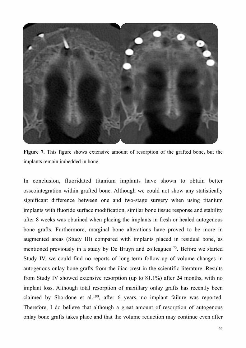
Figure 7. This figure shows extensive amount of resorption of the grafted bone, but the
implants remain imbedded in bone
In conclusion, fluoridated titanium implants have shown to obtain better
osseointegration within grafted bone. Although we could not show any statistically
significant difference between one and two-stage surgery when using titanium
implants with fluoride surface modification, similar bone tissue response and stability
after 8 weeks was obtained when placing the implants in fresh or healed autogenous
bone grafts. Furthermore, marginal bone alterations have proved to be more in
augmented areas (Study III) compared with implants placed in residual bone, as
mentioned previously in a study by De Bruyn and colleagues172. Before we started
Study IV, we could find no reports of long-term follow-up of volume changes in
autogenous onlay bone grafts from the iliac crest in the scientific literature. Results
from Study IV showed extensive resorption (up to 81.1%) after 24 months, with no
implant loss. Although total resorption of maxillary onlay grafts has recently been
claimed by Sbordone et al.180, after 6 years, no implant failure was reported.
Therefore, I do believe that although a great amount of resorption of autogenous
onlay bone grafts takes place and that the volume reduction may continue even after
65

24 months, it is still possible to rehabilitate patients with autogenous onlay bone
blocks from the iliac crest and titanium implants with surface modification.
66

CONCLUSIONS
When using fluoridated moderately rough titanium implants in healed autogenous
onlay bone grafts, a greater amount of BIC can be achieved compared to implants
with turned surfaces. Additionally, a higher degree of stability can be achieved with
fluoridated implants after 8 weeks of healing.
Fluoridated moderately rough titanium implants placed in autogenous onlay grafts
show no significant differences in osseointegration and stability between fresh and
healed bone grafts.
No significant difference was shown in marginal bone-level alteration between block
or particulate bone grafts over a 5-year period. Most of the marginal bone-level
change took place during the initial healing time and the first year of loading.
No significant differences in resorption were seen between autogenous block and
particulate bone from the iliac crest. The volume reduction was extensive at 6 months
and continued until 24 months and may continue to reduce further; however, the
implants will have a good prognosis when titanium implants with a modified surface
structure such as titanium dioxide are used.
67

FUTURE PERSPECTIVES
In the future, a 10-year follow-up with volume and marginal bone-level
measurements in the same patient material would be interesting to examine whether
continued resorption has occurred after 24 months. A clinical evaluation with stability
measurements would show how many of the implants remain in function after 10
years. Moreover, other bone augmentation techniques such as the use of growth
factors in a particulate bone scaffold could be compared with autogenous bone with
no additive growth factors. The drawbacks with radiographic studies is the difficulty
in involving patients due to ethical considerations regarding repeated radiation doses
through CT scans. However, I hope that this problem will be overcome through the
usage of Cone Beam CT by which one can conduct a prospective study where
patients can be followed up after 6, 12 and 24 months and hopefully longer.
68

ACKNOWLEDGEMENTS
I would like to express my sincere gratitude to my supervisor, Prof. Lars Rasmusson who introduced me to the field of research and oral & maxillofacial surgery, for
always being there no matter when and where. Thank your for all your support and
encouragement. I could not have had a better mentor.
Thanks to my second supervisors, Prof. Lars Sennerby and Dr. Andreas Thor for all
the guidance and advise in writing this thesis.
My limitless gratitude to Dr. Annika Ekestubbe whose effort and advise made it
possible to finish Studies III and IV. Thank you for inviting me to your department.
I wish to thank Dr. Hossein Kashani for his efforts in Study I.
It has been a pleasure, getting to know Mr. Carl Rasmusson. Thank you for your
work in Study II.
I wish to thank Mrs. Vivanne Kämpe who provided me with leave of absence for my
research.
Finally, I would like to thank my family for their endless support in whatever
challenge I have taken on. Without your love, support and patience, this thesis would
have never been completed.
69

REFERENCES
1. Müller F, Naharro M, Carlsson GE. What are the prevalence and incidence of tooth loss in the adult and elderely population in Europe? Clin Oral Implants Res 2007; 18:2–14.
2. Brånemark PI, Adell R, Breine U, Hansson BO, Lindström J, Ohlsson A. Intra-osseous anchorage of dental prostheses. I. Experimental studies. Scand J Plast Reconstr Surg. 1969;3(2):81-100.
3. Brånemark PI, Hansson BO, Adell R, Breine U, Lindström J, Hallén O, Ohman A. Osseointegrated implants in the treatment of the edentulous jaw. Experience from a 10-year period. Scand J Plast Reconstr Surg Suppl. 1977;16:1-132.
4. Albrektsson T, Albrektsson B. Osseointegration of bone implants. A review of an alternative mode of fixation. Acta Orthop Scand. 1987;58(5):567-577.
5. Brånemark PI. Introduction to osseointegration. In: Tissue integrated Prostheses. Osseointegration in Clinical Dentistry (Eds. Brånemark PI, Zarb G, Albrektsson T.) Quintessence, Berlin, Chicago, Tokyo 1985:1-76.
6. Kahnberg KE, Nystrom E, Bartholdsson L. Combined use of bone grafts and Brånemark fixtures in the treatment of severely resorbed maxillae. Int J Oral Maxillofac Implants. 1989 Winter;4(4):297-304.
7. Nyström E, Lundgren S, Gunne J, Nilson H. Interpositional bone grafting and Le Fort I osteotomy for reconstruction of the atrophic edentulous maxilla. Int J Oral Maxillofac Surg 1997;26:423-427.
8. Thor A, Wannfors K, Sennerby L, Rasmusson L. Reconstruction of the severely resorbed maxilla with autogenous bone, platelet-rich plasma, and implants: 1-Year Results of a controlled prospective 5-year study. Clin Implant Dent Relat Res 2005;7(4):209-220.
9. Lundgren S, Andersson S, Gualini F, Sennerby L. Bone reformation with sinus membrane elevation: a new surgical technique for maxillary sinus floor augmentation. Clin Implant Dent Relat Res. 2004;6(3):165-173.
10. Hallman M, Hedin M, Sennerby L, Lundgren S. A prospective 1-year clinical and radiographic study of implants placed after maxillary sinus floor augmentation with bovine hydroxyapatite and autogenous bone. J Oral Maxillofac Surg. 2002;60(3):277-284.
11. Striker A, Voss PJ, Gutwald R, et al. Maxillary sinus floor augmentation with autogenous bone grafts to enable placement of SLA-surfaced implants: Preliminary results after 15-40 months. Clin Oral Implants Res. 2003;14:207-212.
70

12. Orsini G, Bianchi AE, Vinci R, Piattelli A. Histologic evaluation of autogenous calvarial bone in maxillary onlay bone grafts: a report of 2 cases. Int J Oral Maxillofac Implants. 2003;18(4):594-598.
13. Isaksson S, Alberius P. Maxillary alveolar ridge augmentation with onlay bone-grafts and immediate endosseous implants. J Craniomaxillofac Surg. 1992;20(1):2-7.
14. Barone A, Ricci M, Mangano F, Covani U. Morbidity associated with iliac crest harvesting in the treatment of maxillary and mandibular atrophies: a 10-year analysis. J Oral Maxillofac Surg. 2011;69(9):2298-2304.
15. Joshi A, Kostakis GC. An investigation of post-operative morbidity following iliac crest graft harvesting. Br Dent J. 2004;196(3):167-171.
16. Eufinger H, Leppänen H. Iliac crest donor site morbidity following open and closed methods of bone harvest for alveolar cleft osteoplasty. J Craniomaxillofac Surg. 2000;28(1):31-38.
17. Beirne JC, Barry HJ, Brady FA, Morris VB. Donor site morbidity of the anterior iliac crest following cancellous bone harvest. Int J Oral Maxillofac Surg. 1996;25(4):268-271.
18. Nkenke E, Weisbach V, Winckler E, Kessler P, Schultze-Mosgau S, Wiltfang J, Neukam FW. Morbidity of harvesting of bone grafts from the iliac crest for preprosthetic augmentation procedures: a prospective study. Int J Oral Maxillofac Surg. 2004;33(2):157-163.
19. Arrington ED, Smith WJ, Chambers HG, Bucknell AL, Davino NA. Complications of iliac crest bone graft harvesting. Clin Orthop Relat Res. 1996;(329):300-309.
20. Burchardt H. The biology of bone graft repair. Clin Orthop Relat Res. 1983;(174):28-42.
21. Goldberg VM, Stevenson S. The biology of bone grafts. Semin Arthroplasty. 1993;4(2):58-63.
22. Sjöström M, Sennerby L, Nilson H, Lundgren S. Reconstruction of the atrophic edentulous maxilla with free iliac crest grafts and implants: a 3-year report of a prospective clinical study. Clin Implant Dent Relat Res. 2007;9(1):46-59.
23. Barone A, Covani U. Maxillary alveolar ridge reconstruction with nonvascularized autogenous block bone: clinical results. J Oral Maxillofac Surg. 2007;65(10):2039-2046.
24. Lundgren S, Sjöström M, Nyström E, Sennerby L. Strategies in reconstruction of the atrophic maxilla with autogenous bone grafts and endosseous implants. Periodontology 2000. 2008;47:143–161.
71
25. Kahnberg KE, Nilsson P, Rasmusson L. Le Fort I osteotomy with interpositional bone grafts and implants for rehabilitation of the severely resorbed maxilla: a 2-stage procedure. Int J Oral Maxillofac Implants. 1999;14(4):571-578.
26. Chiapasco M, Brusati R, Ronchi P. Le Fort I osteotomy with interpositional bone grafts and delayed oral implants for the rehabilitation of extremely atrophied maxillae: a 1-9-year clinical follow-up study on humans. Clin Oral Implants Res. 2007;18(1):74-85.
27. Johansson B, Grepe A, Wannfors K, Hirsch JM. A clinical study of changes in the volume of bone grafts in the atrophic maxilla. Dentomaxillofac Radiol. 2001;30:157-161.
28. Ivanoff CJ, Hallgren C, Widmark G, Sennerby L, Wennerberg A. Histologic evaluation of the bone integration of TiO(2) blasted and turned titanium microimplants in humans. Clin Oral Implants Res. 2001;12(2):128-134.
29. Lundgren S, Rasmusson L, Sjöström M, Sennerby L. Simultaneous or delayed placement of titanium implants in free autogenous iliac bone grafts. Histological analysis of the bone graft-titanium interface in 10 consecutive patients. Int J Oral Maxillofac Surg. 1999;28(1):31-37.
30. Triplett RG, Schow SR. Autologous bone grafts and endosseous implants: complementary techniques. J Oral Maxillofac Surg. 1996;54(4):486-494.
31. Olsen BR, Reginato AM, Wang W. Bone development. 2000;16:191-220.
32. Sommerfeldt DW, Rubin CT. Biology of bone and how it orchestrates the form and function of the skeleton. Eur Spine J. 2001;10:86-95.
33. Knothe Tate ML, Adamson JR, Tami AE, Bauer TW. The osteocyte. Int J Biochem Cell Biol. 2004;36:1-8.
34. Buck DW 2nd, Dumanian GA. Bone biology and physiology: Part I. The fundamentals. Plast Reconstr Surg. 2012;129(6):1314-1320.
35. Clarke B. Normal bone anatomy and physiology. Clin J Am Soc Nephrol. 2008;3:131–139.
36. Buckwalter JA, Glimcher MJ, Cooper RR, Recker R. Bone biology. I: Structure, blood supply, cells, matrix, and mineralization. Instr Course Lect. 1996;45:371-386.
37. Kular J, Tickner J, Chim SM, Xu J. An overview of the regulation of bone remodelling at the cellular level. Clinical Biochemistry. 2012;45:863–887.
38. Ross MH, Romrell LJ, Kaye GI. Bone. In: Coryell PA, ed. Histology. A text and atlas-3. Baltimore, Maryland: Williams & Wilkins, 1995:150-187.
72

39. Mackie EJ. Osteoblasts: novel roles in orchestration of skeletal architecture. Int J Biochem Cell Biol. 2003;35(9):1301-1305.
40. Gehron RP. Bone matrix proteoglycans and glycoproteins. In Bilezikian JP, Raisz LG, Rodan GA, ed. Principles of bone biology. SanDiego: Academic Press, 2002;60:225–237.
41. Anderson H. Matrix vesicles and calcification. Curr Rheumatol Rep. 2003;5:222–226.
42. Yasuda H, Shima N, Nakagawa N et al. Osteoclast differentiation factor is a ligand for osteoprotegerin/osteoclastogenesis-inhibitory factor and is identical to TRANCE/RANKL. Proc Natl Acad Sci U S A. 1998;95(7):3597-3602.
43. Ma YL, Cain RL, Halladay DL, Yang X, Zeng Q, Miles RR, Chandrasekhar S, Martin TJ, Onyia JE. Catabolic effects of continuous human PTH (1-38) in vivo is associated with sustained stimulation of RANKL and inhibition of osteoprotegerin and gene-associated bone formation. Endocrinology. 2001;142(9):4047-4054.
44. Martin TJ. Osteoblast-derived pthrp is a physiological regulator of bone formation. J Clin Invest. 2005;115:2322–2324.
45. Horwood NJ, Elliott J, Martin TJ, Gillespie MT. Osteotropic agents regulate the expression of osteoclast differentiation factor and osteoprotegerin in osteoblastic stromal cells. Endocrinology. 1998;139(11):4743-4746.
46. Ragab AA, Nalepka JL, Bi Y, Greenfield EM. Cytokines synergistically induce osteoclast differentiation: support by immortalized or normal calvarial cells. Am J Physiol Cell Physiol. 2002;283(3):679-687.
47. Suda T, Takahashi N, Martin TJ. Modulation of osteoclast differentiation. Endocr Rev. 1992;13:66–80.
48. Aubin JE, Liu F. The osteoblast lineage. In: Bilezikian J, Raisz L, Rodan G, ed. Principles of bone biology. San Diego: Academic Press, 1996:51–68.
49. Lynch MP, Capparelli C, Stein JL, Stein GS, Lian JB. Apoptosis during bone-like tissue development in vitro. J Cell Biochem. 1998;68(1):31-49.
50. Manolagas SC. Birth and death of bone cells: basic regulatory mechanisms and implications for the pathogenesis and treatment of osteoporosis. Endocr Rev. 2000;21(2):115-137.
51. Bonewald LF. The amazing osteocyte. J Bone Miner Res. 2011;26:229-238.
52. Noble BS, Reeve J. Osteocyte function, osteocyte death and bone fracture resistance. Mol Cell Endocrinol. 2000;159:7-13.
53. Aubin JE. Advances in the osteoblast lineage. Biochem Cell Biol. 1998;76(6):899–910.
73

54. Dudley HR, Spiro D. The fine structure of bone cells. J Cell Biol. 1961;11:627–649.
55. Tanaka-Kamioka K, Kamioka H, Ris H, Lim S-S. Osteocyte shape is dependent on actin filaments and osteocyte processes are unique actin-rich projections. J Bone Miner Res. 1998;13:1555–1568.
56. Aarden EM, Burger EH, Nijweide PJ. Function of osteocytes in bone. J Cell Biochem. 1994;(3):287-299.
57. Burger EH, Klein-Nulend J. Mechanotransduction in bone—role of the lacunocanalicular network. FASEB. 1999;13:101–112.
58. Bonewald LF. Establishment and characterization of an osteocyte-like cell line, MLO-Y4. J Bone Miner Metab. 1999;17:61-65.
59. Miller SC, Jee WS. The bone lining cell: a distinct phenotype? Calcif Tissue Int. 1987;41:1-5.
60. Talmage RV. Morphological and physiological considerations in a new concept of calcium transport in bone. Am J Anat. 1970;129:467-476.
61. Recker RR. Embryology, anatomy and microstructure of bone. In: Coe FL and Favus MJ, ed. Disorders of Bone and Mineral Metaholism. New York: Raven Press, 1992:219-240.
62. Lerner UH. Osteoclast formation and resorption. Matrix Biol. 2000;19:107-120.
63. Roodman GD. Cell biology of the osteoclast. Exp Hematol. 1999;27:1229-1241.
64. Brodsky B, Persikov AV. Molecular structure of the collagen triple helix. Adv Protein Chem. 2005;70:301–339.
65. Sjöström M. On healing of titanium implants in iliac crest bone grafts. Thesis, Umeå University, 2006.
66. Hadjidakis DJ, Androulakis II. Bone remodeling. Ann N Y Acad Sci.2006;1092:385-396.
67. Raisz LG. Physiology and pathophysiology of bone remodeling. Clin Chem. 1999;45:1353-1358.
68. Chambers TJ. Regulation of the differentiation and function of osteoclasts. J Pathol. 2000;192(1):4-13.
69. Rasmusson L. On implant integration in membrane-induced and grafted bone. Thesis, Göteborg University, 1998.
70. Frost HM. Bone remodeling dynamics. Springfield, IL, Charles C Thomas, 1963.
74

71. Parfitt AM. Quantum concept of bone remodeling and turnover: implications for the pathogenesis of osteoporosis. Calcif Tissue Int. 1979;28:1-5.
72. Albrektsson T. Repair of bone grafts. A vital microscopic and histological investigation. Scand J Plast Reconstr Surg. 1980;14:1-12.
73. Shapiro F. Bone development and its relation to fracture repair. The role of mesenchymal osteoblasts and surface osteoblasts. Eur Cell Mater. 2008;15:53-76.
74. Peterson LJ, Ellis E 3rd, Hupp JR, Tucker MR. Wound repair. In: Hupp JR, ed. Contemporary oral and maxillofacial surgery-4. St. Louis, Missouri: Mosby, 2003:49-62.
75. Hing KA. Bone repair in the twenty-first century: biology, chemistry or engineering? Philos Trans A Math Phys Eng Sci. 2004;362(1825):2821-2850.
76. Shapiro F. Cortical bone repair. The relationship of the lacunar-canalicular system and intercellular gap junctions to the repair process. J Bone Joint Surg Am. 1988;70(7):1067-1081.
77. Philips JH, Rahn BA. Fixation effects on membranous and endochondral onlay bone graft resorption. Plast Reconstr Surg. 1988;82:872-877.
78. Hjorting-Hansen E, Worsaae N, Lemons JE. Histologic response after implantation of porous hydroxylapatite ceramic in humans. Int J Oral Maxillofac Implants. 1990;5(3):255-263.
79. Sivolella S, Bressan E, Salata LA, Urrutia ZA, Lang NP, Botticelli D. Osteogenesis at implants without primary bone contact - an experimental study in dogs. Clin Oral Implants Res. 2012;23(5):542-549.
80. Botticelli D, Berglundh T, Buser D, Lindhe J. The jumping distance revisited: An experimental study in the dog. Clin Oral Implants Res. 2003;14(1):35-42.
81. Botticelli D, Berglundh T, Persson LG, Lindhe J. Bone regeneration at implants with turned or rough surfaces in self-contained defects. An experimental study in the dog. J Clin Periodontol. 2005;32(5):448-455.
82. Abrahamsson I, Albouy JP, Berglundh T. Healing at fluoride-modified implants placed in wide marginal defects: an experimental study in dogs. Clin Oral Implants Res. 2008;19(2):153-159.
83. Boëck-Neto RJ, Gabrielli MF, Shibli JA, Marcantonio E, Lia RC, Marcantonio E Jr. Histomorphometric evaluation of human sinus floor augmentation healing responses to placement of calcium phosphate or Ricinus communis polymer associated with autogenous bone. Clin Implant Dent Relat Res. 2005;7(4):181-188.
84. Friedlaender GE. Bone grafts. The basic science rationale for clinical applications. J Bone Joint Surg Am. 1987;69(5):786-790.
75

85. Heiple KG, Chase SW, Herndon CH. A Comparative study of the healing process following different types of bone transplantation. J. Bone and Joint Surg.1963;45:1593-1616.
86. Urist MR, Silverman BF, Buring K, Dubuc FL, Rosenberg JM. The Bone Induction Principle. Clin. Orthop. 1967;53:243-283.
87. Wang HL, Boyapati L. "PASS" principles for predictable bone regeneration. Implant Dent. 2006;15(1):8-17.
88. Dahlin C, Linde A, Gottlow J, Nyman S. Healing of bone defects by guided tissue regeneration. Plast Reconstr Surg. 1988;81(5):672-676.
89. Goldstein M, Boyan BD, Schwartz Z. The palatal advanced flap: a pedicle flap for primary coverage of immediately placed implants. Clin Oral Implants Res. 2002;13(6):644-650.
90. Cha JK, Kim CS, Choi SH, Cho KS, Chai JK, Jung UW. The influence of perforating the autogenous block bone and the recipient bed in dogs. Part II: histologic analysis. Clin Oral Implants Res. 2012;23(8):987-992.
91. Winet, H. The role of microvasculature in normal and perturbed bone healing as revealed by intravital microscopy. Bone. 1996;19:39–57.
92. Glowacki J. Angiogenesis in fracture repair. Clin Orthop Relat Res. 1998;(355 Suppl):82-89.
93. Lu C, Miclau T, Hu D, Marcucio RS. Ischemia leads to delayed union during fracture healing: a mouse model. J Orthop Res. 2007;25(1):51-61.
94. Schmid J, Wallkamm B, Hämmerle CH, Gogolewski S, Lang NP. The significance of angiogenesis in guided bone regeneration. A case report of a rabbit experiment. Clin Oral Implants Res. 1997;8(3):244-248.
95. Mousa SA. Angiogenesis promoters and inhibitors: potential therapeutic implications. Mol Med Today. 1996;2(4):140-142.
96. LaTrenta GS, McCarthy JG, Breitbart AS, May M, Sissons HA. The role of rigid skeletal fixation in bone-graft augmentation of the craniofacial skeleton. Plast Reconstr Surg. 1989;84(4):578-588.
97. Keller EE, van Roekel NB, Desjardins RP, Tolman DE. Prosthetic-surgical reconstruction of the severely resorbed maxilla with iliac bone grafting and tissue-integrated prostheses. Int J Oral Maxillofac Implants. 1987;2:155–165.
98. Misch CM, Misch CE, Resnik R, Ismail YH. Reconstruction of maxillary alveolar defects with mandibular symphysis grafts for dental implants: A preliminary procedural report. Int J Oral Maxillofac Implants. 1992;7:360–366.
76

99. Jensen J, Sindet-Pedersen S, Oliver AJ. Varying treatment strategies for reconstruction of maxillary atrophy with implants: Results in 98 patients. J Oral Maxillofac Surg. 1994;52:210–216.
100. Misch CM. Ridge augmentation using mandibular ramus bone grafts for the placement of dental implants: Presentation of a technique. Pract Periodontics Aesthet Dent. 1996;8:127–135.
101. Braun TW, Sotereanos GC. Autogenous regional bone grafting as an adjunct in orthognathic surgery. J Oral Maxillofac Surg. 1984;42:43–48.
102. Sindet-Pedersen S, Enemark H. Reconstruction of alveolar clefts with mandibular or iliac crest bone grafts. A comparative study. J Oral Maxillofac Surg. 1990;48:554–558.
103. Wood RM, Moore DL. Grafting of the maxillary sinus with intraorally harvested autogenous bone prior to implant placement. Int J Oral Maxillofac Implants. 1988;3:209–214.
104. Williamson RA. Rehabilitation of the resorbed maxilla and mandible using autogenous bone grafts and osseointegrated implants. Int J Oral Maxillofac Implants. 1996;11:476–488.
105. Raghoebar GM, Batenburg RHK, Vissink A, Reintsema H. Augmentation of localized defects of the anterior maxillary ridge with autogenous bone before insertion of implants. J Oral Maxillofac Surg. 1996;54:1180–1185.
106. Misch CM. Comparison of intraoral donor sites for onlay grafting prior to implant placement. Int J Oral Maxillofac Implants. 1997;12(6):767-776.
107. Cawood JL, Howell RA. A classification of the edentulous jaw. Int J Oral Maxillofac Surg. 1988;17:232-336.
108. Lundgren S, Nyström E, Nilson H, Gunne J, Lindhagen O. Bone grafting to the maxillary sinuses, nasal floor and anterior maxilla in the atrophic edentulous maxilla. A two-stage technique. Int J Oral Maxillofac Surg. 1997;26(6):428-434.
109. Whitaker LA. Biological boundaries: a concept in facial skeletal restructuring. Clin Plast Surg. 1989;16(1):1-10.
110. Peer LA. Fate of autogenous human bone grafts. Br J Plast Surg. 1951;3(4):233-243.
111. Zins JE, Whitaker LA. Membranous versus endochondral bone: implications for craniofacial reconstruction. Plast Reconstr Surg. 1983;72(6):778-785.
112. Hardesty RA, Marsh JL. Craniofacial onlay bone grafting: a prospective evaluation of graft morphology, orientation, and embryonic origin. Plast Reconstr Surg. 1990;85(1):5-14.
77

113. Donovan MG, Dickerson NC, Hellstein JW, Hanson LJ. Autologous calvarial and iliac onlay bone grafts in miniature swine. J Oral Maxillofac Surg. 1993;51(8):898-903.
114. Ozaki W, Buchman SR. Volume maintenance of onlay bone grafts in the craniofacial skeleton: micro-architecture versus embryologic origin. Plast Reconstr Surg. 1998;102(2):291-299.
115. Thompson N, Casson JA. Experimental onlay bone grafts to the jaws. A preliminary study in dogs. Plast Reconstr Surg. 1970;46(4):341-349.
116. Smith JD, Abramson M. Membranous vs endochondrial bone autografts. Arch Otolaryngol. 1974;99(3):203-205.
117. Kusiak JF, Zins JE, Whitaker LA. The early revascularization of membranous bone. Plast Reconstr Surg. 1985;76(4):510-516.
118. Levin L, Schwartz-Arad D. The effect of cigarette smoking on dental implants and related surgery. Implant Dent. 2005;14(4):357-361.
119. Bain CA, Moy PK. The association between the failure of dental implants and cigarette smoking. Int J Oral Maxillofac Implants. 1993;8(6):609-615.
120. Baig MR, Rajan M. Effects of smoking on the outcome of implant treatment: a literature review. Indian J Dent Res. 2007;18(4):190-195.
121. Huynh-Ba G, Friedberg JR, Vogiatzi D, Ioannidou E. Implant failure predictors in the posterior maxilla: a retrospective study of 273 consecutive implants. J Periodontol. 2008;79(12):2256-2261.
122. Koldsland OC, Scheie AA, Aass AM. Prevalence of implant loss and the influence of associated factors. J Periodontol. 2009;80(7):1069-1075.
123. DeLuca S, Habsha E, Zarb GA. The effect of smoking on osseointegrated dental implants. Part I: implant survival. Int J Prosthodont. 2006;19(5):491-498.
124. DeLuca S, Zarb G. The effect of smoking on osseointegrated dental implants. Part II: Peri-implant bone loss. Int J Prosthodont. 2006;19(6):560-566.
125. Patel RA, Wilson RF, Patel PA, Palmer RM. The effect of smoking on bone healing: A systematic review. Bone Joint Res. 2013;2(6):102-111.
126. Sloan A, Hussain I, Maqsood M, Eremin O, El-Sheemy M. The effects of smoking on fracture healing. Surgeon. 2010;8(2):111-116.
127. Jorgensen LN, Kallehave F, Christensen E, Siana JE, Gottrup F. Less collagen production in smokers. Surgery. 1998;123(4):450-455.
78

128. Zinser MJ, Randelzhofer P, Kuiper L, Zöller JE, De Lange GL. The predictors of implant failure after maxillary sinus floor augmentation and reconstruction: a retrospective study of 1045 consecutive implants. Oral Surg Oral Med Oral Pathol Oral Radiol. 2013;115(5):571-582.
129. Sørensen LT. Wound healing and infection in surgery. The clinical impact of smoking and smoking cessation: a systematic review and meta-analysis. Arch Surg. 2012;147(4):373-383.
130. Del Fabbro M, Testori T, Francetti L, Weinstein R. Systematic review of survival rates for implants placed in the grafted maxillary sinus. Int J Periodontics Restorative Dent. 2004;24(6):565-577.
131. Chaushu G, Mardinger O, Calderon S, Moses O, Nissan J. The use of cancellous block allograft for sinus floor augmentation with simultaneous implant placement in the posterior atrophic maxilla. J Periodontol. 2009;80(3):422-428.
132. Olson JW, Dent CD, Morris HF, Ochi S. Long-term assessment (5 to 71 months) of endosseous dental implants placed in the augmented maxillary sinus. Ann Periodontol. 2000;5(1):152-156.
133. Nyström E, Legrell PE, Forssell A, Kahnberg KE. Combined use of bone grafts and implants in the severely resorbed maxilla. Postoperative evaluation by computed tomography. Int J Oral Maxillofac Surg. 1995;24:20-25.
134. Nyström E, Kahnberg KE, Gunne J. Bone grafts and Brånemark implants in the treatment of the severely resorbed maxilla: a 2-year longitudinal study. Int J Oral Maxillofac Implants. 1993;8(1):45-53.
135. Nyström E, Ahlqvist J, Gunne J, Kahnberg KE. 10-year follow-up of onlay bone grafts and implants in severely resorbed maxillae. Int J Oral Maxillofac Surg. 2004;33(3):258-262.
136. Van Steenberghe D, Naert I, Bossuyt M, De Mars G, Calberson L, Ghyselen J, Brånemark PI. The rehabilitation of the severely resorbed maxilla by simultaneous placement of autogenous bone grafts and implants: a 10-year evaluation. Clin Oral Investig. 1997;1(3):102-108.
137. Becktor JP, Eckert SE, Isaksson S, Keller EE. The influence of mandibular dentition on implant failures in bone-grafted edentulous maxillae. Int J Oral Maxillofac Implants. 2002;17(1):69-77.
138. Sándor GK. Tissue engineering of bone: Clinical observations with adipose-derived stem cells, resorbable scaffolds, and growth factors. Ann Maxillofac Surg. 2012; 2(1): 8–11.
139. Sándor GK, Tuovinen VJ, Wolff J, Patrikoski M, Jokinen J, Nieminen E, Mannerström B, Lappalainen OP, Seppänen R, Miettinen S. Adipose stem cell tissue-engineered construct used to treat large anterior mandibular defect: a case
79

report and review of the clinical application of good manufacturing practice-level adipose stem cells for bone regeneration. J Oral Maxillofac Surg. 2013;71(5):938-950.
140. Albrektsson T, Brånemark PI, Hansson HA, Lindström J. Osseointegrated titanium implants. Requirements for ensuring a long-lasting, direct bone-to-implant anchorage in man. Acta Orthop Scand. 1981;52(2):155-170.
141. Nygren H, Tengvall P, Lundström I. The initial reactions of TiO2 with blood. J Biomed Mater Res. 1997;34(4):487-492.
142. Palmquist A, Omar OM, Esposito M, Lausmaa J, Thomsen P. Titanium oral implants: surface characteristics, interface biology and clinical outcome. J R Soc Interface. 2010;7:515-527.
143. Sennerby L, Ericson LE, Thomsen P, Lekholm U, Astrand P. Structure of the bone-titanium interface in retrieved clinical oral implants. Clin Oral Implants Res. 1991;2(3):103-111.
144. Sennerby L, Thomsen P, Ericson LE. Early tissue response to titanium implants inserted in rabbit cortical bone. Journal of Materials Science: Materials in Medicine.1993;4:240-250.
145. Nyström E, Kahnberg KE, Albrektsson T. Treatment of the severely resorbed maxillae with bone graft and titanium implants: histologic review of autopsy specimens. Int J Oral Maxillofac Implants. 1993;8(2):167-172.
146. Albrektsson T, Wennerberg A. Oral implant surfaces: Part 1-review focusing on topographic and chemical properties of different surfaces and in vivo responses to them. Int J Prosthodont. 2004;17(5):536-543.
147. Frandsen PA, Christoffersen H, Madsen T. Holding power of different screws in the femoral head. A study in human cadaver hips. Acta Orthop Scand. 1984;55(3):349-351.
148. Albrektsson T. On long-term maintenance of the osseointegrated response. Aust Prosthodont J. 1993;7 Suppl:15-24.
149. Carlsson L, Röstlund T, Albrektsson B, Albrektsson T, Brånemark PI. Osseointegration of titanium implants. Acta Orthop Scand. 1986;57(4):285-289.
150. Adell R, Eriksson B, Lekholm U, Brånemark PI, Jemt T. Long-term follow-up study of osseointegrated implants in the treatment of totally edentulous jaws. Int J Oral Maxillofac Implants. 1990;5(4):347-359.
151. Wennerberg A, Albrektsson T. Effects of titanium surface topography on bone integration: a systematic review. Clin Oral Implants Res. 2009;20 Suppl 4:172-184.
80

152. Shalabi MM, Gortemaker A, Van't Hof MA, Jansen JA, Creugers NH. Implant surface roughness and bone healing: a systematic review. J Dent Res. 2006;85(6):496-500.
153. Cooper LF. A role for surface topography in creating and maintaining bone at titanium endosseous implants. J Prosthet Dent. 2000;84(5):522-534.
154. Davies JE. Mechanisms of endosseous integration. Int J Prosthodont. 1998;11(5):391-401.
155. Ellingsen JE. Pre-treatment of titanium implants with fluoride improves their retention in bone. J of Mater Sci. Mater Med. 1995;6:749-753.
156. Ellingsen JE. Surface configuration of dental implants. Periodontology 2000. 1998;17:36-46.
157. Ellingsen JE, Johansson CB, Wennerberg A, Holmén A. Improved retention and Bone-to-implant contact with fluoride-modified titanium implants. Int J Oral Maxillofac Implants. 2004;19(5):659-666.
158. Meredith N, Alleyne D, Cawley P. Quantitative determination of the stability of the implant-tissue interface using resonance frequency analysis. Clin Oral Implants Res. 1996;7(3):261-267.
159. Meredith N, Book K, Friberg B, Jemt T, Sennerby L. Resonance frequency measurements of implant stability in vivo. A cross-sectional and longitudinal study of resonance frequency measurements on implants in the edentulous and partially dentate maxilla. Clin Oral Implants Res. 1997;8(3):226-233.
160. Sennerby L, Meredith N. Implant stability measurements using resonance frequency analysis: biological and biomechanical aspects and clinical implications. Periodontol 2000. 2008;47:51-66.
161. Phillips JH, Rahn BA. Fixation effects on membranous and endochondral onlay bone graft resorption. Plast Reconstr Surg. 1988;82:872-877.
162. Rasmusson L, Meredith N, Cho IH, Sennerby L. The influence of simultaneous or delayed placement of titanium implants in onlay bone grafts. A histologic and biomechanic study in the rabbit. Int J Oral Maxillofac Surg. 1999;28:224-231.
163. Shirota T, Ohno K, Michi K, Tachikawa T. An experimental study of healing around hydroxylapatite implants installed with autogenous iliac bone grafts for jaw reconstruction. J Oral Maxillofac Surg. 1991;49(12):1310-1315.
164. Sjöström M, Lundgren S, Nilson H, Sennerby L. Monitoring of implant stability in grafted bone using resonance frequency analysis. A clinical study from implant placement to 6 months of loading. Int J Oral Maxillofac Surg. 2005; 34:45-51.
81

165. Farley JR, Wergedal JE, Baylink DJ. Fluoride directly stimulates proliferation and alkaline phosphatase activity of bone-forming cells. Science. 1983;222:330-332.
166. Ellingsen JE. Thomsen P. Lyngstadaas SP. Advances in dental implant materials and tissue regeneration. Periodontology 2000. 2006;136-156.
167. Bellows CG, Heersche JN, Aubin JE. The effects of fluoride on osteoblast progenitors in vitro. J Bone Miner Res. 1990;5 Suppl 1:101-105.
168. Kassem M, Mosekilde L, Eriksen EF. Effects of fluoride on human bone cells in vitro: differences in responsiveness between stromal osteoblast precursors and mature osteoblasts. Eur J Endocrinol. 1994;130(4):381-386.
169. Rasmusson L, Thor A, Sennerby L. Stability evaluation of implants integrated in grafted and nongrafted maxillary bone: a clinical study from implant placement to abutment connection. Clin Implant Dent Relat Res. 2012;14(1):61-66.
170. Wennerberg A, Albrektsson T. On implant surfaces: A review of current knowledge and opinions. Int J Oral Maxillofac Impl. 2009;24:63-74.
171. Lee DW, Choi YS, Park KH, Kim CS, Moon IS. Effect of microthread on the maintenance of marginal bone level: a 3-year prospective study. Clin Oral Implants Res. 2007;18(4):465-470.
172. De Bruyn H, Van de Velde T, Collaert B. Immediate functional loading of TiOblast dental implants in full-arch edentulous mandibles: a 3-year prospective study. Clin Oral Implants Res. 2008;19(7):717-723.
173. Steveling H, Roos J, Rasmusson L. Maxillary implants loaded at 3 months after insertion: results with Astra Tech implants after up to 5 years. Clin Implant Dent Relat Res. 2001;3(3):120-124.
174. Smolka W, Eggensperger N, Carollo V, Ozdoba C, Iizuka T. Changes in the volume and density of calvarial split bone grafts after alveolar ridge augmentation. Clin Oral Implants Res. 2006;17(2):149-155.
175. Wong RW, Rabie AB. A quantitative assessment of the healing of intramembranous and endochondral autogenous bone grafts. Eur J Orthod. 1999;21(2):119-126.
176. Rabie AB, Dan Z, Samman N. Ultrastructural identification of cells involved in the healing of intramembranous and endochondral bones. Int J Oral Maxillofac Surg. 1996;25(5):383-388.
177. Verhoeven JW, Ruijter J, Cune MS, Terlou M, Zoon M. Onlay grafts in combination with endosseous implants in severe mandibular atrophy: one year results of a prospective, quantitative radiological study. Clin Oral Implants Res. 2000;11(6):583-594.
82

178. Swart JG, Allard RH. Subperiosteal onlay augmentation of the mandible: a clinical and radiographic survey. J Oral Maxillofac Surg. 1985;43(3):183-187.
179. Sbordone L, Toti P, Menchini-Fabris GB, Sbordone C, Piombino P, Guidetti F. Volume changes of autogenous bone grafts after alveolar ridge augmentation of atrophic maxillae and mandibles. Int J Oral Maxillofac Surg. 2009;38(10):1059-1065.
180. Sbordone C, Toti P, Guidetti F, Califano L, Santoro A, Sbordone L. Volume changes of iliac crest autogenous bone grafts after vertical and horizontal alveolar ridge augmentation of atrophic maxillas and mandibles: a 6-year computerized tomographic follow-up. J Oral Maxillofac Surg. 2012;70(11):2559-2565.
181. Gotfredsen K, Nimb L, Hjörting-Hansen E, Jensen JS, Holmén A. Histomorphometric and removal torque analysis for TiO2-blasted titanium implants. An experimental study on dogs. Clin Oral Implants Res. 1992;3(2):77-84.
182. Wennerberg A, Albrektsson T, Andersson B, Krol JJ. A histomorphometric and removal torque study of screw-shaped titanium implants with three different surface topographies. Clin Oral Implants Res. 1995;6(1):24-30.
83
Top Related