


Languages
Pages
Legal


Newborn
Hyperbilirubinemia
A Self–Learning Module
2013 (Updated July 2015)
Newborn Hyperbilirubinemia: A Self-Learning Module
© CMNRP 2015 2
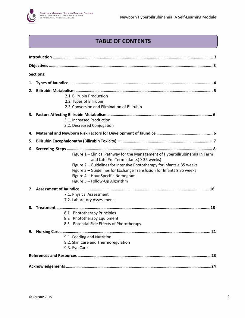
Introduction ……………………………………………………………………………………………………………………………………... 3
Objectives ………………………………………………………………………………………………………………………………………... 3
Sections:
1. Types of Jaundice ……………………………………………………………………………………………………………………….. 4
2. Bilirubin Metabolism ………………………………………………………………………………………………………………….. 5
2.1 Bilirubin Production
2.2 Types of Bilirubin
2.3 Conversion and Elimination of Bilirubin
3. Factors Affecting Bilirubin Metabolism …………………………………………………………………………………….… 6
3.1. Increased Production
3.2. Decreased Conjugation
4. Maternal and Newborn Risk Factors for Development of Jaundice …………………………………………..... 6
5. Bilirubin Encephalopathy (Bilirubin Toxicity) ………………………………………………………………………………. 7
6. Screening Steps …………………………………………………………………………………………………………………………. 8
Figure 1 – Clinical Pathway for the Management of Hyperbilirubinemia in Term
and Late Pre-Term Infants( ≥ 35 weeks)
Figure 2 – Guidelines for Intensive Phototherapy for Infants ≥ 35 weeks
Figure 3 – Guidelines for Exchange Transfusion for Infants ≥ 35 weeks
Figure 4 – Hour Specific Nomogram
Figure 5 – Follow-Up Algorithm
7. Assessment of Jaundice …………………………………………………………………………………………………………… 16
7.1. Physical Assessment
7.2. Laboratory Assessment
8. Treatment ………………………………………………………………………………………………………………………….….….18
8.1 Phototherapy Principles
8.2 Phototherapy Equipment
8.3 Potential Side Effects of Phototherapy
9. Nursing Care……………………………………………………………………………………………………………………………... 21
9.1. Feeding and Nutrition
9.2. Skin Care and Thermoregulation
9.3. Eye Care
References and Resources …………………………………………………………………………………………………………….… 23
Acknowledgements ………………………………………………………………………………………………………………………….24
TABLE OF CONTENTS
Newborn Hyperbilirubinemia: A Self-Learning Module
© CMNRP 2015 3
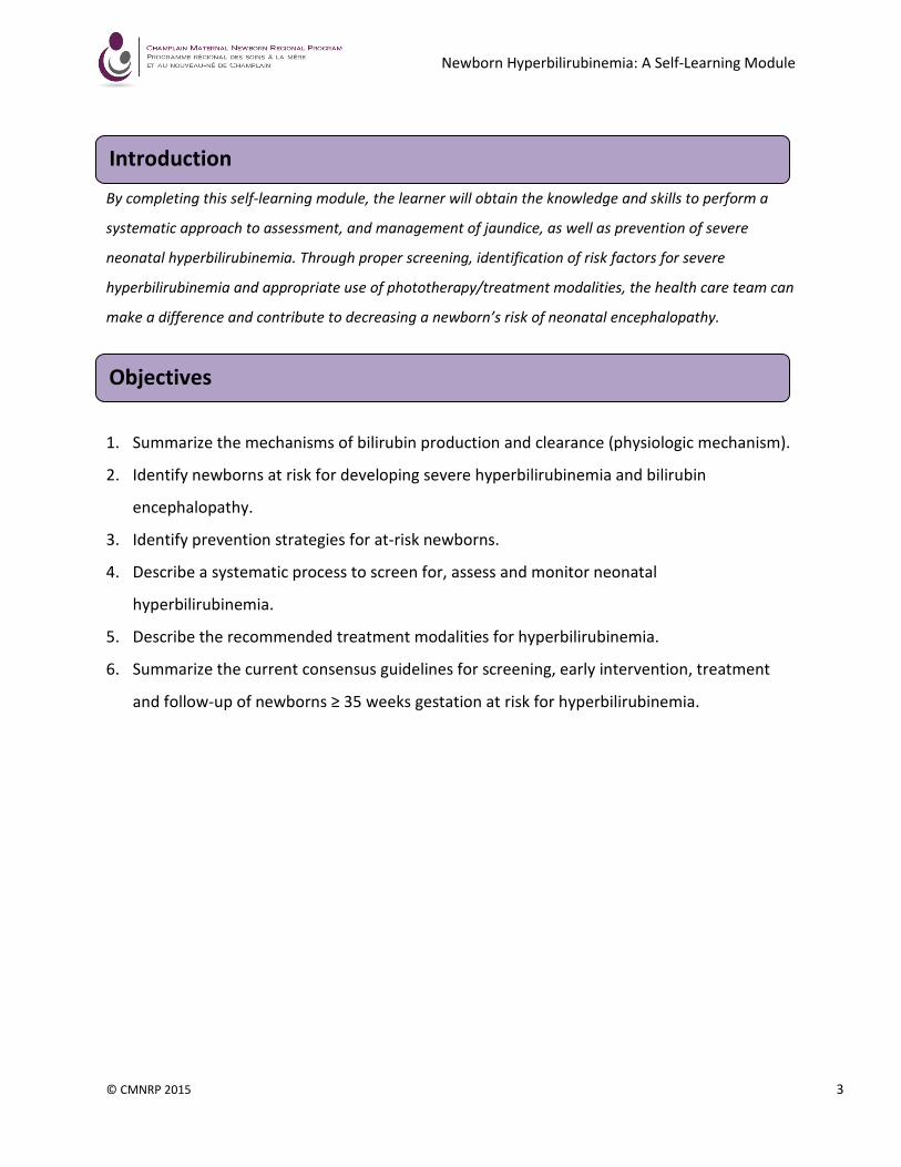
By completing this self-learning module, the learner will obtain the knowledge and skills to perform a
systematic approach to assessment, and management of jaundice, as well as prevention of severe
neonatal hyperbilirubinemia. Through proper screening, identification of risk factors for severe
hyperbilirubinemia and appropriate use of phototherapy/treatment modalities, the health care team can
make a difference and contribute to decreasing a newborn’s risk of neonatal encephalopathy.
1. Summarize the mechanisms of bilirubin production and clearance (physiologic mechanism).
2. Identify newborns at risk for developing severe hyperbilirubinemia and bilirubin
encephalopathy.
3. Identify prevention strategies for at-risk newborns.
4. Describe a systematic process to screen for, assess and monitor neonatal
hyperbilirubinemia.
5. Describe the recommended treatment modalities for hyperbilirubinemia.
6. Summarize the current consensus guidelines for screening, early intervention, treatment
and follow-up of newborns ≥ 35 weeks gestation at risk for hyperbilirubinemia.
Introduction
Objectives
Newborn Hyperbilirubinemia: A Self-Learning Module
© CMNRP 2015 4
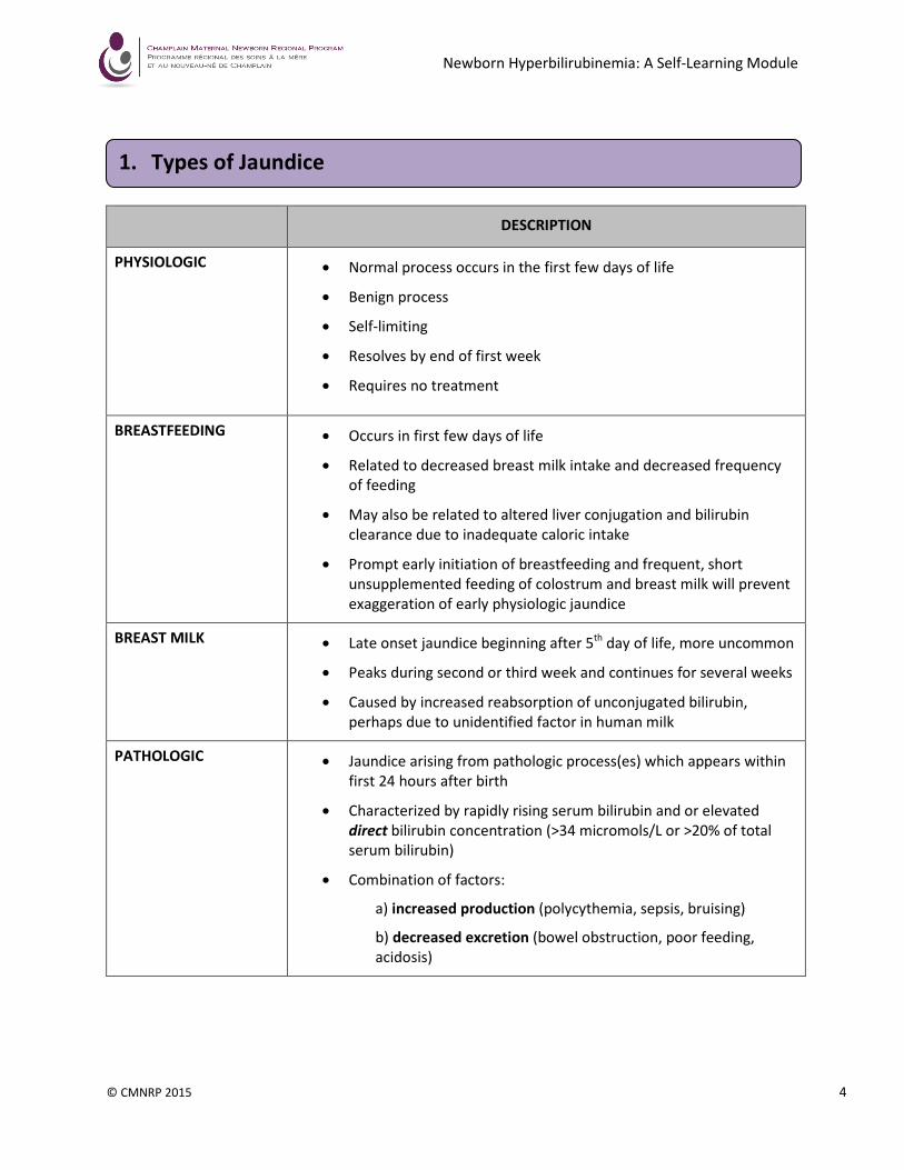
DESCRIPTION
PHYSIOLOGIC
• Normal process occurs in the first few days of life
• Benign process
• Self-limiting
• Resolves by end of first week
• Requires no treatment
BREASTFEEDING • Occurs in first few days of life
• Related to decreased breast milk intake and decreased frequency
of feeding
• May also be related to altered liver conjugation and bilirubin
clearance due to inadequate caloric intake
• Prompt early initiation of breastfeeding and frequent, short
unsupplemented feeding of colostrum and breast milk will prevent
exaggeration of early physiologic jaundice
BREAST MILK • Late onset jaundice beginning after 5
th day of life, more uncommon
• Peaks during second or third week and continues for several weeks
• Caused by increased reabsorption of unconjugated bilirubin,
perhaps due to unidentified factor in human milk
PATHOLOGIC • Jaundice arising from pathologic process(es) which appears within
first 24 hours after birth
• Characterized by rapidly rising serum bilirubin and or elevated
direct bilirubin concentration (>34 micromols/L or >20% of total
serum bilirubin)
• Combination of factors:
a) increased production (polycythemia, sepsis, bruising)
b) decreased excretion (bowel obstruction, poor feeding,
acidosis)
1. Types of Jaundice
Newborn Hyperbilirubinemia: A Self-Learning Module
© CMNRP 2015 5
2.1 Bilirubin Production
Bilirubin is a product of the breakdown of the heme portion of hemoglobin that occurs when red blood
cells are destroyed. Normally, bilirubin is excreted through the body after passing through the liver,
spleen, kidneys and the gastrointestinal tract.
2.2 Types of Bilirubin
There are two types of bilirubin circulating in the blood stream, unconjugated and conjugated.
Unconjugated bilirubin (or indirect bilirubin) can be found in circulating blood either bound to albumin
or not. It is fat-soluble and therefore more potentially toxic since it can bind to the tissues. Most of the
unconjugated bilirubin is bound to albumin and transported to the liver. There, it is converted to
glucuronic acid aided by uridine diphosphate glucuronosyl transferase (UDGT) to produce conjugated
bilirubin. Once it becomes conjugated, it is sent to the gut for excretion via the biliary system. The
unbound, unconjugated bilirubin is most likely to cross the blood-brain barrier and settle in the tissues
where it can cause temporary or permanent neurological damage. Once it settles in the brain, it is there
forever. The unbound bilirubin is difficult to measure but it is thought that it is directly related to the
amount of unconjugated bilirubin.
Conjugated bilirubin (or direct bilirubin) is water-soluble and therefore is a more stable and non-toxic
form. This allows it to be easily excreted from the body in urine and stool. Elevated levels of conjugated
bilirubin may indicate evidence of liver disease.
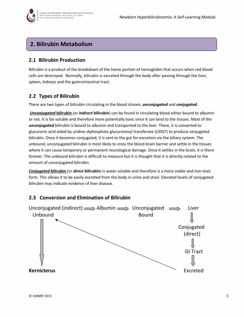
2.3 Conversion and Elimination of Bilirubin
Unconjugated (indirect) Albumin Unconjugated Liver
Unbound Bound
Conjugated
(direct)
GI Tract
Kernicterus Excreted
2. Bilirubin Metabolism
Newborn Hyperbilirubinemia: A Self-Learning Module
© CMNRP 2015 6
3.1 Increased Production
Any disorder which causes an increase in the number of red blood cells such as polycythemia, will
lead to an increase in the amount of bilirubin produced as these cells breakdown. If there is a
decreased amount of albumin available, there will be decreased binding capacity and conversion of
indirect to direct bilirubin in the liver resulting in more indirect bilirubin that could potentially cross
the blood-brain barrier or settle in the tissues. Bruising will also increase the breakdown of RBCs
and increase bilirubin levels.
3.2 Decreased Conjugation
Conditions such as acidosis and hypoxia can also affect the bilirubin/albumin ratio for binding. The
presence of any type of liver disease, metabolic or enzyme disorder will also affect the ability of the
body to convert bilirubin to the direct form to allow for excretion. Because bilirubin is changed in
the gut to urobilinogen with the assistance of the normal intestinal flora, anything that affects
normal gut function can affect the excretion of bilirubin from the body. We know that at birth, the
infant’s gut is not fully developed so that prematurity and/or any disorder of the bowel, as well as
antibiotic therapy, can slow the excretion of bilirubin.
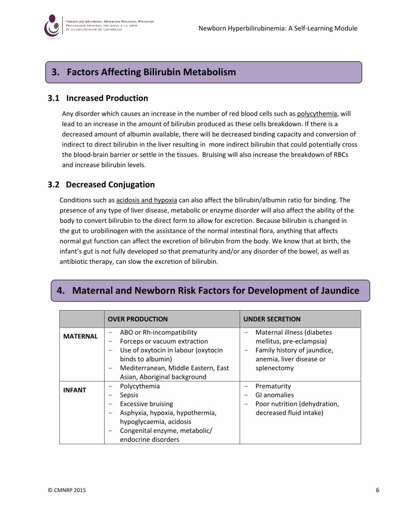
OVER PRODUCTION UNDER SECRETION
MATERNAL - ABO or Rh-incompatibility
- Forceps or vacuum extraction
- Use of oxytocin in labour (oxytocin
binds to albumin)
- Mediterranean, Middle Eastern, East
Asian, Aboriginal background
- Maternal illness (diabetes
mellitus, pre-eclampsia)
- Family history of jaundice,
anemia, liver disease or
splenectomy
INFANT - Polycythemia
- Sepsis
- Excessive bruising
- Asphyxia, hypoxia, hypothermia,
hypoglycaemia, acidosis
- Congenital enzyme, metabolic/
endocrine disorders
- Prematurity
- GI anomalies
- Poor nutrition (dehydration,
decreased fluid intake)
3. Factors Affecting Bilirubin Metabolism
4. Maternal and Newborn Risk Factors for Development of Jaundice
Newborn Hyperbilirubinemia: A Self-Learning Module
© CMNRP 2015 7
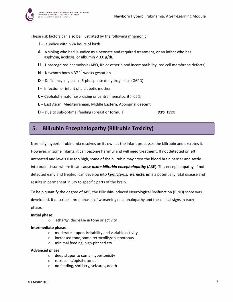
These risk factors can also be illustrated by the following mnemonic:
J - Jaundice within 24 hours of birth
A – A sibling who had jaundice as a neonate and required treatment, or an infant who has
asphyxia, acidosis, or albumin < 3.0 g/dL
U – Unrecognized haemolysis (ABO, Rh or other blood incompatibility, red cell membrane defects)
N – Newborn born < 37 + 0
weeks gestation
D – Deficiency in glucose-6-phosphate dehydrogenase (G6PD)
I – Infection or infant of a diabetic mother
C – Cephalohematoma/bruising or central hematocrit > 65%
E – East Asian, Mediterranean, Middle Eastern, Aboriginal descent
D – Due to sub-optimal feeding (breast or formula) (CPS, 1999)
Normally, hyperbilirubinemia resolves on its own as the infant processes the bilirubin and excretes it.
However, in some infants, it can become harmful and will need treatment. If not detected or left
untreated and levels rise too high, some of the bilirubin may cross the blood brain barrier and settle
into brain tissue where it can cause acute bilirubin encephalopathy (ABE). This encephalopathy, if not
detected early and treated, can develop into kernicterus. Kernicterus is a potentially fatal disease and
results in permanent injury to specific parts of the brain.
To help quantify the degree of ABE, the Bilirubin-Induced Neurological Dysfunction (BIND) score was
developed. It describes three phases of worsening encephalopathy and the clinical signs in each
phase:
Initial phase:
o lethargy, decrease in tone or activity
Intermediate phase:
o moderate stupor, irritability and variable activity
o increased tone, some retrocollis/opisthotonus
o minimal feeding, high-pitched cry
Advanced phase:
o deep stupor to coma, hypertonicity
o retrocollis/opisthotonus
o no feeding, shrill cry, seizures, death
5. Bilirubin Encephalopathy (Bilirubin Toxicity)
Newborn Hyperbilirubinemia: A Self-Learning Module
© CMNRP 2015 8
The content of this section is based on the Ontario Ministry of Health and Long-Term Care (MOHTLC)
Quality-Based Procedure (QBP) titled Hyperbilirubinemia in Term and Late Pre-Term Infants (≥ 35
weeks) (2013). The key objectives of the QBP for Hyperbilirubinemia are to:
- ensure all newborns receive bilirubin screening between 24-72 hours of life (if not
clinically indicated and performed earlier)
- ensure infants receive systematic bilirubin monitoring as per the treatment graph and
risk nomograms recommended by evidence-based guidelines
- utilize health care resources responsibly through avoidance of unnecessary/excessive
testing, timely discharge, appropriate outpatient follow-up and minimization of
preventable readmission
- reduce the incidence of severe hyperbilirubinemia and acute bilirubin encephalopathy
The Provincial Council for Maternal and Child Health (PCMCH) has developed a toolkit to accompany
the MOHLTC’s QBP on hyperbilirubinemia. The toolkit details the clinical pathway and the tools
clinicians can use to implement the QBP. Further information may be downloaded from the PCMCH
website. Information regarding QBPs can be downloaded from the Health System Funding Reform,
Quality Based Procedures portion of the MOHLTC webpage.
6. Screening Steps
Newborn Hyperbilirubinemia: A Self-Learning Module
© CMNRP 2015 9
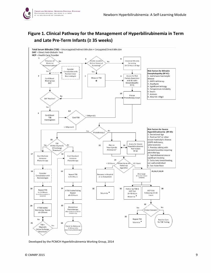
Figure 1. Clinical Pathway for the Management of Hyperbilirubinemia in Term
and Late Pre-Term Infants (≥ 35 weeks)
Developed by the PCMCH Hyperbilirubinemia Working Group, 2014
Newborn Hyperbilirubinemia: A Self-Learning Module
© CMNRP 2015 10
Screening Steps (1 – 32) (Figure 1)
1. Identify newborns of mothers with red cell antibodies (isoimmunization).
2. Newborns of mothers with red cell antibodies should have blood group evaluation and
direct anti-globulin test (DAT).
3. Measure cord blood for hemoglobin and total serum bilirubin (TSB).
4. If cord TSB level ≥ 100 µmol/L this is a critical value and is suggestive of a need for exchange
transfusion. Multiple intensive phototherapy lights should be initiated without delay, while
continuing on pathway (step #17) and initiating consult (step #18).
5. If cord TSB level < 100 µmol/L plot bilirubin on phototherapy graph: Figure 2 (step #10)
using time=0.
6. Clinically assess for jaundice routinely during newborn care. Jaundice in the first 24 hours is
more likely to be significant/pathologic, so multiple clinical assessments in the first 24 hours
are recommended.
7. Measure TSB in all newborns that appear clinically jaundiced in their first 24 hours of life.
8. If not required earlier because of clinical jaundice, TSB should be obtained at the same time
as newborn metabolic screening (between 24-72 hours of age).
9. Assess for presence of any Bilirubin Encephalopathy Risk Factors. These risk factors along
with gestational age are used to identify the low/medium/high treatment threshold lines on
the phototherapy graph (Figure 2).
• Risk factors for bilirubin encephalopathy to consider when determining which line
to follow as cutoff for treatment (threshold line) include:
- isoimmune haemolytic disease
- G6PD deficiency
- asphyxia
- current and significant lethargy
- unresolved temperature instability (requiring active treatment)
- sepsis (current treatment with antibiotics)
- ongoing acidosis (not isolated low cord pH)
- albumin < 30g/L (if measured)
10. Plot TSB on Phototherapy graph (Figure 2) to determine need for phototherapy.
Determination of the treatment line depends on gestational age at birth as well as presence
of bilirubin encephalopathy risk factors from step #9. Plot on phototherapy graph using TSB
(unconjugated + conjugated) and age in hours at the time of the bilirubin was measured.

Newborn Hyperbilirubinemia: A Self-Learning Module
© CMNRP 2015 11
Figure 2. Guidelines for Intensive Phototherapy for Infants ≥ 35 weeks
Adapted with permission from the American Academy of Pediatrics - Subcommittee on Hyperbilirubinemia.
(2004). Management of hyperbilirubinemia in the newborn infant 35 or more weeks of gestation. Pediatrics,
114(1), 297-316.
11. If phototherapy indicated, determine if TSB is within 50µmol/L of the exchange transfusion
line on Exchange Transfusion Graph (Figure 3).
12. If ‘No” in Step #11, start Standard Intensive Phototherapy:
- Begin with high intensity of at least 30 µw/cm²/nm
- Expose maximum skin surface; limiting interruptions to 20 minutes every 3 hours
Newborn Hyperbilirubinemia: A Self-Learning Module
© CMNRP 2015 12
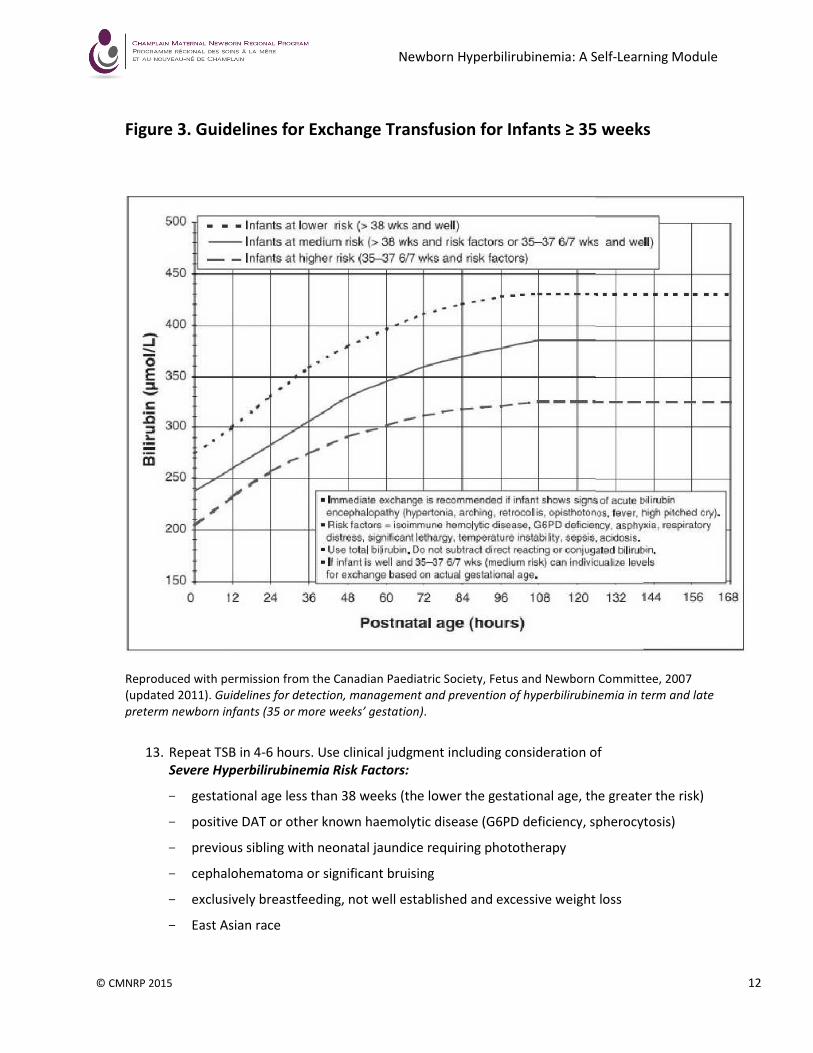
Figure 3. Guidelines for Exchange Transfusion for Infants ≥ 35 weeks
Reproduced with permission from the Canadian Paediatric Society, Fetus and Newborn Committee, 2007
(updated 2011). Guidelines for detection, management and prevention of hyperbilirubinemia in term and late
preterm newborn infants (35 or more weeks’ gestation).
13. Repeat TSB in 4-6 hours. Use clinical judgment including consideration of
Severe Hyperbilirubinemia Risk Factors:
- gestational age less than 38 weeks (the lower the gestational age, the greater the risk)
- positive DAT or other known haemolytic disease (G6PD deficiency, spherocytosis)
- previous sibling with neonatal jaundice requiring phototherapy
- cephalohematoma or significant bruising
- exclusively breastfeeding, not well established and excessive weight loss
- East Asian race
Newborn Hyperbilirubinemia: A Self-Learning Module
© CMNRP 2015 13
14. If TSB is stable or falling continue to repeat TSB every 8 – 24 hours while on phototherapy.
15. Discontinue phototherapy when TSB is below threshold for phototherapy initiation.
16. Check TSB for rebound 12-24 hours after discontinuing phototherapy.
17. If YES in Step #11, start multiple intensive phototherapy modalities including a
phototherapy blanket under the infant to increase exposed surface area. Do not interrupt
phototherapy for feeding or other care.
18. Consider immediate consult with neonatologist. IVIG or exchange transfusion may be
indicated in specific situations.
19. Repeat TSB in 2-6 hours to confirm response to treatment.
20. If TSB stable or decreasing continue to repeat every 6-12 hours.
21. When TSB is more than 50 µmol/L below exchange transfusion threshold return to step #9.
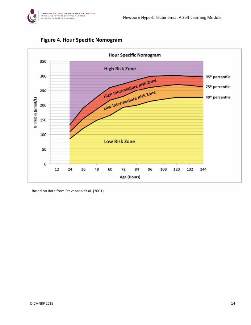
22. If phototherapy is not indicated, plot the TSB on the Hour Specific Nomogram (Figure 4).
23. Assess for presence of any Severe Hyperbilirubinemia Risk Factors (see step #13).
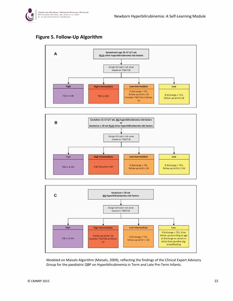
24. Consult Follow-up Algorithm (Figure 5) for management and follow-up according to pre-
discharge TSB.
25. Arrange follow-up TSB measurement, if indicated.
26. If appropriate follow-up cannot be ensured in the presence of elevated risk for developing
severe hyperbilirubinemia, delay discharge.
27. Provide lactation evaluation and support for all breastfeeding mothers.
28. Any infant discharged before 24 hours should be assessed by a health care provider within
24 hours. That care provider should have access to testing and treatment facilities.
29. The infants’ parent or guardian should be provided with written and verbal instructions
regarding the infant’s jaundice follow-up and the timing of that follow-up.
30. The follow-up assessment should include confirmation that:
- Weight loss should be no more than 10% of birth weight
- 4-6 wet diapers and 3-4 stools per day by the fourth day
- Stools in breastfed infants should have changed from meconium to mustard yellow
- Breastfeeding is effective
31. Clinical judgment should be used to determine the need for TSB measurement. Visual
estimation of bilirubin levels can lead to errors, especially in darkly pigmented infants.
32. Any repeat TSB measurement should be plotted in this algorithm in the same manner as the
initial TSB to identify the need for and timing of further clinical follow-up.
Newborn Hyperbilirubinemia: A Self-Learning Module
© CMNRP 2015 14
Figure 4. Hour Specific Nomogram
Based on data from Stevenson et al. (2001).
Newborn Hyperbilirubinemia: A Self-Learning Module
© CMNRP 2015 15
Figure 5. Follow-Up Algorithm
Modeled on Maisels Algorithm (Maisels, 2009), reflecting the findings of the Clinical Expert Advisory
Group for the paediatric QBP on Hyperbilirubinemia in Term and Late Pre-Term Infants.
Newborn Hyperbilirubinemia: A Self-Learning Module
© CMNRP 2015 16
7.1 Physical Assessment
• Visual assessment: Jaundice moves from head to toe, with the eyes affected last.
Serum bilirubin (approx.)
= 85 micromols/L - When yellow tinge first becomes visible
= 150 micromols/L - Yellow tinge appears on trunk
= 200 micromols/L - Yellow tinge appears on legs
= 250 micromols/L - Eyes (sclera) are affected
• Although visual assessment alone cannot determine the degree of jaundice, a general
assessment of the extent of jaundice can be done under bright light. It is important to:
o Blanch skin to determine underlying colour.
o Press over a bony prominence for best results (nose, forehead).
o Check sclera.
NOTE: For dark skinned infants, the colour of the sclera, conjunctiva and oral mucosa is
most reliable indicator of level of jaundice.
NOTE: Petechiae may indicate underlying sepsis or haemolytic disease.
• Level of activity:
o Increasing levels of unconjugated bilirubin in the brain can lead to decreased levels of
consciousness or alertness. Infants may become lethargic and less responsive.
• Level of hydration:
o Monitor intake and output.
o Adequate hydration is necessary to help maintain enough fluid to help with the
absorption and excretion of conjugated bilirubin once it passes through the liver.
• Stools:
o Monitor frequency, type and colour of stools (meconium versus transitional).
o Unconjugated bilirubin can accumulate in stool and thus has the potential to be
reabsorbed.
o Conjugated bilirubin can also become unconjugated in the gut and become reabsorbed
into the blood stream.
7. Assessment of Jaundice
Newborn Hyperbilirubinemia: A Self-Learning Module
© CMNRP 2015 17
7.2 Laboratory Assessment
• Obtain serum bilirubin levels as per algorithm (Figure 2).
• NOTE: When bloodwork is being drawn, phototherapy should be stopped to prevent the sample
from being affected by the lights. The total bilirubin should be interpreted according to the
infant’s age in hours to determine the treatment plan and timing of reassessment.
• Other blood work that may be ordered:
o Serum albumin - to help determine how much albumin is available for binding
o CBC and differential – can help determine level of red blood cell destruction, haemolytic
anemia, sepsis or polycythemia
o Direct Antiglobulin Test (DAT) - to look for presence of maternal antibodies in infant’s
serum. NOTE: Indirect Antibody Test (IAT) is done on maternal serum antenatally
o G6PD (glucose-6-phosphate dehydrogenase) – helps maintain RBC wall integrity; a
deficiency indicates enzyme deficiency and a possible metabolic reason for jaundice
REMEMBER:
It is possible that bilirubin levels could rise once
phototherapy is discontinued although current
methods of weaning phototherapy from high
intensity (intensive) to low intensity (standard)
phototherapy can prevent this. A follow-up serum
bilirubin should be taken 12-24 hours as per
protocol; usually 12–24 hours after therapy is
discontinued.
Newborn Hyperbilirubinemia: A Self-Learning Module
© CMNRP 2015 18
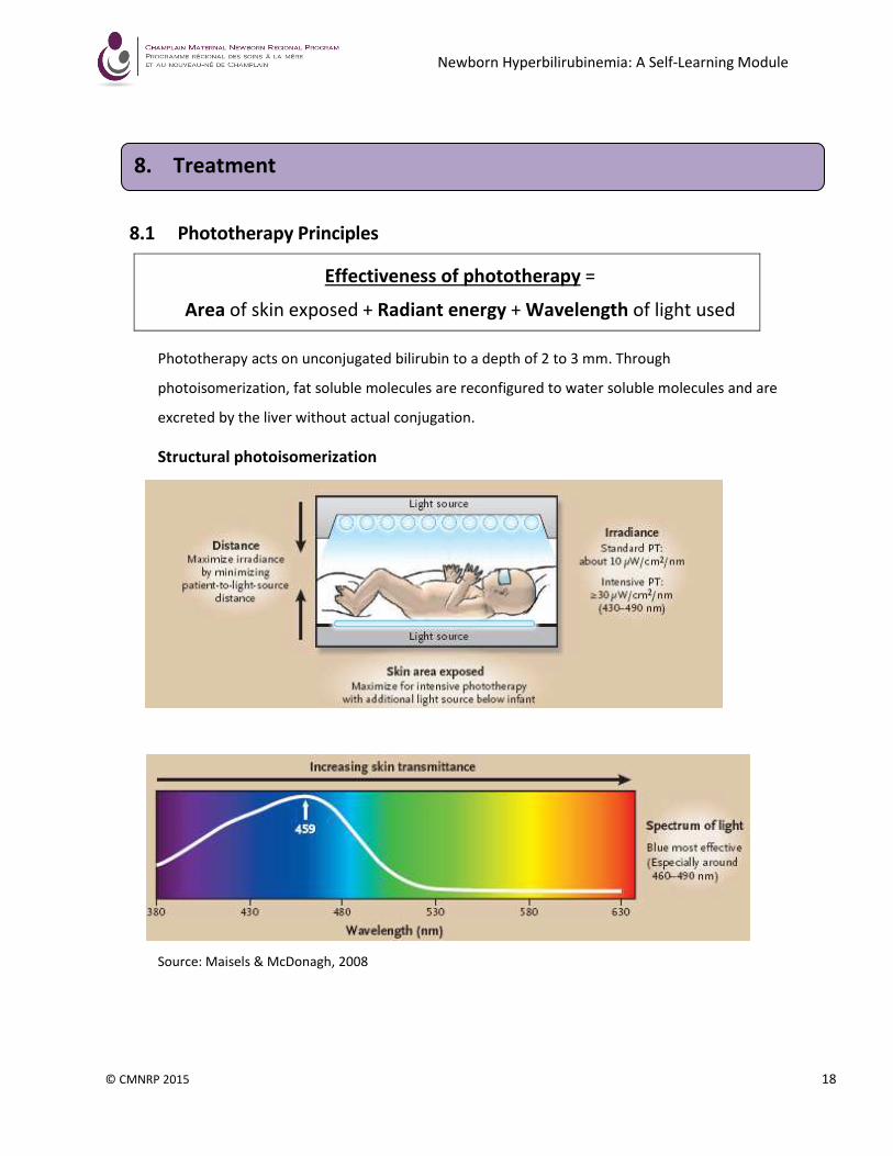
8.1 Phototherapy Principles
Phototherapy acts on unconjugated bilirubin to a depth of 2 to 3 mm. Through
photoisomerization, fat soluble molecules are reconfigured to water soluble molecules and are
excreted by the liver without actual conjugation.
Structural photoisomerization
Source: Maisels & McDonagh, 2008
Effectiveness of phototherapy =
Area of skin exposed + Radiant energy + Wavelength of light used
8. Treatment
Newborn Hyperbilirubinemia: A Self-Learning Module
© CMNRP 2015 19
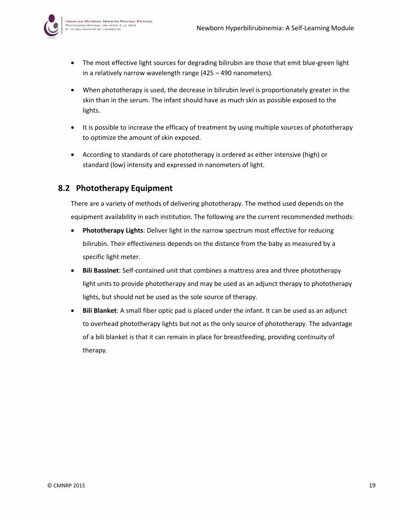
• The most effective light sources for degrading bilirubin are those that emit blue-green light
in a relatively narrow wavelength range (425 – 490 nanometers).
• When phototherapy is used, the decrease in bilirubin level is proportionately greater in the
skin than in the serum. The infant should have as much skin as possible exposed to the
lights.
• It is possible to increase the efficacy of treatment by using multiple sources of phototherapy
to optimize the amount of skin exposed.
• According to standards of care phototherapy is ordered as either intensive (high) or
standard (low) intensity and expressed in nanometers of light.
8.2 Phototherapy Equipment
There are a variety of methods of delivering phototherapy. The method used depends on the
equipment availability in each institution. The following are the current recommended methods:
• Phototherapy Lights: Deliver light in the narrow spectrum most effective for reducing
bilirubin. Their effectiveness depends on the distance from the baby as measured by a
specific light meter.
• Bili Bassinet: Self-contained unit that combines a mattress area and three phototherapy
light units to provide phototherapy and may be used as an adjunct therapy to phototherapy
lights, but should not be used as the sole source of therapy.
• Bili Blanket: A small fiber optic pad is placed under the infant. It can be used as an adjunct
to overhead phototherapy lights but not as the only source of phototherapy. The advantage
of a bili blanket is that it can remain in place for breastfeeding, providing continuity of
therapy.
Newborn Hyperbilirubinemia: A Self-Learning Module
© CMNRP 2015 20
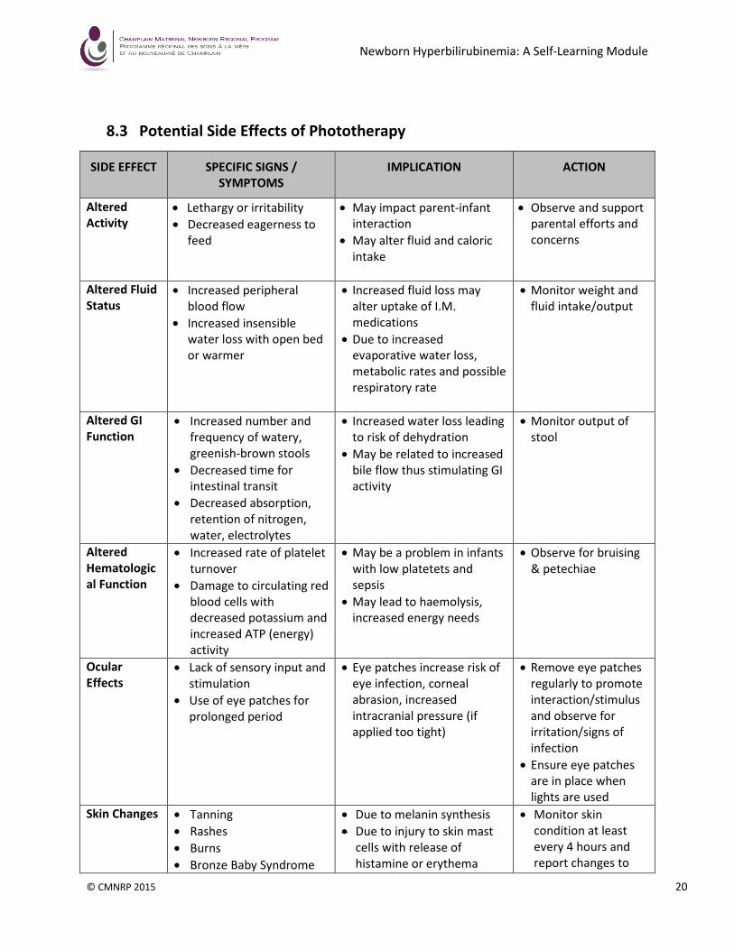
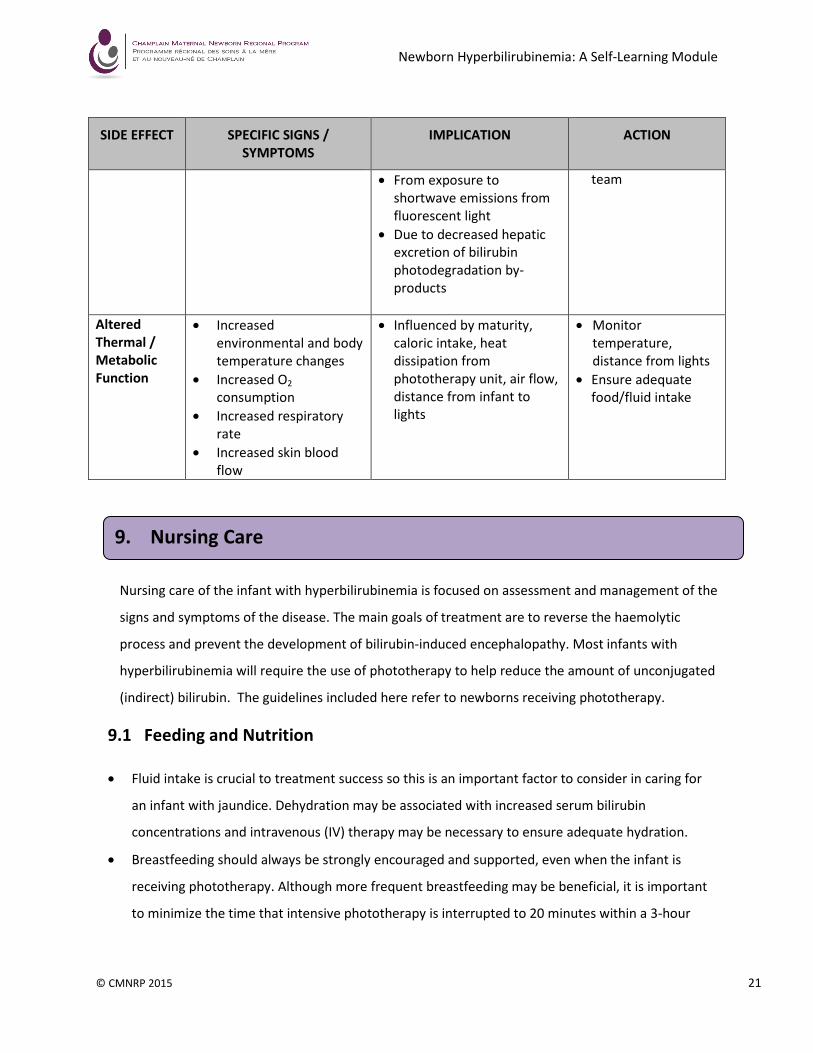
8.3 Potential Side Effects of Phototherapy
SIDE EFFECT SPECIFIC SIGNS /
SYMPTOMS
IMPLICATION ACTION
Altered
Activity
• Lethargy or irritability
• Decreased eagerness to
feed
• May impact parent-infant
interaction
• May alter fluid and caloric
intake
• Observe and support
parental efforts and
concerns
Altered Fluid
Status
• Increased peripheral
blood flow
• Increased insensible
water loss with open bed
or warmer
• Increased fluid loss may
alter uptake of I.M.
medications
• Due to increased
evaporative water loss,
metabolic rates and possible
respiratory rate
• Monitor weight and
fluid intake/output
Altered GI
Function
• Increased number and
frequency of watery,
greenish-brown stools
• Decreased time for
intestinal transit
• Decreased absorption,
retention of nitrogen,
water, electrolytes
• Increased water loss leading
to risk of dehydration
• May be related to increased
bile flow thus stimulating GI
activity
• Monitor output of
stool
Altered
Hematologic
al Function
• Increased rate of platelet
turnover
• Damage to circulating red
blood cells with
decreased potassium and
increased ATP (energy)
activity
• May be a problem in infants
with low platetets and
sepsis
• May lead to haemolysis,
increased energy needs
• Observe for bruising
& petechiae
Ocular
Effects
• Lack of sensory input and
stimulation
• Use of eye patches for
prolonged period
• Eye patches increase risk of
eye infection, corneal
abrasion, increased
intracranial pressure (if
applied too tight)
• Remove eye patches
regularly to promote
interaction/stimulus
and observe for
irritation/signs of
infection
• Ensure eye patches
are in place when
lights are used
Skin Changes • Tanning
• Rashes
• Burns
• Bronze Baby Syndrome
• Due to melanin synthesis
• Due to injury to skin mast
cells with release of
histamine or erythema
• Monitor skin
condition at least
every 4 hours and
report changes to
Newborn Hyperbilirubinemia: A Self-Learning Module
© CMNRP 2015 21
SIDE EFFECT SPECIFIC SIGNS /
SYMPTOMS
IMPLICATION ACTION
• From exposure to
shortwave emissions from
fluorescent light
• Due to decreased hepatic
excretion of bilirubin
photodegradation by-
products
team
Altered
Thermal /
Metabolic
Function
• Increased
environmental and body
temperature changes
• Increased O2
consumption
• Increased respiratory
rate
• Increased skin blood
flow
• Influenced by maturity,
caloric intake, heat
dissipation from
phototherapy unit, air flow,
distance from infant to
lights
• Monitor
temperature,
distance from lights
• Ensure adequate
food/fluid intake
Nursing care of the infant with hyperbilirubinemia is focused on assessment and management of the
signs and symptoms of the disease. The main goals of treatment are to reverse the haemolytic
process and prevent the development of bilirubin-induced encephalopathy. Most infants with
hyperbilirubinemia will require the use of phototherapy to help reduce the amount of unconjugated
(indirect) bilirubin. The guidelines included here refer to newborns receiving phototherapy.
9.1 Feeding and Nutrition
• Fluid intake is crucial to treatment success so this is an important factor to consider in caring for
an infant with jaundice. Dehydration may be associated with increased serum bilirubin
concentrations and intravenous (IV) therapy may be necessary to ensure adequate hydration.
• Breastfeeding should always be strongly encouraged and supported, even when the infant is
receiving phototherapy. Although more frequent breastfeeding may be beneficial, it is important
to minimize the time that intensive phototherapy is interrupted to 20 minutes within a 3-hour
9. Nursing Care
Newborn Hyperbilirubinemia: A Self-Learning Module
© CMNRP 2015 22
period. If possible, provide a referral to a Lactation Consultant for a more specific assessment of
breastfeeding.
• Accurate recording of intake and output, including stool pattern, is necessary. If the infant is
receiving IV therapy, usual standards of care should be followed.
9.2 Skin Care and Thermoregulation
• Skin plays an important role in the regulation of body temperature and serves as a route of water
excretion.
• Skin care includes observing colour, rashes and excoriation; cleaning the skin with warm water,
especially the perineal area after stooling; changing position every 2 hours. NOTE: Prompt
cleansing of perianal area after stooling is important as photo-oxidation of bilirubin results in
loose green caustic stools and can result in excoriation.
• Phototherapy is most effective when the exposed skin area is maximized, so infants in isolettes
should be exposed as much as possible; otherwise, diapers to catch urine and stool should be
used.
• The infant’s temperature must be monitored at least every 4 hours to maintain a safe
environment.
9.3 Eye Care
• The infant’s eyes must be protected from the phototherapy lights to prevent retinal damage.
Place the pads correctly over the eyes:
o Use the appropriate size of the eye pad or prefabricated eye protection.
o Make sure the eyes are closed first to help prevent corneal abrasion, irritation and/or
infection
o Check frequently to make sure the eye pads remain in place and are not obstructing the
nares.
• Eye protection should be removed every 2 to 4 hours and eyes cleansed with normal saline to
reduce irritation and promote eye contact, socialization and attachment.
NOTE: Eye patches are not necessary if only the Bili Blanket is being used since the infant’s eyes are not
directly exposed to the blue lights.
Newborn Hyperbilirubinemia: A Self-Learning Module
© CMNRP 2015 23
American Academy of Pediatrics. (2004). Management of hyperbilirubinemia in the newborn infant 35
or more weeks of gestation. Clinical Practice Guideline. Pediatrics, 114(4), 297–316.
AWHONN. (2005). Hyperbilirubinemia in the neonate: Risk assessment, screening and management
(2nd
Edition). Washington: Author.
Bhutani, V.K., Johnson, L., & Sivieri, E. (1999). Predictive ability of a predischarge hour-specific serum
bilirubin for subsequent hyperbilirubinemia in healthy term and near-term newborns.
Pediatrics, 103(1), 6-14.
Canadian Paediatric Society - Fetus and Newborn Committee. (updated 2011). Guidelines for
detection, management and prevention of hyperbilirubinemia in term and late preterm newborn
infants (35 or more weeks’ gestation). Retrieved from
http://www.cps.ca/en/documents/position/hyperbilirubinemia-newborn
Champlain Maternal Newborn Regional Program. (2012). Hyperbilirubinemia Toolkit. Retrieved from
http://www.cmnrp.ca/en/cmnrp/Hyperbilirubinemia_Toolkit_p3951.html
Cohen, S.M. (2006). Jaundice in the full-term newborn. Pediatric Nursing, 32(3), 202-208.
Hockenberry, M. (2005). Hyperbilirubinemia. In Wong’s Essentials of Pedioatric Nursing, 7th
Ed. St.
Louis: Elsevier-Mosby, 262–268.
Maisels, M.J., Bhutani, V.K., Bogen, D., Newman, T.B., Starl, A.R., & Watchko, J.F. (2009).
Hyperbilirubinemia in the newborn infant ≥ 35 weeks’ gestation: An update with clarifications.
Pediatrics, 124(4), 1193-1198.
Maisels, M.J., & McDonagh, A.F. (2008). Phototherapy for jaundice. New England Journal of Medicine,
358, 920-928.
Provincial Council for Maternal and Child Health. (2014). Quality-based procedure. Hyperbilirubinemia
in term and late pre-term infants (≥ 35 weeks) toolkit. Retrieved from
http://www.pcmch.on.ca/initiatives/qbp-hyperbilirubinemia-term-and-late-pre-term-infants
Sgro, M., Campbell, D., & Shah, V. (2006). Incidence and causes of severe neonatal hyperbilirubinemia
in Canada. Canadian Medical Association Journal, 175(6), 587–90.
Stevenson, D.K. et al. (2001). Prediction of hyperbilirubinemia in near-term and term infants.
Pediatrics, 108, 31-39.
Stokowski, L. (2011). Fundamentals of phototherapy for neonatal jaundice. Advances in Neonatal
Care, 11(5S), S10-S21.
Zettle, R. (2006). Neonatal hyperbilirubinemia and the prevention of kernicterus. The HIROC
Connection, 9(1), 7–11.
10. References and Resources
Newborn Hyperbilirubinemia: A Self-Learning Module
© CMNRP 2015 24
The Champlain Maternal Newborn Regional Program (CMNRP) would like to thank the
members of the Joint Orientation Sub-Committee for their work on the development of the
Newborn Hyperbilirubinemia Self-Learning Module.
CMNRP also acknowledges the work of the following health care professionals who have
provided feedback and expertise:
� Members of the Interprofessional Education & Research Committee (IERC)
� Members of the Jaundice Working Group
� Pediatricians and Neonatologists
� CMNRP Perinatal Consultants and Neonatal Nurse Practitioners
Acknowledgments
Top Related