


Languages
Pages
Legal


Acute Pancreatitis
University Hospital Berne, Clinic for Visceral Surgery and Medicine
Jan Hendrik
Niess

How do you diagnose acute pancreatitis (AP)?
Presence of two of the following three criteria
- abdominal pain consistent with disease
- lipase or serum amylase greater than three times
upper the normal limit
- characteristic findings of abdominal imaging

- renal diseases
- appendicitis
- cholecystitis
- diabetics
When is the lipase false positive ?

What abdominal imaging modality needs to be
performed in early AP?
- Transabdominal ultrasound
- Search for biliary aetiology
- needs to be performed on admission (since sludge can develop
during AP evolution)
- sensitivity for GB lithiasis 95%
- sensitivity for CBD lithiasis 30-80%
- (pancreatic swelling only in 25 – 50%; bowel gases may mask the
pancreatic region)

What problems does CE-CT have at early AP?
- High sensitivity (90%) and specificity for AP
- In most cases AP can be diagnosed without imaging modality
- Has a low sensitivity and specificity for the detection of lithiasis
- Necrosis are not marked at early time points (prior to 48 to 72h)

When should CE-CT carried out?
In patients not improving with 72 hours (persistent pain, fever, nausea,
unable to begin oral feeding) to assess local complications

What is the aetiology of AP?
• Gallstones (40-70%)
• Alcohol (25-35%)
• Medications (http://www.mucosalimmunology.ch/images/content/PPT-presentations_free_access/bible_class/Drug-inducedpancreatitis.pdf)
• (i.e. azathioprine, furosemide, valproic acid)
• Primary and secondary hypertriglyceridemia ( > 1,000 mg/dl)
• Hypercalcemia and hyperparathyroidism
• Autoimmune
• Infectious (coxackie, mumps, CMV, HSV, HAC, HBV, HCV)
• Post -ERCP
• SPINK, PRSS1, CFTR mutations
• Congenital anomaly of the pancreas (Pancreas divisum)
• 5-14% pf patients with a benign or malign mass present with AP

Work-up of patients with AP?
- Ultrasound at admission
- Lab tests
- Ca2+
- ALT, AST, Bili g-GT
- Fasting TG (if > 1000 mg/dl, repeat 4 weeks after resolution of AP)
- Immunoglobulins
- Consider genetic testing in young patients (< 30 years) and with a
positive family history of pancreatic diseases.
- Consider anomalies of the pancreas
- Consider in patients > 40 years a benign or maligne mass as cause
for the pancreatitis
- Abdominal imaging after resolution of pancreatitis

How is the severity of AP defined?
Note:
dynamics
of OF
Am J Gastroenterol. 2013 Sep;108(9):1400-15

How can severe AP predicted?
- There is no lab parameter, which predicts severe AP
- AP-specific scoring systems (BISAP, APACHE etc.) are of importance for clinical
studies, but have limited value within the first 48 hours of AP
- Clinical assessment is most important in monitoring AP patients
Patient characteristics
Age > 55 years
Obesity (BMI > 30 kg/m2)
Altered mental status
Comorbidities

How can severe AP predicted?
The systemic inflammatory response syndrome (SIRS)
Pulls > 90 beats/min
Respiration > 20/min
Temperature <36; > 38
Laboratory findings
blood urea nitrogen (BNU) > 20 mg/dl
Haematocrit > 44
Radiology findings
pleural effusions
pulmonary infiltrates
Multiple or extrapancreatic collections
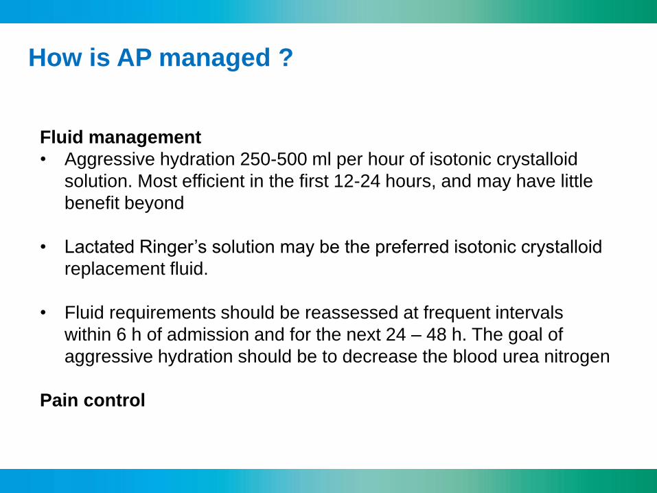
How is AP managed ?
Fluid management
• Aggressive hydration 250-500 ml per hour of isotonic crystalloid
solution. Most efficient in the first 12-24 hours, and may have little
benefit beyond
• Lactated Ringer’s solution may be the preferred isotonic crystalloid
replacement fluid.
• Fluid requirements should be reassessed at frequent intervals
within 6 h of admission and for the next 24 – 48 h. The goal of
aggressive hydration should be to decrease the blood urea nitrogen
Pain control

When should ERCP performed?
Am J Gastroenterol. 2013
Sep;108(9):1400-15

What are risk factors for stones ?
N Engl J Med. 2014 May 15;370(20):1955

How should antibiotics used ?
Consider
Mortality
Sterile necrosis 10%
Infected necrosis 30%
but it takes approximately 7 to 10 days before necrosis will become infected
This means
- Antibiotics should be given for the treatment of extrapancreatic infections
(cholangitis, pneumonia, urinary tract infections ……..).
- Routine use of prophylactic antibiotics in patients with severe
AP is not recommended (any longer)
- There are only few antibiotics that penetrate into necrotic regions
(carbapenems, quinolones, metronidazole and 4th generation cephalosporins)
- Consider the development of pancreatitis for the choice of the antibiotic used
for the treatment of extrapancreatic infections in early AP

How should nutrition be managed in AP patients?
N Engl J Med. 2014 Nov 20;371(21):1983-93
• Intravenous fluids in the first 72 hours
If patients actively asks for food an oral diet can be offered
• Start on demand oral diet after 72 hours
• If an oral diet is not tolerated within 96 hours after onset of pancreatitis,
then place a naso-enteral feeding tube for nutrition
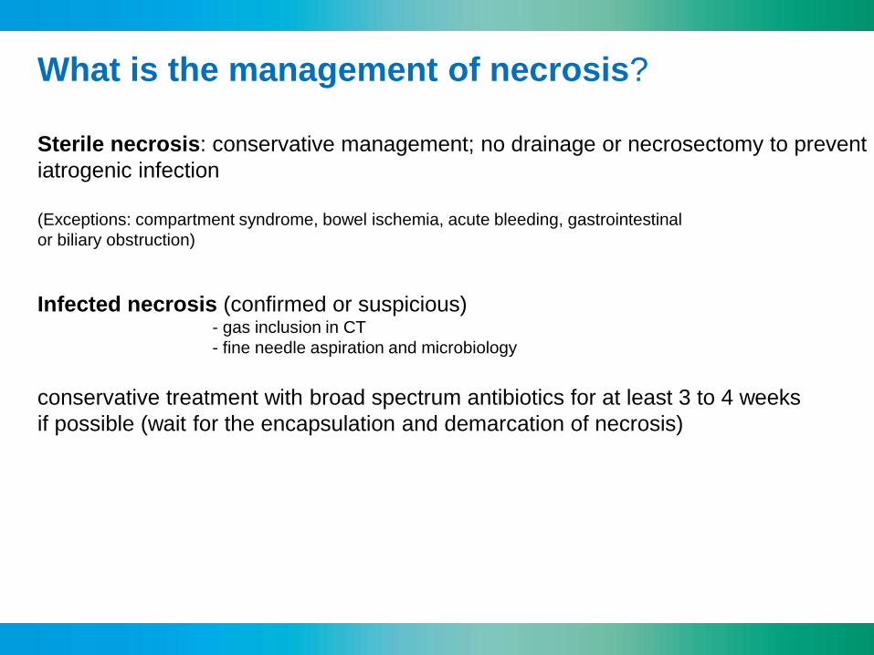
What is the management of necrosis?
Sterile necrosis: conservative management; no drainage or necrosectomy to prevent
iatrogenic infection
(Exceptions: compartment syndrome, bowel ischemia, acute bleeding, gastrointestinal
or biliary obstruction)
Infected necrosis (confirmed or suspicious) - gas inclusion in CT
- fine needle aspiration and microbiology
conservative treatment with broad spectrum antibiotics for at least 3 to 4 weeks
if possible (wait for the encapsulation and demarcation of necrosis)

Invasive approaches for the treatment of
infected necrosis?
• Percutaneous catheter drainage
• Endoscopic transluminal drainage
• Minimally invasive retroperitoneal necrosectomy
• Minimally invasive laparoscopic necrosectomy
• Endoscopic (transluminal) necrosectomy
• Open necrosectomy

CLINICAL GASTROENTEROLOGY AND HEPATOLOGY 2012;10:1190–1201
Minimale invasive retroperitoneal necrosectomy

Endoscopic transluminal drainage and necrosectomy
CLINICAL GASTROENTEROLOGY
AND HEPATOLOGY 2012;10:1190–1201
Axios
Stent System
Gastrointestinal Endoscopy
2012; 75:870–876

CLINICAL GASTROENTEROLOGY
AND HEPATOLOGY 2012;10:1190–
1201
Summary – Management of acute pancreatitis
Top Related