


Languages
Pages
Legal

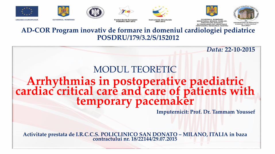
AD-COR Program inovativ de formare in domeniul cardiologiei pediatricePOSDRU/179/3.2/S/152012
Data: 22-10-2015
MODUL TEORETIC
Arrhythmias in postoperative paediatriccardiac critical care and care of patients with
temporary pacemakerImputernicit: Prof. Dr. Tammam Youssef
Activitate prestata de I.R.C.C.S. POLICLINICO SAN DONATO – MILANO, ITALIA in baza contractului nr. 18/22144/29.07.2015
Acest material a fost documentat/ validat/ prezentat la sesiunile de formare în
cadrul proiectului „AD-COR Program inovativ de formare în domeniul cardiologiei
pediatrice” - POSDRU/179/3.2/S/152012, proiect cofinanțat din Fondul Social
Operațional Sectorial Dezvoltarea Resurselor Umane 2007-2013.
Beneficiar: Universitatea de Medicină și Farmacie „Carol Davila” București
Conținutul acestui material nu reprezintă în mod obligatoriu poziția oficială a Uniunii Europene sau a Guvernului României
RobinAid Foundation Angrés M 10_2015 Bucharest Marie Curie Children‘s Hospital
AD-COR Program inovativ de formare in domeniul cardiologiei pediatricePOSDRU/179/3.2/S/152012
Lecture II
Bucharest, October 22, 2015
Matthias Angrés, MD, PhD
RobinAid Foundation
Hamburg / Germany
RobinAid Foundation Angrés M 10_2015 Bucharest Marie Curie Children‘s Hospital
Let‘s continue with the next topic
Arrhythmias in postoperative paediatric
cardiac critical care and care of patients
with temporary pacemaker
RobinAid Foundation Angrés M 10_2015 Bucharest Marie Curie Children‘s Hospital
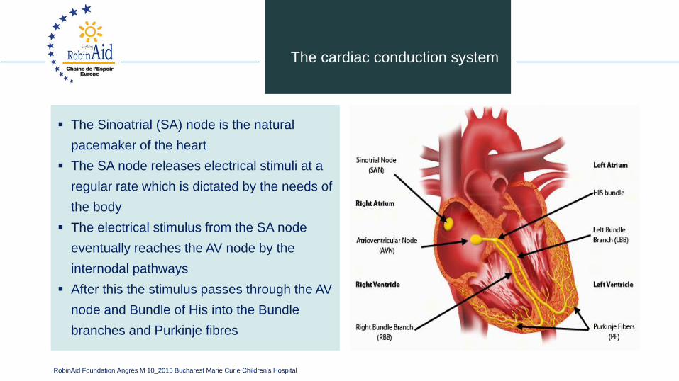
The cardiac conduction system
▪ The Sinoatrial (SA) node is the natural
pacemaker of the heart
▪ The SA node releases electrical stimuli at a
regular rate which is dictated by the needs of
the body
▪ The electrical stimulus from the SA node
eventually reaches the AV node by the
internodal pathways
▪ After this the stimulus passes through the AV
node and Bundle of His into the Bundle
branches and Purkinje fibres
RobinAid Foundation Angrés M 10_2015 Bucharest Marie Curie Children‘s Hospital
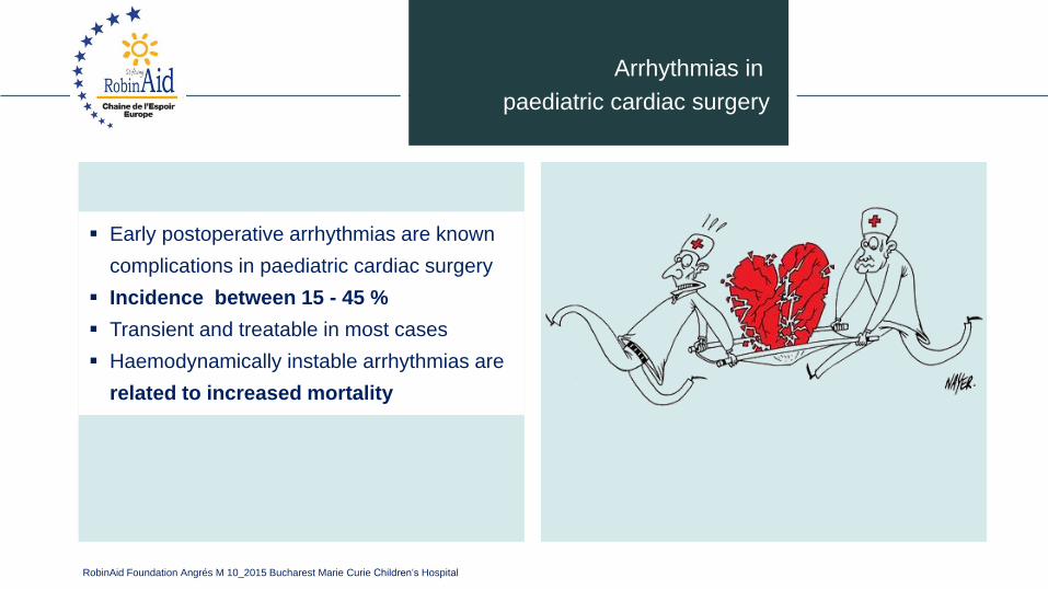
▪ Early postoperative arrhythmias are known
complications in paediatric cardiac surgery
▪ Incidence between 15 - 45 %
▪ Transient and treatable in most cases
▪ Haemodynamically instable arrhythmias are
related to increased mortality
Arrhythmias in
paediatric cardiac surgery
RobinAid Foundation Angrés M 10_2015 Bucharest Marie Curie Children‘s Hospital
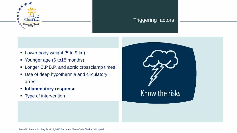
Triggering factors
▪ Lower body weight (5 to 9 kg)
▪ Younger age (6 to18 months)
▪ Longer C.P.B.P. and aortic crossclamp times
▪ Use of deep hypothermia and circulatory
arrest
▪ Inflammatory response
▪ Type of intervention
RobinAid Foundation Angrés M 10_2015 Bucharest Marie Curie Children‘s Hospital
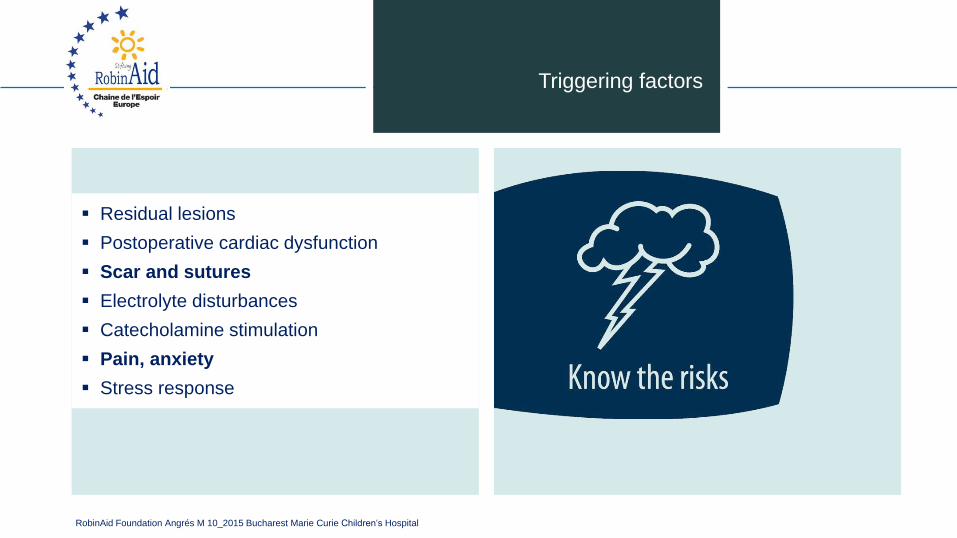
Triggering factors
▪ Residual lesions
▪ Postoperative cardiac dysfunction
▪ Scar and sutures
▪ Electrolyte disturbances
▪ Catecholamine stimulation
▪ Pain, anxiety
▪ Stress response
RobinAid Foundation Angrés M 10_2015 Bucharest Marie Curie Children‘s Hospital
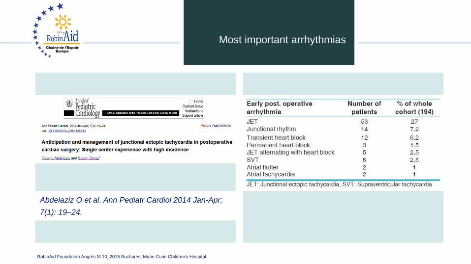
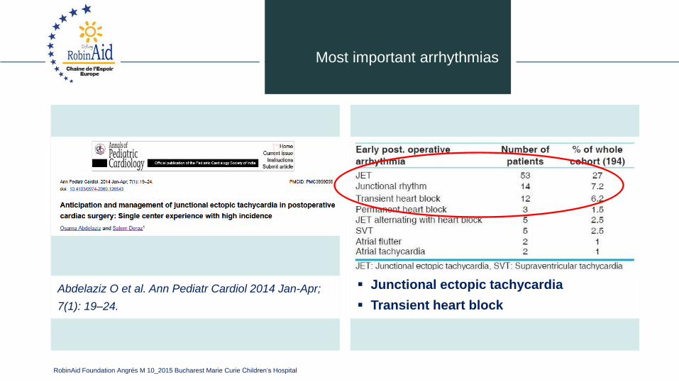
Abdelaziz O et al. Ann Pediatr Cardiol 2014 Jan-Apr;
7(1): 19–24.
Most important arrhythmias
RobinAid Foundation Angrés M 10_2015 Bucharest Marie Curie Children‘s Hospital
Abdelaziz O et al. Ann Pediatr Cardiol 2014 Jan-Apr;
7(1): 19–24.
Most important arrhythmias
▪ Junctional ectopic tachycardia
▪ Transient heart block
RobinAid Foundation Angrés M 10_2015 Bucharest Marie Curie Children‘s Hospital
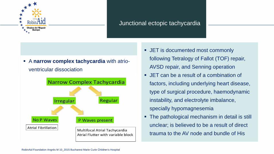
Junctional ectopic tachycardia
▪ A narrow complex tachycardia with atrio-
ventricular dissociation
▪ JET is documented most commonly
following Tetralogy of Fallot (TOF) repair,
AVSD repair, and Senning operation
▪ JET can be a result of a combination of
factors, including underlying heart disease,
type of surgical procedure, haemodynamic
instability, and electrolyte imbalance,
specially hypomagnesemia
▪ The pathological mechanism in detail is still
unclear; is believed to be a result of direct
trauma to the AV node and bundle of His
RobinAid Foundation Angrés M 10_2015 Bucharest Marie Curie Children‘s Hospital
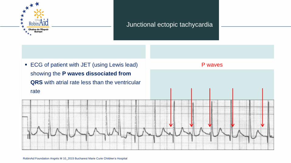
Junctional ectopic tachycardia
▪ ECG of patient with JET (using Lewis lead)
showing the P waves dissociated from
QRS with atrial rate less than the ventricular
rate
P waves
RobinAid Foundation Angrés M 10_2015 Bucharest Marie Curie Children‘s Hospital
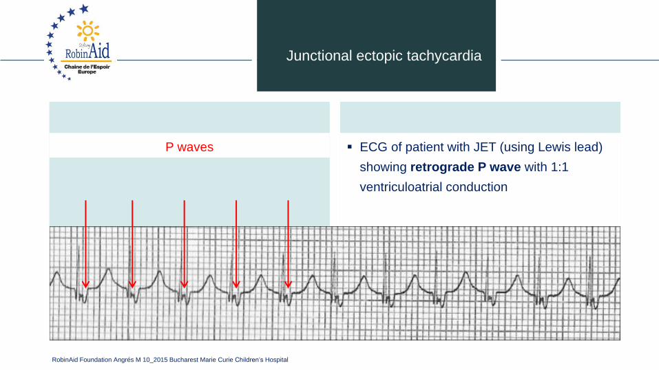
▪ ECG of patient with JET (using Lewis lead)
showing retrograde P wave with 1:1
ventriculoatrial conduction
Junctional ectopic tachycardia
P waves
RobinAid Foundation Angrés M 10_2015 Bucharest Marie Curie Children‘s Hospital
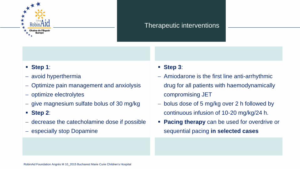
▪ Step 1:
avoid hyperthermia
Optimize pain management and anxiolysis
optimize electrolytes
give magnesium sulfate bolus of 30 mg/kg
▪ Step 2:
decrease the catecholamine dose if possible
especially stop Dopamine
▪ Step 3:
Amiodarone is the first line anti-arrhythmic
drug for all patients with haemodynamically
compromising JET
bolus dose of 5 mg/kg over 2 h followed by
continuous infusion of 10-20 mg/kg/24 h.
▪ Pacing therapy can be used for overdrive or
sequential pacing in selected cases
Therapeutic interventions
RobinAid Foundation Angrés M 10_2015 Bucharest Marie Curie Children‘s Hospital
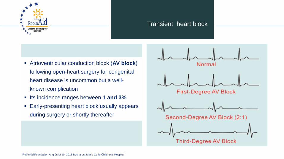
▪ Atrioventricular conduction block (AV block)
following open-heart surgery for congenital
heart disease is uncommon but a well-
known complication
▪ Its incidence ranges between 1 and 3%
▪ Early-presenting heart block usually appears
during surgery or shortly thereafter
Transient heart block
RobinAid Foundation Angrés M 10_2015 Bucharest Marie Curie Children‘s Hospital
▪ The greatest risk for complete AV block is
associated with corrective surgical
procedures for:
Transient heart block
ventricular septal defects (VSD), usually as
part of more complex congenital heart
disease
atrioventricular septal defects (AVSD),
left ventricular outflow tract obstruction
L-transposition of the great arteries
Tetralogy of Fallot (TOF)
discordant atrioventricular connections
RobinAid Foundation Angrés M 10_2015 Bucharest Marie Curie Children‘s Hospital
Transient heart block
▪ Most children who require pacing for
surgically induced AV block are less than 1
year old
▪ Spontaneous resolution of complete AV
block in the early postoperative period most
often occurring between 7 and 14 days
▪ Complete AV block may resolve
spontaneously in 43 - 92% of children
(Gross et al. 2006)
▪ Factors associated with a spontaneous
recovery of AV nodal function are currently
not known
RobinAid Foundation Angrés M 10_2015 Bucharest Marie Curie Children‘s Hospital
▪ Atrial and ventricular pacing wires are
essentials in every open heart surgery
▪ Having a sequential pacemaker available,
ready, and knowing how it functioned are
also basic essentials in paediatric cardiac
critical care
Therapeutic interventions
RobinAid Foundation Angrés M 10_2015 Bucharest Marie Curie Children‘s Hospital
Temporary pacemakers
▪ Objectives:
explain the situations when temporary
pacemakers are indicated
describe the principles of pacing
illustrate normal and abnormal pacemaker
behavior
discuss the steps to be taken in
troubleshooting a temporary pacemaker
RobinAid Foundation Angrés M 10_2015 Bucharest Marie Curie Children‘s Hospital
Indications for temporary pacing
▪ Bradyarrhythmias
▪ AV conduction block
▪ Slow sinus or junctional rhythm
▪ Suppression of ectopy
▪ Drugs, electrolyte imbalances (Sick Sinus
Syndrome)
▪ Secondary to pronounced atrial stretch
▪ Old TGA s/p Senning or Mustard procedure
RobinAid Foundation Angrés M 10_2015 Bucharest Marie Curie Children‘s Hospital
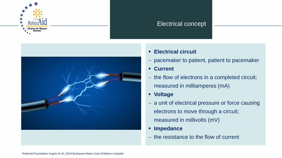
Electrical concept
▪ Electrical circuit
pacemaker to patient, patient to pacemaker
▪ Current
the flow of electrons in a completed circuit;
measured in milliamperes (mA)
▪ Voltage
a unit of electrical pressure or force causing
electrons to move through a circuit;
measured in millivolts (mV)
▪ Impedance
the resistance to the flow of current
RobinAid Foundation Angrés M 10_2015 Bucharest Marie Curie Children‘s Hospital
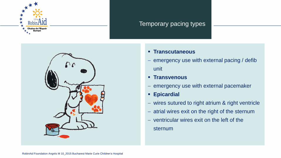
Temporary pacing types
▪ Transcutaneous
emergency use with external pacing / defib
unit
▪ Transvenous
emergency use with external pacemaker
▪ Epicardial
wires sutured to right atrium & right ventricle
atrial wires exit on the right of the sternum
ventricular wires exit on the left of the
sternum
RobinAid Foundation Angrés M 10_2015 Bucharest Marie Curie Children‘s Hospital
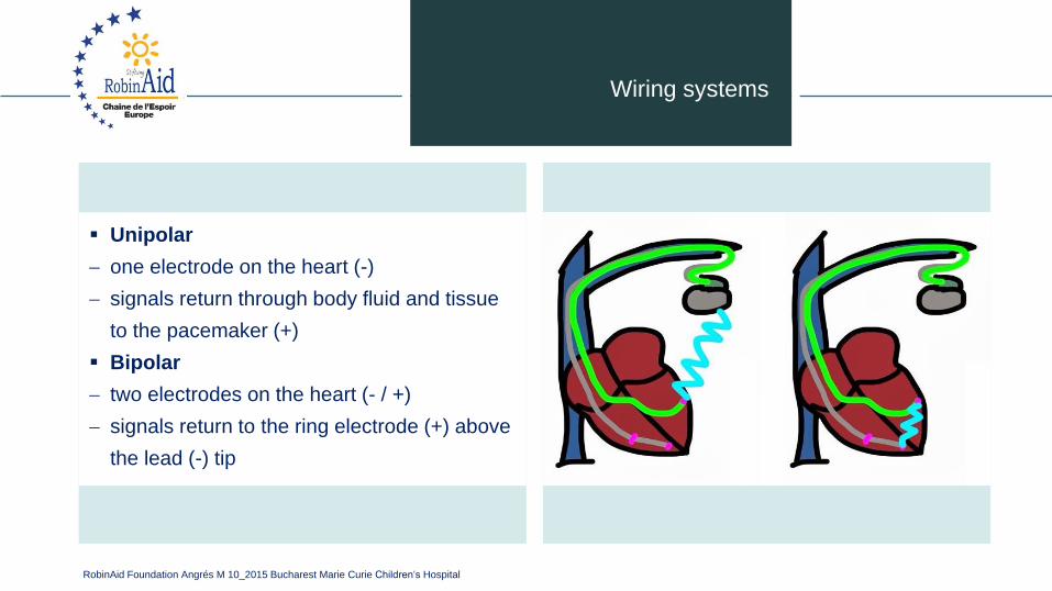
Wiring systems
▪ Unipolar
one electrode on the heart (-)
signals return through body fluid and tissue
to the pacemaker (+)
▪ Bipolar
two electrodes on the heart (- / +)
signals return to the ring electrode (+) above
the lead (-) tip
RobinAid Foundation Angrés M 10_2015 Bucharest Marie Curie Children‘s Hospital
Overview of terminology
RobinAid Foundation Angrés M 10_2015 Bucharest Marie Curie Children‘s Hospital
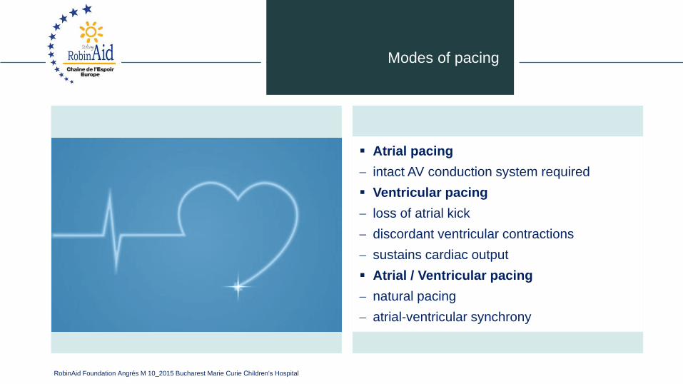
Modes of pacing
▪ Atrial pacing
intact AV conduction system required
▪ Ventricular pacing
loss of atrial kick
discordant ventricular contractions
sustains cardiac output
▪ Atrial / Ventricular pacing
natural pacing
atrial-ventricular synchrony
RobinAid Foundation Angrés M 10_2015 Bucharest Marie Curie Children‘s Hospital
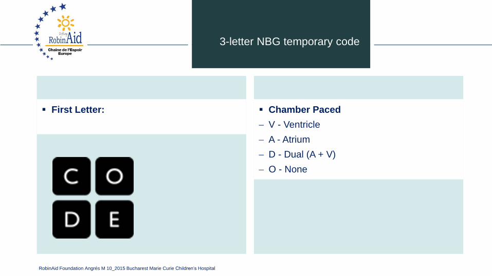
▪ First Letter: ▪ Chamber Paced
V - Ventricle
A - Atrium
D - Dual (A + V)
O - None
3-letter NBG temporary code

RobinAid Foundation Angrés M 10_2015 Bucharest Marie Curie Children‘s Hospital
▪ Chamber Sensed
V - Ventricle
A - Atrium
D - Dual (A & V)
O - None
▪ Second letter:
3-letter NBG temporary code
RobinAid Foundation Angrés M 10_2015 Bucharest Marie Curie Children‘s Hospital
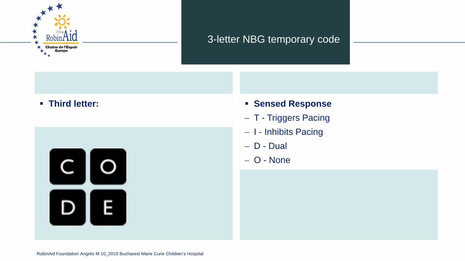
▪ Sensed Response
T - Triggers Pacing
I - Inhibits Pacing
D - Dual
O - None
▪ Third letter:
3-letter NBG temporary code
RobinAid Foundation Angrés M 10_2015 Bucharest Marie Curie Children‘s Hospital
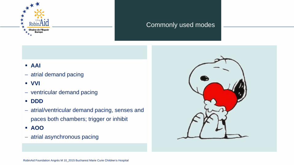
Commonly used modes
▪ AAI
atrial demand pacing
▪ VVI
ventricular demand pacing
▪ DDD
atrial/ventricular demand pacing, senses and
paces both chambers; trigger or inhibit
▪ AOO
atrial asynchronous pacing
RobinAid Foundation Angrés M 10_2015 Bucharest Marie Curie Children‘s Hospital
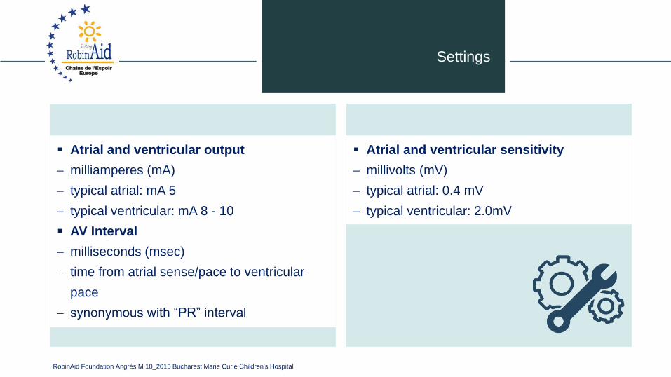
Settings
▪ Atrial and ventricular sensitivity
millivolts (mV)
typical atrial: 0.4 mV
typical ventricular: 2.0mV
▪ Atrial and ventricular output
milliamperes (mA)
typical atrial: mA 5
typical ventricular: mA 8 - 10
▪ AV Interval
milliseconds (msec)
time from atrial sense/pace to ventricular
pace
synonymous with “PR” interval
RobinAid Foundation Angrés M 10_2015 Bucharest Marie Curie Children‘s Hospital
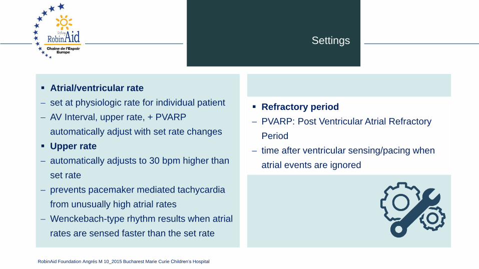
▪ Refractory period
PVARP: Post Ventricular Atrial Refractory
Period
time after ventricular sensing/pacing when
atrial events are ignored
▪ Atrial/ventricular rate
set at physiologic rate for individual patient
AV Interval, upper rate, + PVARP
automatically adjust with set rate changes
▪ Upper rate
automatically adjusts to 30 bpm higher than
set rate
prevents pacemaker mediated tachycardia
from unusually high atrial rates
Wenckebach-type rhythm results when atrial
rates are sensed faster than the set rate
Settings
RobinAid Foundation Angrés M 10_2015 Bucharest Marie Curie Children‘s Hospital
Very important
▪ Pacemaker care & cleaning
batteries
bridging cables
pacemakers
▪ Electrical Safety
microshock
accidental de-wiring
taping wires
securing pacemaker
▪ Removal of pacing wires
potential myocardial trauma
bleeding
pericardial effusion / tamponade
haemothorax
ventricular arrhythmias
RobinAid Foundation Angrés M 10_2015 Bucharest Marie Curie Children‘s Hospital
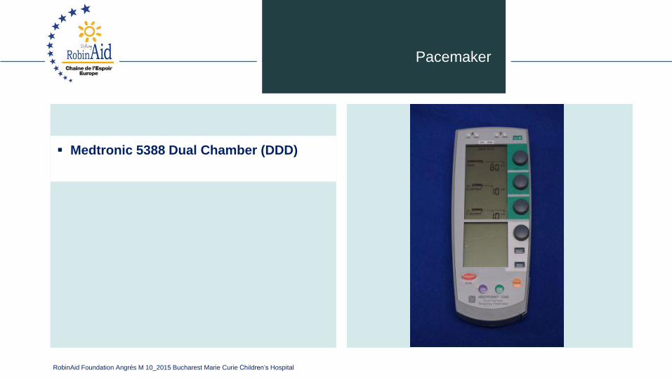
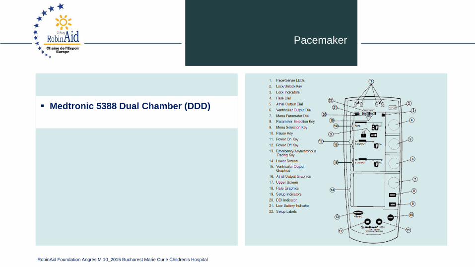
Pacemaker
▪ Medtronic 5388 Dual Chamber (DDD)
RobinAid Foundation Angrés M 10_2015 Bucharest Marie Curie Children‘s Hospital
Pacemaker
▪ Medtronic 5388 Dual Chamber (DDD)
RobinAid Foundation Angrés M 10_2015 Bucharest Marie Curie Children‘s Hospital
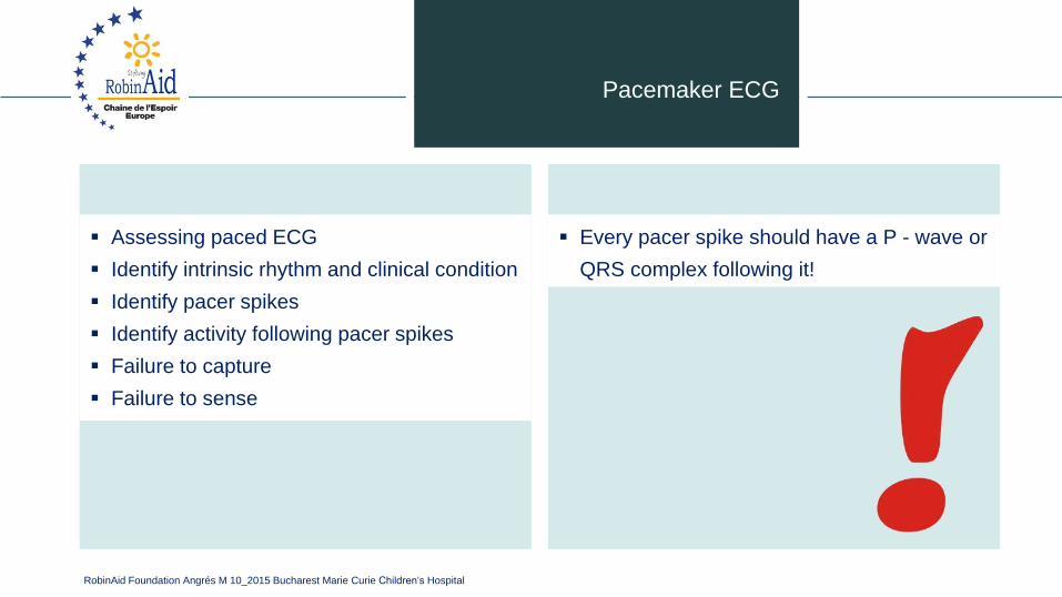
Pacemaker ECG
▪ Every pacer spike should have a P - wave or
QRS complex following it!
▪ Assessing paced ECG
▪ Identify intrinsic rhythm and clinical condition
▪ Identify pacer spikes
▪ Identify activity following pacer spikes
▪ Failure to capture
▪ Failure to sense
RobinAid Foundation Angrés M 10_2015 Bucharest Marie Curie Children‘s Hospital
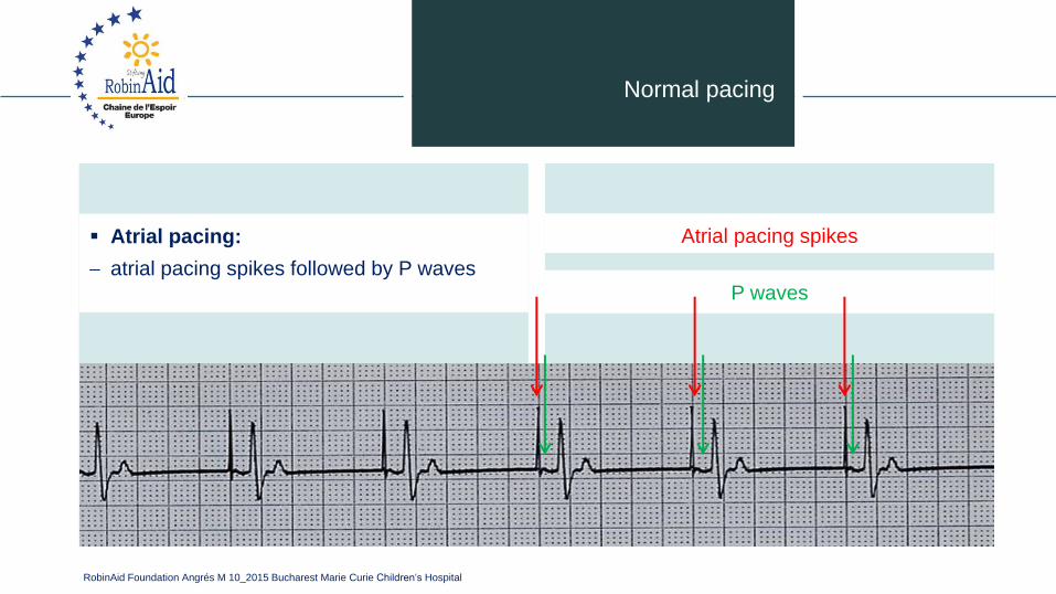
P waves
Normal pacing
▪ Atrial pacing:
atrial pacing spikes followed by P waves
Atrial pacing spikes
RobinAid Foundation Angrés M 10_2015 Bucharest Marie Curie Children‘s Hospital
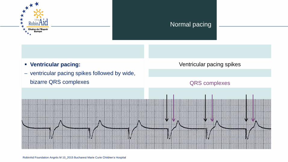
QRS complexes
Normal pacing
▪ Ventricular pacing:
ventricular pacing spikes followed by wide,
bizarre QRS complexes
Ventricular pacing spikes
RobinAid Foundation Angrés M 10_2015 Bucharest Marie Curie Children‘s Hospital
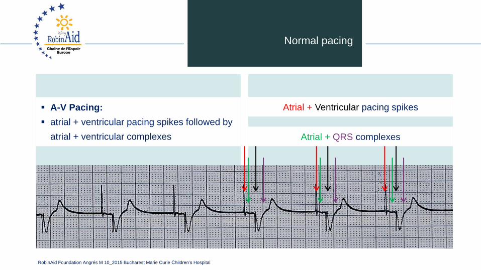
Atrial + QRS complexes
Normal pacing
▪ A-V Pacing:
▪ atrial + ventricular pacing spikes followed by
atrial + ventricular complexes
Atrial + Ventricular pacing spikes
RobinAid Foundation Angrés M 10_2015 Bucharest Marie Curie Children‘s Hospital
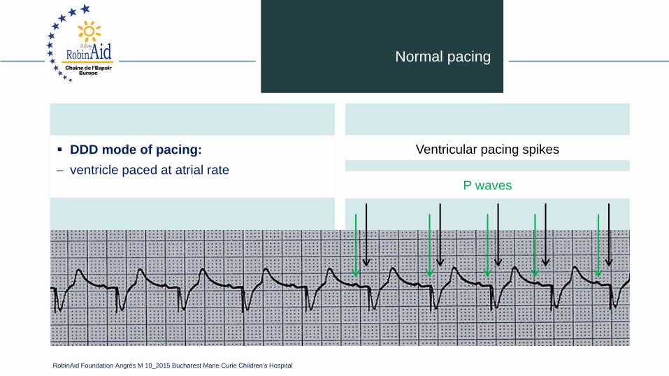
Normal pacing
▪ DDD mode of pacing:
ventricle paced at atrial rate
Ventricular pacing spikes
P waves
RobinAid Foundation Angrés M 10_2015 Bucharest Marie Curie Children‘s Hospital
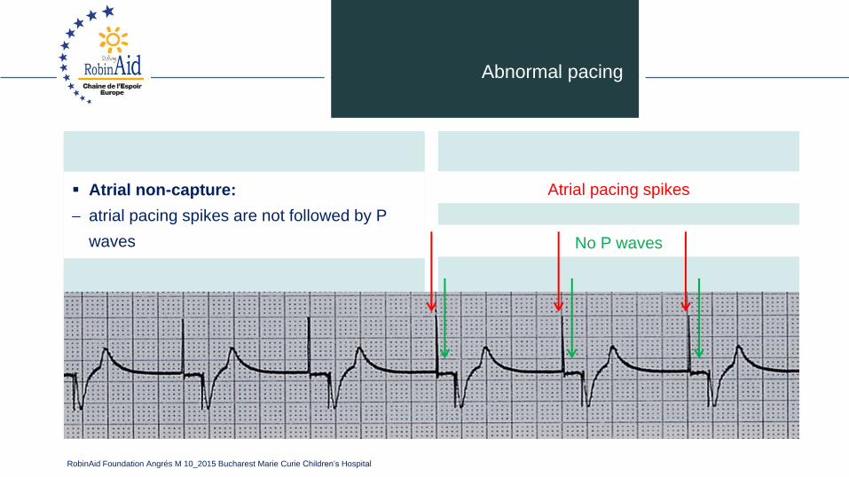
Abnormal pacing
▪ Atrial non-capture:
atrial pacing spikes are not followed by P
waves No P waves
Atrial pacing spikes
RobinAid Foundation Angrés M 10_2015 Bucharest Marie Curie Children‘s Hospital
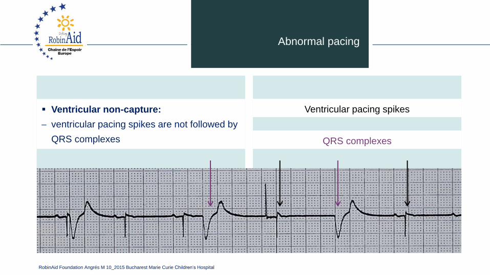
Abnormal pacing
▪ Ventricular non-capture:
ventricular pacing spikes are not followed by
QRS complexes
Ventricular pacing spikes
QRS complexes
RobinAid Foundation Angrés M 10_2015 Bucharest Marie Curie Children‘s Hospital
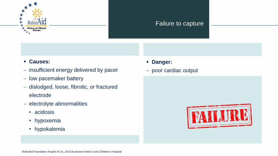
Failure to capture
▪ Causes:
insufficient energy delivered by pacer
low pacemaker battery
dislodged, loose, fibrotic, or fractured
electrode
electrolyte abnormalities
• acidosis
• hypoxemia
• hypokalemia
▪ Danger:
poor cardiac output
RobinAid Foundation Angrés M 10_2015 Bucharest Marie Curie Children‘s Hospital
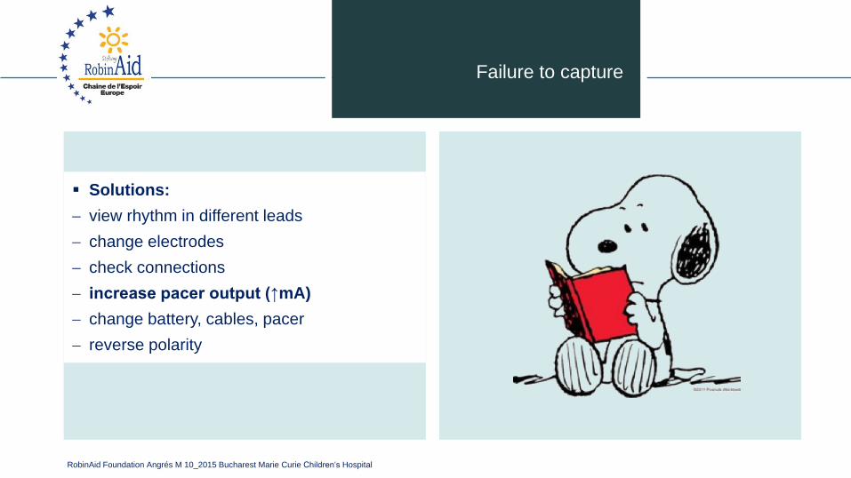
Failure to capture
▪ Solutions:
view rhythm in different leads
change electrodes
check connections
increase pacer output (↑mA)
change battery, cables, pacer
reverse polarity
RobinAid Foundation Angrés M 10_2015 Bucharest Marie Curie Children‘s Hospital
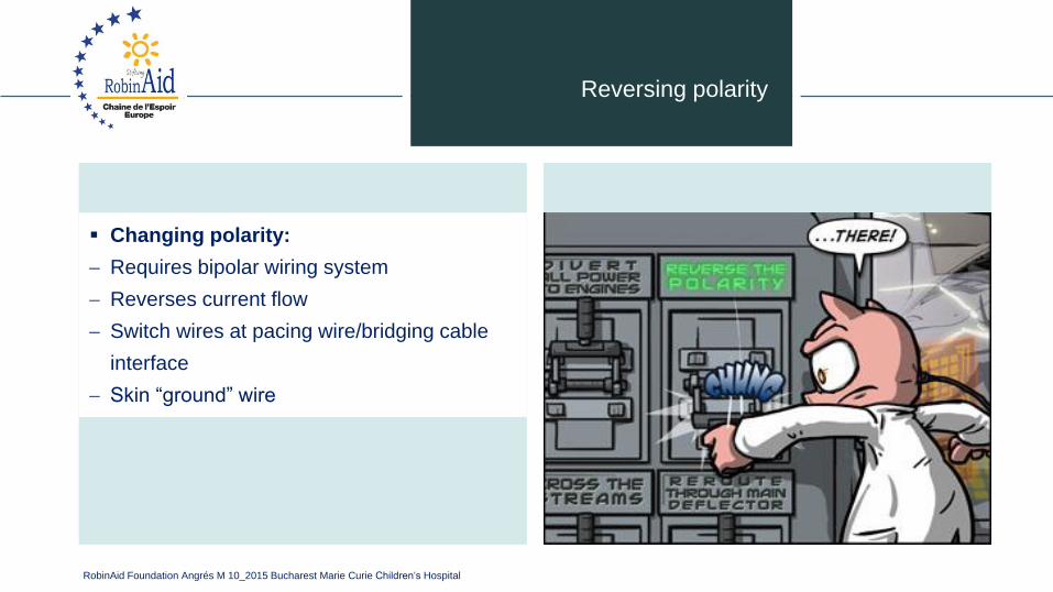
Reversing polarity
▪ Changing polarity:
Requires bipolar wiring system
Reverses current flow
Switch wires at pacing wire/bridging cable
interface
Skin “ground” wire
RobinAid Foundation Angrés M 10_2015 Bucharest Marie Curie Children‘s Hospital
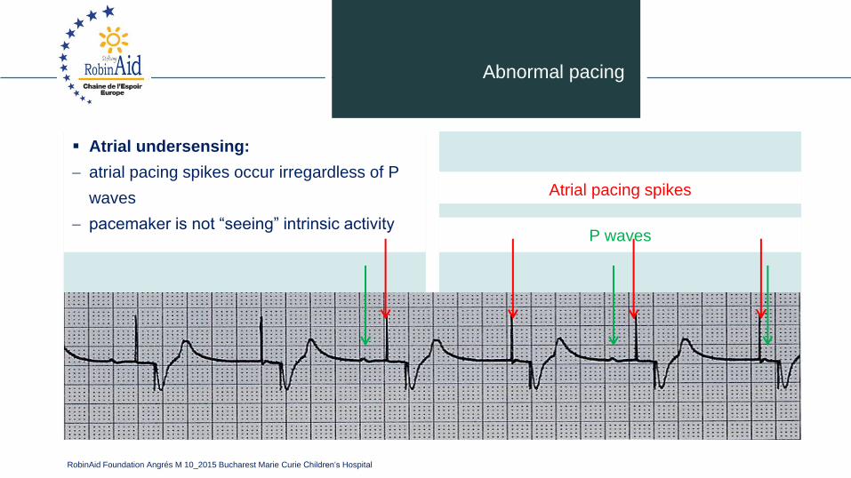
Abnormal pacing
▪ Atrial undersensing:
atrial pacing spikes occur irregardless of P
waves
pacemaker is not “seeing” intrinsic activityP waves
Atrial pacing spikes
RobinAid Foundation Angrés M 10_2015 Bucharest Marie Curie Children‘s Hospital
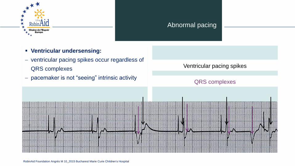
Abnormal pacing
Ventricular pacing spikes
QRS complexes
▪ Ventricular undersensing:
ventricular pacing spikes occur regardless of
QRS complexes
pacemaker is not “seeing” intrinsic activity
RobinAid Foundation Angrés M 10_2015 Bucharest Marie Curie Children‘s Hospital
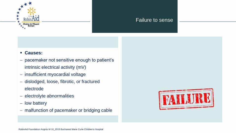
Failure to sense
▪ Causes:
pacemaker not sensitive enough to patient’s
intrinsic electrical activity (mV)
insufficient myocardial voltage
dislodged, loose, fibrotic, or fractured
electrode
electrolyte abnormalities
low battery
malfunction of pacemaker or bridging cable
RobinAid Foundation Angrés M 10_2015 Bucharest Marie Curie Children‘s Hospital
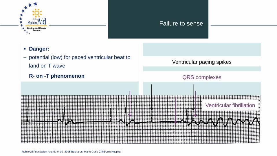
Failure to sense
▪ Danger:
potential (low) for paced ventricular beat to
land on T wave
R- on -T phenomenon
Ventricular pacing spikes
QRS complexes
Ventricular fibrillation

RobinAid Foundation Angrés M 10_2015 Bucharest Marie Curie Children‘s Hospital
Failure to sense
▪ Solution:
view rhythm in different leads
change electrodes
check connections
increase pacemaker’s sensitivity (↓mV)
change cables, battery, pacemaker
reverse polarity
check electrolytes
unipolar pacing with subcutaneous “ground
wire”
RobinAid Foundation Angrés M 10_2015 Bucharest Marie Curie Children‘s Hospital
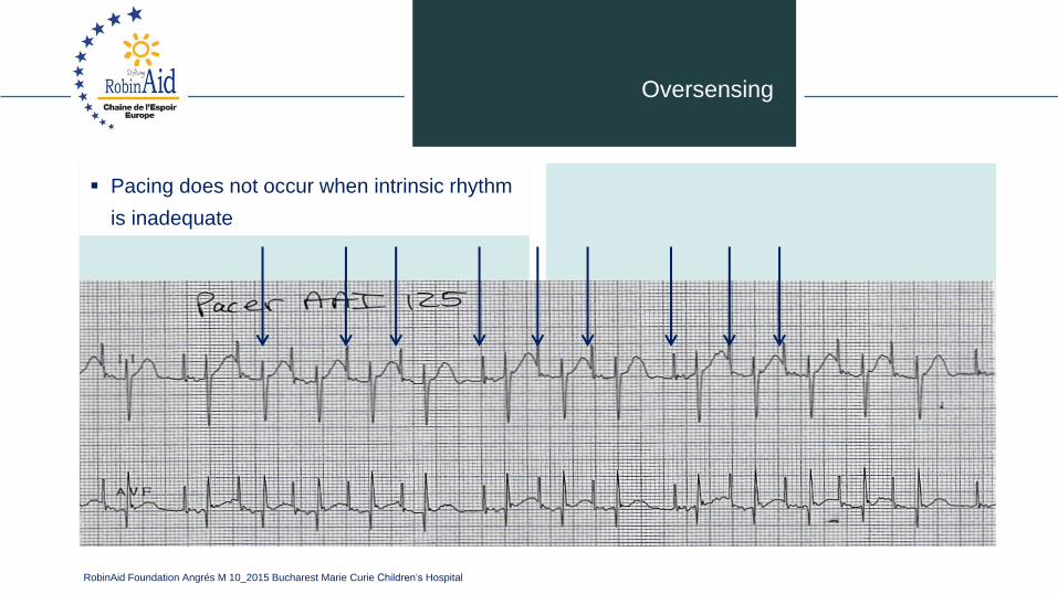
Oversensing
▪ Pacing does not occur when intrinsic rhythm
is inadequate
RobinAid Foundation Angrés M 10_2015 Bucharest Marie Curie Children‘s Hospital
Oversensing
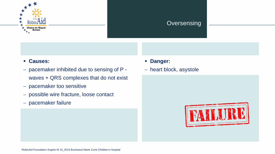
▪ Causes:
pacemaker inhibited due to sensing of P -
waves + QRS complexes that do not exist
pacemaker too sensitive
possible wire fracture, loose contact
pacemaker failure
▪ Danger:
heart block, asystole
RobinAid Foundation Angrés M 10_2015 Bucharest Marie Curie Children‘s Hospital
Oversensing
▪ Solution:
view rhythm in different leads
change electrodes
check connections
decrease pacemaker sensitivity (↑mV)
change cables, battery, pacemaker
reverse polarity
check electrolytes
unipolar pacing with subcutaneous “ground
wire”
RobinAid Foundation Angrés M 10_2015 Bucharest Marie Curie Children‘s Hospital
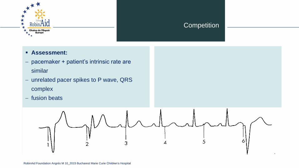
Competition
▪ Assessment:
pacemaker + patient’s intrinsic rate are
similar
unrelated pacer spikes to P wave, QRS
complex
fusion beats
RobinAid Foundation Angrés M 10_2015 Bucharest Marie Curie Children‘s Hospital
Competition
▪ Causes:
asynchronous pacing
failure to sense
mechanical failure: wires, bridging cables,
pacemaker
loose connections
▪ Danger:
impaired cardiac output
potential (low) for paced ventricular beat to
land on T wave
RobinAid Foundation Angrés M 10_2015 Bucharest Marie Curie Children‘s Hospital
Competition
▪ Solution:
Assess underlying rhythm
slowly turn pacer rate down
troubleshoot as for failure to sense
increase pacemaker sensitivity (↓mV)
increase pacemaker rate
RobinAid Foundation Angrés M 10_2015 Bucharest Marie Curie Children‘s Hospital
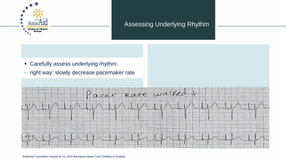
Assessing Underlying Rhythm
▪ Carefully assess underlying rhythm:
right way: slowly decrease pacemaker rate
RobinAid Foundation Angrés M 10_2015 Bucharest Marie Curie Children‘s Hospital
Stimulation threshold
▪ Definition:
minimum current necessary to capture +
stimulate the heart
RobinAid Foundation Angrés M 10_2015 Bucharest Marie Curie Children‘s Hospital
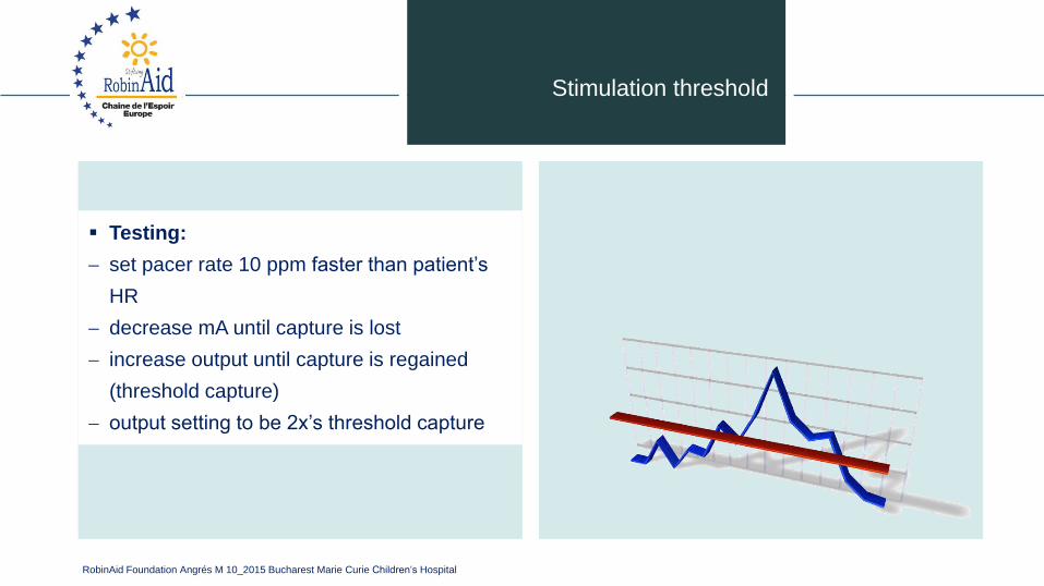
Stimulation threshold
▪ Testing:
set pacer rate 10 ppm faster than patient’s
HR
decrease mA until capture is lost
increase output until capture is regained
(threshold capture)
output setting to be 2x’s threshold capture
RobinAid Foundation Angrés M 10_2015 Bucharest Marie Curie Children‘s Hospital
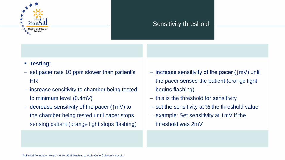
Sensitivity threshold
▪ Definition:
minimum level of intrinsic electric activity
generated by the heart detectable by the
pacemaker
RobinAid Foundation Angrés M 10_2015 Bucharest Marie Curie Children‘s Hospital
Sensitivity threshold
▪ Testing:
set pacer rate 10 ppm slower than patient’s
HR
increase sensitivity to chamber being tested
to minimum level (0.4mV)
decrease sensitivity of the pacer (↑mV) to
the chamber being tested until pacer stops
sensing patient (orange light stops flashing)
increase sensitivity of the pacer (↓mV) until
the pacer senses the patient (orange light
begins flashing).
this is the threshold for sensitivity
set the sensitivity at ½ the threshold value
example: Set sensitivity at 1mV if the
threshold was 2mV

RobinAid Foundation Angrés M 10_2015 Bucharest Marie Curie Children‘s Hospital
Mode of pacing:
rhythm / problem / solution
What does it show?
RobinAid Foundation Angrés M 10_2015 Bucharest Marie Curie Children‘s Hospital
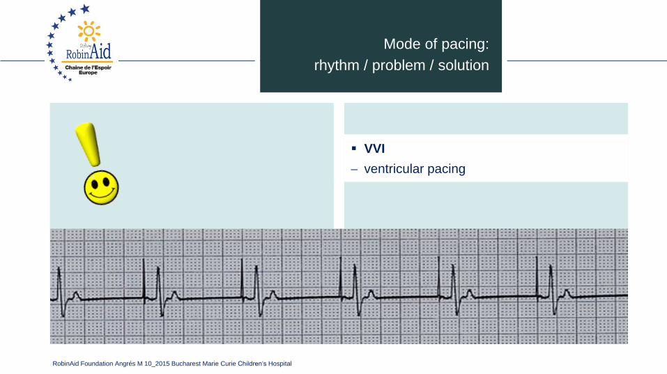
Mode of pacing:
rhythm / problem / solution
▪ VVI
ventricular pacing
RobinAid Foundation Angrés M 10_2015 Bucharest Marie Curie Children‘s Hospital
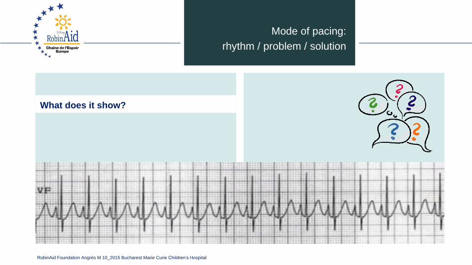
What does it show?
Mode of pacing:
rhythm / problem / solution
RobinAid Foundation Angrés M 10_2015 Bucharest Marie Curie Children‘s Hospital
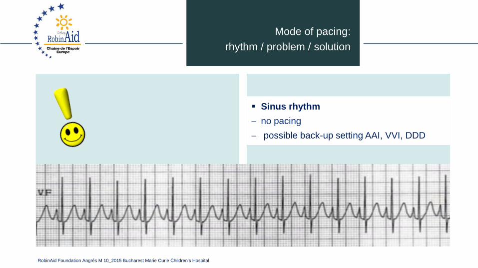
▪ Sinus rhythm
no pacing
possible back-up setting AAI, VVI, DDD
Mode of pacing:
rhythm / problem / solution

RobinAid Foundation Angrés M 10_2015 Bucharest Marie Curie Children‘s Hospital
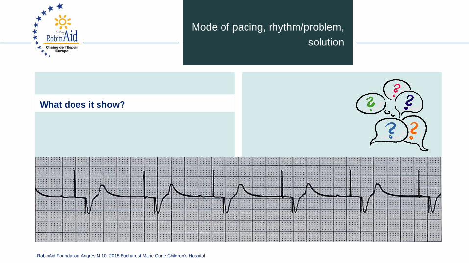
Mode of pacing, rhythm/problem,
solution
What does it show?
RobinAid Foundation Angrés M 10_2015 Bucharest Marie Curie Children‘s Hospital
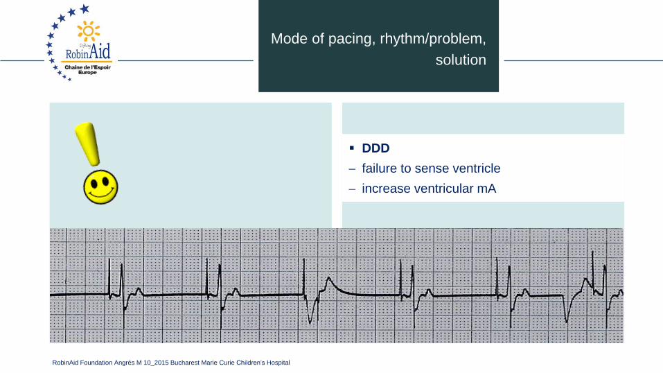
▪ DDD
failure to sense ventricle
increase ventricular mA
Mode of pacing, rhythm/problem,
solution
RobinAid Foundation Angrés M 10_2015 Bucharest Marie Curie Children‘s Hospital
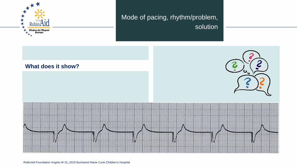
Mode of pacing, rhythm/problem,
solution
What does it show?
RobinAid Foundation Angrés M 10_2015 Bucharest Marie Curie Children‘s Hospital
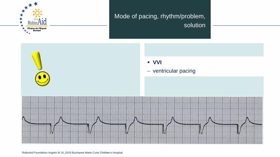
▪ VVI
ventricular pacing
Mode of pacing, rhythm/problem,
solution
RobinAid Foundation Angrés M 10_2015 Bucharest Marie Curie Children‘s Hospital
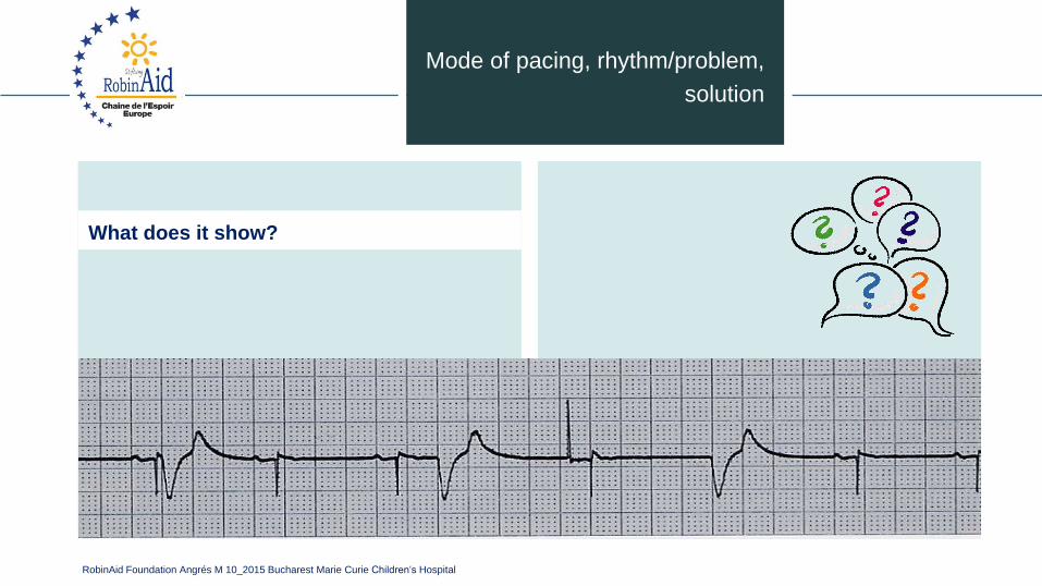
Mode of pacing, rhythm/problem,
solution
What does it show?
RobinAid Foundation Angrés M 10_2015 Bucharest Marie Curie Children‘s Hospital
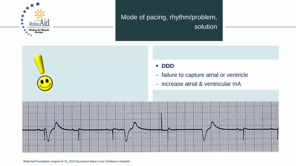
▪ DDD
failure to capture atrial or ventricle
increase atrial & ventricular mA
Mode of pacing, rhythm/problem,
solution

RobinAid Foundation Angrés M 10_2015 Bucharest Marie Curie Children‘s Hospital
Mode of pacing, rhythm/problem,
solution
What does it show?
RobinAid Foundation Angrés M 10_2015 Bucharest Marie Curie Children‘s Hospital
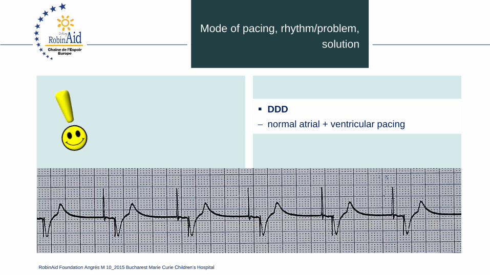
▪ DDD
normal atrial + ventricular pacing
Mode of pacing, rhythm/problem,
solution
RobinAid Foundation Angrés M 10_2015 Bucharest Marie Curie Children‘s Hospital
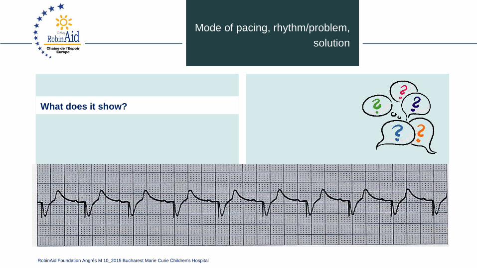
Mode of pacing, rhythm/problem,
solution
What does it show?
RobinAid Foundation Angrés M 10_2015 Bucharest Marie Curie Children‘s Hospital
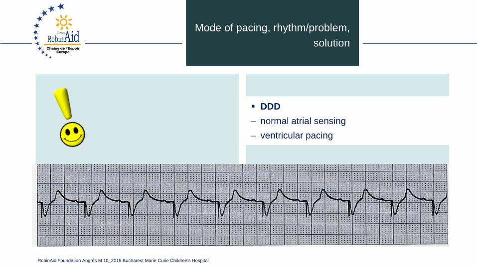
▪ DDD
normal atrial sensing
ventricular pacing
Mode of pacing, rhythm/problem,
solution
RobinAid Foundation Angrés M 10_2015 Bucharest Marie Curie Children‘s Hospital
Mode of pacing, rhythm/problem,
solution
What does it show?
RobinAid Foundation Angrés M 10_2015 Bucharest Marie Curie Children‘s Hospital
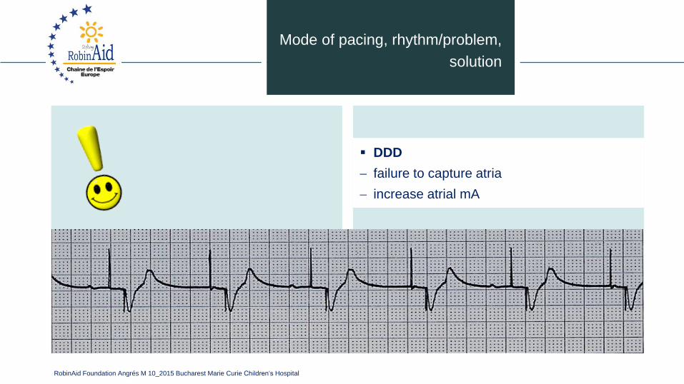
▪ DDD
failure to capture atria
increase atrial mA
Mode of pacing, rhythm/problem,
solution
RobinAid Foundation Angrés M 10_2015 Bucharest Marie Curie Children‘s Hospital
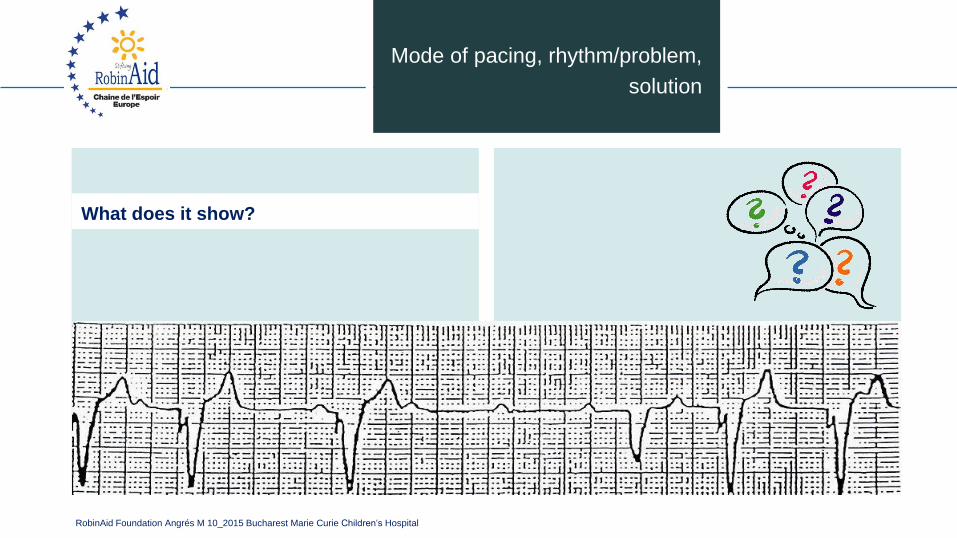
Mode of pacing, rhythm/problem,
solution
What does it show?
RobinAid Foundation Angrés M 10_2015 Bucharest Marie Curie Children‘s Hospital
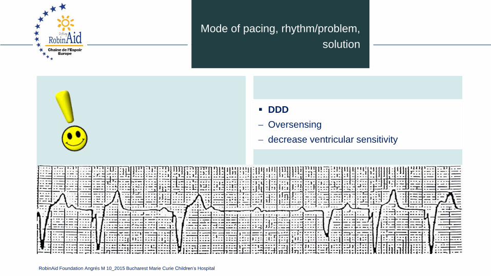
▪ DDD
Oversensing
decrease ventricular sensitivity
Mode of pacing, rhythm/problem,
solution
RobinAid Foundation Angrés M 10_2015 Bucharest Marie Curie Children‘s Hospital
Thank you for your attention