


Languages
Pages
Legal

MANAGEMENT OF BILE DUCT INJURY
Recognition and Operative Treatment
Kellie McFarlin, MD FACSSenior Staff Surgeon
Henry Ford Hospital
Assistant Professor of Surgery
Wayne State University School of Medicine
NMA 2014
Nothing to Disclose
Background
• About 750,000 Laparoscopic Cholecystectomies in
United States
• Bile Duct injury rate remains 0.2% to 0.4% versus
open cholecystectomy rate of 0.1% to 0.2%
• Single Site Cholecystectomy as high as 0.72%
• BDI is most litigated laparoscopic gastrointestional
surgery

Causes of Iatrogenic Bile Duct Injury
• Misidentification of Biliary Anatomy
• Misperception of Biliary Structures
• Aberrant Biliary Anatomy
• Aberrant Arterial Anatomy
• Complicated Cholecystitis with significant
inflammation, scarring, perforation or Mirizzi
syndrome
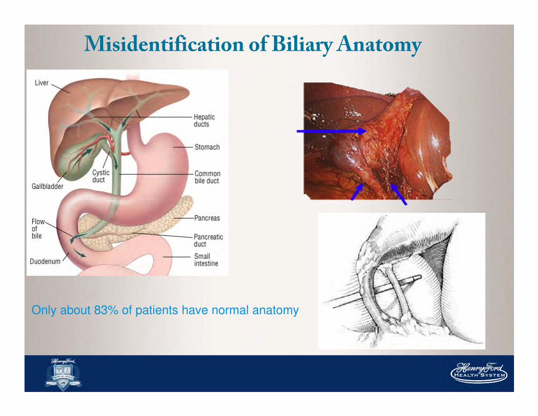
Misidentification of Biliary Anatomy
Only about 83% of patients have normal anatomy
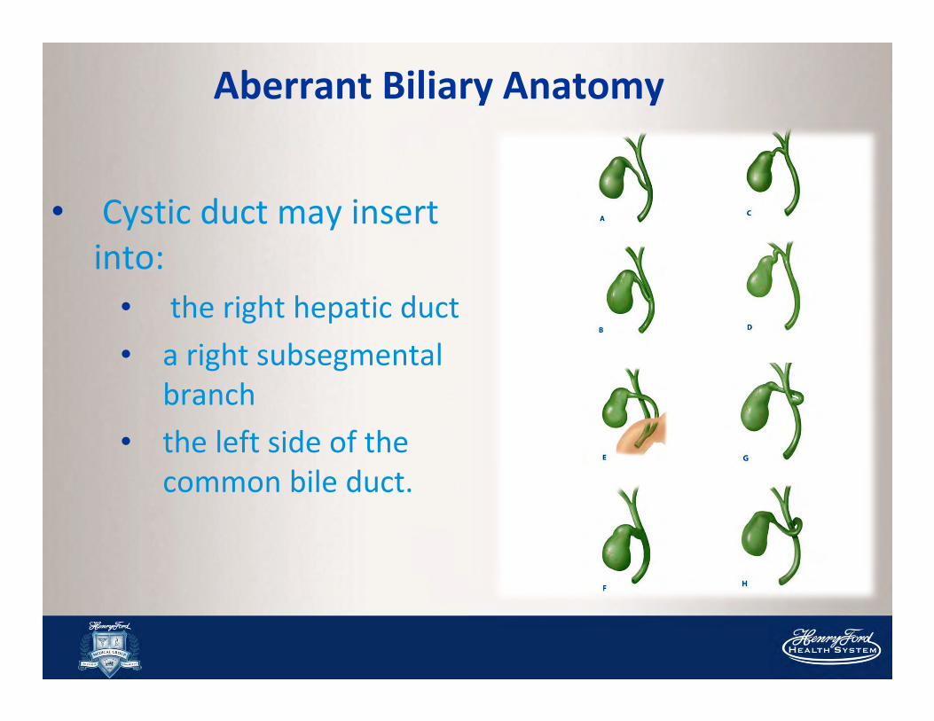
Aberrant Biliary Anatomy
• Cystic duct may insert
into:
• the right hepatic duct
• a right subsegmental
branch
• the left side of the
common bile duct.
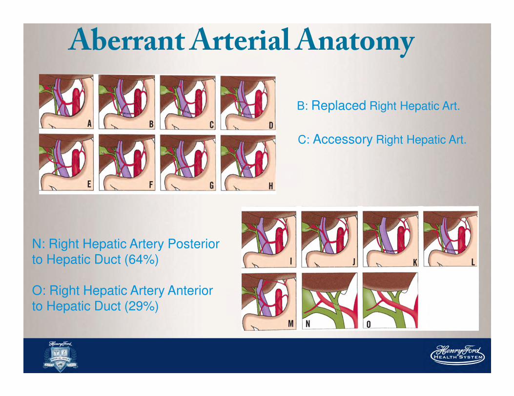
AberrantArterial Anatomy
B: Replaced Right Hepatic Art.
C: Accessory Right Hepatic Art.
O: Right Hepatic Artery Anterior to Hepatic Duct (29%)
N: Right Hepatic Artery Posterior to Hepatic Duct (64%)
Technical Errors• Failure to control the retained side of the cystic duct
• Inadequate clip apposition, clip “scissoring,”
• Thermal injury with excessive or blind application of electrocautery, laser
dissection, or ultrasonic scalpel
• Tran-section of the cystic duct with cautery can lead to arcing to previously placed
clips and result in thermal injury.
• Excessive dissection of the common bile duct may cause local ischemia and
stricturing.
• Injudicious application of clips to control bleeding can lead to obstruction of a part
of the biliary tree or the entire extrahepatic bile duct.
• Excessive lateral traction of the gallbladder can result in ‘tenting”
• Too Deep dissection in the gallbladder fossa
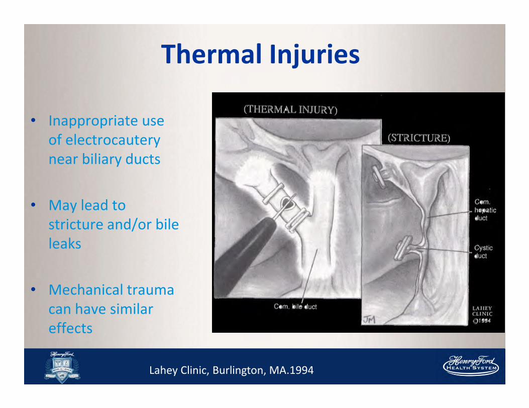
Thermal Injuries
• Inappropriate use
of electrocautery
near biliary ducts
• May lead to
stricture and/or bile
leaks
• Mechanical trauma
can have similar
effects
Lahey Clinic, Burlington, MA.1994
At Risk Patients
• Male
• Advanced Age
• Multiple Comorbidities
• Inflammation of the Gallbladder
Sequelae of BDI
• Biloma formation/ Biliary Fistula
• Cholangitis
• Sepsis
• Bile Duct Stricturing
• Malnutrition (from external
loss of bile salts)
• Biliary cirrhosis, or liver atrophy
• Liver Transplantation
• Mortality
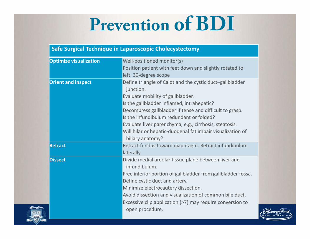
Prevention of BDISafe Surgical Technique in Laparoscopic Cholecystectomy
Optimize visualization Well-positioned monitor(s)
Position patient with feet down and slightly rotated to
left. 30-degree scope
Orient and inspect Define triangle of Calot and the cystic duct–gallbladder
junction.
Evaluate mobility of gallbladder.
Is the gallbladder inflamed, intrahepatic?
Decompress gallbladder if tense and difficult to grasp.
Is the infundibulum redundant or folded?
Evaluate liver parenchyma, e.g., cirrhosis, steatosis.
Will hilar or hepatic-duodenal fat impair visualization of
biliary anatomy?
Retract Retract fundus toward diaphragm. Retract infundibulum
laterally.
Dissect Divide medial areolar tissue plane between liver and
infundibulum.
Free inferior portion of gallbladder from gallbladder fossa.
Define cystic duct and artery.
Minimize electrocautery dissection.
Avoid dissection and visualization of common bile duct.
Excessive clip application (>7) may require conversion to
open procedure.
Complications
• Overall Mortality from BDI is 0% to 4.2%
• Operative Mortality from Repair 1.8%
• Postoperative Complications in over 40%- Wound infection (8%) - Cholangitis (5.7%)
- Intraabdominal Abscess/Biloma (2.9%)
- Stent Related (5.7%
Predictors of Operative Failure
• History of multiple previous repairs
• High-level injury involving the bifurcation
• Incomplete excision of scarred duct remnant
• Use of nonabsorbable suture
• Use of a two-layer anastomosis
• Failure to eradicate infection before repair
Classification
• Location of injury
• Mechanism & type of injury
• Effect on biliary continuity
• Timing of identification
Each plays significant role in determining
appropriate management & operative repair
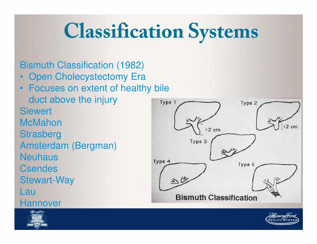
Classification Systems
Bismuth Classification (1982)
• Open Cholecystectomy Era
• Focuses on extent of healthy bile
duct above the injury
Siewert
McMahon
Strasberg
Amsterdam (Bergman)
Neuhaus
Csendes
Stewart-Way
Lau
Hannover
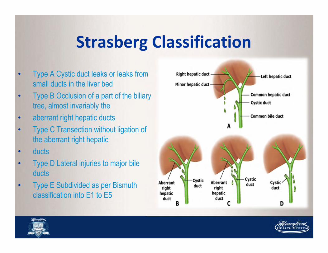
• Type A Cystic duct leaks or leaks from
small ducts in the liver bed
• Type B Occlusion of a part of the biliary
tree, almost invariably the
• aberrant right hepatic ducts
• Type C Transection without ligation of
the aberrant right hepatic
• ducts
• Type D Lateral injuries to major bile
ducts
• Type E Subdivided as per Bismuth
classification into E1 to E5
Strasberg Classification
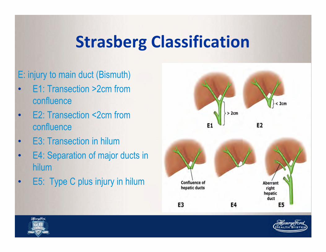
E: injury to main duct (Bismuth)
• E1: Transection >2cm from
confluence
• E2: Transection <2cm from
confluence
• E3: Transection in hilum
• E4: Separation of major ducts in
hilum
• E5: Type C plus injury in hilum
Strasberg Classification
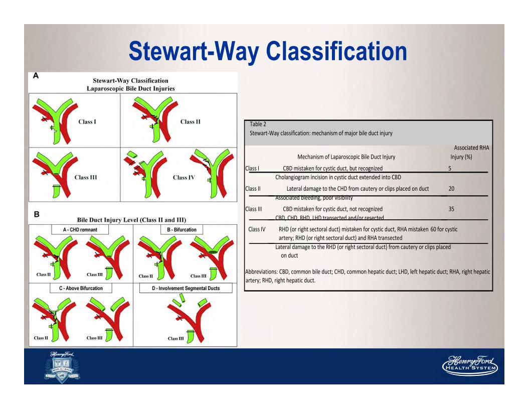
Stewart-Way Classification
Table 2 Stewart-Way classification: mechanism of major bile duct injury
Associated RHA Mechanism of Laparoscopic Bile Duct Injury Injury (%)
Class I CBD mistaken for cystic duct, but recognized 5 Cholangiogram incision in cystic duct extended into CBD
Class II Lateral damage to the CHD from cautery or clips placed on duct 20 Associated bleeding, poor visibility
Class III CBD mistaken for cystic duct, not recognized 35 CBD, CHD, RHD, LHD transected and/or resected
Class IV RHD (or right sectoral duct) mistaken for cystic duct, RHA mistaken 60 for cystic
artery; RHD (or right sectoral duct) and RHA transected Lateral damage to the RHD (or right sectoral duct) from cautery or clips placed
on duct
Abbreviations: CBD, common bile duct; CHD, common hepatic duct; LHD, left hepatic duct; RHA, right hepatic
artery; RHD, right hepatic duct.
Timing of Identification
Intra-op
• Unexpected ductal structures seen bile leak into
field from lacerated or transected duct
Post-op
• Depends on continuity of bile duct & presence or
absence of bile leak
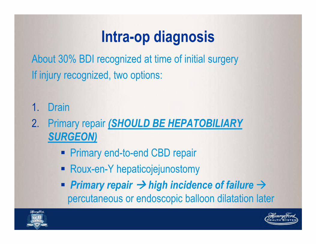
Intra-op diagnosis
About 30% BDI recognized at time of initial surgery
If injury recognized, two options:
1. Drain
2. Primary repair (SHOULD BE HEPATOBILIARY
SURGEON)
� Primary end-to-end CBD repair
� Roux-en-Y hepaticojejunostomy
� Primary repair ���� high incidence of failure �
percutaneous or endoscopic balloon dilatation later
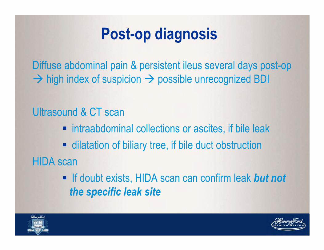
Post-op diagnosis
Diffuse abdominal pain & persistent ileus several days post-op
� high index of suspicion � possible unrecognized BDI
Ultrasound & CT scan
� intraabdominal collections or ascites, if bile leak
� dilatation of biliary tree, if bile duct obstruction
HIDA scan
� If doubt exists, HIDA scan can confirm leak but not
the specific leak site
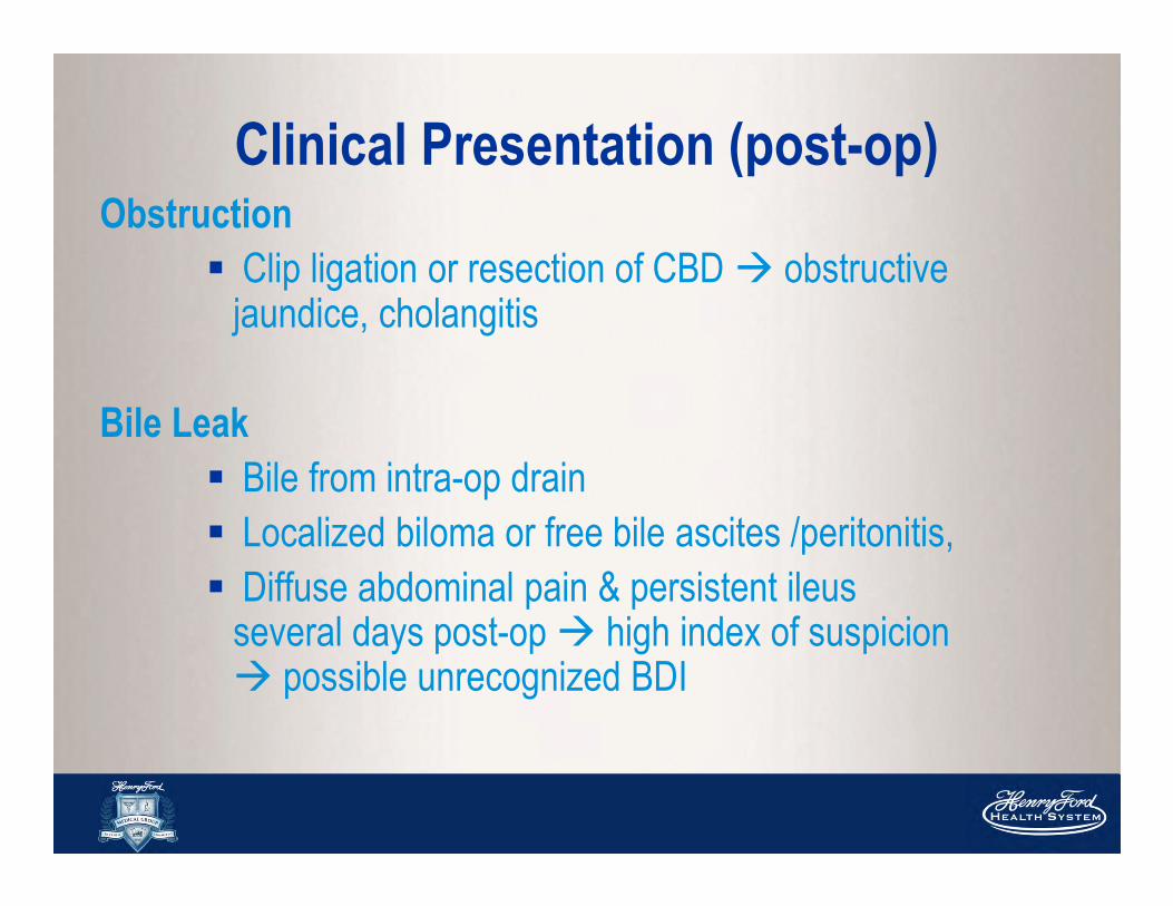
Clinical Presentation (post-op)Obstruction
� Clip ligation or resection of CBD � obstructive jaundice, cholangitis
Bile Leak
� Bile from intra-op drain
� Localized biloma or free bile ascites /peritonitis,
� Diffuse abdominal pain & persistent ileus several days post-op � high index of suspicion � possible unrecognized BDI
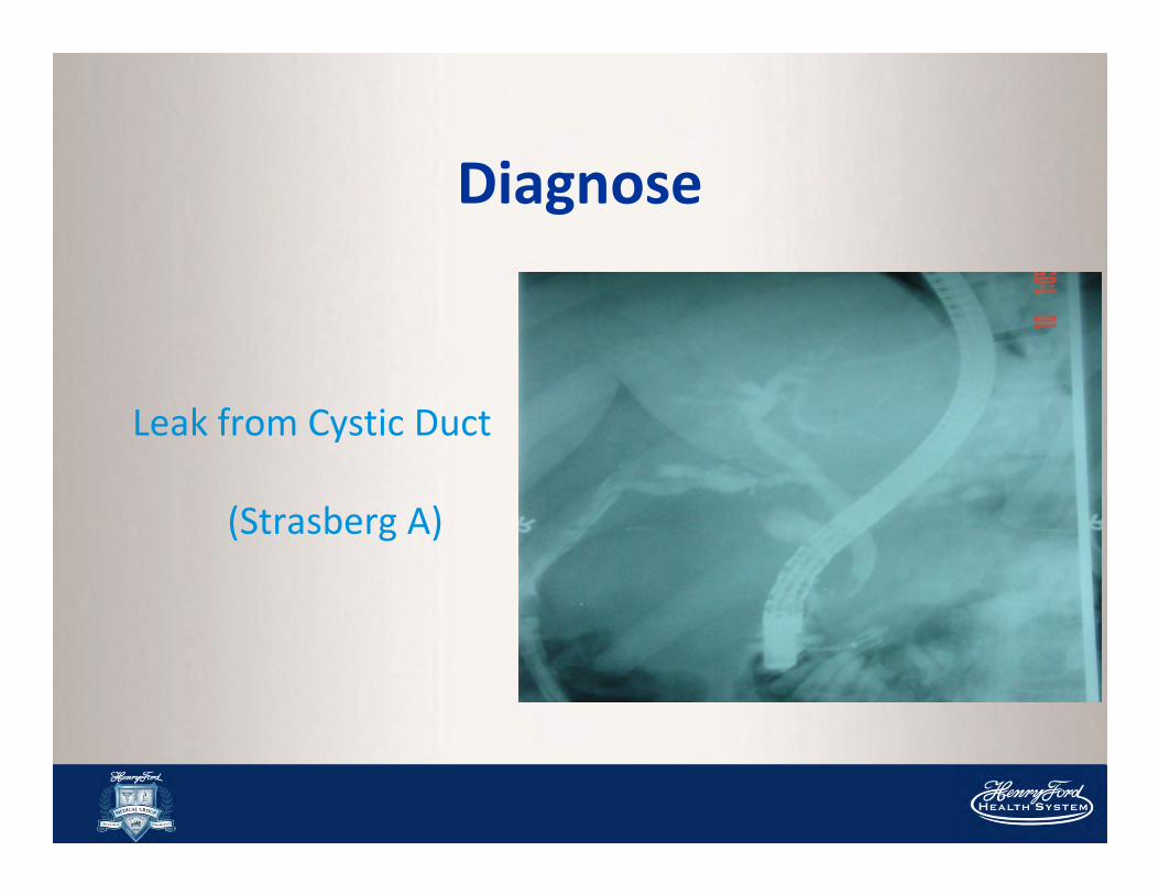
Diagnose
Leak from Cystic Duct
(Strasberg A)
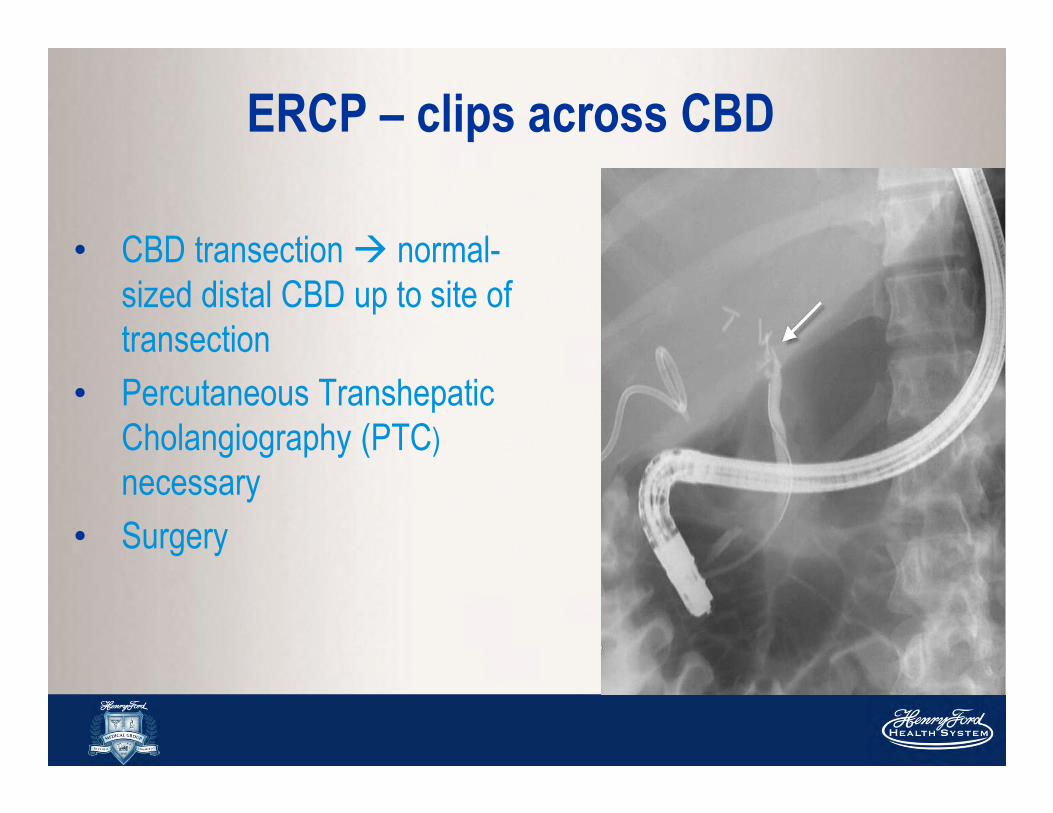
ERCP – clips across CBD
• CBD transection � normal-
sized distal CBD up to site of
transection
• Percutaneous Transhepatic
Cholangiography (PTC)
necessary
• Surgery
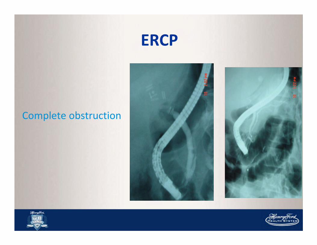
ERCP
Complete obstruction
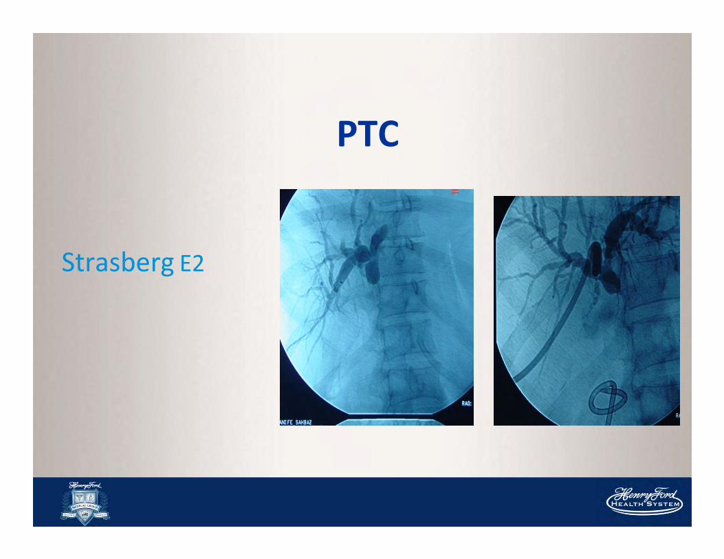
PTC
Strasberg E2
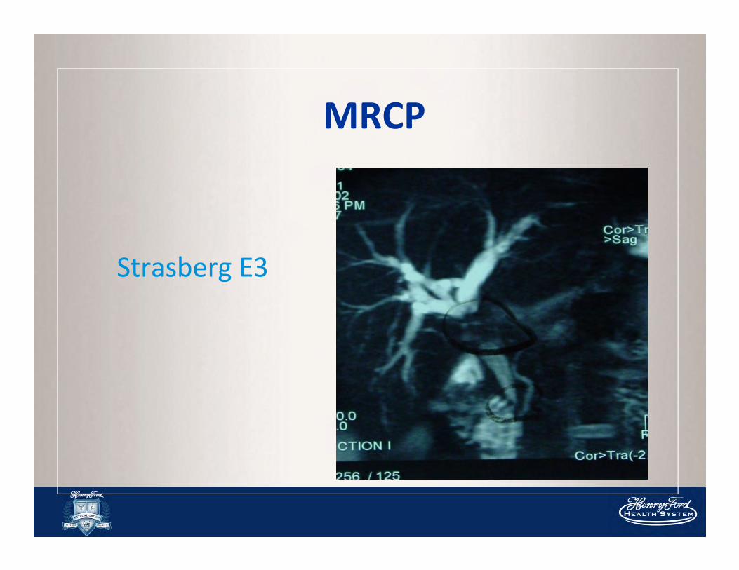
MRCP
Strasberg E3
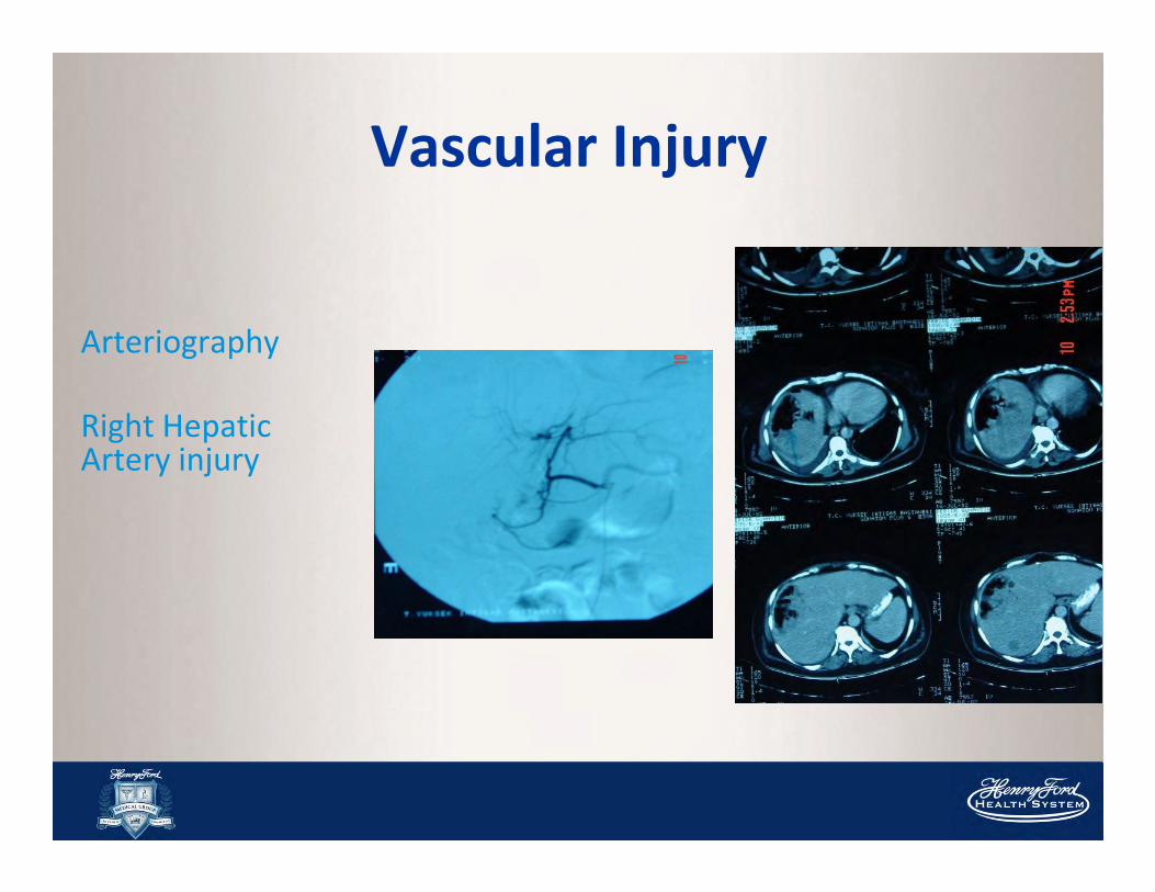
Vascular Injury
Arteriography
Right HepaticArtery injury
Definitive management
Goal
� reestablishment of bile flow into
proximal gastrointestinal tract
� prevent cholangitis, sludge or stone
formation, re-stricturing & progressive
liver injury
Bile duct intact & simply narrowed �
percutaneous or endoscopic dilatation
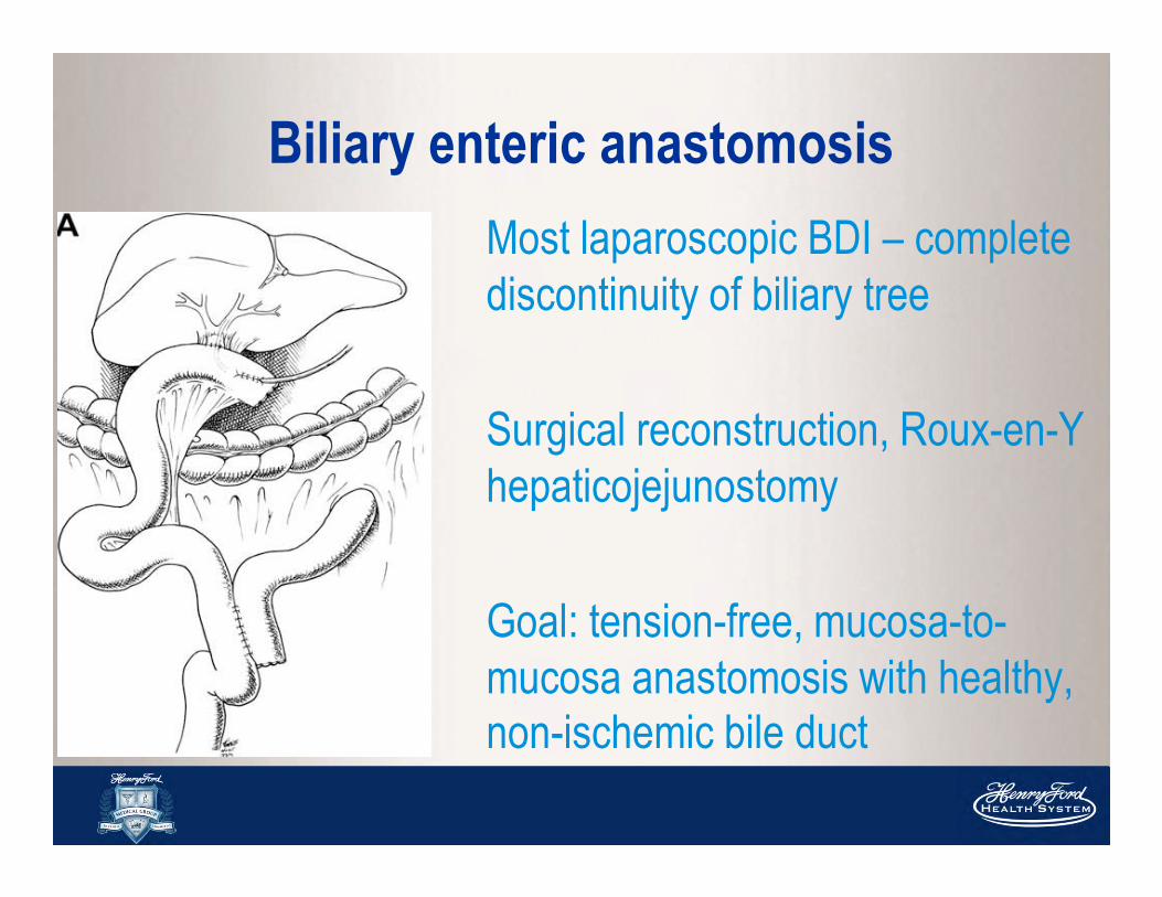
Biliary enteric anastomosis
Most laparoscopic BDI – complete
discontinuity of biliary tree
Surgical reconstruction, Roux-en-Y
hepaticojejunostomy
Goal: tension-free, mucosa-to-
mucosa anastomosis with healthy, non-ischemic bile duct
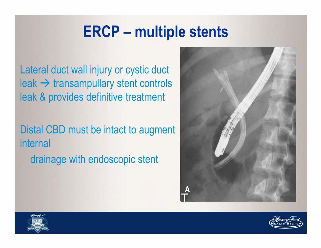
ERCP – multiple stents
Lateral duct wall injury or cystic duct
leak � transampullary stent controls
leak & provides definitive treatment
Distal CBD must be intact to augment
internal
drainage with endoscopic stent
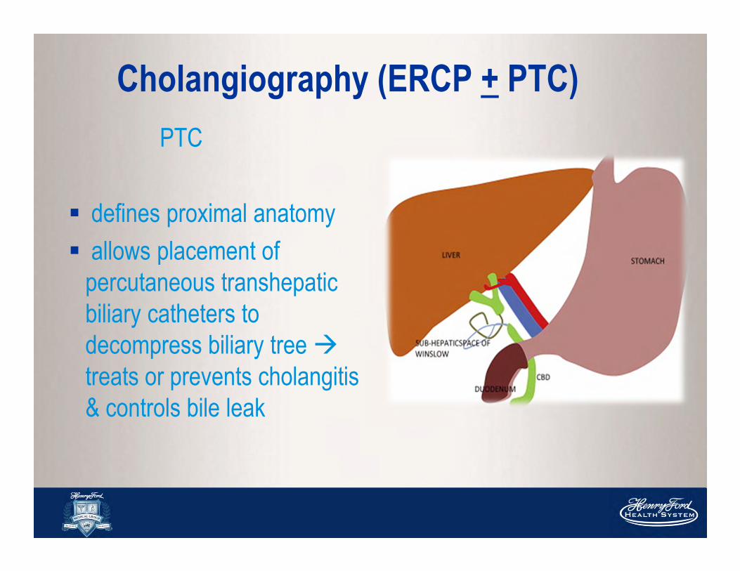
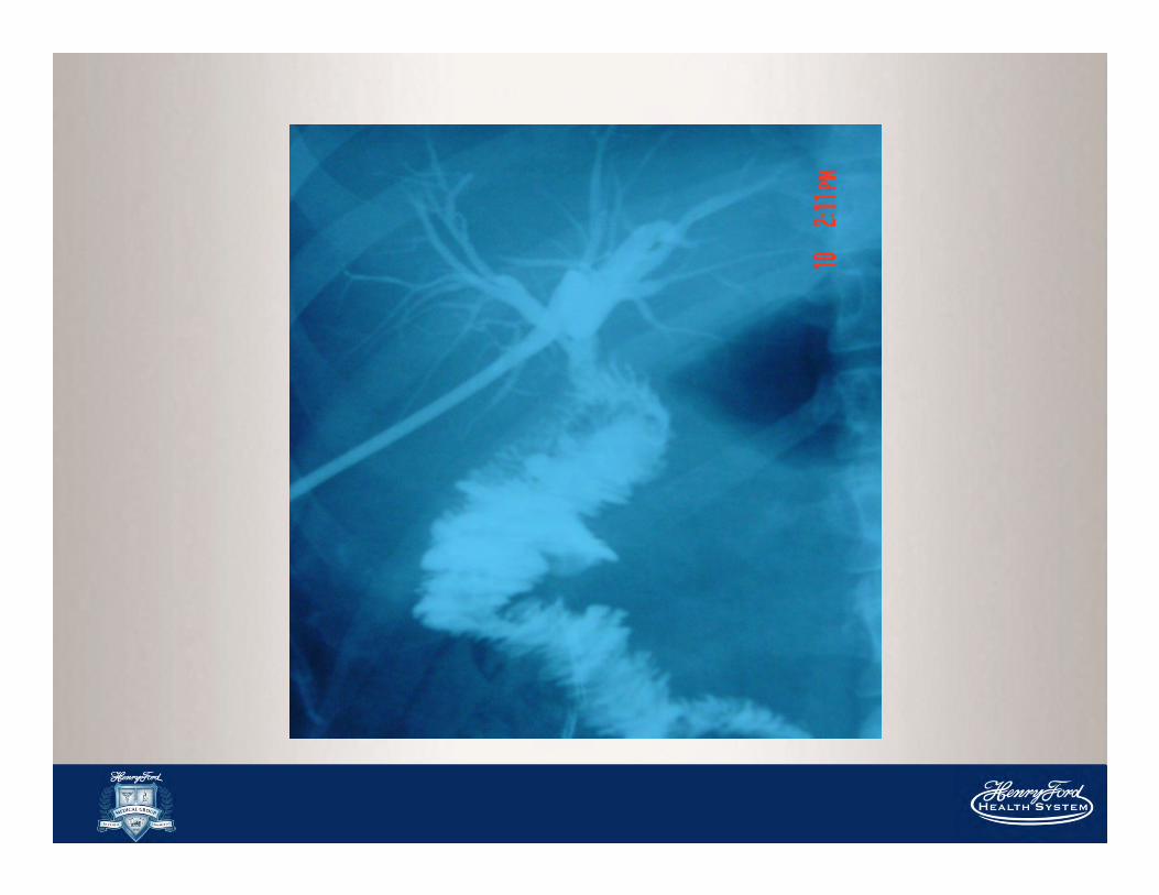
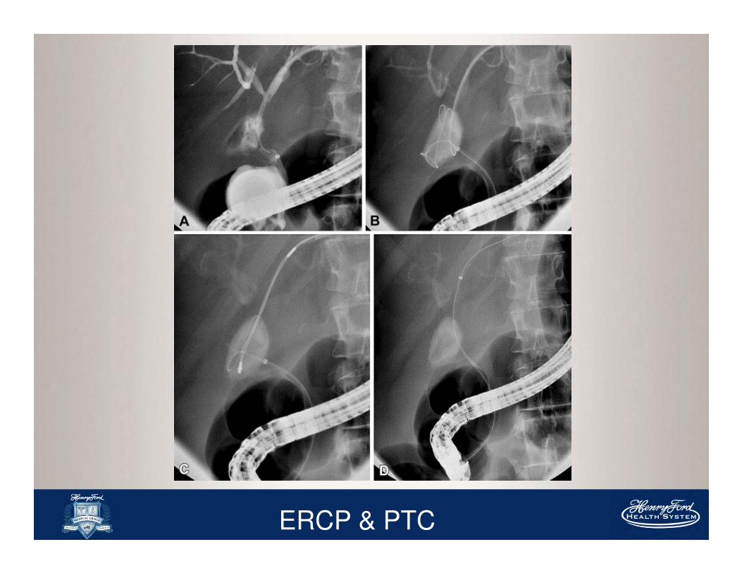
Cholangiography (ERCP + PTC)
PTC
� defines proximal anatomy
� allows placement of
percutaneous transhepatic
biliary catheters to
decompress biliary tree �
treats or prevents cholangitis
& controls bile leak
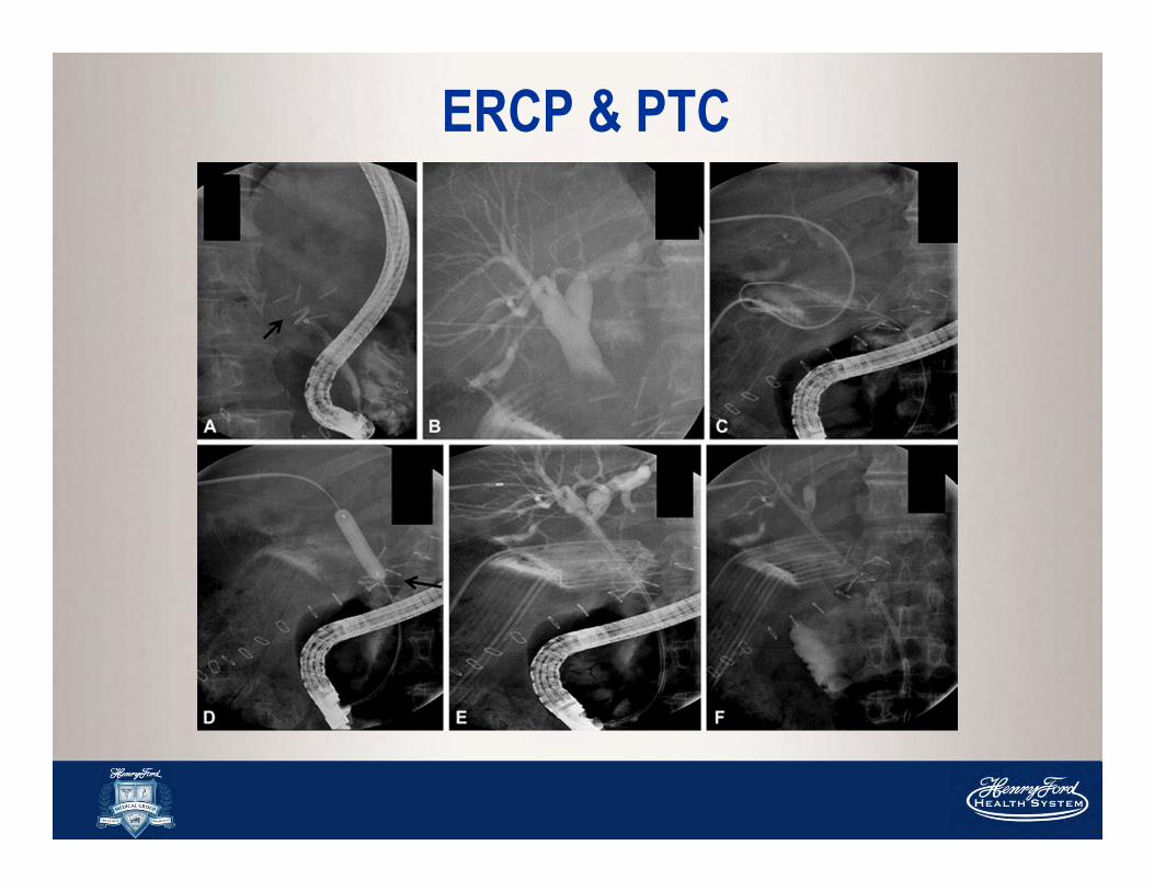
ERCP & PTC
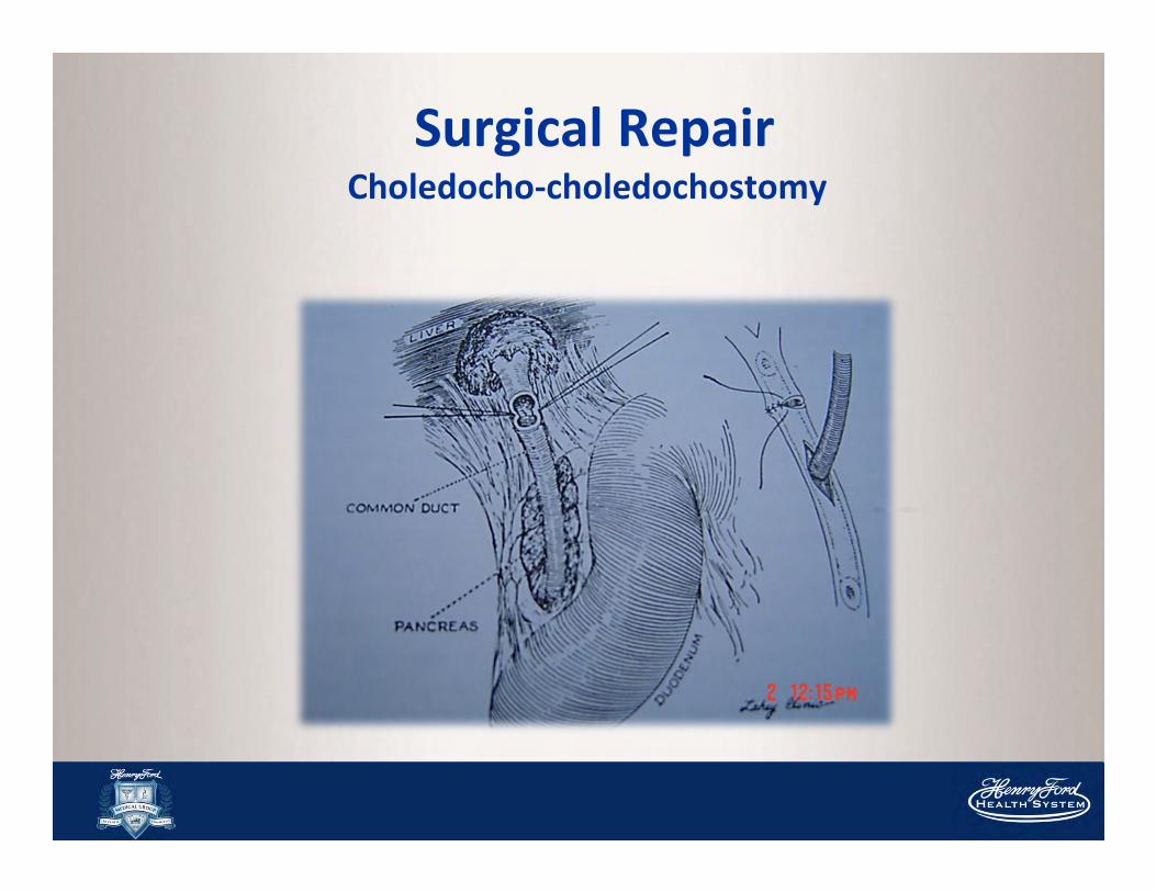
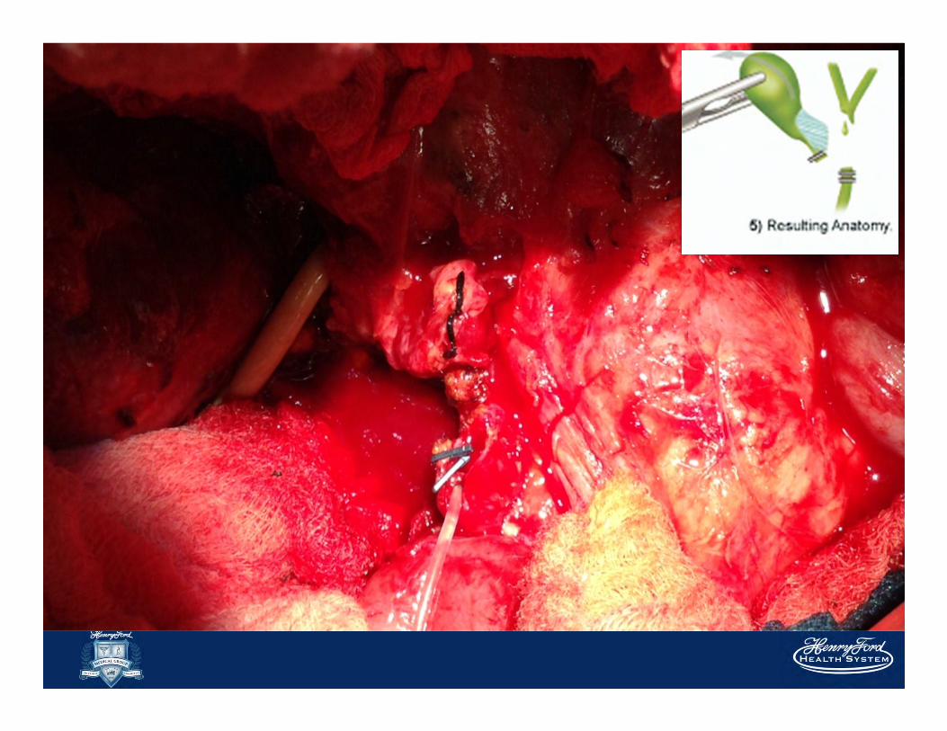
Surgical RepairCholedocho-choledochostomy
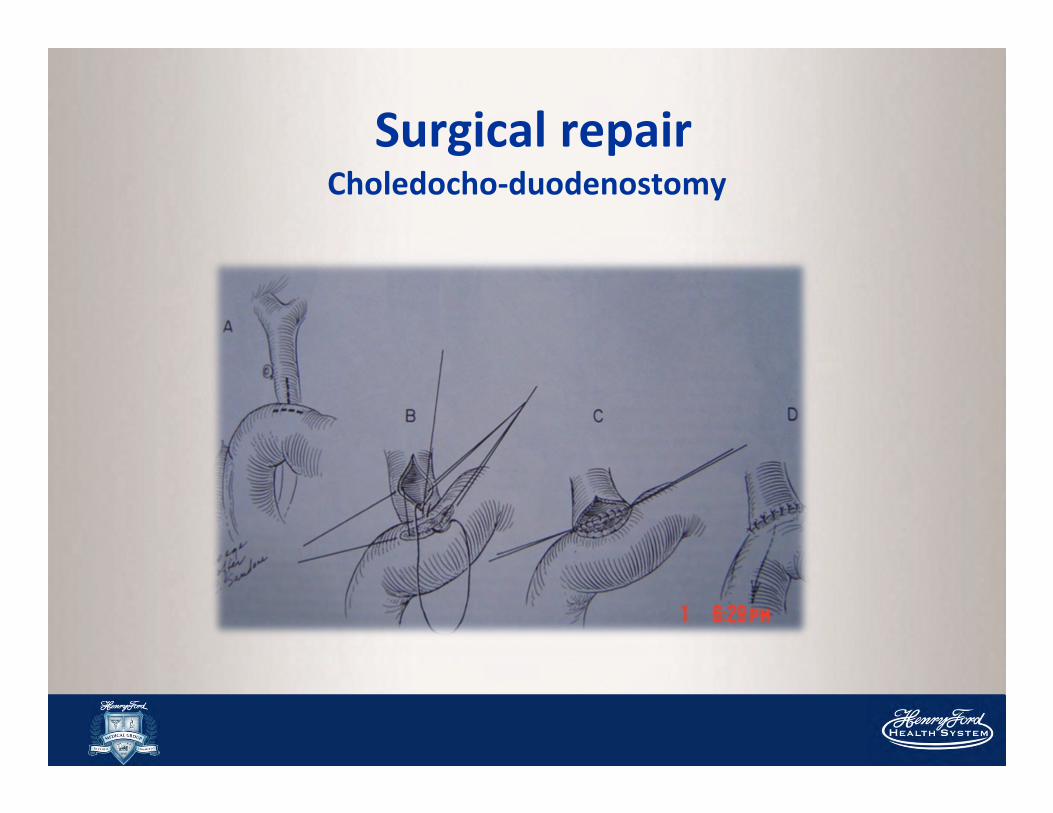
Surgical repair Choledocho-duodenostomy
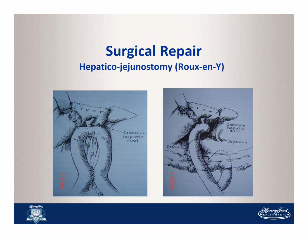
Surgical Repair Hepatico-jejunostomy (Roux-en-Y)
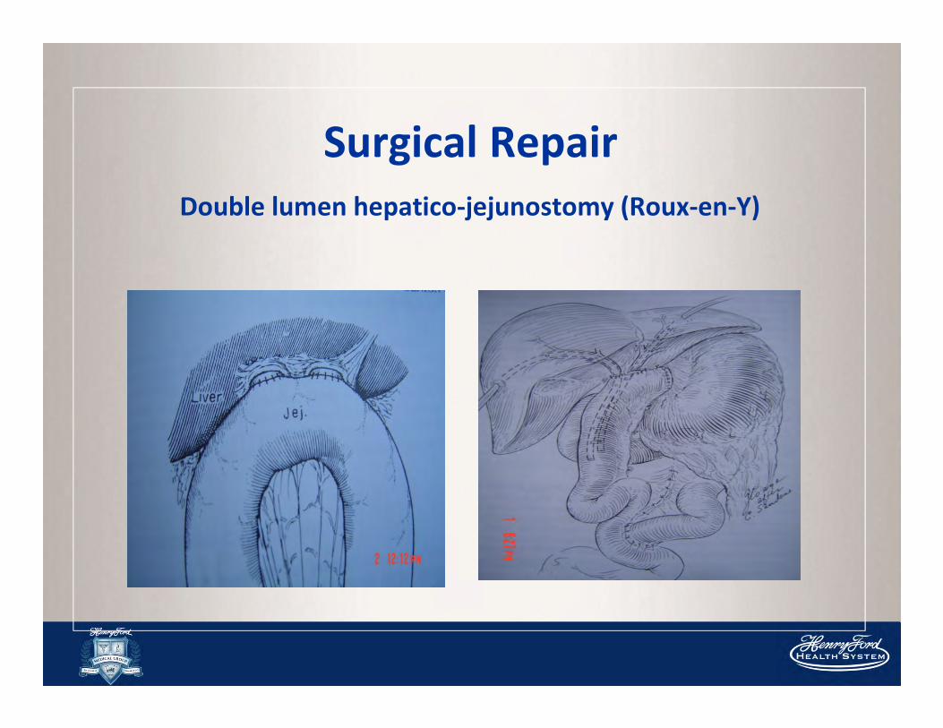
Surgical Repair
Double lumen hepatico-jejunostomy (Roux-en-Y)
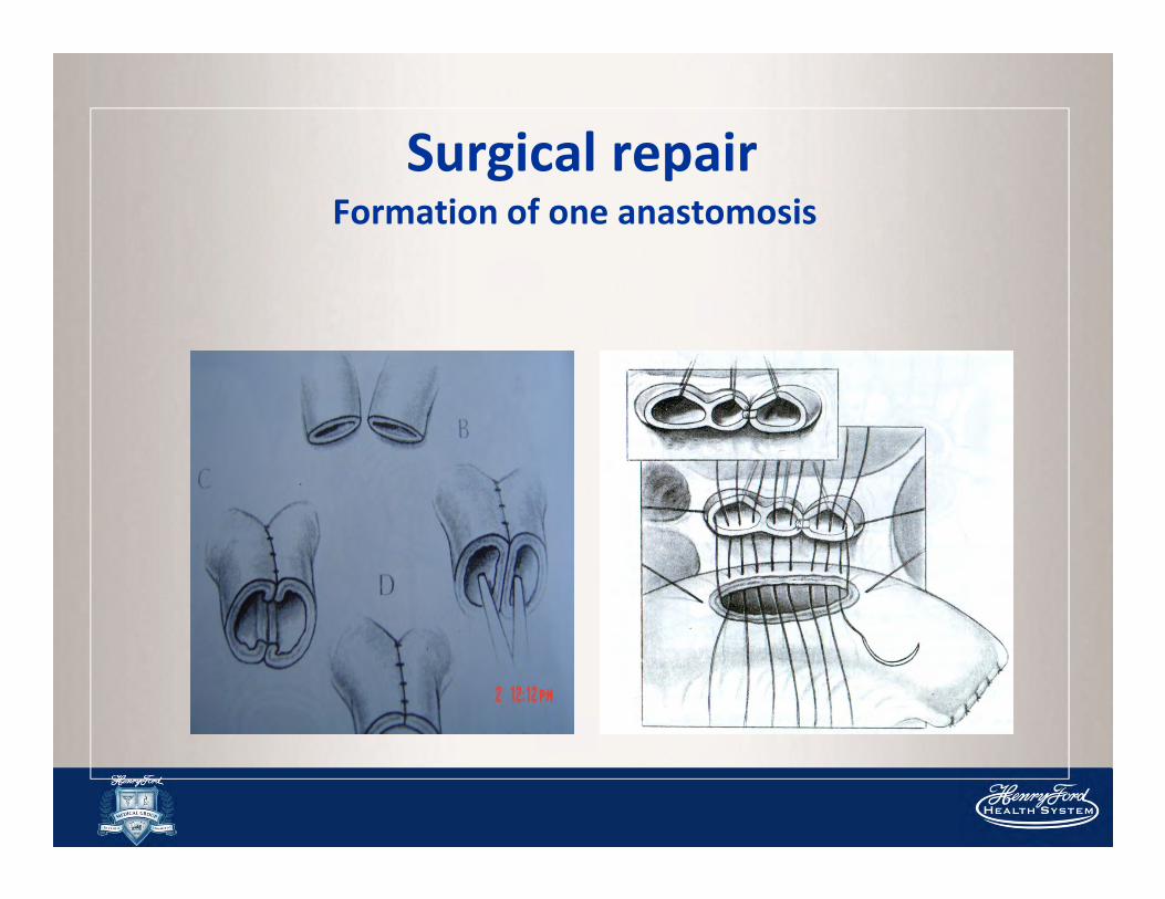
Surgical repair Formation of one anastomosis
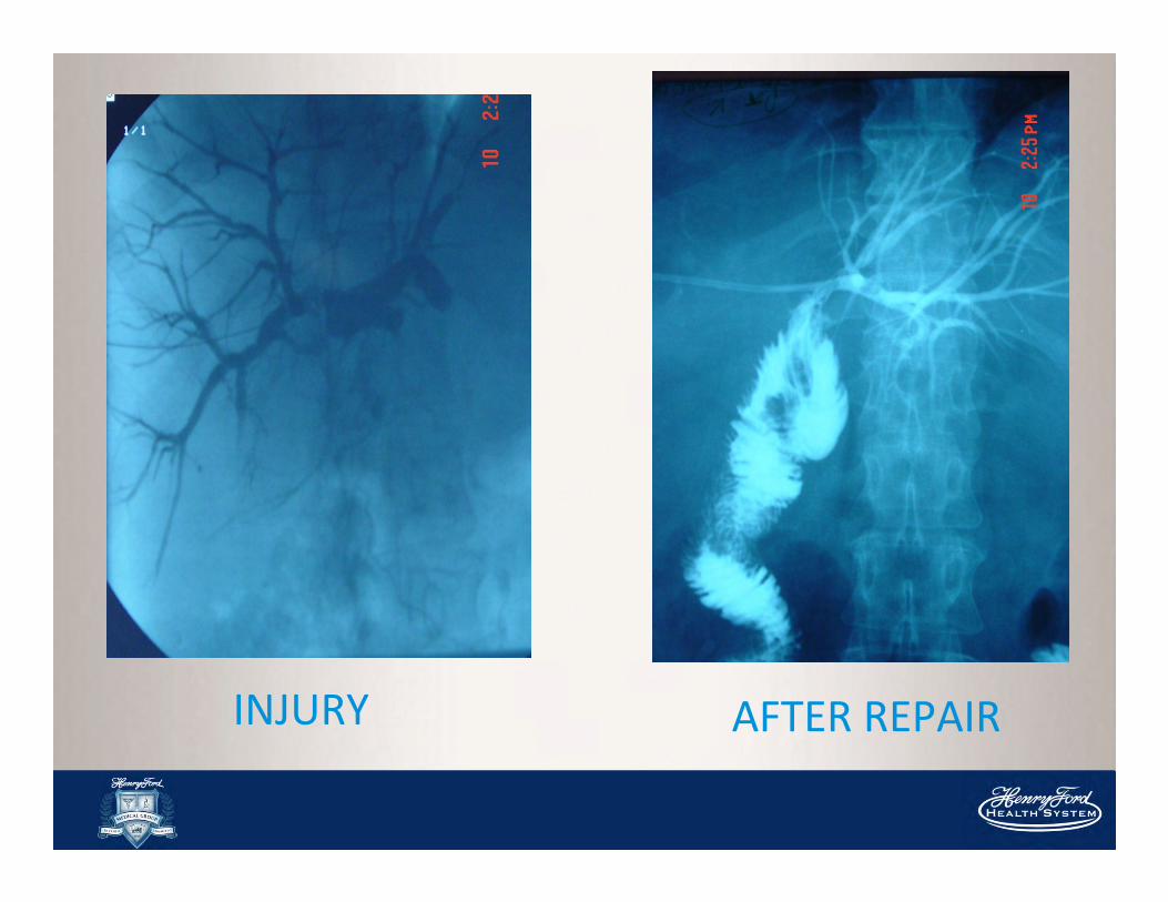
AFTER REPAIRINJURY
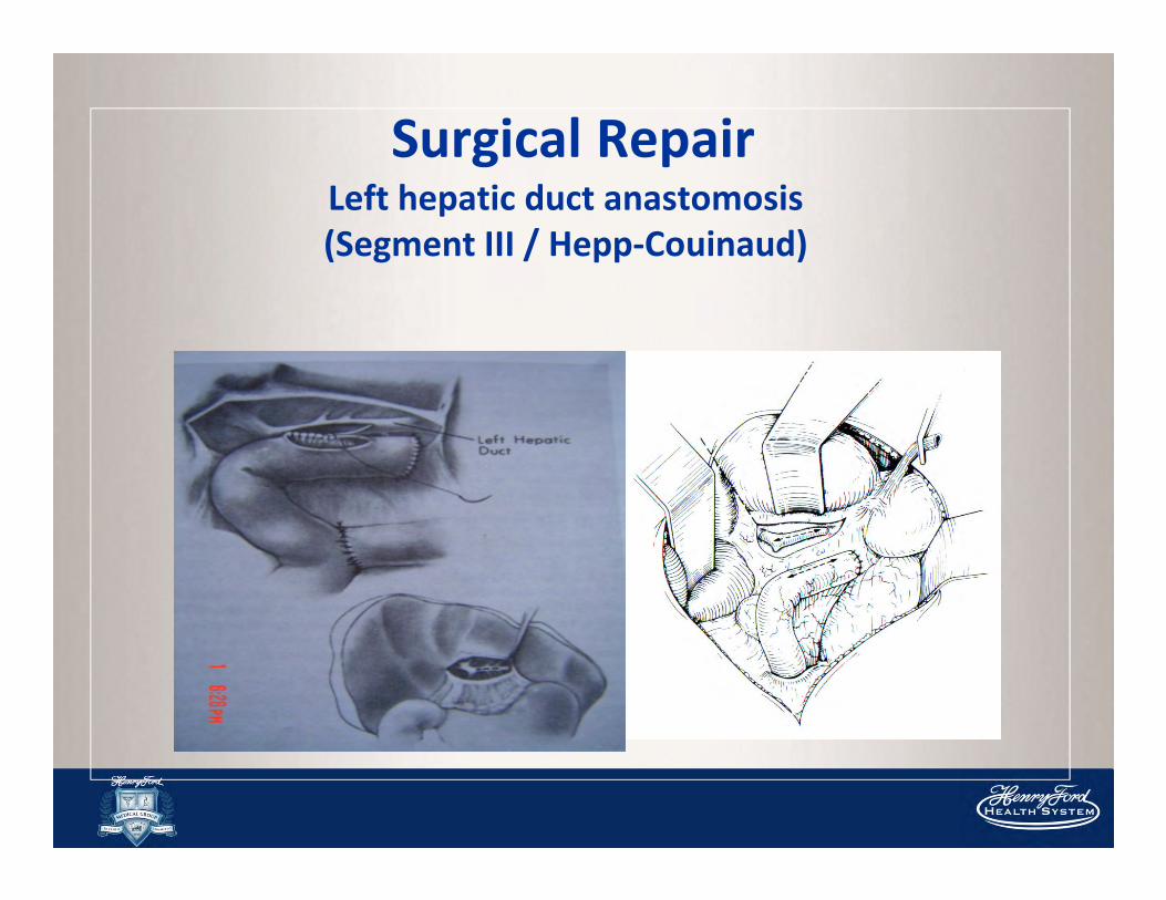
Surgical Repair Left hepatic duct anastomosis
(Segment III / Hepp-Couinaud)
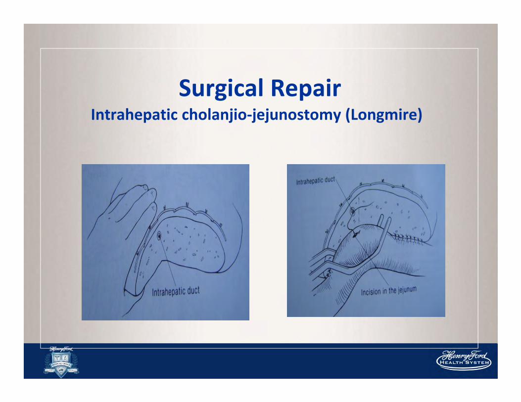
Surgical Repair Intrahepatic cholanjio-jejunostomy (Longmire)
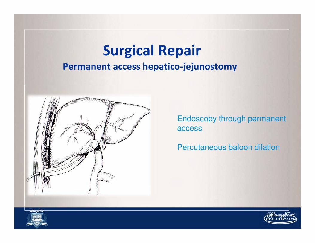
Surgical Repair Permanent access hepatico-jejunostomy
Endoscopy through permanent access
Percutaneous baloon dilation

ERCP & PTC
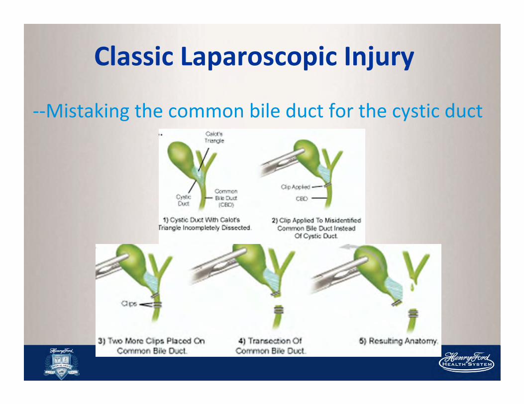
Classic Laparoscopic Injury
--Mistaking the common bile duct for the cystic duct
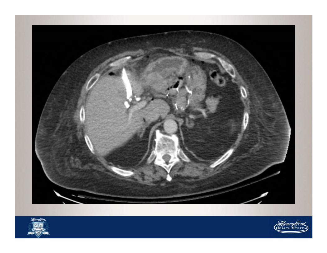
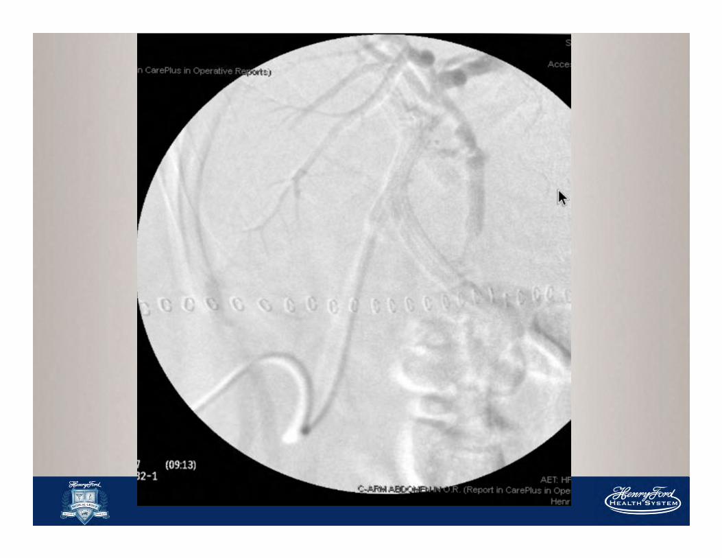
Case
65 yo male transferred from outside hospital with
suspected bile duct injury
PSHx: Gastric bypass surgery
Laparoscopic converted to open with T-tube placement.
Clinical course: Patient arrived stable and non toxic; LFT’s
normal; bile drain to gravity and cholangiogram catheter
stitched in place.
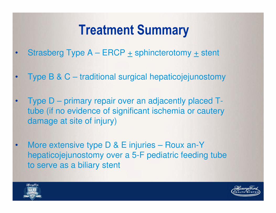
Treatment Summary
• Strasberg Type A – ERCP + sphincterotomy + stent
• Type B & C – traditional surgical hepaticojejunostomy
• Type D – primary repair over an adjacently placed T-
tube (if no evidence of significant ischemia or cautery
damage at site of injury)
• More extensive type D & E injuries – Roux an-Y
hepaticojejunostomy over a 5-F pediatric feeding tube
to serve as a biliary stent
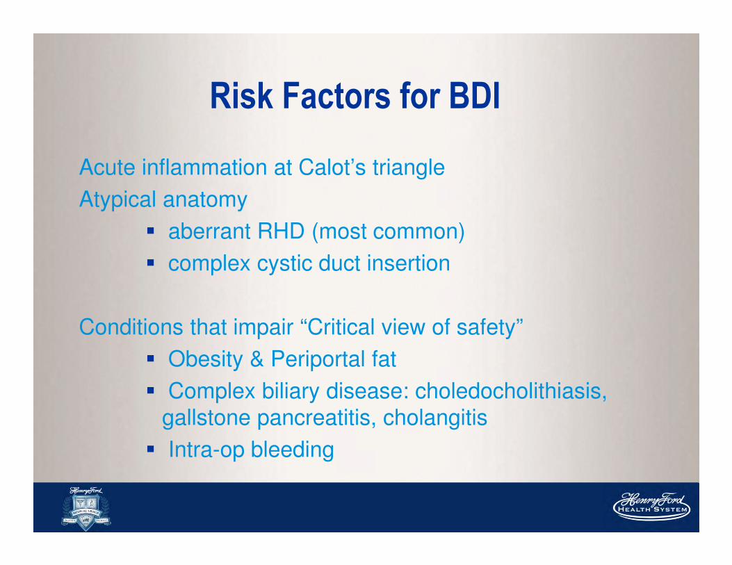
Risk Factors for BDI
Acute inflammation at Calot’s triangle
Atypical anatomy
� aberrant RHD (most common)
� complex cystic duct insertion
Conditions that impair “Critical view of safety”
� Obesity & Periportal fat
� Complex biliary disease: choledocholithiasis,
gallstone pancreatitis, cholangitis
� Intra-op bleeding
Intraoperative Detection
• Only 25-33% of injures are recognized intraoperatively
• If experienced, convert to Open Procedure and perform
Cholangiography (determine extent of injury)
• If not experienced, perform the cholangiogram laparoscopically with
intent of referring patient (placement of drains)
• Consult an experienced hepatobiliary surgeon
• CONTROL INFECTION BEFORE REPAIR
• Acute Management
– Biliary catheter for decompression of biliary tract and control of
bile leaks
– Percutaneous drainage of intraperitoneal bile collection
Preventative Measures
• Attention to operative details (insufficient close or
deep plane)
• Stasberg’s critical view of safety
• Appropriate Handling of Gallbladder
• Careful use of diathermy
• Recognition of Biliary and Vasculature Anomalies
Take Home Message
• Iatrogenic bile duct injury with subsequent biliary fistula or
obstruction has significant economic, legal, mental, and
physical consequences.
• Preoperative planning, safe operative technique, routine use
of IOC or intraoperative ultrasound, and application of
“stopping rules” can decrease the risk of biliary injury.
• Early recognition and involvement of a multidisciplinary team
specialized in biliary disease can result in successful long-
term repair of the biliary injury. After identifying the injury with
high-quality imaging (CT or MRCP), ERCP, and/or PTC, the
appropriate intervention is determined.
Take Home Message
• Endoscopic stenting is a viable treatment option in selected
patients.
• Definitive repair may require biliary-enteric anastomosis
• Postoperative complications are increased with hepatic
artery injury, and the most common longterm complication is
anastomotic stricture
• Overall, patients will have good surgical outcomes if treated
by a multidisciplinary team of hepatobiliary specialists, but
may still have significant negative long-term psychological
effects.
MANAGEMENT OF BILE DUCT INJURY
Recognition and Operative Treatment
Kellie McFarlin, MD FACSSenior Staff Surgeon
Henry Ford Hospital
Assistant Professor of Surgery
Wayne State University School of Medicine
NMA 2014
THANK YOU
Top Related