


Languages
Pages
Legal


Liver Function Tests
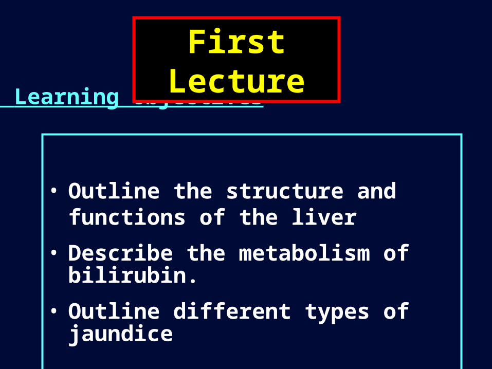
• Outline the structure and functions of the liver
• Describe the metabolism of bilirubin.
• Outline different types of jaundice
Learning objectives:
First Lecture
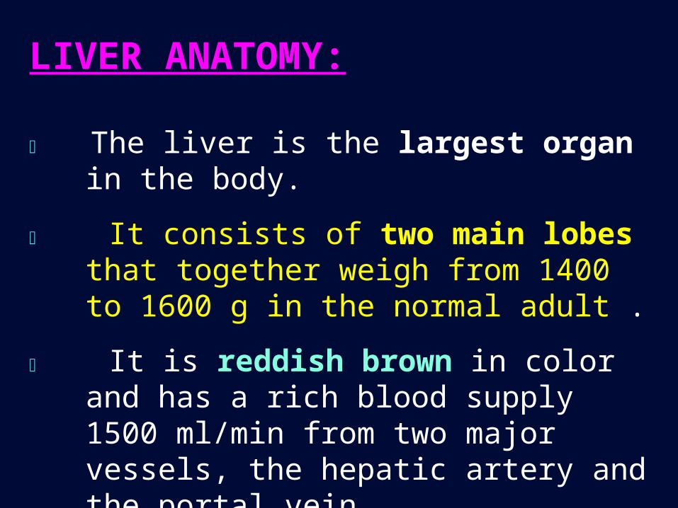
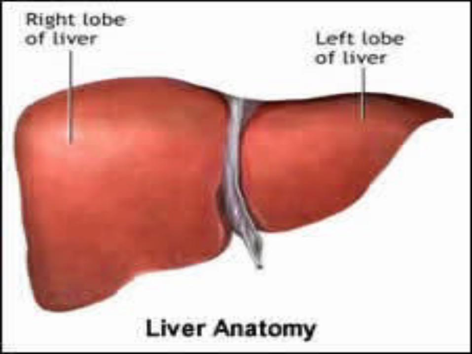
LIVER ANATOMY:
The liver is the largest organ in the body.
It consists of two main lobes that together weigh from 1400 to 1600 g in the normal adult .
It is reddish brown in color and has a rich blood supply 1500 ml/min from two major vessels, the hepatic artery and the portal vein
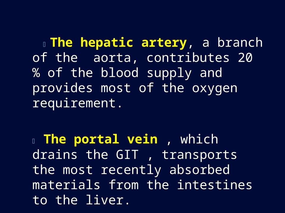
The hepatic artery, a branch of the aorta, contributes 20 % of the blood supply and provides most of the oxygen requirement.
The portal vein , which drains the GIT , transports the most recently absorbed materials from the intestines to the liver.
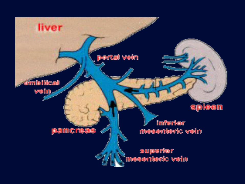
The Portal Circulation
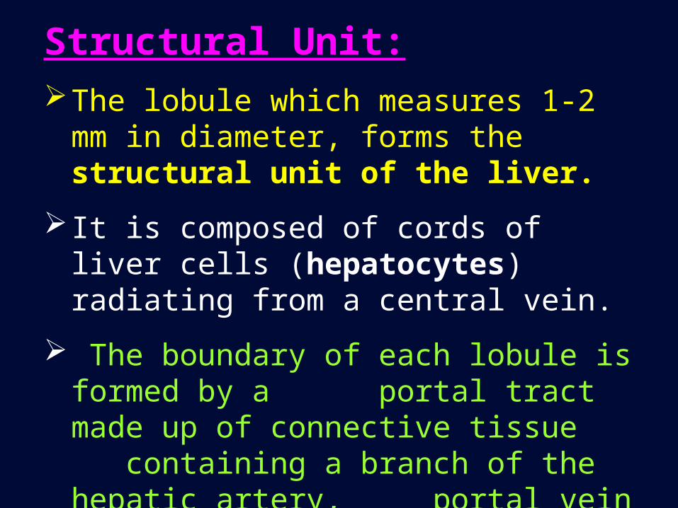
Structural Unit:The lobule which measures 1-2 mm in
diameter, forms the structural unit of the liver.
It is composed of cords of liver cells (hepatocytes) radiating from a central vein.
The boundary of each lobule is formed by a portal tract made up of connective tissue containing a branch of the hepatic artery, portal vein and bile duct.
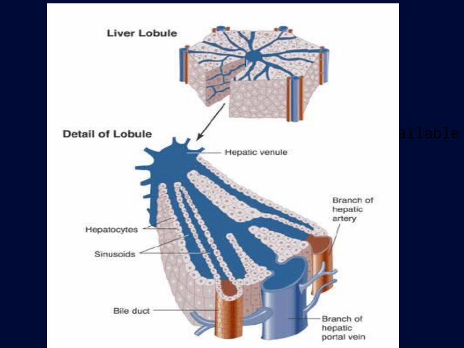
No higher resolution available.
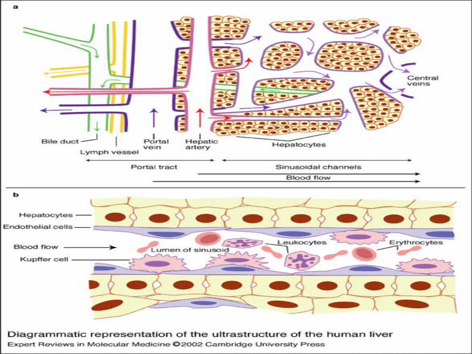
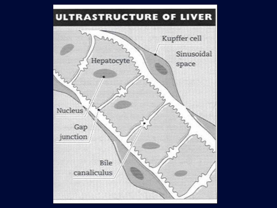
Between the cords of the liver cells are vascular spaces, called sinusoids, that are lined by Kupffer’s cells
The Kupffer’s cells are phagocytic macrophages capable of ingesting bacteria or other foreign material from the blood that flows through the sinusoids
Hepatocytes form 80% of liver , Kupffer’s cells ,vascular and supporting cells form the 20% .
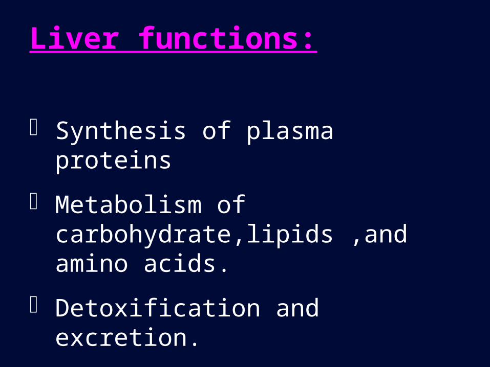
Liver functions:
Synthesis of plasma proteins
Metabolism of carbohydrate,lipids ,and amino acids.
Detoxification and excretion.
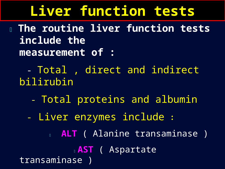
The routine liver function tests include the measurement of :
- Total , direct and indirect bilirubin
- Total proteins and albumin
- Liver enzymes include :
ALT ( Alanine transaminase )
AST ( Aspartate transaminase )
ALP ( Alkaline phosphatase )
GGT ( - Glutamyl transferase )
Liver function tests
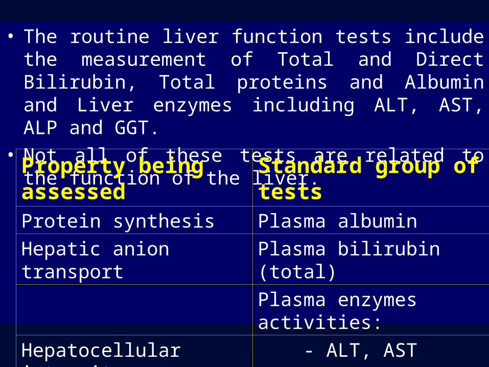
• The routine liver function tests include the measurement of Total and Direct Bilirubin, Total proteins and Albumin and Liver enzymes including ALT, AST, ALP and GGT.
• Not all of these tests are related to the function of the liver.
Standard group of testsProperty being assessedPlasma albuminProtein synthesis
Plasma bilirubin (total)Hepatic anion transport
Plasma enzymes activities:
- ALT, ASTHepatocellular integrity
- ALP, GGTPresence of cholestasis
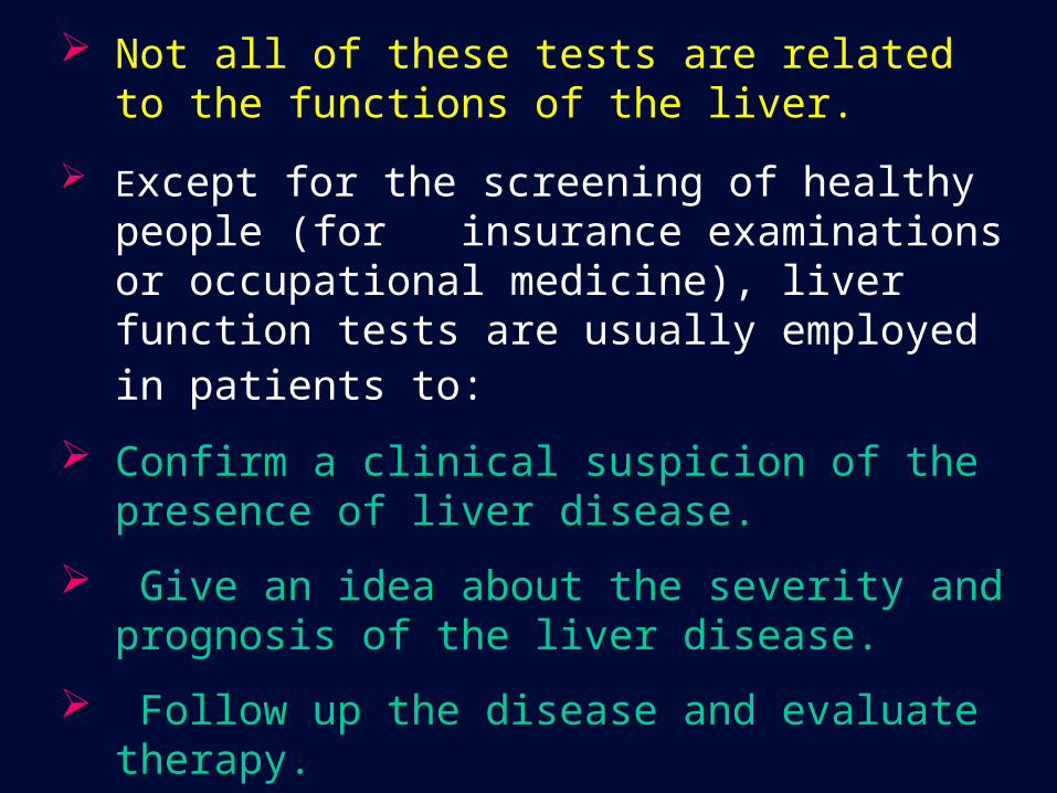
Not all of these tests are related to the functions of the liver.
Except for the screening of healthy people (for insurance examinations or occupational medicine), liver function tests are usually employed in patients to:
Confirm a clinical suspicion of the presence of liver disease.
Give an idea about the severity and prognosis of the liver disease.
Follow up the disease and evaluate therapy.
Arrive at a differential diagnosis (e.g. cholestatic vs hepatocellular liver disease).
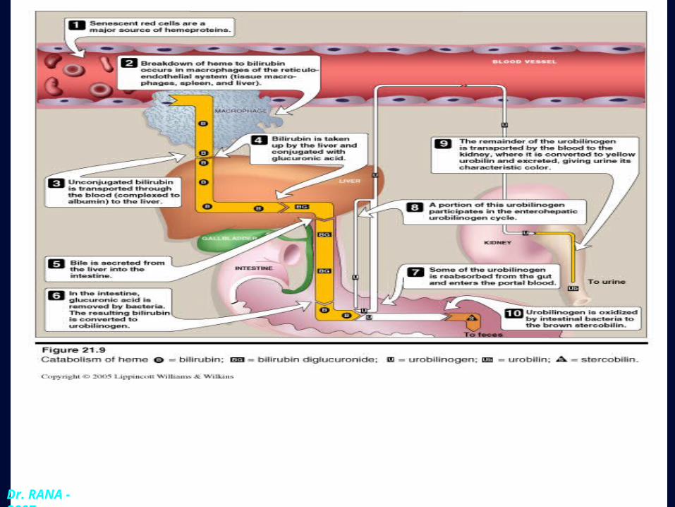
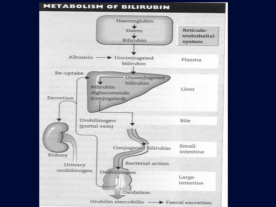
Bilirubin Metabolism1.Production of bilirubin in RES:
80% of bilirubin formed from haem arise from red blood cells.
The remaining 20% comes from red cell precursors destroyed in the bone marrow (ineffective erythropoiesis), and from other haem proteins such as myoglobin, cytochromes, catalase and peroxidase.
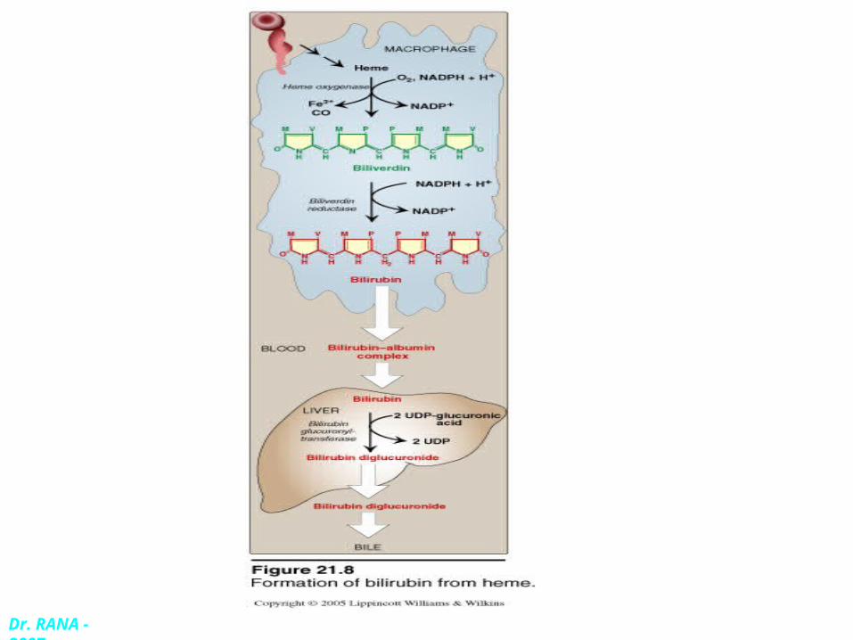
Iron is removed from the haem molecule and the porphyrin ring is opened to form bilirubin.
Dr. RANA - 2007
2, transport of bilirubin in the plasma:
bilirubin is soluble in lipid solvents but almost insoluble in water., so it is carried in plasma by protein-binding mainly to albumin forming indirect or unconjugated bilirubin.
So the bound form does not readily enter most tissues, nor it is filtered at the glomerulus.
The maximum capacity of albumin for bilirubin is 340 mol/L. the excess ,free, unconjugated bilirubin crosses the BBB and dissolves in the lipid –rich brain tissue and leads to brain damage to the baby( kernicterus )
3. IN THE LIVER:
a) Hepatic uptake:
The bilirubin-albumin complex appears to be associated by receptors on the plasma membrane of the hepatocytes, bilirubin taken up by a specific carrier (facilitated diffusion), leaving albumin in the plasma.
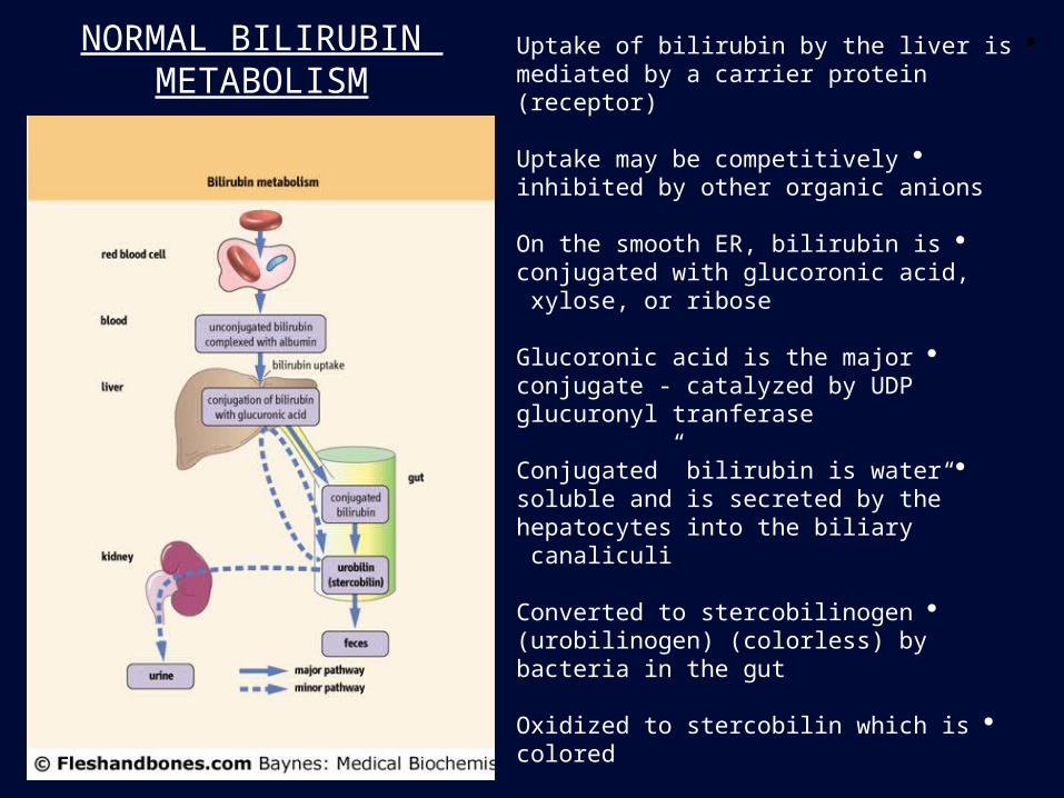
NORMAL BILIRUBIN METABOLISM
Uptake of bilirubin by the liver is mediated by a carrier protein (receptor)
Uptake may be competitively inhibited by other organic anions
On the smooth ER, bilirubin is conjugated with glucoronic acid, xylose, or ribose
Glucoronic acid is the major conjugate - catalyzed by UDP glucuronyl tranferase
“Conjugated” bilirubin is water soluble and is secreted by the hepatocytes into the biliary canaliculi
Converted to stercobilinogen (urobilinogen) (colorless) by bacteria in the gut
Oxidized to stercobilin which is colored
Excreted in feces
Some stercobilin may be re-adsorbed by the gut and re-excreted by either the liver or kidney

b) Conjugation:
Conjugation of bilirubin within the hepatocytes makes it water-soluble. The enzyme is Bilirubin-UDP-glucuronyl transferase forms bilirubin –diglucuronide (direct or conjugated bilirubin).
c) Secretion of bilirubin into bile:
Occurs against a high concentration gradient, a carrier mediated energy dependant process (active secretion).
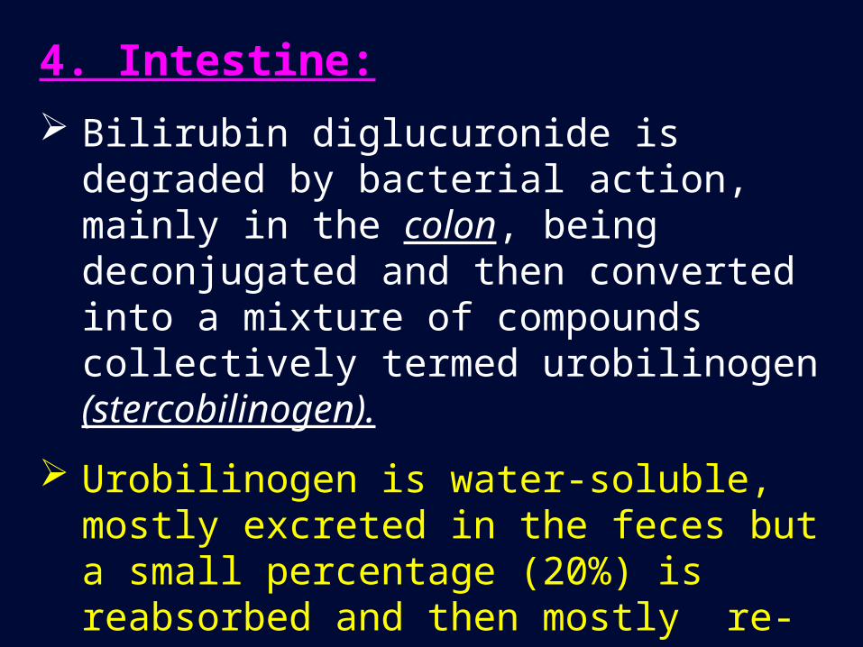
4. Intestine:
Bilirubin diglucuronide is degraded by bacterial action, mainly in the colon, being deconjugated and then converted into a mixture of compounds collectively termed urobilinogen (stercobilinogen).
Urobilinogen is water-soluble, mostly excreted in the feces but a small percentage (20%) is reabsorbed and then mostly re-excreted by the liver.
After excretion, urobilinogen (colorless) is oxidized to urobilin (stercobilin) which is brown gives stools its color.
Some of the reabsorbed urobilinogen passes through the liver into the systemic circulation and is then excreted in the urine (urobilin) gives the urine its yellow color.
Dr. RANA - 2007
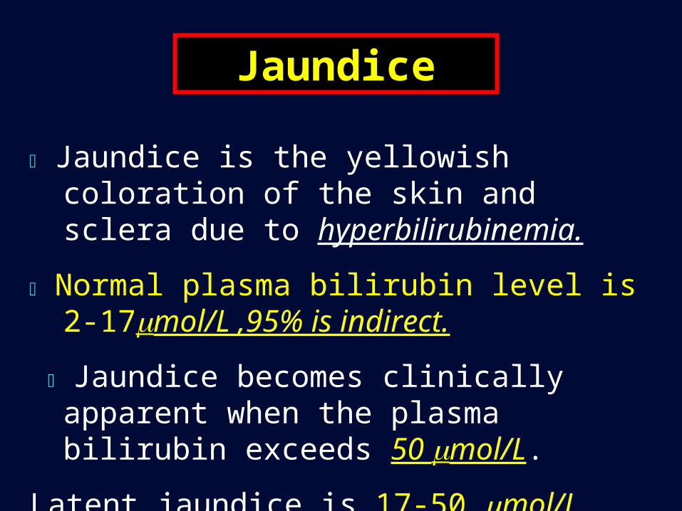
Jaundice is the yellowish coloration of the skin and sclera due to hyperbilirubinemia.
Normal plasma bilirubin level is 2-17mol/L ,95% is indirect.
Jaundice becomes clinically apparent when the plasma bilirubin exceeds 50 mol/L.
Latent jaundice is 17-50 mol/L
Jaundice
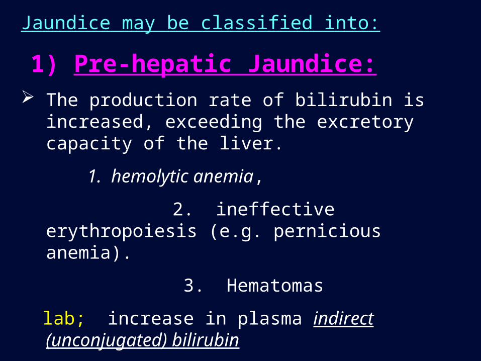
Jaundice may be classified into:
1) Pre-hepatic Jaundice: The production rate of bilirubin is increased, exceeding the
excretory capacity of the liver.
1. hemolytic anemia,
2. ineffective erythropoiesis (e.g. pernicious anemia).
3. Hematomas
lab; increase in plasma indirect (unconjugated) bilirubin
. Bilirubin is not excreted in urine.
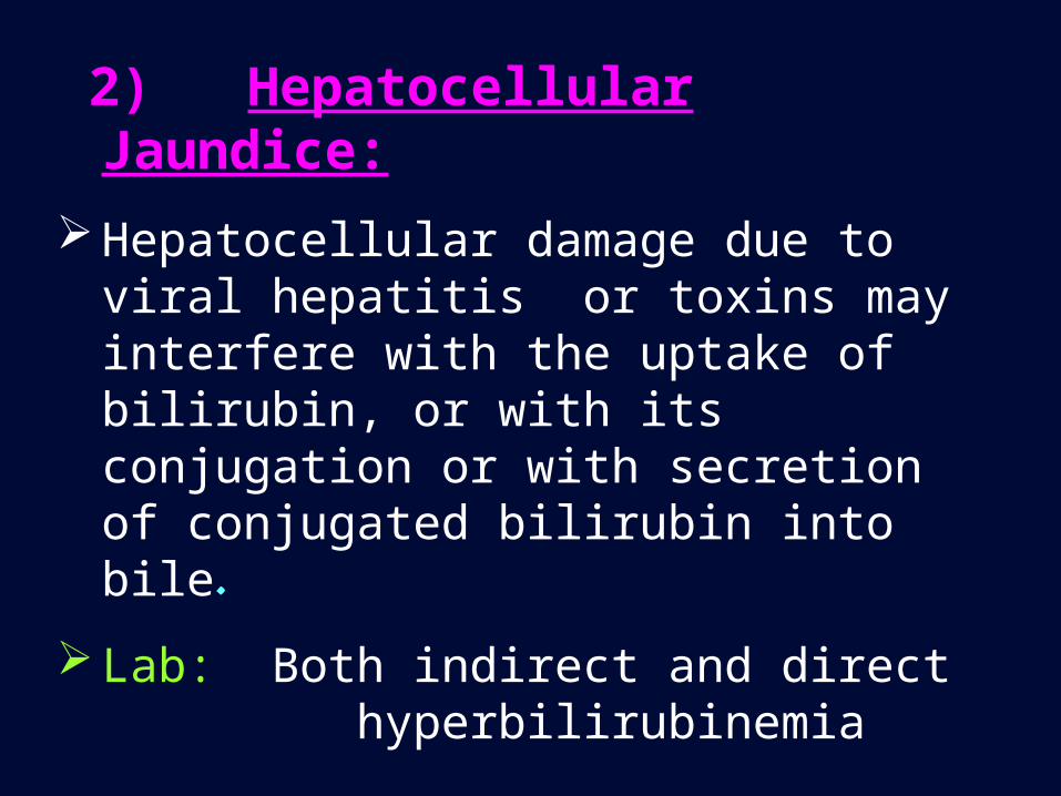
2) Hepatocellular Jaundice:
Hepatocellular damage due to viral hepatitis or toxins may interfere with the uptake of bilirubin, or with its conjugation or with secretion of conjugated bilirubin into bile.
Lab: Both indirect and direct hyperbilirubinemia
Bilirubin is found in urine(bilirubinuria).
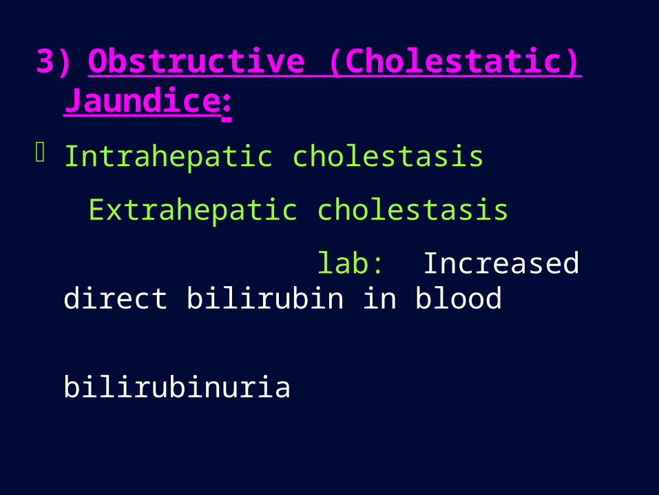
3) Obstructive (Cholestatic) Jaundice:
Intrahepatic cholestasis
Extrahepatic cholestasis
lab: Increased direct bilirubin in blood
bilirubinuria

Congenital Hyperbilirubinemias:
They are all due to inherited defects in the mechanism of bilirubin transport.
1) Gilbert’s Disease :
A common congenital disorder (autosomal dominant) of bilirubin transport affecting approximately 2% of the population, males more affected than females.
Gilbert’s disease is a benign condition and life expectancy is normal.
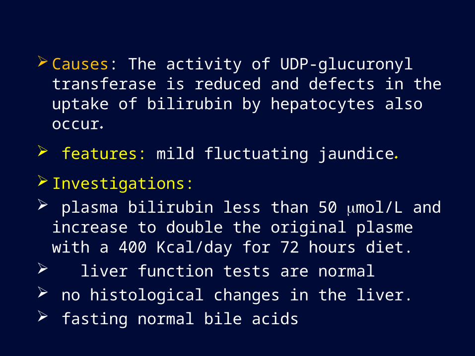
Causes: The activity of UDP-glucuronyl transferase is reduced and defects in the uptake of bilirubin by hepatocytes also occur.
features: mild fluctuating jaundice.
Investigations: plasma bilirubin less than 50 mol/L and increase to
double the original plasme with a 400 Kcal/day for 72 hours diet.
liver function tests are normal no histological changes in the liver. fasting normal bile acids

Second Lecture
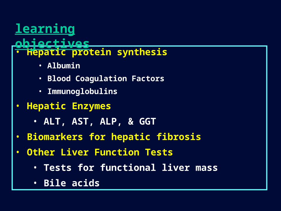
• Hepatic protein synthesis • Albumin
• Blood Coagulation Factors
• Immunoglobulins
• Hepatic Enzymes
• ALT, AST, ALP, & GGT
• Biomarkers for hepatic fibrosis
• Other Liver Function Tests
• Tests for functional liver mass
• Bile acids
learning objectives
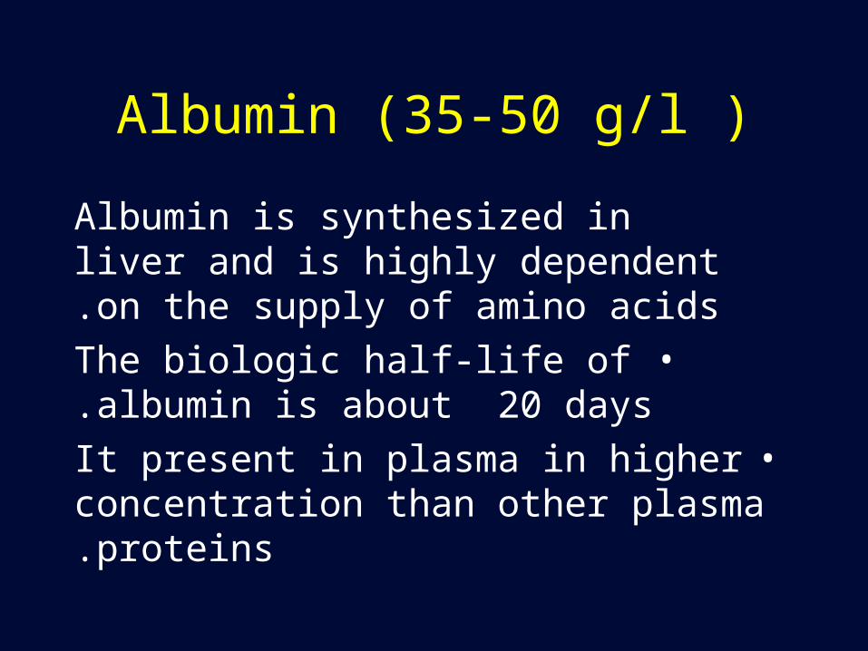
Albumin (35-50 g/l )
Albumin is synthesized in liver and is highly dependent on the supply of amino acids.
•The biologic half-life of albumin is about 20 days.
•It present in plasma in higher concentration than other plasma proteins.
Functions of AlbuminThe main functions of albumin are::
1.Oncotic pressure. Albumin is responsible for approximately 80% of the plasma oncotic pressure (the osmotic pressure due to proteins).This is a major determinant of the distribution of fluid between the intravascular and extravascular compartments and thus plasma volume.
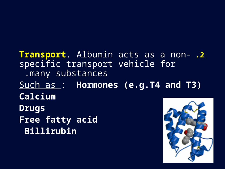
2 .Transport. Albumin acts as a non-specific transport vehicle for many
substances .Such as : Hormones (e.g.T4 and T3)
Calcium Drugs
Free fatty acid Billirubin
Hyperalbuminaemia is rare and is usually caused by dehydration.
Reduced serum albumin levels are common , occurring in many conditions.
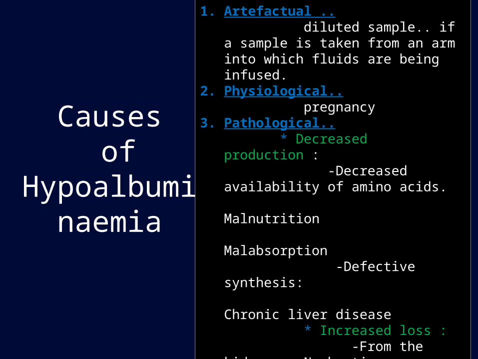
Causes of
Hypoalbuminaemia
1. Artefactual .. diluted sample.. if a sample is taken from
an arm into which fluids are being infused.2. Physiological.. pregnancy3. Pathological.. * Decreased production : -Decreased availability of amino acids. Malnutrition Malabsorption -Defective synthesis: Chronic liver disease * Increased loss : -From the kidney Nephrotic
syndrome - Increased catabolism Trauma Surgery Infection
II- Coagulation factors:
• In liver disease the synthesis of prothrombin and other clotting factors is diminished, prolonged prothrombin Time (PT).
• This may be one of the earliest abnormalities seen in hepatocellular damage, since prothrombin has a short half-life (~ 6h).
• Deficiency of fat soluble vitamin K due to failure of absorption of lipids prolonged pT.
• In vit. K deficiency the coagulation defect can often be corrected by parentral administration of vit. K.
III- Immunoglobulins:
• Plasma Ig measurements are of little value in liver disease, because the changes are of low specificity.
• In most types of cirrhosis plasma IgA.
• In primary biliary cirrhosis plasma IgM.
• In chronic active hepatitis plasma IgG.
a) Aminotransferases (ALT & AST):
ALT is cytomplasmic enzyme ,liver specific, raises at an early stage in hepatic injury.
AST is cytoplamic and mitochondrial enzyme, less liver specific, raises to greater degree in chronic hepatitis.
amounts of both transaminases leak from inflamed or damaged hepatocytes due to acute or chronic hepatitis.
Hepatic Enzymes
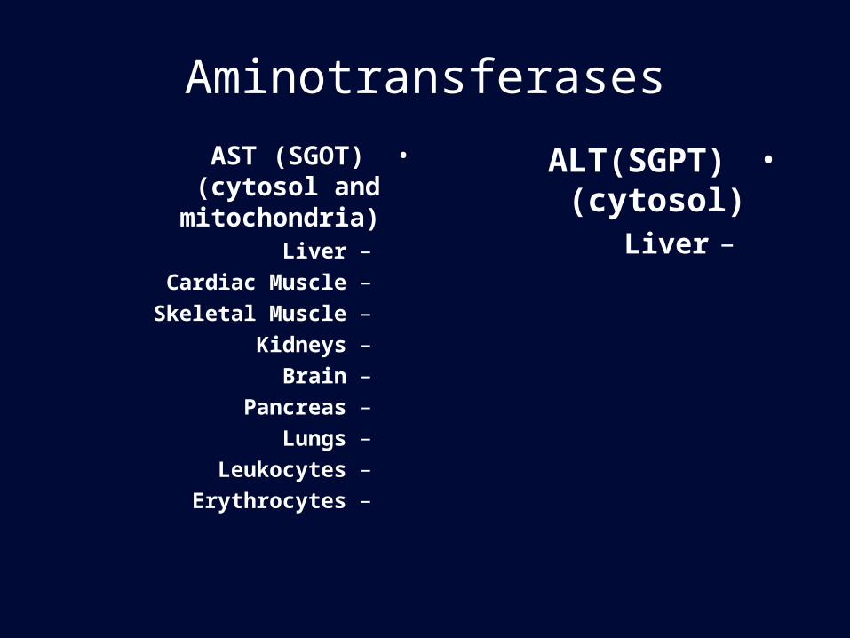
Aminotransferases
•AST (SGOT) (cytosol and mitochondria)
–Liver
–Cardiac Muscle
–Skeletal Muscle
–Kidneys
–Brain
–Pancreas
–Lungs
–Leukocytes
–Erythrocytes
•ALT(SGPT) (cytosol)
–Liver
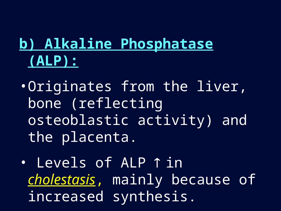
b) Alkaline Phosphatase (ALP):
• Originates from the liver, bone (reflecting osteoblastic activity) and the placenta.
• Levels of ALP in cholestasis, mainly because of increased synthesis.
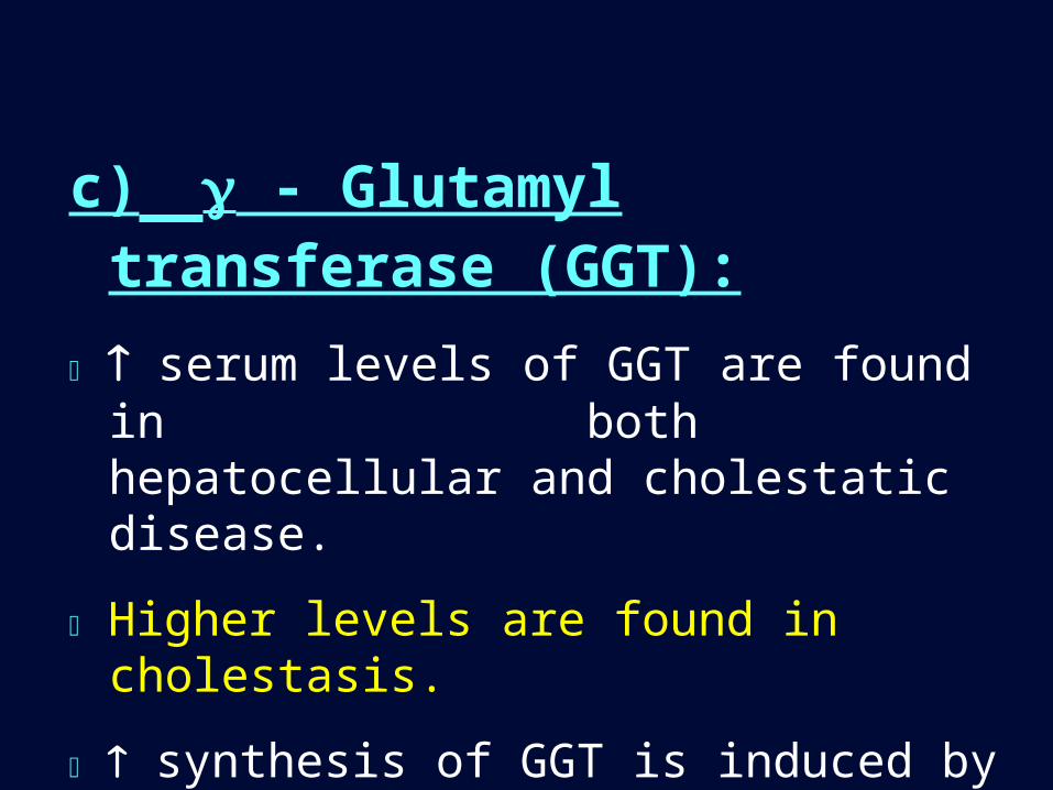
c) - Glutamyl transferase (GGT):
serum levels of GGT are found in both hepatocellular and cholestatic disease.
Higher levels are found in cholestasis.
synthesis of GGT is induced by excessive ethanol intake.
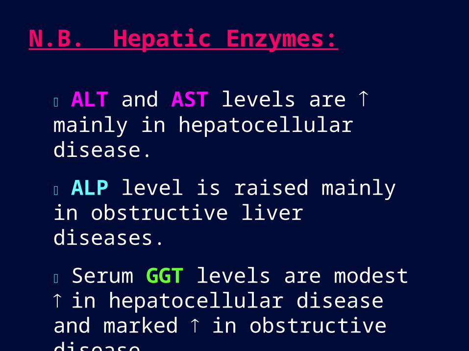
N.B. Hepatic Enzymes:
ALT and AST levels are mainly in hepatocellular disease.
ALP level is raised mainly in obstructive liver diseases.
Serum GGT levels are modest in hepatocellular disease and marked in obstructive disease.
• A Varity of markers have been described that
may be of help in measurement of hepatic fibrosis.
• Procollagen type III terminal peptide and hyaluronic acid (hyaluronin) are the most commonly used tests.
Makers of fibrosis
A. A number of liver function tests have been described to give an indication of the functional liver mass.
• These tests are not often used but include :– the aminopyrine breath tests. – the galactose elimination test.
Other Liver Function Tests
B. Bile Acids :
• bile acids measurement is the most sensitive
test for early detection of liver disease used in:
– Investigation of hepatic dysfunction associated with
pregnancy (increased)
investigation of Gilbert’s syndrome(normal)

The End
Top Related