


Languages
Pages
Legal

Limbic Structures, Emotion, and MemoryET Rolls, Oxford Centre for Computational Neuroscience, Oxford, United Kingdom; and University of Warwick, Coventry,United Kingdom
� 2017 Elsevier Inc. All rights reserved.
Introduction 2Historical Background to the Concept of a Limbic System 2Brain Systems Involved in Emotion: The Orbitofrontal Cortex, Amygdala, and Anterior Cingulate Cortex 2
Emotions Defined 2An Anatomical and Functional Framework for Understanding the Neural Basis of Emotion 3The Orbitofrontal Cortex 5Anatomical and Functional Connectivity 5Effects of Damage to the Orbitofrontal Cortex on Emotion and Emotion-Related Learning 7Reward Outcome Value for Taste, Olfaction, Flavor, Oral Texture, and Oral Temperature in the Orbitofrontal Cortex 8Outcome Value and Somatosensory and Temperature Inputs to the Orbitofrontal Cortex 10Expected Value Visual Inputs to the Orbitofrontal Cortex, Visual Stimulus–Reinforcement Association Learning and Reversal, and Negative RewardPrediction Error Neurons 10Orbitofrontal Cortex Neurons Compared to Dopamine Neurons 11Face-Selective Processing in the Orbitofrontal Cortex 12Top-Down Effects of Cognition and Attention on Taste, Olfactory, Flavor, Somatosensory, and Visual Processing: Cognitive Enhancement of theValue of Affective Stimuli 12Representations of Specific Reward Value on a Common Scale but With No Common Currency, and Emotion 14Absolute Value and Relative Value are Both Represented in the Orbitofrontal Cortex 15Abstract Monetary Reward Value, Social Value, and Attractiveness Are Represented in the Orbitofrontal Cortex 15A Representation of Novel Visual Stimuli in the Orbitofrontal Cortex 15
The Amygdala 15Connections 16Effects of Amygdala Lesions 16Amygdala Neuronal Activity 17Amygdala Damage in Humans 18
The Anterior Cingulate Cortex 18Connections 18Activations and Neuronal Activity 18Effects of Anterior Cingulate Cortex Lesions in Humans 21
The Insular Cortex 21The Insular Primary Taste Cortex 22The Visceral/Autonomic Insular Cortex 22The Somatosensory Insula 22
A Hippocampal Limbic System for Memory and Spatial Function 22Connections 23Effects of Hippocampal System Damage in Primates Including Humans 24Neuronal Representations in the Primate Hippocampus 25Spatial View Neurons in the Primate Hippocampus 25Object-Place Neurons in the Primate Hippocampus 25Recall-Related Neurons in the Primate Hippocampus 25Reward-Place Neurons in the Primate Hippocampus 26Grid Cells in the Entorhinal Cortex 26Neuronal Representations of Space “out there” for Episodic Memory, and Parietal Inputs to the Hippocampus 27
Neural Network Computations in the Hippocampus for Episodic Memory 27Different Computations for the “Emotional Limbic System” From Those in the “Memory Limbic System” 28Conclusions and Summary: Separate Limbic Structures or Systems for Emotion and for Memory, but No Single LimbicSystem 29Acknowledgments 30References 30
Reference Module in Neuroscience and Biobehavioral Psychology http://dx.doi.org/10.1016/B978-0-12-809324-5.06857-7 1
Introduction
Limbic structures include cortical structures such as the hippocampus (and entorhinal cortex), cingulate cortex, and olfactory cortex;and structures to which they are connected such as the mammillary bodies (via the fornix), septal area (including the nucleusaccumbens), and amygdala. The orbitofrontal cortex is closely connected with these limbic structures and can be consideredwith them. The hypothalamus is also closely connected to limbic structures.
These structures are involved in emotion, motivation, episodic memory, and spatial processing.This section of the Reference Module in Neuroscience and Biobehavioral Psychology includes articles that describe the neuroscience of
limbic structures, of emotion, and of memory. This section also includes articles on learning and memory in invertebrates.
Historical Background to the Concept of a Limbic System
The use of the term “limbic” has changed over time, and although the concept of a limbic system is still in use (Mesulam, 2000;Catani et al., 2013), the concept is being replaced by the concept that there are different limbic structures, with different functionsthat include emotion and episodic memory (Rolls, 2015b). The term “limbic”was introduced by ThomasWillis (1664) to designatea cortical border encircling the brain stem (limbus, Latin for “border”). Paul Broca (1878) held the view that “le grand lobe limbi-que”was mainly an olfactory structure common to all mammalian brains, although he argued that its functions were not limited toolfaction. Limbic structures are frequently taken to include cortical structures such as the hippocampus, cingulate cortex, and pyri-form cortex; and structures to which they are connected such as the mammillary bodies, septal area, and amygdala (Isaacson, 1982).After Broca’s publication, the accumulation of experimental evidence from ablation studies in animals broadened the role of limbicstructures to include other aspects of behavior such as controlling social interactions and behavior (Brown and Schäfer, 1888),consolidating memories (Bechterew, 1900), and forming emotions (Cannon, 1927).
Anatomical and physiological advances led James Papez (1937) to describe a neural circuit for linking action and perceptionto emotion. The Papez circuit consists of the hippocampus connecting via the fornix to the mammillary body, which connects viathe mammillothalamic tract to the anterior nuclei of the thalamus and thus back to the cingulate cortex. According to Papez,emotion arises either from cognition entering the circuit from the cortex through the hippocampus or from visceral and somaticperceptions entering the circuit through the hypothalamus. Some of Papez’ evidence on his circuit and emotion was that in rabieswhere the disease appears to have a predilection for the hippocampus and cerebellum, the patient is subject to anxiety, appre-hensiveness, and paroxysms of rage or terror. Papez held that “the cortex of the cingular gyrus may be looked on as the receptiveregion for the experiencing of emotion as the result of impulses coming from the hypothalamic region or the hippocampalformation” (Papez, 1937). A decade later, Paul Yakovlev (1948) proposed that the orbitofrontal cortex, insula, amygdala, andanterior temporal lobe form a network underlying emotion and motivation. Paul MacLean crystallized previous works by incor-porating both Papez’ and Yakovlev’s views into a model of the limbic system (MacLean, 1949, 1952). MacLean concluded thatthe limbic cortex, together with the limbic subcortical structures, is a functionally integrated system involved especially inemotion. Robert Isaacson assembled evidence on the functions of this system in emotion and memory in a book entitled TheLimbic System (Isaacson, 1982).
In the remainder of this Introduction to the section of the Reference Module in Neuroscience and Biobehavioral Psychology on LimbicStructures, Emotion, and Memory, I describe evidence that there are separate systems in the brain for emotion and for episodicmemory, each involving limbic structures; describe a hypothesis about the nature of the links between these systems; and showthat the computations in the two systems are very different (Rolls, 2015b). In doing this, I also provide an introduction to Emotionand Memory, which are included in this section of the Reference Module in Neuroscience and Biobehavioral Psychology.
Brain Systems Involved in Emotion: The Orbitofrontal Cortex, Amygdala, and Anterior Cingulate Cortex
Emotions Defined
A very useful working definition of emotions is that they are states elicited by rewards and punishers, that is, by instrumental rein-forcers (Weiskrantz, 1968; Gray, 1975; Rolls, 2005, 2013a, 2014). Instrumental reinforcers are rewards and punishers that are ob-tained as a result of an action instrumental in gaining the reward or avoiding the punisher. This approach is supported by manyconsiderations (Rolls, 2013a, 2014), including the following three. First, the definition is conceptually acceptable, in that it is diffi-cult to think of exceptions to the rule that rewards and punishers are associated with emotional states and to the rule that emotionalstates are produced by rewards and punishers (Rolls, 2014). Second, the definition is powerful in an evolutionary and explanatorysense, in that the functions of emotion can be conceived of as related to processes involved in obtaining goals, and in states that areproduced when goals are received. Indeed, my evolutionary Darwinian account states that the adaptive value of rewards andpunishers is that they are gene-specified goals for action, and that it is much more effective for genes to specify rewards andpunishers, the goals for action, than to attempt to specify actions (Rolls, 2013a, 2014). Examples of such primary (ie, unlearnedor gene-specified) reinforcers include the taste of food, pain, stimuli that promote reproductive success, and face expression. Otherstimuli become secondary reinforcers by learned associations with primary reinforcers in parts of the brain involved in emotionsuch as the orbitofrontal cortex and amygdala. An example is the sight of food, which by learned association with a primary
2 Limbic Structures, Emotion, and Memory

reinforcer, taste, becomes a secondary reinforcer. Third, this approach provides a principled way to analyze the brain mechanisms ofemotion, by examination of where in the brain stimuli are represented by their reward value (Rolls, 2014, 2016c).
An Anatomical and Functional Framework for Understanding the Neural Basis of Emotion
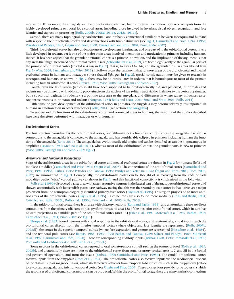
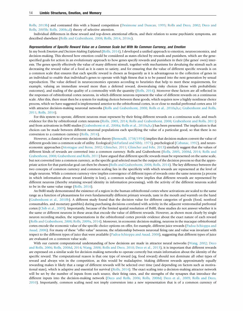
I now provide a framework for understanding some of the brain structures involved in emotion, and at the same time contrast themwith the structures that in terms of connectivity and function precede them and succeed them in the anatomical and functionalhierarchy moving from left to right in Fig. 1 (Rolls, 2014).
In Tier 1 (Fig. 1), information is processed to a level at which the neurons represent “what” the stimulus is, independently of thereward or punishment value of the stimulus. Thus neurons in the primary taste cortex represent what the taste is, and its intensity,but not its reward value (Rolls, 2014, 2016c). In the inferior temporal visual cortex, the representation is of objects, invariantly with
Behavior:Habit
Autonomicand endocrineresponses
Cingulate CortexBehavior:Action-Outcome
Dopamine
V1
ThalamusReceptors solitary tract VPMpc nucleus
VISION
TasteTASTE
Bulb
Frontal operculum/Insula
visual cortexInferior temporal
(Primary Taste Cortex)
Nucleus of the
Amygdala
Gate
Lateral
function
by e.g. glucose utilization,stomach distension or bodyweight
Gate
OrbitofrontalCortex
Hypothalamus
Hunger neuron controlled
TOUCH
OLFACTION
ThalamusVPL
Olfactory
Primary somatosensory cortex (1.2.3)
Olfactory (Pyriform)Cortex
Insula
Striatum
PregenCing
Medial PFC area 10Choice valueDecision-making
'what' decision-making / output
. . . . . . . . . . . . . . . . . . . . . . . . . . . . . . . . . . . . . . . . . . . . . . . . . . . . . . Tier 1 Tier 2 Tier 3
Lateral PFC
V2 V4
Figure 1 Schematic diagram showing some of the connections of the taste, olfactory, somatosensory, and visual pathways in the brain. V1,primary visual (striate) cortex; V2 and V4, further cortical visual areas; PFC, prefrontal cortex; VPL, ventroposterolateral nucleus of the thalamus,which conveys somatosensory information to the primary somatosensory cortex (areas 1, 2, and 3); VPMpc, ventroposteromedial nucleus pars par-vocellularis of the thalamus, which conveys taste information to the primary taste cortex; Pregen Cing, pregenual cingulate cortex. For purposes ofdescription, the stages can be described as Tier 1, representing what object is present independently of reward value; Tier 2 in which reward value isrepresented; and Tier 3 in which decisions between stimuli of different value are taken and in which value is interfaced to behavioral output systems.
Limbic Structures, Emotion, and Memory 3

respect to the exact position on the retina, size, and even view. Forming invariant representations involves a great deal of corticalcomputation in the hierarchy of visual cortical areas from the primary visual cortex V1 to the inferior temporal visual cortex (Rolls,2008d, 2012a, 2016c). The fundamental advantage of this separation of “what” processing in Tier 1 from reward value processing inTier 2 is that any learning in Tier 2 of the value of an object or face seen in one location on the retina, size, and view will generalize toother views etc. In rodents, there is no such clear separation of “what” from “value” representations. For example, in the taste system,satiety influences taste processing at the first central synapse in the taste system (Rolls and Scott, 2003), and this property makes theprocessing in rodents not only different from that in primates including humans, but also much more difficult to analyze (Rolls,2014, 2015c, 2016c).
There are brain mechanisms in Tier 2 in the orbitofrontal cortex that are involved in computing the reward value of primary(unlearned) reinforcers, as shown by devaluation experiments in which, for example, a food is fed to satiety (Rolls et al., 1989a;Critchley and Rolls, 1996a; Kringelbach et al., 2003; Rolls and Grabenhorst, 2008) and by neuroeconomics experiments whichshow that the amount and quality of each commodity are encoded by orbitofrontal cortex neurons (Padoa-Schioppa and Assad,2008; Grabenhorst and Rolls, 2011; Padoa-Schioppa, 2011). The primary reinforcers include taste, touch (both pleasant touchand pain), and to some extent smell, and perhaps certain visual stimuli such as face expression. There is evidence that there is a repre-sentation of the (reward/punishment) value of many primary reinforcers in the orbitofrontal cortex, including taste, positive touchand pain, face expression, face beauty, and auditory consonance/dissonance. In neuroeconomics, these are termed “outcome value”representations (Rolls, 2014). Further evidence for value representations is that orbitofrontal cortex activations in humans to thesestimuli are linearly related to the subjectively reported pleasantness of stimuli (medially) or to their unpleasantness (laterally)(Rolls, 2014).
Brain regions in Tier 2 are also concerned with learning associations between previously neutral stimuli, such as the sight ofobjects or of individuals’ faces, with primary reinforcers. These brain regions include the amygdala and orbitofrontal cortex,with the orbitofrontal cortex being especially important in the rapid, one-trial learning and reversal of stimulus–reinforcer associ-ations. In neuroeconomics, these are termed “expected value” representations. Once the Tier 2 brain regions have determinedwhether the input is reinforcing, whether primary or secondary, the signal is passed directly to output regions of the brain, withno need to produce and then feed back peripheral body or autonomic responses to the brain.
In the orbitofrontal cortex in Tier 2, the representation is of the value of stimuli, and actions are not represented. The values ofvery many different types of stimuli, events, or goals are represented separately at the neuronal level, providing the basis for choicebetween stimuli, and the selection at later stages of processing of an appropriate action to obtain the chosen goal.
Whereas the orbitofrontal cortex in Tier 2 represents the value of stimuli (potential goals for action) on a continuous scale, anarea anterior to this, medial prefrontal cortex area 10 (in Tier 3), is implicated in decision-making between stimuli, in which a selec-tion or choice must be made, moving beyond a representation of value on a continuous scale toward a decision between goodsbased on their value (Rolls et al., 2008b; Grabenhorst et al., 2011; Rolls, 2014).
The brain regions in which the reinforcing, and hence emotional, value of stimuli are represented interface to three main types ofoutput system:
The first is the autonomic and endocrine system, for producing such changes as increased heart rate and release of adrenaline, whichprepare the body for action. Structures receiving from the orbitofrontal cortex, amygdala, and anterior cingulate cortex (ACC)that provide a route for these autonomic effects include the hypothalamus and parts of the anterior insula close to the insulartaste cortex (Critchley and Harrison, 2013; Rolls, 2014).
The second type of output is to brain systems concerned with performing actions unconsciously or implicitly, to obtain rewards oravoid punishers. These brain systems include the basal ganglia for habit (“stimulus-response”) behavior, and the ACC for action-outcome learning. (The “outcome” is the reward or punisher that is or is not obtained when the action is performed.) The ACCcontains representations of reward and punisher value, and thus of outcome, which are essential for learning associationsbetween actions and the outcomes that follow actions. The midcingulate area contains representations of actions.
The third type of output is to a system capable of planning many steps ahead and, for example, deferring short-term rewards toexecute a long-term plan. This system may use syntactic processing to perform the planning and is therefore part of a linguisticsystem which performs explicit (conscious) processing, as described more fully elsewhere (Rolls, 2014, 2016c).
It is notable that the orbitofrontal cortex and amygdala do not receive inputs from the dorsal visual “where” processing areas suchas the parietal cortex including the retrosplenial cortex (which is part of the posterior cingulate cortex) that provide inputs via parahip-pocampal areas TF/TH to the hippocampus for its spatial (“where”) functions in memory, which are described in the section A Hippo-campal Limbic System For Memory And Spatial Function. In a complementary way, the hippocampus and parahippocampal areas donot contain value representations of stimuli, except insofar as value may be part of a memory such as reward-place memory (Rolls andXiang, 2005, 2006; Rolls, 2010b, 2016c). This is part of the evidence that the emotional and episodic memory systems have differentconnections and functions, as described in the section AHippocampal Limbic System ForMemory And Spatial Function and elsewhere(Rolls and Xiang, 2005, 2006; Rolls, 2008d, 2010b, 2016c), and thus that there is no single and unified limbic system.
Because of the intended relevance to understanding human emotion and its disorders, the focus of the research described here ison humans and macaques. This is important for many of the brain systems that are involved in emotion have undergone consider-able development in primates (eg, monkeys and humans) (Rolls, 2014, 2016c), as summarized next.
First, the temporal lobe has undergone great development in primates, and several systems in the temporal lobe either areinvolved in emotion (eg, the amygdala) or provide some of the main sensory inputs to brain systems involved in emotion and
4 Limbic Structures, Emotion, and Memory
motivation. For example, the amygdala and the orbitofrontal cortex, key brain structures in emotion, both receive inputs from thehighly developed primate temporal lobe cortical areas, including those involved in invariant visual object recognition, and faceidentity and expression processing (Rolls, 2000b, 2008d, 2011a, 2012a, 2016c).
Second, there are many topological, cytoarchitectural, and probably connectional similarities between macaques and humanswith respect to the orbitofrontal cortex and its connections with limbic structures (see Fig. 1; Carmichael and Price, 1994, 1995a;Petrides and Pandya, 1995; Öngür and Price, 2000; Kringelbach and Rolls, 2004; Price, 2006, 2007).
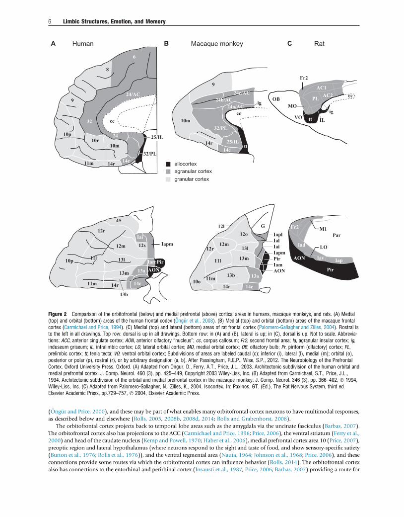
Third, the prefrontal cortex has also undergone great development in primates, and one part of it, the orbitofrontal cortex, is verylittle developed in rodents, yet is one of the major brain areas involved in emotion and motivation in primates including humans.Indeed, it has been argued that the granular prefrontal cortex is a primate innovation, and the implication of the argument is thatany areas that might be termed orbitofrontal cortex in rats (Schoenbaum et al., 2009) are homologous only to the agranular parts ofthe primate orbitofrontal cortex (shaded mid gray in Fig. 2), that is, to areas 13a, 14c, and the agranular insular areas labeled Ia inFig. 2 (Wise, 2008; Passingham and Wise, 2012). It follows from that argument that for most areas of the orbitofrontal and medialprefrontal cortex in humans and macaques (those shaded light gray in Fig. 2), special consideration must be given to research inmacaques and humans. As shown in Fig. 2, there may be no cortical area in rodents that is homologous to most of the primateincluding human orbitofrontal cortex (Preuss, 1995; Wise, 2008; Passingham and Wise, 2012).
Fourth, even the taste system (which might have been supposed to be phylogenetically old and preserved) of primates androdents may be different, with obligatory processing from the nucleus of the solitary tract via the thalamus to the cortex in primates,but a subcortical pathway in rodents via a pontine taste area to the amygdala, and differences in where satiety influences taste-responsive neurons in primates and rodents (Norgren, 1984; Rolls and Scott, 2003; Small and Scott, 2009; Rolls, 2014).
Fifth, with the great development of the orbitofrontal cortex in primates, the amygdala may become relatively less important inhumans in emotion than in other vertebrates (Rolls, 2014) (see section The Amygdala).
To understand the functions of the orbitofrontal cortex and connected areas in humans, the majority of the studies describedhere were therefore performed with macaques or with humans.
The Orbitofrontal Cortex
The first structure considered is the orbitofrontal cortex, and although not a limbic structure such as the amygdala, has similarconnections to the amygdala, is connected to the amygdala, and has considerably eclipsed in primates including humans the func-tions of the amygdala (Rolls, 2014). The amygdala has evolutionarily old origins and can be identified, as can the hippocampus, inamphibia (Isaacson, 1982; Medina et al., 2011), whereas most of the orbitofrontal cortex, the granular parts, is new to primates(Wise, 2008; Passingham and Wise, 2012; Fig. 2).
Anatomical and Functional ConnectivityMaps of the architectonic areas in the orbitofrontal cortex and medial prefrontal cortex are shown in Fig. 2 for humans (left) andmonkeys (middle) (Carmichael and Price, 1994; Öngür et al., 2003). The connections of the orbitofrontal cortex (Carmichael andPrice, 1994, 1995b; Barbas, 1995; Petrides and Pandya, 1995; Pandya and Yeterian, 1996; Öngür and Price, 2000; Price, 2006,2007) are summarized in Fig. 3. Conceptually, the orbitofrontal cortex can be thought of as receiving from the ends of eachmodality-specific “what” cortical pathway as shown in Fig. 1, and this functional connectivity is emphasized in the following.
Rolls et al. (1990) discovered a taste area with taste-responsive neurons in the lateral part of themacaque orbitofrontal cortex andshowed anatomically with horseradish peroxidase pathway tracing that this was the secondary taste cortex in that it receives a majorprojection from the neurophysiologically identified primary taste cortex (Baylis et al., 1995). This region projects on to more ante-rior areas of the orbitofrontal cortex (Baylis et al., 1995). Taste neurons are also found more medially (Rolls and Baylis, 1994;Critchley and Rolls, 1996b; Rolls et al., 1996b; Pritchard et al., 2005; Rolls, 2008b).
In the midorbitofrontal cortex, there is an area with olfactory neurons (Rolls and Baylis, 1994), and anatomically there are directconnections from the primary olfactory cortex, pyriform cortex, to area 13a of the posterior orbitofrontal cortex, which in turn hasonward projections to a middle part of the orbitofrontal cortex (area 13) (Price et al., 1991; Morecraft et al., 1992; Barbas, 1993;Carmichael et al., 1994; Price, 2007; see Fig. 1).
Thorpe et al. (1983) found neurons with visual responses in the orbitofrontal cortex, and anatomically, visual inputs reach theorbitofrontal cortex directly from the inferior temporal cortex (where object and face identity are represented (Rolls, 2007b,2008d)), the cortex in the superior temporal sulcus (where face expression and gesture are represented (Hasselmo et al., 1989)),and the temporal pole cortex (see Barbas, 1988, 1993, 1995; Barbas and Pandya, 1989; Seltzer and Pandya, 1989; Morecraftet al., 1992; Carmichael and Price, 1995b). There are corresponding auditory inputs (Barbas, 1988, 1993; Romanski et al., 1999;Romanski and Goldman-Rakic, 2001; Rolls et al., 2006b).
Some neurons in the orbitofrontal cortex respond to oral somatosensory stimuli such as the texture of food (Rolls et al., 1999,2003b), and anatomically there are inputs to the orbitofrontal cortex from somatosensory cortical areas 1, 2, and SII in the frontaland pericentral operculum, and from the insula (Barbas, 1988; Carmichael and Price, 1995b). The caudal orbitofrontal cortexreceives inputs from the amygdala (Price et al., 1991). The orbitofrontal cortex also receives inputs via the mediodorsal nucleusof the thalamus, pars magnocellularis, which itself receives afferents from temporal lobe structures such as the prepyriform (olfac-tory) cortex, amygdala, and inferior temporal cortex (see Öngür and Price, 2000). These connections provide some routes via whichthe responses of orbitofrontal cortex neurons can be produced. Within the orbitofrontal cortex, there are many intrinsic connections
Limbic Structures, Emotion, and Memory 5
(Öngür and Price, 2000), and these may be part of what enables many orbitofrontal cortex neurons to have multimodal responses,as described below and elsewhere (Rolls, 2005, 2008b, 2008d, 2014; Rolls and Grabenhorst, 2008).
The orbitofrontal cortex projects back to temporal lobe areas such as the amygdala via the uncinate fasciculus (Barbas, 2007).The orbitofrontal cortex also has projections to the ACC (Carmichael and Price, 1996; Price, 2006), the ventral striatum (Ferry et al.,2000) and head of the caudate nucleus (Kemp and Powell, 1970; Haber et al., 2006), medial prefrontal cortex area 10 (Price, 2007),preoptic region and lateral hypothalamus (where neurons respond to the sight and taste of food, and show sensory-specific satiety(Burton et al., 1976; Rolls et al., 1976)), and the ventral tegmental area (Nauta, 1964; Johnson et al., 1968; Price, 2006), and theseconnections provide some routes via which the orbitofrontal cortex can influence behavior (Rolls, 2014). The orbitofrontal cortexalso has connections to the entorhinal and perirhinal cortex (Insausti et al., 1987; Price, 2006; Barbas, 2007) providing a route for
9
10p10r
10m
11m 14r 14c32/PL
24
32
8
6
A B CHuman Macaque monkey Rat
24/AC
IG24a
24b/AC24c/AC
9
32/PL
25/IL14c
14r
10m
24a/ACcc
tt
ig
agranular cortexgranular cortex
allocortex
Iapm
10p
12r
45
12m
Iai
Pir13l
13m 13a
13b
11m 14r 14c
Iam
12s
11l
12l12o
12r
10o 11m
11l
12m13l
13m
13b 13a
Iam
IapmPir
AON
IaiIalIapl
G
14r 14c
Fr2 M1
Iad LO
Iav Iap
Par
Fr2
AC1
PL
IL
MO
VO
AC2 ccOB
ttig
Pir
AON
cc
25/IL
AON
Figure 2 Comparison of the orbitofrontal (below) and medial prefrontal (above) cortical areas in humans, macaque monkeys, and rats. (A) Medial(top) and orbital (bottom) areas of the human frontal codex (Öngür et al., 2003). (B) Medial (top) and orbital (bottom) areas of the macaque frontalcortex (Carmichael and Price, 1994). (C) Medial (top) and lateral (bottom) areas of rat frontal cortex (Palomero-Gallagher and Zilles, 2004). Rostral isto the left in all drawings. Top row: dorsal is up in all drawings. Bottom row: in (A) and (B), lateral is up; in (C), dorsal is up. Not to scale. Abbrevia-tions: ACC, anterior cingulate cortex; AON, anterior olfactory “nucleus”; cc, corpus callosum; Fr2, second frontal area; Ia, agranular insular cortex; ig,induseum griseum; IL, infralimbic cortex; LO, lateral orbital cortex; MO, medial orbital cortex; OB, olfactory bulb; Pr, piriform (olfactory) cortex; PL,prelimbic cortex; tt, tenia tecta; VO, ventral orbital cortex; Subdivisions of areas are labeled caudal (c); inferior (i), lateral (l), medial (m); orbital (o),posterior or polar (p), rostral (r), or by arbitrary designation (a, b). After Passingham, R.E.P., Wise, S.P., 2012. The Neurobiology of the PrefrontalCortex. Oxford University Press, Oxford. (A) Adapted from Ongur, D., Ferry, A.T., Price, J.L., 2003. Architectonic subdivision of the human orbital andmedial prefrontal cortex. J. Comp. Neurol. 460 (3), pp. 425–449, Copyright 2003 Wiley-Liss, Inc. (B) Adapted from Carmichael, S.T., Price, J.L.,1994. Architectonic subdivision of the orbital and medial prefrontal cortex in the macaque monkey. J. Comp. Neurol. 346 (3), pp. 366–402, � 1994,Wiley-Liss, Inc. (C) Adapted from Palomero-Gallagher, N., Zilles, K., 2004. Isocortex. In: Paxinos, GT. (Ed.), The Rat Nervous System, third ed.Elsevier Academic Press, pp.729–757, � 2004, Elsevier Academic Press.
6 Limbic Structures, Emotion, and Memory
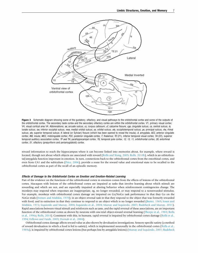
reward information to reach the hippocampus where it can become linked into memories about, for example, where reward islocated, though not about which objects are associated with reward (Rolls and Xiang, 2005; Rolls, 2010b), which is an orbitofron-tal/amygdala function important in emotion. In turn, connections back to the orbitofrontal cortex from the entorhinal cortex, andeven from CA1 and the subiculum (Price, 2006), provide a route for the reward value and emotional state to be recalled to theorbitofrontal cortex as part of the recall of an episodic memory.
Effects of Damage to the Orbitofrontal Cortex on Emotion and Emotion-Related LearningPart of the evidence on the functions of the orbitofrontal cortex in emotion comes from the effects of lesions of the orbitofrontalcortex. Macaques with lesions of the orbitofrontal cortex are impaired at tasks that involve learning about which stimuli arerewarding and which are not, and are especially impaired at altering behavior when reinforcement contingencies change. Themonkeys may respond when responses are inappropriate, eg, no longer rewarded, or may respond to a nonrewarded stimulus.For example, monkeys with orbitofrontal cortex damage are impaired on Go/NoGo task performance in that they Go on theNoGo trials (Iversen and Mishkin, 1970); in an object reversal task in that they respond to the object that was formerly rewardedwith food; and in extinction in that they continue to respond to an object which is no longer rewarded (Butter, 1969; Jones andMishkin, 1972; Izquierdo and Murray, 2004; Izquierdo et al., 2004; Murray and Izquierdo, 2007; Rudebeck and Murray, 2011).Rapid associations between visual stimuli and reinforcers such as taste, and the rapid reversal of these associations, are an importantfunction of the orbitofrontal cortex as shown by neurons with one-trial object-reward reversal learning (Thorpe et al., 1983; Rollset al., 1996a; Rolls, 2014). Consistent with this, in humans, rapid reversal is impaired by orbitofrontal cortex damage (Rolls et al.,1994; Fellows and Farah, 2003; Hornak et al., 2004).
Orbitofrontal cortex damage affects reward value as also shown by devaluation investigations. Sensory-specific satiety (a methodof reward devaluation in which a food is fed to satiety), which is implemented neuronally in the orbitofrontal cortex (Rolls et al.,1989a), is impaired by orbitofrontal cortex lesions (but perhaps less by amygdala lesions) (Murray and Izquierdo, 2007; Rudebeck
28
ACC
MCC
PCC
1014
11
12
13
cc
as
fros
ps
sts
ios
mts
cgs
ots
cf
rhs
46
cs ips
TG
TA
lf
TE 21
ls
Medial Inverted
Lateral
Ventral view oforbitofrontal cortex
TGlos
mos
os
A
T
TFTH
22
olfactory
visualTE20taste
auditory
oryam
sotosens
51
Insula
ros
Figure 3 Schematic diagram showing some of the gustatory, olfactory, and visual pathways to the orbitofrontal cortex and some of the outputs ofthe orbitofrontal cortex. The secondary taste cortex and the secondary olfactory cortex are within the orbitofrontal cortex. V1, primary visual cortex;V4, visual cortical area V4. Abbreviations: as, arcuate sulcus; cc, corpus callosum; cf, calcarine fissure; cgs, cingulate sulcus; cs, central sulcus; ls,lunate sulcus; ios, inferior occipital sulcus; mos, medial orbital sulcus; os, orbital sulcus; ots, occipitotemporal sulcus; ps, principal sulcus; rhs, rhinalsulcus; sts, superior temporal sulcus; lf, lateral (or Sylvian) fissure (which has been opened to reveal the insula); A, amygdala; ACC, anterior cingulatecortex; INS, insula; MCC, midcingulate cortex; PCC, posterior cingulate cortex; T, thalamus; TE (21), inferior temporal visual cortex; TA (22), superiortemporal auditory association cortex; TF and TH, parahippocampal cortex; TG, temporal pole cortex; 12, 13, 11, orbitofrontal cortex; 28, entorhinalcortex; 51, olfactory (prepyriform and periamygdaloid) cortex.
Limbic Structures, Emotion, and Memory 7
and Murray, 2011). In relation to neuroeconomics, the estimation of expected reward value as influenced by reward size, and delayto reward, or both, is impaired by orbitofrontal cortex lesions in macaques (Simmons et al., 2010).
It is suggested that difficulty in processing reinforcers, and especially in rapid visual discrimination reversal learning, under-lies some of the impairments in emotion produced by damage to the orbitofrontal cortex (Rolls, 2014). In humans, euphoria,irresponsibility, lack of affect, and impulsiveness can follow frontal lobe damage (Damasio, 1994; Rolls, 1999a; Kolb andWhishaw, 2003; Zald and Rauch, 2006), particularly orbitofrontal cortex damage (Rolls et al., 1994; Hornak et al., 1996,2003; Rolls, 1999a, 2014; Berlin et al., 2004, 2005). These emotional changes may be related at least in part to a failure torapidly update the reinforcement associations of stimuli when the contingencies are changed as in a visual discriminationreversal task (Rolls et al., 1994; Rolls, 1999c, 2014; Fellows and Farah, 2003; Berlin et al., 2004; Hornak et al., 2004; Fellows,2007, 2011). Similar mechanisms may contribute at least in part to the poor performance of humans with ventromedialprefrontal cortex damage on the Iowa Gambling Task (Bechara et al., 2000; Maia and McClelland, 2004). It is of interestthat the patients with bilateral orbitofrontal cortex damage who were impaired at the visual discrimination reversal taskhad high scores on parts of a Social Behavior Questionnaire in which the patients were rated on behaviors such as emotionrecognition in others (eg, their sad, angry, or disgusted mood); in interpersonal relationships (such as not caring what othersthink, and not being close to the family); emotional empathy (eg, when others are happy, is not happy for them); interpersonalrelationships (eg, does not care what others think, and is not close to his family); public behavior (is uncooperative); antisocialbehavior (is critical of and impatient with others); impulsivity (does things without thinking); and sociability (is notsociable and has difficulty making or maintaining close relationships) (Hornak et al., 2003, 2004), all of which could reflectless behavioral sensitivity to different types of punishment and reward. Further, in a Subjective Emotional Change Question-naire in which the patients reported on any changes in the intensity and/or frequency of their own experience of emotions, thebilateral orbitofrontal cortex lesion patients with deficits in the visual discrimination reversal task reported a number ofchanges, including changes in sadness, anger, fear, and happiness (Hornak et al., 2003).
Reward Outcome Value for Taste, Olfaction, Flavor, Oral Texture, and Oral Temperature in the Orbitofrontal CortexTaste and Oral Texture
One of the discoveries that have helped us to understand the functions of the orbitofrontal cortex in behavior is that it containsa major cortical representation of taste (see Rolls et al., 1990; Rolls, 1995a, 1997, 2014, 2015a, 2016b; Rolls and Scott, 2003;Kadohisa et al., 2005b; cf. Fig. 1). Given that taste can act as a primary reinforcer, that is, without learning as a reward or punisher,we now have the start for a fundamental understanding of the function of the orbitofrontal cortex in stimulus–reinforcer asso-ciation learning (Rolls, 1999a, 2004, 2008d, 2014). We know how one class of primary reinforcers reaches and is representedin the orbitofrontal cortex. A representation of primary reinforcers is essential for a system that is involved in learning associationsbetween previously neutral stimuli and primary reinforcers, eg, between the sight of an object and its taste.
The representation in the orbitofrontal cortex (shown by analyzing the responses of single neurons in macaques) is for themajority of neurons the reward value of taste (Rolls et al., 1990, 1996b, 1998a; Baylis and Rolls, 1991; Rolls, 1995a, 1997,2000d; Rolls and Scott, 2003; Kadohisa et al., 2005b) and oral texture including viscosity (Rolls et al., 2003b), fat texture (Rollset al., 1999; Verhagen et al., 2003), and astringency as exemplified by tannic acid (Critchley and Rolls, 1996b). The evidence forthis is that the responses of orbitofrontal cortex taste neurons are modulated by hunger (as is the reward value or palatability ofa taste). In particular, it has been shown that orbitofrontal cortex taste neurons gradually stop responding to the taste of a foodas the monkey is fed to satiety, but not to the taste of other foods, revealing a mechanism for sensory-specific satiety and rewarddevaluation (Rolls et al., 1989a, 1996b). In contrast, the representation of taste in the primary taste cortex (Scott et al., 1986; Yaxleyet al., 1990) is not modulated by hunger (Rolls et al., 1988; Yaxley et al., 1988). Thus in the primate including human primary tastecortex, the reward value of taste is not represented, and instead the identity and intensity of the taste are represented (Grabenhorstand Rolls, 2008; Grabenhorst et al., 2008b; Rolls, 2008d, 2014).
Additional evidence that the reward value of food is represented in the orbitofrontal cortex is that monkeys work for electricalstimulation of this brain region if they are hungry, but not if they are satiated (Mora et al., 1979; Rolls, 2005). Further, neurons inthe orbitofrontal cortex are activated from many brain-stimulation reward sites (Mora et al., 1980; Rolls et al., 1980). Thus there isclear evidence that it is the reward value of taste that is represented in the orbitofrontal cortex (see further Rolls, 1999a, 2000a,2014), and this is further supported by the finding that feeding to satiety decreases the activation of the human orbitofrontal cortexto the food eaten to satiety in a sensory-specific way (Kringelbach et al., 2003). Some orbitofrontal cortex neurons respond to the“taste” of water in the mouth (Rolls et al., 1990), and their responses occur only when thirsty and not when satiated (Rolls et al.,1989a); and correspondingly in humans the subjective pleasantness or affective value of the taste of water in the mouth is repre-sented in the orbitofrontal cortex (de Araujo et al., 2003b). This is part of the evidence for the separation of a “what” tier of pro-cessing, which in this case is the primary taste cortex, from a reward and affect-related representation in the orbitofrontal cortex tierof processing, as shown in Fig. 1.
Functional neuroimaging studies in humans have shown that the most medial part of the human orbitofrontal cortex is acti-vated by taste, oral texture, and olfactory stimuli (Francis et al., 1999; O’Doherty et al., 2000; Small et al., 2001; de Araujo et al.,2003a,c, 2005; Rolls et al., 2003a; de Araujo and Rolls, 2004; Small et al., 2005; Gottfried et al., 2006; McCabe and Rolls, 2007;Rolls and McCabe, 2007) and that the activations correlate with ratings of subjective pleasantness and so are in the domain ofaffective representations (Kringelbach and Rolls, 2004; Rolls, 2014). This most medial part of the human orbitofrontal cortex mayhave moved medially when compared with the representation in macaques, probably because of the extensive development of the
8 Limbic Structures, Emotion, and Memory

dorsolateral prefrontal cortex in humans (Rolls, 2008b; Rolls and Grabenhorst, 2008). Affectively pleasant stimuli are often rep-resented medially, and unpleasant or aversive stimuli laterally, in the human orbitofrontal cortex. Evidence consistent with thishas been found for taste (O’Doherty et al., 2001a; de Araujo et al., 2003a), pleasant touch (Francis et al., 1999; Rolls et al., 2003c),and pleasant versus aversive olfactory stimuli (Francis et al., 1999; O’Doherty et al., 2000; Rolls, 2000a; Rolls et al., 2003a) (seefurther Kringelbach and Rolls, 2004; Grabenhorst and Rolls, 2011). An important point for those seeking to understand thehedonic topology of the human orbitofrontal cortex is that it should not be assumed to be the same as that in macaques.
An Olfactory Reward Representation in the Orbitofrontal Cortex
For 35% of orbitofrontal cortex olfactory neurons, the odors to which a neuron responded were influenced by the taste value(glucose or saline) with which the odor was associated (Critchley and Rolls, 1996c). Thus the odor representation for 35% of orbi-tofrontal neurons appeared to be built by olfactory-to-taste association learning. This possibility was confirmed by reversing thetaste with which an odor was associated in the reversal of an olfactory discrimination task. It was found that 68% of the sampleof neurons analyzed altered the way in which they responded to odor when the taste reinforcement association of the odor wasreversed (Rolls et al., 1996a). The olfactory-to-taste reversal was quite slow, both neurophysiologically and behaviorally, oftenrequiring 20–80 trials, consistent with the need for some stability of flavor representations formed by a combination of odorand taste inputs.
To analyze the nature of the olfactory representation in the orbitofrontal cortex, Critchley and Rolls (1996a) measured theresponses of olfactory neurons that responded to food while they fed the monkey to satiety. They found that the majority of orbi-tofrontal olfactory neurons decreased their responses to the odor of the food with which the monkey was fed to satiety. Thus forthese neurons, the reward value of the odor is what is represented in the orbitofrontal cortex (cf. Rolls and Rolls, 1997), and thisparallels the changes in the relative pleasantness of different foods after a food is eaten to satiety (Rolls et al., 1981a,b; Rolls, 1997;see Rolls, 1999a, 2000a, 2014). The subjective pleasantness or reward or affective value of odor is represented in the orbitofrontalcortex, in that feeding humans to satiety decreases the activation found to the odor of that food, and this effect is relatively specific tothe food eaten in the meal (Francis et al., 1999; O’Doherty et al., 2000; cf. Morris and Dolan, 2001). Further, the human medialorbitofrontal cortex has activation that is related to the subjective pleasantness of a set of odors, and a more lateral area has acti-vation that is related to the degree of subjective unpleasantness of odors (Rolls et al., 2003a). An fMRI investigation in humansshowed that whereas in the orbitofrontal cortex the pleasantness versus unpleasantness of odors is represented, this was not thecase in primary olfactory cortical areas, where instead the activations reflected the intensity of the odors (Rolls et al., 2003a),providing a further example of the hierarchy of “what” followed by reward processing shown in Fig. 1.
Convergence of Taste and Olfactory Inputs in the Orbitofrontal Cortex: The Representation of Flavor
In the orbitofrontal cortex, not only unimodal taste neurons, but also unimodal olfactory neurons are found. In addition, somesingle neurons respond to both gustatory and olfactory stimuli, often with correspondence between the two modalities (Rollsand Baylis, 1994). It is probably here in the orbitofrontal cortex of primates including humans that these two modalities convergeto produce the representation of flavor (Rolls and Baylis, 1994; de Araujo et al., 2003c), for neurons in the primary taste cortex in theinsular/frontal opercular cortex do not respond to olfactory (or visual) stimuli (Verhagen et al., 2004).
The importance of the combination of taste and smell for producing affectively pleasant and rewarding representations ofsensory stimuli is exemplified by findings with umami, the delicious taste or flavor that is associated with combinations of compo-nents that include meat, fish, milk, tomatoes, and mushrooms, all of which are rich in umami-related substances such as glutamateor inosine 50-monophosphate. Umami taste is produced by glutamate acting on a fifth taste system (Chaudhari et al., 2000; Zhaoet al., 2003; Maruyama et al., 2006). However, glutamate presented alone as a taste stimulus is not highly pleasant and does not actsynergistically with other tastes (sweet, salt, bitter, and sour). However, when glutamate is given in combination with a consonant,savory, odor (vegetable), the resulting flavor can be much more pleasant (McCabe and Rolls, 2007). We showed using functionalbrain imaging with fMRI that this glutamate taste and savory odor combination produced much greater activation of the medialorbitofrontal cortex and pregenual cingulate cortex than the sum of the activations by the taste and olfactory components presentedseparately (McCabe and Rolls, 2007). Supralinear effects were much less (and significantly less) evident for sodium chloride andvegetable odor. Further, activations in these brain regions were correlated with the subjective pleasantness and fullness of the flavor,and with the consonance of the taste and olfactory components. Supralinear effects of glutamate taste and savory odor were notfound in the insular primary taste cortex. We thus proposed that glutamate acts by the nonlinear effects it can produce whencombined with a consonant odor in multimodal cortical taste-olfactory convergence regions. We suggested that umami can bethought of as a rich and delicious flavor that is produced by a combination of glutamate taste and a consonant savory odor (Rolls,2009c). Glutamate is thus a flavor enhancer because of the way that it can combine supralinearly with consonant odors in corticalareas where the taste and olfactory pathways converge far beyond the receptors (McCabe and Rolls, 2007).
Oral Texture and Temperature
A population of orbitofrontal cortex neurons responds when a fatty food such as cream is in the mouth. These neurons can also beactivated by pure fat such as glyceryl trioleate and by nonfat substances with a fatlike texture such as paraffin oil (hydrocarbon) andsilicone oil [(Si(CH3)2O)n]. These neurons thus provide information by somatosensory pathways that a fatty food is in the mouth(Rolls et al., 1999). These inputs are perceived as pleasant when hungry, because of the utility of ingestion of foods that are likely to
Limbic Structures, Emotion, and Memory 9
contain essential fatty acids and to have a high calorific value (Rolls, 2000a, 2014). Satiety produced by eating a fatty food, cream,can decrease the responses of orbitofrontal cortex neurons to the texture of fat in the mouth (Rolls et al., 1999).
We have shown that the orbitofrontal cortex receives inputs from a number of different oral texture channels, which togetherprovide a rich sensory representation of what is in the mouth (Rolls, 2011c, 2012b). Using a set of stimuli in which viscositywas systematically altered (carboxymethylcellulose with viscosity in the range 10–10 000 centipoise), we have shown that someorbitofrontal cortex neurons encode fat texture independently of viscosity (by a physical parameter that varies with the slicknessof fat) (Verhagen et al., 2003); that other orbitofrontal cortex neurons encode the viscosity of the texture in the mouth (withsome neurons tuned to viscosity and others showing increasing or decrease firing rates as viscosity increases) (Rolls et al.,2003b); and that other neurons have responses that indicate the presence of texture stimuli (such as grittiness and capsaicin) inthe mouth independently of viscosity and slickness (Rolls et al., 2003b). Ensemble (ie, population, distributed) encoding of allthese variables is found (Rolls et al., 2010d; Rolls and Treves, 2011). In a complementary human functional neuroimaging study,it has been shown that activation of parts of the orbitofrontal cortex, primary taste cortex, and mid-insular somatosensory regionposterior to the insular taste cortex has activations that are related to the viscosity of what is in the mouth and that there is in addi-tion a medial prefrontal/cingulate area where the mouth feel of fat is represented (de Araujo and Rolls, 2004). Moreover, the subjec-tive pleasantness of fat is represented in the orbitofrontal cortex and a region to which it projects the pregenual cingulate cortex(Grabenhorst et al., 2010a).
An overlapping population of orbitofrontal cortex neurons represents the temperature of what is in the mouth (Kadohisa et al.,2004), and this is supported by a human fMRI study (Guest et al., 2007).
Outcome Value and Somatosensory and Temperature Inputs to the Orbitofrontal CortexIn addition to these oral somatosensory inputs to the orbitofrontal cortex, there are also somatosensory inputs from other parts ofthe body (Rolls, 2010c), and indeed an fMRI investigation we have performed in humans indicates that pleasant and painfultouch stimuli to the hand produce greater activation of the orbitofrontal cortex relative to the somatosensory cortex than do affec-tively neutral stimuli (Francis et al., 1999; Rolls et al., 2003c). In an fMRI investigation in humans, it was found that the mid-orbitofrontal and pregenual cingulate cortex and a region to which they project, the ventral striatum, have activations that arecorrelated with the subjective pleasantness ratings made to warm (41�C) and cold (12�C) stimuli, and combinations of warmand cold stimuli, applied to the hand (Rolls et al., 2008b). Activations in the lateral and some more anterior parts of the orbito-frontal cortex were correlated with the unpleasantness of the stimuli. In contrast, activations in the somatosensory cortex andventral posterior insula were correlated with the intensity but not the pleasantness of the thermal stimuli. Further, cognitivemodulators of affective value such as the description of cream being rubbed on the arm as “rich and moisturizing” increase acti-vations to the sight of rubbing of the arm in the orbitofrontal and pregenual cingulate cortex, and increased correlations therewith the subjectively rated pleasantness of the touch (McCabe et al., 2008).
A principle thus appears to be that processing related to the affective value and associated subjective emotional experience ofthermal stimuli that are important for survival is performed in different brain areas to those where activations are related to sensoryproperties of the stimuli such as their intensity. This conclusion appears to be the case for processing in a number of sensory modal-ities, including taste (Grabenhorst and Rolls, 2008; Grabenhorst et al., 2008b) and olfaction (Anderson et al., 2003; Rolls et al.,2003a; Grabenhorst et al., 2007), and the finding with such prototypical stimuli as warm and cold (Rolls et al., 2008b) providesstrong support for this principle (see Fig. 1).
Nonglabrous skin such as that on the forearm contains C fiber tactile afferents that respond to light moving touch (Olaussonet al., 2002). The orbitofrontal cortex is implicated in some of the affectively pleasant aspects of touch that may be mediatedthrough C fiber tactile afferents, in that it is activated more by light touch to the forearm than by light touch to the glabrousskin (palm) of the hand (McCabe et al., 2008).
Expected Value Visual Inputs to the Orbitofrontal Cortex, Visual Stimulus–Reinforcement Association Learning and Reversal, andNegative Reward Prediction Error NeuronsWe have been able to show that there is a major visual input to many neurons in the orbitofrontal cortex and that what is repre-sented by these neurons is in many cases the reinforcement association of visual stimuli. The visual input is from the ventral,temporal lobe, visual stream concerned with “what” object is being seen (see Rolls, 2000b, 2012a). Many neurons in these temporalcortex visual areas have responses to objects or faces that are invariant with respect to size, position on the retina, and even view(Rolls, 2000b, 2007b, 2008a,d, 2009d, 2012a, 2016c), making these neurons ideal as an input to a system that may learn aboutthe reinforcement association properties of objects and faces, for after a single learning trial, the learning then generalizes correctlyto other views etc. (see Rolls, 2000b, 2008d, 2012a, 2014). Using this object-related information, orbitofrontal cortex visualneurons frequently respond differentially to objects or images depending on their reward association (Thorpe et al., 1983; Rollset al., 1996a). The primary reinforcer that has been used is taste, and correlates of visual to taste association learning have beendemonstrated in the human orbitofrontal cortex with fMRI (O’Doherty et al., 2002). Many of these neurons show visual-tastereversal in one or a very few trials. (In a visual discrimination task, they will reverse the stimulus to which they respond, from,eg, a triangle to a square, in one trial when the taste delivered for a behavioral response to that stimulus is reversed (Thorpeet al., 1983).) This reversal learning probably occurs in the orbitofrontal cortex, for it does not occur one synapse earlier in the visualinferior temporal cortex (Rolls et al., 1977), and it is in the orbitofrontal cortex that there is convergence of visual and taste pathwaysonto the same single neurons (Thorpe et al., 1983; Rolls and Baylis, 1994; Rolls et al., 1996a).
10 Limbic Structures, Emotion, and Memory
The probable mechanism for this learning is an associative modification of synapses conveying visual input onto taste-responsive neurons, implementing a pattern association network (Rolls and Treves, 1998; Rolls and Deco, 2002; Rolls, 2008d,2014; see Fig. 8), with the reversal facilitated by a rule for which stimulus is currently rewarded held in short-term memory(Deco and Rolls, 2005a).
The visual and olfactory neurons in primates that respond to the sight or smell of stimuli that are primary reinforcers such as tasteclearly signal an expectation of reward that is based on previous stimulus–reinforcement associations (Thorpe et al., 1983; Rollset al., 1996a). So do the conditional reward neurons which reflect the reward value only for one of a pair of stimuli (Thorpeet al., 1983; Rolls et al., 1996a; Rolls and Grabenhorst, 2008). With visual–taste association learning and reversal in primates, inwhich the orbitofrontal cortex neurons and the behavior can change in one trial (Thorpe et al., 1983; Rolls et al., 1996a), thechanging responses of the orbitofrontal cortex neurons can contribute to the reversed behavior, a view of course supported bythe impaired reversal learning produced in primates including humans by orbitofrontal cortex damage (eg, Rolls et al., 1994;Fellows and Farah, 2003; Berlin et al., 2004; Hornak et al., 2004; Murray and Izquierdo, 2007).
To analyze the nature of the visual representation of food-related stimuli in the orbitofrontal cortex, Critchley and Rolls (1996a)measured the responses of neurons that responded to the sight of food while they fed the monkey to satiety in a devaluation inves-tigation. They found that the majority of orbitofrontal visual food-related neurons decreased their responses to the sight of the foodwith which the monkey was fed to satiety. Thus for these neurons, the expected reward value of the sight of food is what is repre-sented in the orbitofrontal cortex.
In addition to these neurons that encode the reward association of visual stimuli, other, “error,” neurons in the orbitofrontalcortex detect nonreward, in that they respond, for example, when an expected reward is not obtained when a visual discriminationtask is reversed (Thorpe et al., 1983), or when reward is no longer made available in a visual discrimination task. These may becalled “negative reward prediction error neurons” (Rolls and Grabenhorst, 2008; Rolls, 2014). Evidence that there may be similarerror neurons in the human orbitofrontal cortex is that in a model of social learning, orbitofrontal cortex activation occurred ina visual discrimination reversal task at the time when the face of one person no longer was associated with a smile, but becameassociated with an angry expression, indicating on such error trials that reversal of choice to the other individual’s face should occur(Kringelbach and Rolls, 2003).
The orbitofrontal cortex negative reward prediction error neurons respond to a mismatch between the reward expected and thereward that is obtained. Both signals are represented in the orbitofrontal cortex, in the form of, for example, neurons that respondto the sight of a learned reinforcer such as the sight of a stimulus paired with taste, and neurons that respond to the primary reinforcer,the taste (or texture or temperature). The orbitofrontal cortex is the probable brain region for this computation, because both thesignals required to compute negative reward prediction error are present in the orbitofrontal cortex, so are the negative reward predic-tion error neurons, and lesions of the orbitofrontal cortex impair tasks such as visual discrimination reversal in which this type of nega-tive reward prediction error is needed (see above and Rolls, 2014). In a new theory of depression, it has been proposed thatoversensitive nonreward attractor networks in the lateral orbitofrontal cortex contribute to depression, with in addition reduced func-tional connectivity of the medial orbitofrontal cortex reward-related system with hippocampal memory systems (Rolls, 2016a).
Orbitofrontal Cortex Neurons Compared to Dopamine NeuronsThe dopamine neurons in the midbrain that respond to positive reward prediction error (a greater reward than expected) may notbe able to provide a good representation of negative reward prediction error, because their spontaneous firing rates are so low(Schultz, 2004) that much further reduction would provide only a small signal. In any case, the dopamine neurons would notappear to be in a position to compute a negative reward prediction error, as they are not known to receive inputs that signal expectedreward, and the actual reward (outcome) that is obtained, and indeed do not represent the reward obtained (or “outcome”), in thatthey stop responding to a taste reward outcome if it is predictable. Although some dopamine neurons do appear to represent a posi-tive reward prediction error signal (responding if a greater than expected reward is obtained) (Schultz, 2004, 2006, 2013), they donot appear to have the signals required to compute this, that is, the expected reward, and the reward outcome obtained, so evena positive reward prediction error must be computed elsewhere. The orbitofrontal cortex does contain representations of thesetwo signals, the expected reward and the reward outcome, and has projections to the ventral striatum, which in turn projects tothe region of the midbrain dopamine neurons, and so this is one possible pathway along which the firing of positive reward predic-tion error might be computed (see Fig. 1; Rolls, 2014). Consistent with this, activations in parts of the human ventral striatum arerelated to positive reward prediction error (Hare et al., 2008; Rolls et al., 2008a). Thus the dopamine projections to the prefrontalcortex and other areas are not likely to convey information about reward to the prefrontal cortex, which instead is likely to bedecoded by the neurons in the orbitofrontal cortex that represent primary reinforcers, and the orbitofrontal cortex neurons thatlearn associations of other stimuli to the primary reinforcers to represent expected value (Thorpe et al., 1983; Rolls et al., 1996a,2008a; Rolls, 2008d). Although it has been suggested that the firing of dopamine neurons may reflect the earliest signal ina task that indicates reward and could be used as a reward prediction error signal during learning (see Schultz et al., 2000; Schultz,2006), it is likely, partly on the basis of the above evidence, though an interesting topic for future investigation, that any error infor-mation to which dopamine neurons fire originates from representations in the orbitofrontal cortex that encode expected value andreward outcome, and which connect to the ventral striatum (Rolls, 2008d, 2009a, 2014). A further problem is that some dopamineneurons respond to aversive or salient stimuli (Matsumoto and Hikosaka, 2009; Bromberg-Martin et al., 2010), and overall thepopulation may not code a reward prediction error (Rolls, 2014).
Limbic Structures, Emotion, and Memory 11
Face-Selective Processing in the Orbitofrontal CortexAnother type of visual information represented in the orbitofrontal cortex is information about faces. There is a population of orbi-tofrontal cortex neurons that respond in many ways similarly to those in the temporal cortical visual areas (Rolls, 1984, 1992b,1996a, 2000b, 2007b, 2008a,d, 2011a, 2012a; Rolls and Deco, 2002). The orbitofrontal cortex face-responsive neurons, firstobserved by Thorpe et al. (1983), then by Rolls et al. (2006b), tend to respond with longer latencies than temporal lobe neurons(140–200 ms typically, compared to 80–100 ms); also convey information about which face is being seen, by having differentresponses to different faces, and are typically rather harder to activate strongly than temporal cortical face-selective neurons, inthat many of them respond much better to real faces than to two-dimensional images of faces on a video monitor (Rolls et al.,2006b; Rolls, 2011a; cf. Rolls and Baylis, 1986). Some of the orbitofrontal cortex face-selective neurons are responsive to faceexpression, gesture, or movement (Rolls et al., 2006b). The findings are consistent with the likelihood that these neurons are acti-vated via the inputs from the temporal cortical visual areas in which face-selective neurons are found (see Fig. 1). The significance ofthe neurons is likely to be related to the fact that faces convey information that is important in social reinforcement in at leasttwo ways that could be implemented by these neurons. The first is that some may encode face expression (Rolls et al., 2006b;cf. Hasselmo et al., 1989), which can indicate reinforcement. The second way is that they encode information about which indi-vidual is present (Rolls et al., 2006b), which by stimulus–reinforcement association learning is important in evaluating and utilizinglearned reinforcing inputs in social situations, eg, about the current reinforcement value as decoded by stimulus–reinforcementassociation, to a particular individual. Between them, these neurons represent whose face has a particular expression, and this isimportant in social situations.
This system has also been shown to be present in humans. For example, Kringelbach and Rolls (2003) showed that activation ofa part of the human orbitofrontal cortex occurs during a face discrimination reversal task. In the task, the faces of two different indi-viduals are shown, and when the correct face is selected, the expression turns into a smile. (The expression turns to angry if thewrong face is selected.) After a period of correct performance, the contingencies reverse, and the other face must be selected to obtaina smile expression as a reinforcer. It was found that activation of a part of the orbitofrontal cortex occurred specifically in relation tothe reversal, that is, when a formerly correct face was chosen, but an angry face expression was obtained. In a control task, it wasshown that the activations were not related just to showing an angry face expression. Thus in humans, there is a part of the orbito-frontal cortex that responds selectively in relation to face expression specifically when it indicates that behavior should change, andthis activation is error-related (Kringelbach and Rolls, 2003) and occurs when the error neurons in the orbitofrontal cortex becomeactive (Thorpe et al., 1983).
Also prompted by the neuronal recording evidence of face and auditory neurons in the orbitofrontal cortex (Rolls et al., 2006b),it has further been shown that there are impairments in the identification of facial and vocal emotional expression in a group ofpatients with ventral frontal lobe damage who had socially inappropriate behavior (Hornak et al., 1996). The expression identifi-cation impairments could occur independently of perceptual impairments in facial recognition, voice discrimination, or environ-mental sound recognition. Poor performance on both expression tests was correlated with the degree of alteration of emotionalexperience reported by the patients. There was also a strong positive correlation between the degree of altered emotional experienceand the severity of the behavioral problems (eg, disinhibition) found in these patients (Hornak et al., 1996). A comparison group ofpatients with brain damage outside the ventral frontal lobe region, without these behavioral problems, was unimpaired on the faceexpression identification test, was significantly less impaired at vocal expression identification, and reported little subjectiveemotional change (Hornak et al., 1996). It has further been shown that patients with discrete surgical lesions of restricted partsof the orbitofrontal cortex may have face and/or voice expression identification impairments, and these are likely to contributeto their difficulties in social situations (Hornak et al., 2003).
Top-Down Effects of Cognition and Attention on Taste, Olfactory, Flavor, Somatosensory, and Visual Processing: CognitiveEnhancement of the Value of Affective StimuliHow does cognition influence affective value? How does cognition influence the way that we feel emotionally? Do cognition andemotion interact in regions that are high in the brain’s hierarchy of processing, for example, in areas where language processingoccurs, or do cognitive influences descend down anatomically to influence the first regions that represent the affective value ofstimuli?
An fMRI study to address these fundamental issues in brain design has shown that cognitive effects can reach down into thehuman orbitofrontal cortex and influence activations produced by odors (de Araujo et al., 2005). In this study, a standard testodor, isovaleric acid with a small amount of cheese flavor, was delivered through an olfactometer. (The odor alone, like theodor of brie, might have been interpreted as pleasant, or perhaps as unpleasant.) On some trials the test odor was accompaniedwith the visually presented word label “cheddar cheese,” and on other trials with the word label “body odor.” It was found thatthe activation in the medial orbitofrontal cortex to the standard test odor was much greater when the word label was cheddar cheesethan when it was body odor. (Controls with clean air were run to show that the effect could not be accounted for by the word labelalone.) Moreover, the word labels influenced the subjective pleasantness ratings to the test odor, and the changing pleasantnessratings were correlated with the activations in the human medial orbitofrontal cortex. Part of the interest and importance of thisfinding is that it shows that cognitive influences, originating here purely at the word level, can reach down andmodulate activationsin the first stage of cortical processing that represents the affective value of sensory stimuli (de Araujo et al., 2005; Rolls, 2014).
Also important is how cognition influences the affective brain representations of the taste and flavor of a food. This is importantnot only for understanding top-down influences in the brain, but also in relation to the topical issues of appetite control and obesity
12 Limbic Structures, Emotion, and Memory

(Rolls, 2007c,d, 2010a, 2011b, 2012b). In an fMRI study, it was shown that activations related to the affective value of umami taste andflavor (as shown by correlations with pleasantness ratings) in the orbitofrontal cortex were modulated by word-level descriptors (eg,“rich and delicious flavor”) (Grabenhorst et al., 2008b). Affect-related activations to taste weremodulated in a region that receives fromthe orbitofrontal cortex, the pregenual cingulate cortex, and to taste and flavor in another region that receives from the orbitofrontalcortex, the ventral striatum. Affect-related cognitive modulations were not found in the insular taste cortex, where the intensity but notthe pleasantness of the taste was represented. Thus the top-down language-level cognitive effects reach far down into the earliestcortical areas that represent the appetitive value of taste and flavor. This is an important way anatomically in which cognition influ-ences the neural mechanisms that control appetite and emotion.
When we see a person being touched, we may empathize the feelings being produced by the touch. Interestingly, cognitivemodulation of this effect can be produced. When subjects were informed by word labels that a cream seen being rubbed ontothe forearm was a “rich moisturizing cream” versus “basic cream,” these cognitive labels influenced activations in the orbitofron-tal/pregenual cingulate cortex and ventral striatum to the sight of touch and their correlations with the pleasantness ratings (McCabeet al., 2008). Some evidence for top-down cognitive modulation of the somatosensory effects produced by the subject being rubbedwith the cream was found in brain regions such as the orbitofrontal and pregenual cingulate cortex and ventral striatum, but someeffects were found in other brain regions, perhaps reflecting backprojections from the orbitofrontal cortex (McCabe et al., 2008;Rolls, 2010c).
What may be a fundamental principle of how top-down attention can influence affective versus nonaffective processing hasrecently been discovered. For an identical taste stimulus, paying attention to pleasantness activated some brain systems (includingemotion-related limbic structures), and paying attention to intensity, which reflected the physical and not the affective properties ofthe stimulus, activated other brain systems (Grabenhorst and Rolls, 2008). In an fMRI investigation, when subjects were instructedto remember and rate the pleasantness of a taste stimulus, 0.1 M monosodium glutamate, activations were greater in the medialorbitofrontal and pregenual cingulate cortex than when subjects were instructed to remember and rate the intensity of the taste.When the subjects were instructed to remember and rate the intensity, activations were greater in the insular taste cortex. Thus,depending on the context in which tastes are presented and whether affect is relevant, the brain responds to a taste differently. Thesefindings show that when attention is paid to affective value, the brain systems engaged to represent the sensory stimulus of taste aredifferent from those engaged when attention is directed to the physical properties of a stimulus such as its intensity. This differentialbiasing of brain regions engaged in processing a sensory stimulus depending on whether the attentional demand is for affect-relatedversus more sensory-related processing may be an important aspect of cognition and attention. This has many implications forunderstanding attentional effects to affective value not only on taste, but also on other sensory stimuli (Ge et al., 2012; Luoet al., 2013; Rolls, 2013b, 2014, 2016c).
Indeed, the concept has been validated in the olfactory system too. In an fMRI investigation, when subjects were instructed toremember and rate the pleasantness of a jasmine odor, activations were greater in the medial orbitofrontal and pregenual cingulatecortex than when subjects were instructed to remember and rate the intensity of the odor (Rolls et al., 2008d). When the subjectswere instructed to remember and rate the intensity, activations were greater in the inferior frontal gyrus. These top-down effectsoccurred not only during odor delivery, but started in a preparation period after the instruction before odor delivery and continuedafter termination of the odor in a short-term memory period. Thus, depending on the context in which odors are presented andwhether affect is relevant, the brain prepares itself, responds to, and remembers an odor differently. These findings show thatwhen attention is paid to affective value, the brain systems, engaged to prepare for, represent, and remember a sensory stimulus,are different from those engaged when attention is directed to the physical properties of a stimulus such as its intensity. This differ-ential biasing of brain regions engaged in processing a sensory stimulus depending on whether the cognitive/attentional demand isfor affect-related versus more sensory-related processing may be important for understanding how the context can influence how weprocess stimuli that may have affective properties, how different people may respond differently to stimuli if they process the stimuliin different ways, and more generally, how attentional set can influence the processing of affective stimuli by influencing processingin, for example, the orbitofrontal cortex and related areas (Rolls, 2013b, 2014).
The principle thus appears to be that top-down attentional and cognitive effects on affective value influence representationsselectively in cortical areas that process the affective value and associated subjective emotional experience of taste (Grabenhorstand Rolls, 2008; Grabenhorst et al., 2008b) and olfactory (Anderson et al., 2003; Rolls et al., 2003a; Grabenhorst et al., 2007)stimuli in brain regions such as the orbitofrontal cortex; whereas top-down attentional and cognitive effects on intensity influencerepresentations in brain areas that process the intensity and identity of the stimulus such as the primary taste and olfactory corticalareas (Anderson et al., 2003; Rolls et al., 2003a; Grabenhorst et al., 2007, 2008b; Grabenhorst and Rolls, 2008). This is compu-tationally appropriate in top-down models of attention (Rolls and Deco, 2002; Deco and Rolls, 2005c; Rolls, 2008d, 2013b,2016c).
To investigate the anatomical source of the top-down modulatory effects on attentional processing, we utilized fMRI psycho-physiological interaction connectivity analyses (Friston et al., 1997) with taste stimuli when attention was being paid to the pleas-antness or to the intensity (Grabenhorst and Rolls, 2010). We showed that, in the anterior lateral prefrontal cortex at Y ¼ 53 mm,the correlation with activity in orbitofrontal cortex and pregenual cingulate cortex seed regions was greater when attention was topleasantness compared to when attention was to intensity. Conversely, we showed that, in a more posterior region of lateralprefrontal cortex at Y ¼ 34, the correlation with activity in the anterior insula seed region was greater when attention was to inten-sity compared to when attention was to pleasantness (Grabenhorst and Rolls, 2010; Ge et al., 2012; Luo et al., 2013).We proposed a biased activation theory of selective attention to account for the findings (Grabenhorst and Rolls, 2010;
Limbic Structures, Emotion, and Memory 13
Rolls, 2013b) and contrasted this with a biased competition (Desimone and Duncan, 1995; Rolls and Deco, 2002; Deco andRolls, 2005b; Rolls, 2008c,d) theory of selective attention.
Individual differences in these reward and top-down attentional effects, and their relation to some psychiatric symptoms, aredescribed elsewhere (Rolls and Grabenhorst, 2008; Rolls, 2014, 2016c).
Representations of Specific Reward Value on a Common Scale but With No Common Currency, and EmotionIn my book Emotion and Decision-Making Explained (Rolls, 2014), I developed a unified approach to emotion, neuroeconomics, anddecision-making. This showed how emotion could be considered as states elicited by rewards and punishers, which are the gene-specified goals for action in an evolutionary approach to how genes specify rewards and punishers in their (the genes’ own) inter-ests. The genes specify effectively the value of many different stimuli, together with mechanisms for devaluing the stimuli such asdecreasing the reward value of a food as it is eaten to satiety, and for ensuring that the value of different specific rewards is ona common scale that ensures that each specific reward is chosen as frequently as it is advantageous to the collection of genes inan individual to enable that individual’s genes to operate with high fitness that is to be passed into the next generation by sexualreproduction. The value defined in neuroeconomics operates according to heuristics that help to meet these requirements, forexample, valuing an immediate reward more than a deferred reward, downvaluing risky choices (those with probabilisticoutcomes), and trading of the quality of a commodity with the quantity (Rolls, 2014). Moreover these factors are all reflected inthe responses of orbitofrontal cortex neurons, in which different neurons represent the value of different rewards on a continuousscale. After this, there must then be a system for making choices between these goods, which requires now a highly nonlinear choiceprocess, which we have suggested is implemented anterior to the orbitofrontal cortex, in or close to medial prefrontal cortex area 10with attractor decision-making neuronal networks (Rolls and Grabenhorst, 2008; Rolls et al., 2010a,b,c; Grabenhorst and Rolls,2011; Rolls, 2014).
For this system to operate, different neurons must represent by their firing different rewards on a continuous scale, and muchevidence for this by orbitofrontal cortex neurons (Rolls, 2005, 2014; Rolls and Grabenhorst, 2008; Grabenhorst and Rolls, 2011)and from activations in fMRI studies (Grabenhorst et al., 2008a; Rolls et al., 2010a,b,c) has been presented. The implication is thatchoices can be made between different neuronal populations each specifying the value of a particular good, so that there is noconversion to a common currency (Rolls, 2014).
However, a classical view of economic decision theory (Bernoulli, 1738/1954) implies that decision-makers convert the value ofdifferent goods into a common scale of utility. Ecological (McFarland and Sibly, 1975), psychological (Cabanac, 1992), and neuro-economic approaches (Montague and Berns, 2002; Glimcher, 2011; Glimcher and Fehr, 2013) similarly suggest that the values ofdifferent kinds of rewards are converted into a common currency. Rolls and Grabenhorst (Rolls, 2005, 2008d, 2014; Rolls andGrabenhorst, 2008; Grabenhorst and Rolls, 2011) have argued that different specific rewards must be represented on the same scale,but not converted into a common currency, as the specific goal selected must be the output of the decision process so that the appro-priate action for that particular goal can then be chosen (Rolls and Grabenhorst, 2008; Rolls, 2014). The key difference between thetwo concepts of common currency and common scaling lies in the specificity with which rewards are represented at the level ofsingle neurons. While a common currency view implies convergence of different types of rewards onto the same neurons (a processin which information about reward identity is lost), a common scaling view implies that different rewards are represented bydifferent neurons (thereby retaining reward identity in information processing), with the activity of the different neurons scaledto be in the same value range (Rolls, 2014).
An fMRI study demonstrated the existence of a region in the human orbitofrontal cortex where activations are scaled to the samerange as a function of pleasantness for even fundamentally different primary rewards, taste in the mouth and warmth on the hand(Grabenhorst et al., 2010b). A different study found that the decision value for different categories of goods (food, nonfoodconsumables, and monetary gambles) during purchasing decisions correlated with activity in the adjacent ventromedial prefrontalcortex (Chib et al., 2009). Importantly, because of the limited spatial resolution of fMRI, these studies do not answer whether it isthe same or different neurons in these areas that encode the value of different rewards. However, as shown most clearly by singleneuron recording studies, the representations in the orbitofrontal cortex provide evidence about the exact nature of each reward(Rolls and Grabenhorst, 2008; Rolls, 2009a, 2014). Moreover, in economic decision-making, neurons in the macaque orbitofrontalcortex encode the economic value of the specific choice options on offer, for example, different juice rewards (Padoa-Schioppa andAssad, 2006). For many of these “offer value” neurons, the relationship between neuronal firing rate and value was invariant withrespect to the different types of juice that were available (Padoa-Schioppa and Assad, 2008), suggesting that different types of juiceare evaluated on a common value scale.
With our current computational understanding of how decisions are made in attractor neural networks (Wang, 2002; Decoand Rolls, 2006; Rolls, 2008d, 2014; Wang, 2008; Rolls and Deco, 2010; Deco et al., 2013), it is important that different rewardsare expressed on a similar scale for decision-making networks to operate correctly but retain information about the identity of thespecific reward. The computational reason is that one type of reward (eg, food reward) should not dominate all other types ofreward and always win in the competition, as this would be maladaptive. Making different rewards approximately equallyrewarding makes it likely that a range of different rewards will be selected over time (and depending on factors such as motiva-tional state), which is adaptive and essential for survival (Rolls, 2014). The exact scaling into a decision-making attractor networkwill be set by the number of inputs from each source, their firing rates, and the strengths of the synapses that introduce thedifferent inputs into the decision-making network (Deco and Rolls, 2006; Rolls, 2008d; Deco et al., 2009; Rolls and Deco,2010). Importantly, common scaling need not imply conversion into a new representation that is of a common currency of
14 Limbic Structures, Emotion, and Memory
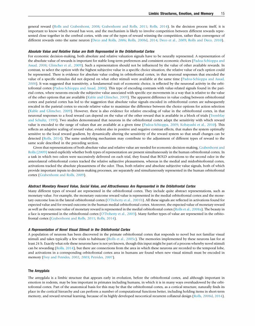
general reward (Rolls and Grabenhorst, 2008; Grabenhorst and Rolls, 2011; Rolls, 2014). In the decision process itself, it isimportant to know which reward has won, and the mechanism is likely to involve competition between different rewards repre-sented close together in the cerebral cortex, with one of the types of reward winning the competition, rather than convergence ofdifferent rewards onto the same neuron (Deco and Rolls, 2006; Rolls, 2008d, 2014; Deco et al., 2009; Rolls and Deco, 2010).
Absolute Value and Relative Value are Both Represented in the Orbitofrontal CortexFor economic decision-making, both absolute and relative valuation signals have to be neurally represented. A representation ofthe absolute value of rewards is important for stable long-term preferences and consistent economic choices (Padoa-Schioppa andAssad, 2008; Glimcher et al., 2009). Such a representation should not be influenced by the value of other available rewards. Incontrast, to select the option with the highest subjective value in a specific choice situation, the relative value of each option couldbe represented. There is evidence for absolute value coding in orbitofrontal cortex, in that neuronal responses that encoded thevalue of a specific stimulus did not depend on what other stimuli were available at the same time (Padoa-Schioppa and Assad,2008). It was suggested that transitivity, a fundamental trait of economic choice, is reflected by the neuronal activity in the orbi-tofrontal cortex (Padoa-Schioppa and Assad, 2008). This type of encoding contrasts with value-related signals found in the pari-etal cortex, where neurons encode the subjective value associated with specific eye movements in a way that is relative to the valueof the other options that are available (Kable and Glimcher, 2009). The apparent difference in value coding between orbitofrontalcortex and parietal cortex has led to the suggestion that absolute value signals encoded in orbitofrontal cortex are subsequentlyrescaled in the parietal cortex to encode relative value to maximize the difference between the choice options for action selection(Kable and Glimcher, 2009). However, there is also evidence for relative encoding of value in the orbitofrontal cortex, in thatneuronal responses to a food reward can depend on the value of the other reward that is available in a block of trials (Tremblayand Schultz, 1999). Two studies demonstrated that neurons in the orbitofrontal cortex adapt the sensitivity with which rewardvalue is encoded to the range of values that are available at a given time (Padoa-Schioppa, 2009; Kobayashi et al., 2010). Thisreflects an adaptive scaling of reward value, evident also in positive and negative contrast effects, that makes the system optimallysensitive to the local reward gradient, by dynamically altering the sensitivity of the reward system so that small changes can bedetected (Rolls, 2014). The same underlying mechanism may contribute to the adjustment of different types of reward to thesame scale described in the preceding section.
Given that representations of both absolute value and relative value are needed for economic decision-making, Grabenhorst andRolls (2009) tested explicitly whether both types of representation are present simultaneously in the human orbitofrontal cortex. Ina task in which two odors were successively delivered on each trial, they found that BOLD activations to the second odor in theanterolateral orbitofrontal cortex tracked the relative subjective pleasantness, whereas in the medial and midorbitofrontal cortex,activations tracked the absolute pleasantness of the odor. Thus, both relative and absolute subjective value signals, both of whichprovide important inputs to decision-making processes, are separately and simultaneously represented in the human orbitofrontalcortex (Grabenhorst and Rolls, 2009).
Abstract Monetary Reward Value, Social Value, and Attractiveness Are Represented in the Orbitofrontal CortexMany different types of reward are represented in the orbitofrontal cortex. They include quite abstract representations, such asmonetary value. For example, the monetary outcome reward value is represented in the medial orbitofrontal cortex and the mone-tary outcome loss in the lateral orbitofrontal cortex (O’Doherty et al., 2001b). All these signals are reflected in activations found forexpected value and for reward outcome in the humanmedial orbitofrontal cortex. Moreover, the expected value of monetary rewardas well as the outcome value of monetary reward is represented in the medial orbitofrontal cortex (Rolls et al., 2008a). The beauty ina face is represented in the orbitofrontal cortex (O’Doherty et al., 2003). Many further types of value are represented in the orbito-frontal cortex (Grabenhorst and Rolls, 2011; Rolls, 2014).
A Representation of Novel Visual Stimuli in the Orbitofrontal CortexA population of neurons has been discovered in the primate orbitofrontal cortex that responds to novel but not familiar visualstimuli and takes typically a few trials to habituate (Rolls et al., 2005c). The memories implemented by these neurons last for atleast 24 h. Exactly what role these neurons have is not yet known, though this input might be part of a process whereby novel stimulican be rewarding (Rolls, 2014), but there are connections from the area in which these neurons are recorded to the temporal lobe,and activations in a corresponding orbitofrontal cortex area in humans are found when new visual stimuli must be encoded inmemory (Frey and Petrides, 2002, 2003; Petrides, 2007).
The Amygdala
The amygdala is a limbic structure that appears early in evolution, before the orbitofrontal cortex, and although important inemotion in rodents, may be less important in primates including humans, in which it is in many ways overshadowed by the orbi-tofrontal cortex. Part of the anatomical basis for this may be that the orbitofrontal cortex, as a cortical structure, naturally finds itsplace in the cortical hierarchy and can perform a number of computational functions better, including holding items in short-termmemory, and reward reversal learning, because of its highly developed neocortical recurrent collateral design (Rolls, 2008d, 2014).
Limbic Structures, Emotion, and Memory 15
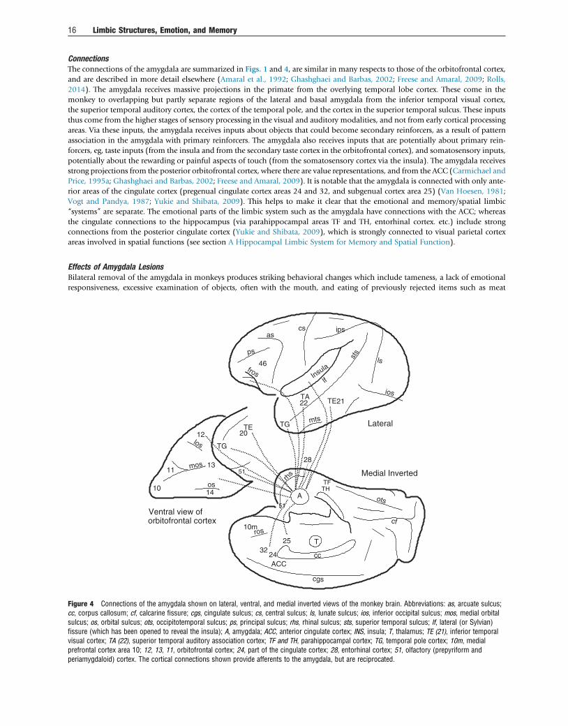
ConnectionsThe connections of the amygdala are summarized in Figs. 1 and 4, are similar in many respects to those of the orbitofrontal cortex,and are described in more detail elsewhere (Amaral et al., 1992; Ghashghaei and Barbas, 2002; Freese and Amaral, 2009; Rolls,2014). The amygdala receives massive projections in the primate from the overlying temporal lobe cortex. These come in themonkey to overlapping but partly separate regions of the lateral and basal amygdala from the inferior temporal visual cortex,the superior temporal auditory cortex, the cortex of the temporal pole, and the cortex in the superior temporal sulcus. These inputsthus come from the higher stages of sensory processing in the visual and auditory modalities, and not from early cortical processingareas. Via these inputs, the amygdala receives inputs about objects that could become secondary reinforcers, as a result of patternassociation in the amygdala with primary reinforcers. The amygdala also receives inputs that are potentially about primary rein-forcers, eg, taste inputs (from the insula and from the secondary taste cortex in the orbitofrontal cortex), and somatosensory inputs,potentially about the rewarding or painful aspects of touch (from the somatosensory cortex via the insula). The amygdala receivesstrong projections from the posterior orbitofrontal cortex, where there are value representations, and from the ACC (Carmichael andPrice, 1995a; Ghashghaei and Barbas, 2002; Freese and Amaral, 2009). It is notable that the amygdala is connected with only ante-rior areas of the cingulate cortex (pregenual cingulate cortex areas 24 and 32, and subgenual cortex area 25) (Van Hoesen, 1981;Vogt and Pandya, 1987; Yukie and Shibata, 2009). This helps to make it clear that the emotional and memory/spatial limbic“systems” are separate. The emotional parts of the limbic system such as the amygdala have connections with the ACC; whereasthe cingulate connections to the hippocampus (via parahippocampal areas TF and TH, entorhinal cortex. etc.) include strongconnections from the posterior cingulate cortex (Yukie and Shibata, 2009), which is strongly connected to visual parietal cortexareas involved in spatial functions (see section A Hippocampal Limbic System for Memory and Spatial Function).
Effects of Amygdala LesionsBilateral removal of the amygdala in monkeys produces striking behavioral changes which include tameness, a lack of emotionalresponsiveness, excessive examination of objects, often with the mouth, and eating of previously rejected items such as meat
10m
ACC
14
11
12
13
cc
as
fros
psst
s
ios
mts
ros
cgs
ots
cf
rhs
46
cs ips
TG
lf
ls
Medial Inverted
Lateral
Ventral view of
TG
TE
los
mos
os
A
T
TFTH
TA22 21TE
20
orbitofrontal cortex
51
51
Insula
25
2432
28
10
Figure 4 Connections of the amygdala shown on lateral, ventral, and medial inverted views of the monkey brain. Abbreviations: as, arcuate sulcus;cc, corpus callosum; cf, calcarine fissure; cgs, cingulate sulcus; cs, central sulcus; ls, lunate sulcus; ios, inferior occipital sulcus; mos, medial orbitalsulcus; os, orbital sulcus; ots, occipitotemporal sulcus; ps, principal sulcus; rhs, rhinal sulcus; sts, superior temporal sulcus; lf, lateral (or Sylvian)fissure (which has been opened to reveal the insula); A, amygdala; ACC, anterior cingulate cortex; INS, insula; T, thalamus; TE (21), inferior temporalvisual cortex; TA (22), superior temporal auditory association cortex; TF and TH, parahippocampal cortex; TG, temporal pole cortex; 10m, medialprefrontal cortex area 10; 12, 13, 11, orbitofrontal cortex; 24, part of the cingulate cortex; 28, entorhinal cortex; 51, olfactory (prepyriform andperiamygdaloid) cortex. The cortical connections shown provide afferents to the amygdala, but are reciprocated.
16 Limbic Structures, Emotion, and Memory
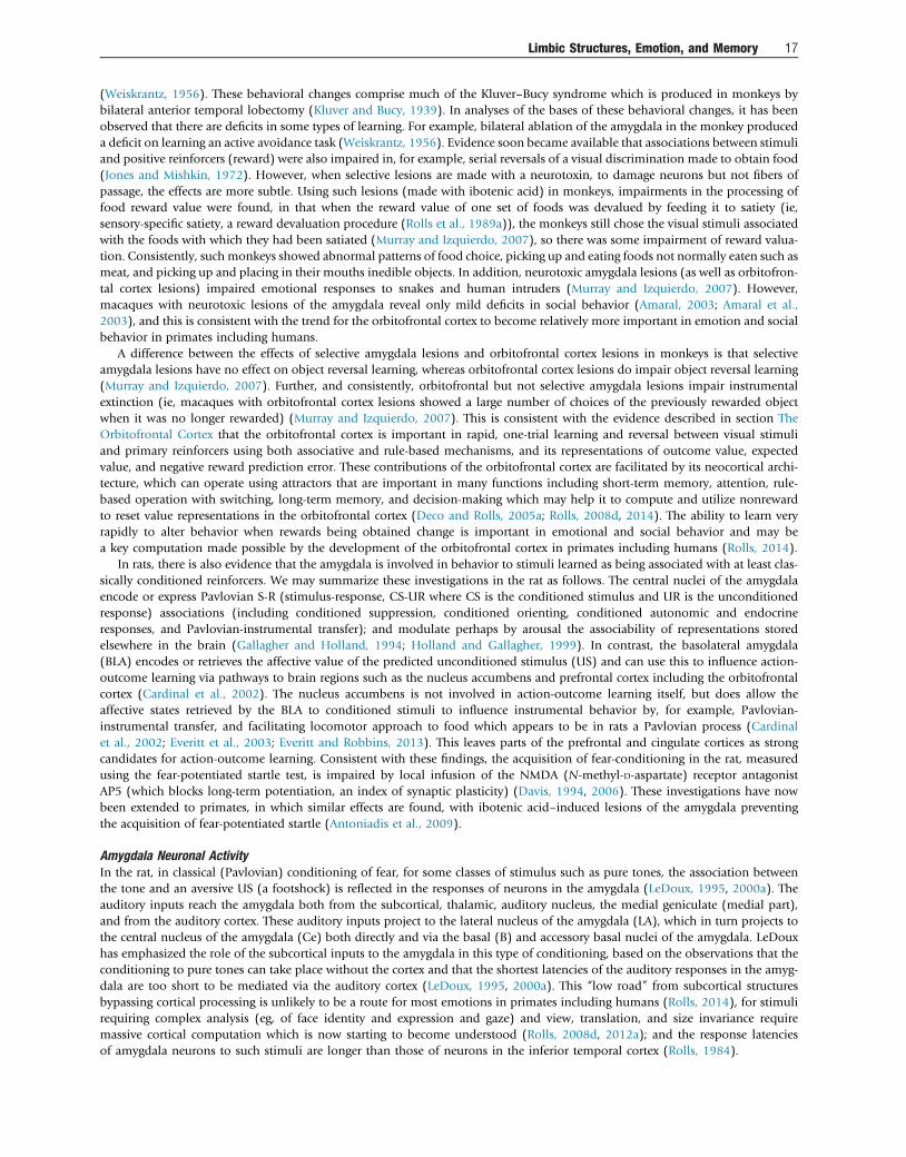
(Weiskrantz, 1956). These behavioral changes comprise much of the Kluver–Bucy syndrome which is produced in monkeys bybilateral anterior temporal lobectomy (Kluver and Bucy, 1939). In analyses of the bases of these behavioral changes, it has beenobserved that there are deficits in some types of learning. For example, bilateral ablation of the amygdala in the monkey produceda deficit on learning an active avoidance task (Weiskrantz, 1956). Evidence soon became available that associations between stimuliand positive reinforcers (reward) were also impaired in, for example, serial reversals of a visual discrimination made to obtain food(Jones and Mishkin, 1972). However, when selective lesions are made with a neurotoxin, to damage neurons but not fibers ofpassage, the effects are more subtle. Using such lesions (made with ibotenic acid) in monkeys, impairments in the processing offood reward value were found, in that when the reward value of one set of foods was devalued by feeding it to satiety (ie,sensory-specific satiety, a reward devaluation procedure (Rolls et al., 1989a)), the monkeys still chose the visual stimuli associatedwith the foods with which they had been satiated (Murray and Izquierdo, 2007), so there was some impairment of reward valua-tion. Consistently, such monkeys showed abnormal patterns of food choice, picking up and eating foods not normally eaten such asmeat, and picking up and placing in their mouths inedible objects. In addition, neurotoxic amygdala lesions (as well as orbitofron-tal cortex lesions) impaired emotional responses to snakes and human intruders (Murray and Izquierdo, 2007). However,macaques with neurotoxic lesions of the amygdala reveal only mild deficits in social behavior (Amaral, 2003; Amaral et al.,2003), and this is consistent with the trend for the orbitofrontal cortex to become relatively more important in emotion and socialbehavior in primates including humans.
A difference between the effects of selective amygdala lesions and orbitofrontal cortex lesions in monkeys is that selectiveamygdala lesions have no effect on object reversal learning, whereas orbitofrontal cortex lesions do impair object reversal learning(Murray and Izquierdo, 2007). Further, and consistently, orbitofrontal but not selective amygdala lesions impair instrumentalextinction (ie, macaques with orbitofrontal cortex lesions showed a large number of choices of the previously rewarded objectwhen it was no longer rewarded) (Murray and Izquierdo, 2007). This is consistent with the evidence described in section TheOrbitofrontal Cortex that the orbitofrontal cortex is important in rapid, one-trial learning and reversal between visual stimuliand primary reinforcers using both associative and rule-based mechanisms, and its representations of outcome value, expectedvalue, and negative reward prediction error. These contributions of the orbitofrontal cortex are facilitated by its neocortical archi-tecture, which can operate using attractors that are important in many functions including short-term memory, attention, rule-based operation with switching, long-term memory, and decision-making which may help it to compute and utilize nonrewardto reset value representations in the orbitofrontal cortex (Deco and Rolls, 2005a; Rolls, 2008d, 2014). The ability to learn veryrapidly to alter behavior when rewards being obtained change is important in emotional and social behavior and may bea key computation made possible by the development of the orbitofrontal cortex in primates including humans (Rolls, 2014).
In rats, there is also evidence that the amygdala is involved in behavior to stimuli learned as being associated with at least clas-sically conditioned reinforcers. We may summarize these investigations in the rat as follows. The central nuclei of the amygdalaencode or express Pavlovian S-R (stimulus-response, CS-UR where CS is the conditioned stimulus and UR is the unconditionedresponse) associations (including conditioned suppression, conditioned orienting, conditioned autonomic and endocrineresponses, and Pavlovian-instrumental transfer); and modulate perhaps by arousal the associability of representations storedelsewhere in the brain (Gallagher and Holland, 1994; Holland and Gallagher, 1999). In contrast, the basolateral amygdala(BLA) encodes or retrieves the affective value of the predicted unconditioned stimulus (US) and can use this to influence action-outcome learning via pathways to brain regions such as the nucleus accumbens and prefrontal cortex including the orbitofrontalcortex (Cardinal et al., 2002). The nucleus accumbens is not involved in action-outcome learning itself, but does allow theaffective states retrieved by the BLA to conditioned stimuli to influence instrumental behavior by, for example, Pavlovian-instrumental transfer, and facilitating locomotor approach to food which appears to be in rats a Pavlovian process (Cardinalet al., 2002; Everitt et al., 2003; Everitt and Robbins, 2013). This leaves parts of the prefrontal and cingulate cortices as strongcandidates for action-outcome learning. Consistent with these findings, the acquisition of fear-conditioning in the rat, measuredusing the fear-potentiated startle test, is impaired by local infusion of the NMDA (N-methyl-D-aspartate) receptor antagonistAP5 (which blocks long-term potentiation, an index of synaptic plasticity) (Davis, 1994, 2006). These investigations have nowbeen extended to primates, in which similar effects are found, with ibotenic acid–induced lesions of the amygdala preventingthe acquisition of fear-potentiated startle (Antoniadis et al., 2009).
Amygdala Neuronal ActivityIn the rat, in classical (Pavlovian) conditioning of fear, for some classes of stimulus such as pure tones, the association betweenthe tone and an aversive US (a footshock) is reflected in the responses of neurons in the amygdala (LeDoux, 1995, 2000a). Theauditory inputs reach the amygdala both from the subcortical, thalamic, auditory nucleus, the medial geniculate (medial part),and from the auditory cortex. These auditory inputs project to the lateral nucleus of the amygdala (LA), which in turn projects tothe central nucleus of the amygdala (Ce) both directly and via the basal (B) and accessory basal nuclei of the amygdala. LeDouxhas emphasized the role of the subcortical inputs to the amygdala in this type of conditioning, based on the observations that theconditioning to pure tones can take place without the cortex and that the shortest latencies of the auditory responses in the amyg-dala are too short to be mediated via the auditory cortex (LeDoux, 1995, 2000a). This “low road” from subcortical structuresbypassing cortical processing is unlikely to be a route for most emotions in primates including humans (Rolls, 2014), for stimulirequiring complex analysis (eg, of face identity and expression and gaze) and view, translation, and size invariance requiremassive cortical computation which is now starting to become understood (Rolls, 2008d, 2012a); and the response latenciesof amygdala neurons to such stimuli are longer than those of neurons in the inferior temporal cortex (Rolls, 1984).
Limbic Structures, Emotion, and Memory 17
There are separate output pathways in the rat from the amygdala for different fear-related classically conditioned responses.Lesions of the lateral hypothalamus (which receives from the central nucleus of the amygdala) blocked conditioned heart rate (auto-nomic) responses. Lesions of the central gray of the midbrain (which also receives from the central nucleus of the amygdala)blocked the conditioned freezing but not the conditioned autonomic response, and lesions of the stria terminalis blocked theneuroendocrine responses (LeDoux, 2000b).
In primates, the amygdala contains neurons that respond to taste, and oral texture including viscosity, fat texture, and capsaicin, andto somatosensory stimuli (Sanghera et al., 1979; Rolls, 1992a, 2000c; Kadohisa et al., 2005b; Kadohisa et al., 2005a). These are allpotentially primary reinforcers, and these neurons could represent outcome value. Neurons in the primate amygdala also respondto the sight of instrumental reinforcers in a visual discrimination task (Sanghera et al., 1979; Wilson and Rolls, 2005) and to odors.These neurons could signal expected value. However, in reward reversal learning, many amygdala neurons do not reverse theirresponses (Sanghera et al., 1979; Wilson and Rolls, 2005), and if any reversal is found, it is slow, taking many trials (Sangheraet al., 1979; Wilson and Rolls, 2005; Paton et al., 2006), whereas orbitofrontal neurons show rule-based reversal in one trial (Thorpeet al., 1983). Further, devaluation by feeding to satiety produced only a partial reduction in amygdala neurons’ responses to taste(Yan and Scott, 1996), and little reduction in responses to visual stimuli associated with food (Sanghera et al., 1979), compared tothe complete reduction found for orbitofrontal cortex taste (Rolls et al., 1989a) and visual food (Critchley and Rolls, 1996a) neurons.Thus by both reversal learning and devaluation measures of neuronal responses, the amygdala in primates appears to make a less important contri-bution to reward value representations than the orbitofrontal cortex.
Other amygdala neurons respond to faces (Sanghera et al., 1979; Leonard et al., 1985; Rolls, 1992a, 2000c, 2011a), includingface expression and identity (Gothard et al., 2007), both of which are represented in the inferior temporal cortex areas that projectinto the amygdala (Hasselmo et al., 1989). The presence of these neurons in the primate amygdala does emphasize that as theamygdala has evolved in primates, it does represent information that becomes highly developed in the primate inferior temporalcortical areas, and that is important for social and emotional responses to individuals, though the orbitofrontal cortex of coursealso represents similar information (Rolls et al., 2006b).
Amygdala Damage in HumansThe greater importance of the orbitofrontal cortex in emotion in humans is emphasized by a comparison with the effects of bilateralamygdala damage in humans, which although producing demonstrable deficits in face processing (Adolphs et al., 2005; Spezio et al.,2007), decision-making with linked autonomic deficits (Bechara et al., 1999; Brand et al., 2007), and autonomic conditioning(Phelps and LeDoux, 2005), may not (in contrast with the orbitofrontal cortex) produce major changes in emotion that are readilyapparent in everyday behavior (Phelps and LeDoux, 2005; Rolls, 2008d; Seymour and Dolan, 2008; Whalen and Phelps, 2009).
The Anterior Cingulate Cortex
The ACC is a limbic structure involved in emotion, with major inputs from structures such as the amygdala and orbitofrontal cortex,and activations that are correlated with the pleasantness or unpleasantness of stimuli. The ACC uses these value (including outcomevalue) representations in its function with the midcingulate cortex in action-outcome learning. The ACC can be conceived asa system that in primates links the orbitofrontal cortex representations of the value of stimuli (including reward and punisheroutcomes) with actions (Rolls, 2014).
ConnectionsThe connections of the ACC (Vogt, 2009; Yukie and Shibata, 2009) are illustrated in Fig. 5. The ACC includes area 32, area 25 thesubgenual cingulate cortex, and part of area 24. The cortex anterior to the genu (knee, at the front) of the corpus callosum isreferred to as the pregenual cingulate cortex. The caudal orbitofrontal cortex and amygdala project to the ACC (Carmichaeland Price, 1996; Price, 2006), and especially the orbitofrontal cortex appears to influence the ACC strongly, for activations relatedto the pleasantness or unpleasantness of many stimuli are present in both (Grabenhorst and Rolls, 2011; Rolls, 2014). It is ofinterest that the anterior but not posterior cingulate cortex receives projections from the amygdala (with a similar trend forthe caudal orbitofrontal cortex) (Morecraft and Tanji, 2009; Yukie and Shibata, 2009), consistent with the evidence that thereare separate limbic structures including different parts of the cingulate cortex for emotion and memory/spatial function. Havingsaid this, the entorhinal cortex (area 28), the perirhinal cortex area 35/36, and the parahippocampal cortex (areas TF and TH)(areas illustrated in Fig. 6 and providing the gateway to the hippocampus) do have reciprocal connections with the anterior,mid, and posterior cingulate cortex, and the interpretation provided in this paper is that emotional/reward/value informationfrom the anterior cingulate and orbitofrontal cortex may need to be stored in the hippocampus as part of an episodic memoryinvolving spatial and also usually object information; and then subsequently recalled from the hippocampus via the backprojec-tions to the anterior cingulate and orbitofrontal cortex. The ACC also has connections with areas through which it can influenceautonomic function, including the anterior insula, hypothalamus, and brain stem autonomic nuclei (Vogt and Derbyshire, 2009;Critchley and Harrison, 2013).
Activations and Neuronal ActivityThe orbitofrontal cortex projects to the pregenual cingulate cortex (Carmichael and Price, 1996; Price, 2006), and both theseareas have reward and punishment value representations that correlate on a continuous scale with the subjective
18 Limbic Structures, Emotion, and Memory
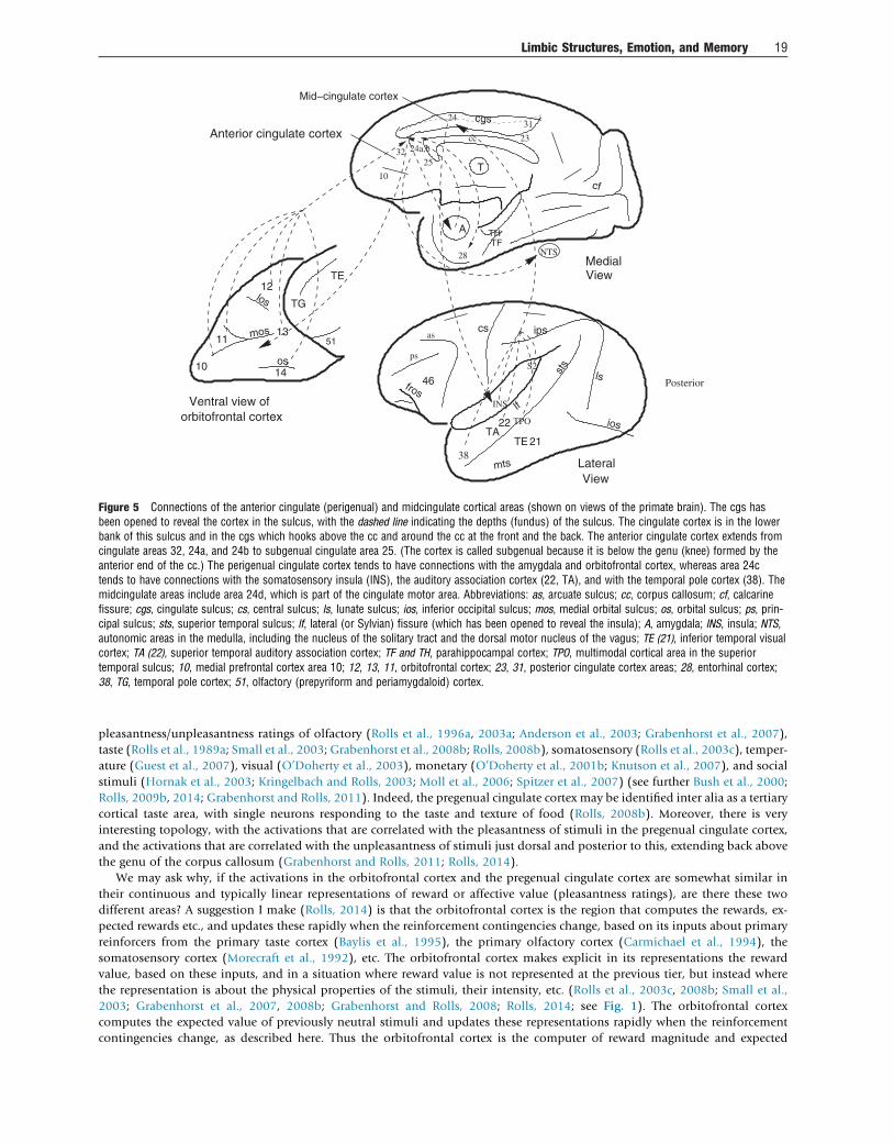
pleasantness/unpleasantness ratings of olfactory (Rolls et al., 1996a, 2003a; Anderson et al., 2003; Grabenhorst et al., 2007),taste (Rolls et al., 1989a; Small et al., 2003; Grabenhorst et al., 2008b; Rolls, 2008b), somatosensory (Rolls et al., 2003c), temper-ature (Guest et al., 2007), visual (O’Doherty et al., 2003), monetary (O’Doherty et al., 2001b; Knutson et al., 2007), and socialstimuli (Hornak et al., 2003; Kringelbach and Rolls, 2003; Moll et al., 2006; Spitzer et al., 2007) (see further Bush et al., 2000;Rolls, 2009b, 2014; Grabenhorst and Rolls, 2011). Indeed, the pregenual cingulate cortex may be identified inter alia as a tertiarycortical taste area, with single neurons responding to the taste and texture of food (Rolls, 2008b). Moreover, there is veryinteresting topology, with the activations that are correlated with the pleasantness of stimuli in the pregenual cingulate cortex,and the activations that are correlated with the unpleasantness of stimuli just dorsal and posterior to this, extending back abovethe genu of the corpus callosum (Grabenhorst and Rolls, 2011; Rolls, 2014).
We may ask why, if the activations in the orbitofrontal cortex and the pregenual cingulate cortex are somewhat similar intheir continuous and typically linear representations of reward or affective value (pleasantness ratings), are there these twodifferent areas? A suggestion I make (Rolls, 2014) is that the orbitofrontal cortex is the region that computes the rewards, ex-pected rewards etc., and updates these rapidly when the reinforcement contingencies change, based on its inputs about primaryreinforcers from the primary taste cortex (Baylis et al., 1995), the primary olfactory cortex (Carmichael et al., 1994), thesomatosensory cortex (Morecraft et al., 1992), etc. The orbitofrontal cortex makes explicit in its representations the rewardvalue, based on these inputs, and in a situation where reward value is not represented at the previous tier, but instead wherethe representation is about the physical properties of the stimuli, their intensity, etc. (Rolls et al., 2003c, 2008b; Small et al.,2003; Grabenhorst et al., 2007, 2008b; Grabenhorst and Rolls, 2008; Rolls, 2014; see Fig. 1). The orbitofrontal cortexcomputes the expected value of previously neutral stimuli and updates these representations rapidly when the reinforcementcontingencies change, as described here. Thus the orbitofrontal cortex is the computer of reward magnitude and expected
32
10
28
Anterior cingulate cortex
Mid−cingulate cortex
31
1014
11
12
13
Ventral view of
TG
TE
los
mos
os
51
cf
THTF
ios
sts
mts
cs ips
TA
lf
21
22
lsfros
46
cc
T
ps
TPO
S2
INS
A
cgs
38
NTS
TE
Lateral
Posterior
View
View
as
Medial
24a,b
25
24
orbitofrontal cortex
23
Figure 5 Connections of the anterior cingulate (perigenual) and midcingulate cortical areas (shown on views of the primate brain). The cgs hasbeen opened to reveal the cortex in the sulcus, with the dashed line indicating the depths (fundus) of the sulcus. The cingulate cortex is in the lowerbank of this sulcus and in the cgs which hooks above the cc and around the cc at the front and the back. The anterior cingulate cortex extends fromcingulate areas 32, 24a, and 24b to subgenual cingulate area 25. (The cortex is called subgenual because it is below the genu (knee) formed by theanterior end of the cc.) The perigenual cingulate cortex tends to have connections with the amygdala and orbitofrontal cortex, whereas area 24ctends to have connections with the somatosensory insula (INS), the auditory association cortex (22, TA), and with the temporal pole cortex (38). Themidcingulate areas include area 24d, which is part of the cingulate motor area. Abbreviations: as, arcuate sulcus; cc, corpus callosum; cf, calcarinefissure; cgs, cingulate sulcus; cs, central sulcus; ls, lunate sulcus; ios, inferior occipital sulcus; mos, medial orbital sulcus; os, orbital sulcus; ps, prin-cipal sulcus; sts, superior temporal sulcus; lf, lateral (or Sylvian) fissure (which has been opened to reveal the insula); A, amygdala; INS, insula; NTS,autonomic areas in the medulla, including the nucleus of the solitary tract and the dorsal motor nucleus of the vagus; TE (21), inferior temporal visualcortex; TA (22), superior temporal auditory association cortex; TF and TH, parahippocampal cortex; TPO, multimodal cortical area in the superiortemporal sulcus; 10, medial prefrontal cortex area 10; 12, 13, 11, orbitofrontal cortex; 23, 31, posterior cingulate cortex areas; 28, entorhinal cortex;38, TG, temporal pole cortex; 51, olfactory (prepyriform and periamygdaloid) cortex.
Limbic Structures, Emotion, and Memory 19
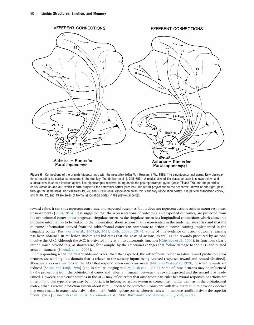
reward value. It can thus represent outcomes, and expected outcomes, but it does not represent actions such as motor responsesor movements (Rolls, 2014). It is suggested that the representations of outcomes, and expected outcomes, are projected fromthe orbitofrontal cortex to the pregenual cingulate cortex, as the cingulate cortex has longitudinal connections which allow thisoutcome information to be linked to the information about actions that is represented in the midcingulate cortex and that theoutcome information derived from the orbitofrontal cortex can contribute to action-outcome learning implemented in thecingulate cortex (Rushworth et al., 2007a,b, 2011; Rolls, 2008d, 2014). Some of this evidence on action-outcome learninghas been obtained in rat lesion studies and indicates that the costs of actions, as well as the rewards produced by actions,involve the ACC. Although the ACC is activated in relation to autonomic function (Critchley et al., 2004), its functions clearlyextend much beyond this, as shown also, for example, by the emotional changes that follow damage to the ACC and relatedareas in humans (Hornak et al., 2003).
In responding when the reward obtained is less than that expected, the orbitofrontal cortex negative reward prediction errorneurons are working in a domain that is related to the sensory inputs being received (expected reward and reward obtained).There are also error neurons in the ACC that respond when errors are made (Niki and Watanabe, 1979), or when rewards arereduced (Shima and Tanji, 1998) (and in similar imaging studies, Bush et al., 2002). Some of these neurons may be influencedby the projections from the orbitofrontal cortex and reflect a mismatch between the reward expected and the reward that is ob-tained. However, some error neurons in the ACC may reflect errors that arise when particular behavioral responses or actions arein error, and this type of error may be important in helping an action system to correct itself, rather than, as in the orbitofrontalcortex, when a reward prediction system about stimuli needs to be corrected. Consistent with this, many studies provide evidencethat errors made in many tasks activate the anterior/midcingulate cortex, whereas tasks with response conflict activate the superiorfrontal gyrus (Rushworth et al., 2004; Matsumoto et al., 2007; Rushworth and Behrens, 2008; Vogt, 2009).
Figure 6 Connections of the primate hippocampus with the neocortex (After Van Hoesen, G.W., 1982. The parahippocampal gyrus. New observa-tions regarding its cortical connections in the monkey. Trends Neurosci. 5, 345–350.). A medial view of the macaque brain is shown below, anda lateral view is shown inverted above. The hippocampus receives its inputs via the parahippocampal gyrus (areas TF and TH), and the perirhinalcortex (areas 35 and 36), which in turn project to the entorhinal cortex (area 28). The return projections to the neocortex (shown on the right) passthrough the same areas. Cortical areas 19, 20, and 21 are visual association areas, 22 is auditory association cortex, 7 is parietal association cortex,and 9, 46, 12, and 13 are areas of frontal association cortex in the prefrontal cortex.
20 Limbic Structures, Emotion, and Memory
A neuroimaging study in humans illustrates how nonreward/error signals relevant to emotion and social behavior are foundin the ACC. Kringelbach et al. (2003) used the faces of two different people, and if one face was selected, then that face smiled,and if the other was selected, the face showed an angry expression. After good performance was acquired, there were repeatedreversals of the visual discrimination task. Kringelbach et al. (2003) found that activation of a lateral part of the orbitofrontalcortex and in a dorsal part of the ACC in the fMRI study was produced on the error trials, that is, when the human chose a face-and did not obtain the expected reward. An interesting aspect of this study that makes it relevant to human social behavior isthat the conditioned stimuli were faces of different individuals and the unconditioned stimuli were face expressions. Thus theassociation that was being reversed in this study was between a representation of face identity and a representation of faceexpression. Moreover, the study reveals that the human orbitofrontal cortex and ACC are very sensitive to social feedbackwhen it must be used to change behavior (Kringelbach and Rolls, 2003, 2004).
Effects of Anterior Cingulate Cortex Lesions in HumansDevinsky et al. (1995) and Cardinal et al. (2002) review evidence that anterior cingulate lesions in humans produce apathy, auto-nomic dysregulation, and emotional instability.
An investigation in patients with selective surgical lesions has shown that patients with unilateral lesions of the anteroventralpart of the ACC and/or medial area 9 were in some cases impaired on voice and face expression identification, had some changein social behavior (such as inappropriateness, being less likely to notice when other people were angry, not being close to his or herfamily, and doing things without thinking), and had significant changes in their subjective emotional state (Hornak et al., 2003).Unilateral lesions were sufficient to produce these effects, and there were no strong laterality effects.
The results of Hornak et al. (2003) also confirmed that damage restricted to the orbitofrontal cortex can produce impair-ments in face and voice expression identification, which may be primary reinforcers. The system is sensitive, in that evenpatients with unilateral orbitofrontal cortex lesions may be impaired. The impairment is not a generic impairment of the abilityto recognize any emotions in others, in that frequently voice but not face expression identification was impaired, and vice versa.This implies some functional specialization for visual versus auditory emotion–related processing in the human orbitofrontalcortex. The results also show that the changes in social behavior can be produced by damage restricted to the orbitofrontalcortex. The patients were particularly likely to be impaired on emotion recognition (they were less likely to notice when otherswere sad, or happy, or disgusted); on emotional empathy (they were less likely to comfort those who are sad, or afraid, or tofeel happy for others who are happy); on interpersonal relationships (not caring what others think and not being close to his/her family); and were less likely to cooperate with others; were impatient and impulsive; and had difficulty in making andkeeping close relationships. The results also show that changes in subjective emotional state (including frequently sadness,anger, and happiness) can be produced by damage restricted to the orbitofrontal cortex (Hornak et al., 2003). In addition,the patients with bilateral orbitofrontal cortex lesions were impaired on the probabilistic reversal learning task (Hornaket al., 2004). The findings overall thus make clear the types of deficit found in humans with orbitofrontal cortexdamage and can be easily related to underlying fundamental processes in which the orbitofrontal cortex is involved asdescribed by Rolls (1999a, 2005), including decoding and representing primary reinforcers, being sensitive to changes in rein-forcers, and rapidly readjusting behavior to stimuli when the reinforcers available change. The implication is that some of theinputs to the ACC which produce similar deficits may come from the orbitofrontal cortex. Consistent with this, unilaterallesions of the ACC (including some of medial area 9) can produce voice and/or face expression identification deficits, changesin social behavior, and marked changes in subjective emotional state (Hornak et al., 2003).
In summary, the primate including human ACC has connections with other limbic structures such as the amygdala and limbic-related structures such as the caudal orbitofrontal cortex, receives reward and punishment-related information from these brainregions, and appears to be involved in using this reward and punishment information to learn about actions to obtain goals, takinginto account the costs of the actions (Rolls, 2009b, 2014; Grabenhorst and Rolls, 2011; Rushworth et al., 2011). Appropriate actionsto emotion-provoking stimuli may not be made after ACC damage. The ACC can thus be seen as a limbic structure involved inemotion that forms part of a system with other limbic structures involved in emotion, the amygdala and orbitofrontal cortex. Struc-tures that receive input from these three structures are involved in linking to habit responses (striatum including ventral striatumand nucleus accumbens), autonomic and endocrine output (insula, hypothalamus, brain stem autonomic nuclei), etc. (Rolls, 2014,2016c).
These three limbic and related structures, the amygdala, orbitofrontal cortex, and ACC, could be thought of as an emotion limbicsystem, but not the limbic system, as different limbic structures related to the hippocampus are involved primarily in episodicmemory and related spatial function, and not in emotion. Damage to the emotion limbic system or structures is not describedas impairing episodic memory or spatial function.
The Insular Cortex
The insula has sometimes been lumped in with limbic structures (Yakovlev, 1948; Catani et al., 2013), and parts of it are involved inautonomic/visceral function (Critchley and Harrison, 2013), so it is considered briefly here, and in more detail elsewhere (Rolls,2014, 2015c).
Limbic Structures, Emotion, and Memory 21
The Insular Primary Taste CortexThe primary taste cortex is in the dorsal part of the anterior insula and adjoining frontal operculum (Pritchard et al., 1986), and thisregion projects to the orbitofrontal cortex (Baylis et al., 1995). Neurons in the primary taste cortex represent what the taste is(including sweet, salt, sour, bitter, and umami) (Scott et al., 1986; Yaxley et al., 1990; Baylis and Rolls, 1991; Rolls, 2009c), butdo not represent reward value in that their responses are not decreased by feeding to satiety (Rolls et al., 1988; Yaxley et al.,1988). Neurons in the insular primary taste cortex do represent oral texture including fat texture (de Araujo and Rolls, 2004;Verhagen et al., 2004; Kadohisa et al., 2005b) and oral temperature (Verhagen et al., 2004; Kadohisa et al., 2005b; Guest et al.,2007). Neurons in the macaque primary taste cortex do not have olfactory responses (Verhagen et al., 2004), and consistently ina human fMRI investigation of olfactory and taste convergence in the brain, it was shown that an agranular more anterior pastof the insula does show convergence between taste and odor to represent flavor (de Araujo et al., 2003c).
The Visceral/Autonomic Insular CortexA region of the anterior insular cortex just ventral to the primary taste cortex has strong projections to the orbitofrontal cortex(Baylis et al., 1995) and is putatively the visceral/autonomic region of the insula (Critchley and Harrison, 2013; Rolls, 2015c).This anterior insular region probably receives inputs from the orbitofrontal cortex and ACC (Price, 2006, 2007), which decodeand represent the reward and punishment-related signals that can produce autonomic/visceral responses (Rolls, 2014). It issuggested (Rolls, 2014) that when the anterior insula is activated by emotion-related stimuli and events such as face expres-sions of disgust (Phillips et al., 1998, 2004) and unfair offers (which are aversive) in an ultimatum game (Sanfey et al.,2003), the activations reflect the effects produced by regions such as the orbitofrontal and anterior cingulate cortices thatcan produce emotional and autonomic responses, rather than face expression decoding (which is performed elsewhere, inthe cortex in the superior temporal sulcus and in the orbitofrontal cortex (Hasselmo et al., 1989; Rolls, 2007b, 2011a,2012a)) or economic computations. Consistent with this, in a neuroeconomics study with monetary reward, it was foundthat expected value was negatively correlated with activations in the anterior insula [�38 24 16] in a region that has beenimplicated in disgust, and interestingly, the activations here were also correlated with the uncertainty of the magnitude ofthe reward that would be obtained (Rolls et al., 2008a). Effectively there was more insula activation in situations that mightbe described as aversive. This part of the insula has activations that are related to visceral/autonomic function, for example, toheart and stomach responses during disgust-associated nausea (Critchley and Harrison, 2013). Further, electrical stimulation inthe anteroventral insula produced feelings related to disgust, including viscero-autonomic feelings (Krolak-Salmon et al.,2003). Moreover, it is of course to be expected, and is the case, that the autonomic output and the corresponding visceralinsular activity will be different for different emotional states, eg, when eating a food versus when reacting to the disgustingbitter taste of quinine or to pain or the sight of aversive or unpleasant stimuli. Menon and Uddin (2010) have suggestedthat the insula is part of a “saliency” network. They postulate that the insula is sensitive to salient events and that its core func-tion is to mark such events for additional processing and initiate appropriate control signals. However, given the inputs to theanterior insula from the orbitofrontal and anterior cingulate cortices, which decode stimuli and events such as rewards,punishers, nonreward, and novel stimuli, it is suggested (Rolls, 2014, 2015c) that the anterior insula may respond to thoughnot compute such “salient” stimuli and events, and its activation may reflect autonomic and related responses which are ofcourse elicited by these “salient” stimuli. A related point (Rolls, 2014, 2015c) is relevant to Damasio’s somatic marker hypoth-esis (Damasio, 1996).
The Somatosensory InsulaThe mid- and posterior insula has somatosensory representations of the body (Mufson and Mesulam, 1982). A property thatmay be special about these somatosensory cortical representations is that activations are produced by touch to the body but, incontrast to many other somatosensory cortical areas, not by the sight of touch (McCabe et al., 2008). It was therefore suggestedthat insular cortex activation thus allows an individual to know that it is touch to the person’s body, and not that someoneelse’s body is about to be touched (McCabe et al., 2008). The insular somatosensory cortex may thus provide evidence aboutwhat is happening to one’s own body (Rolls, 2010c). The same might be said of the insular primary taste cortex, which whenactivated leaves no doubt that one is tasting, and not seeing someone else tasting. So, in a sense, feelings associated with acti-vations of the insular cortex (and they are the subjective intensity of taste is linearly related to the activation of the primary tastecortex (Grabenhorst and Rolls, 2008)) do inform one about the state of one’s own body, and this relates to Craig’s suggestions(Craig, 2009, 2011) about the importance of the insula in interoceptive feelings. However, this does not mean that the insularcortex is necessary for body feelings, and indeed that seems to be ruled out by the finding that a patient with extensive bilateraldamage to the insular cortex reported normal body/emotional feelings (Damasio et al., 2013).
A Hippocampal Limbic System for Memory and Spatial Function
Evidence will be described that the hippocampus and its connected structures are involved in episodic memory, and not in emotion.This is thus a separate system from the amygdala/orbitofrontal cortex/ACC emotional system. Moreover, the computational prin-ciples of operation of these two systems are very different.
22 Limbic Structures, Emotion, and Memory
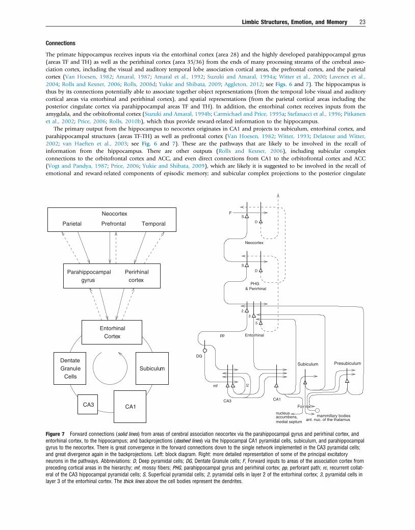
Connections
The primate hippocampus receives inputs via the entorhinal cortex (area 28) and the highly developed parahippocampal gyrus(areas TF and TH) as well as the perirhinal cortex (area 35/36) from the ends of many processing streams of the cerebral asso-ciation cortex, including the visual and auditory temporal lobe association cortical areas, the prefrontal cortex, and the parietalcortex (Van Hoesen, 1982; Amaral, 1987; Amaral et al., 1992; Suzuki and Amaral, 1994a; Witter et al., 2000; Lavenex et al.,2004; Rolls and Kesner, 2006; Rolls, 2008d; Yukie and Shibata, 2009; Aggleton, 2012; see Figs. 6 and 7). The hippocampus isthus by its connections potentially able to associate together object representations (from the temporal lobe visual and auditorycortical areas via entorhinal and perirhinal cortex), and spatial representations (from the parietal cortical areas including theposterior cingulate cortex via parahippocampal areas TF and TH). In addition, the entorhinal cortex receives inputs from theamygdala, and the orbitofrontal cortex (Suzuki and Amaral, 1994b; Carmichael and Price, 1995a; Stefanacci et al., 1996; Pitkanenet al., 2002; Price, 2006; Rolls, 2010b), which thus provide reward-related information to the hippocampus.
The primary output from the hippocampus to neocortex originates in CA1 and projects to subiculum, entorhinal cortex, andparahippocampal structures (areas TF-TH) as well as prefrontal cortex (Van Hoesen, 1982; Witter, 1993; Delatour and Witter,2002; van Haeften et al., 2003; see Fig. 6 and 7). These are the pathways that are likely to be involved in the recall ofinformation from the hippocampus. There are other outputs (Rolls and Kesner, 2006), including subicular complexconnections to the orbitofrontal cortex and ACC, and even direct connections from CA1 to the orbitofrontal cortex and ACC(Vogt and Pandya, 1987; Price, 2006; Yukie and Shibata, 2009), which are likely it is suggested to be involved in the recall ofemotional and reward-related components of episodic memory; and subicular complex projections to the posterior cingulate
rc
CortexEntorhinal
DentateGranule
CellsSubiculum
CA1
Neocortex
CA3
gyrusParahippocampal
cortexPerirhinal
Fo nixr
Parietal Prefrontal Temporal
23
DG
rhinalEntopp
mf
Neocortex
FS
D
D
S
5
accumbens,medial septum
nucleus
CA1CA3
& PerirhinalPHG
ant. nuc. of the thalamusmammillary bodies
Subiculum Presubiculum
Figure 7 Forward connections (solid lines) from areas of cerebral association neocortex via the parahippocampal gyrus and perirhinal cortex, andentorhinal cortex, to the hippocampus; and backprojections (dashed lines) via the hippocampal CA1 pyramidal cells, subiculum, and parahippocampalgyrus to the neocortex. There is great convergence in the forward connections down to the single network implemented in the CA3 pyramidal cells;and great divergence again in the backprojections. Left: block diagram. Right: more detailed representation of some of the principal excitatoryneurons in the pathways. Abbreviations: D, Deep pyramidal cells; DG, Dentate Granule cells; F, Forward inputs to areas of the association cortex frompreceding cortical areas in the hierarchy; mf, mossy fibers; PHG, parahippocampal gyrus and perirhinal cortex; pp, perforant path; rc, recurrent collat-eral of the CA3 hippocampal pyramidal cells; S, Superficial pyramidal cells; 2, pyramidal cells in layer 2 of the entorhinal cortex; 3, pyramidal cells inlayer 3 of the entorhinal cortex. The thick lines above the cell bodies represent the dendrites.
Limbic Structures, Emotion, and Memory 23
cortex (Vogt and Pandya, 1987; Yukie and Shibata, 2009), which are likely it is suggested to be involved in the recall of spatialcomponents of episodic memory.
In addition, there are subcortical connections that form Papez’ circuit (Papez, 1937). The subiculum (subicular complex)projects via the fornix to the mammillary bodies (Fig. 7), which then project via the mammillothalamic tract to the anteriorthalamic nuclei, which project most strongly to the posterior cingulate cortex (Shibata and Yukie, 2009), which in turn via thecingulum projects back toward the hippocampus via the parahippocampal cortex (Yukie and Shibata, 2009). The fornix alsoconveys the cholinergic input from the septal nuclei, classed as limbic structures, to the hippocampus, and this must be takeninto account when considering the effects on memory of damage to the hippocampus, for acetylcholine facilitates synaptic modi-fication and regulates recurrent collateral efficacy in the CA3 system (Giocomo and Hasselmo, 2007; Rolls, 2010b).
Effects of Hippocampal System Damage in Primates Including Humans
In humans, episodic memory, the memory of a particular episode, requires the ability to remember particular events, and todistinguish them from other events, and is impaired by damage to the hippocampal system. An event consists of a set of itemsthat occur together, such as seeing a particular object or person’s face in a particular place. An everyday example might beremembering where one was for dinner, who was present, what was eaten, what was discussed, and the time at which itoccurred. The spatial context is almost always an important part of an episodic memory (Dere et al., 2008), and it may bepartly for this reason that episodic memory is linked to the functions of the hippocampal system, which is involved in spatialprocessing and memory. A famous case is that of HM, who after surgery for epilepsy that removed bilaterally parts of thetemporal lobe including parts of the hippocampus could no longer form memories of events that occurred after the damage(anterograde amnesia), but could recall memories of events prior to the hippocampal damage (Corkin, 2002), and similarimpairments are found in other patients with damage to the hippocampus and connected structures (Squire and Wixted,2011). Indeed, section of the fornix in humans, at one time a side effect of surgery on a third ventricle cyst, produces similaramnesia (Gaffan and Gaffan, 1991), as can damage to the mammillary body/mammillothalamic tract/anterior nucleus of thethalamus pathway (see Aggleton, 2012). In humans, functional neuroimaging shows that the hippocampal system is activatedby allocentric spatial processing and episodic memory (Burgess et al., 2002; Burgess, 2008; Hassabis et al., 2009; Chadwicket al., 2010).
Damage to the hippocampus or to some of its connections such as the fornix in monkeys produces deficits in learningabout the places of objects and about the places where responses should be made (Buckley and Gaffan, 2000). For example,macaques and humans with damage to the hippocampal system or fornix are impaired in object-place memory tasks in whichnot only the objects seen, but where they were seen, must be remembered (Smith and Milner, 1981; Gaffan and Saunders,1985; Parkinson et al., 1988; Gaffan, 1994; Burgess et al., 2002; Crane and Milner, 2005). Posterior parahippocampal lesionsin macaques impair even a simple type of object-place learning in which the memory load is just one pair of trial-uniquestimuli (Malkova and Mishkin, 2003). (It is further predicted that a more difficult object-place learning task with non–trial-unique stimuli and with many object-place pairs would be impaired by neurotoxic hippocampal lesions.) Further, neuro-toxic lesions that selectively damage the primate hippocampus impair spatial scene memory, tested by the ability to rememberwhere in a scene to touch to obtain reward (Murray et al., 1998). Also, fornix lesions impair conditional left–right discrim-ination learning, in which the visual appearance of an object specifies whether a response is to be made to the left or the right(Rupniak and Gaffan, 1987). A comparable deficit is found in humans (Petrides, 1985). Fornix-sectioned monkeys are alsoimpaired in learning on the basis of a spatial cue which object to choose (eg, if two objects are on the left, choose objectA, but if the two objects are on the right, choose object B) (Gaffan and Harrison, 1989a). Monkeys with fornix damage arealso impaired in using information about their place in an environment. For example, there are learning impairmentswhen which of two or more objects the monkey had to choose depended on the position of the monkey in the room (Gaffanand Harrison, 1989b).
More recently, Lavenex and colleagues have described deficits produced by hippocampal damage in monkeys performing allo-centric spatial memory tasks (Banta Lavenex and Lavenex, 2009). One such task involved freely moving in an environment usingallocentric spatial room cues to remember the locations of inverted cups that contained food. This is a food reward – allocentricplace association task. In contrast, the perirhinal cortex, area 35, with its connections to inferior temporal cortex areas involvedin object perception (Rolls, 2008d, 2012a), is involved in recognitionmemory (Buckley, 2005) and indeed contains neurons relatedto long-term familiarity memory (Hölscher et al., 2003b; Rolls et al., 2005b; Rolls, 2008d).
Rats with hippocampal lesions are also impaired in using environmental spatial cues to remember particular places (O’Keefeand Nadel, 1978; Jarrard, 1993; Cassaday and Rawlins, 1997; Martin et al., 2000; Kesner et al., 2004, 2012), to utilize spatial cuesor to bridge delays (Rawlins, 1985; Kesner, 1998; Kesner and Rolls, 2001; Kesner et al., 2004; Rolls and Kesner, 2006), to performobject-place memory (Rolls and Kesner, 2006; Kesner et al., 2012), or to perform relational operations on remembered material(Eichenbaum, 1997).
It is notable that emotional changes, reward-related learning and reversal impairments, and devaluation impairments of stimuliare not produced by damage to this hippocampal system. This is further evidence for one of the theses of this paper that we shouldno longer be considering the operation of limbic and related structures as being part of a single limbic system. There is a doubledissociation of functions, with the orbitofrontal/amygdala/ACC limbic system being involved in emotion but not episodicmemory, and the hippocampal system being involved in memory but not in emotion.
24 Limbic Structures, Emotion, and Memory
Neuronal Representations in the Primate Hippocampus
The systems-level neurophysiology of the hippocampus shows what information could be stored or processed by the hippo-campus. To understand how the hippocampus works, it is not sufficient to state just that it can store information – one needsto know what information. The systems-level neurophysiology of the primate hippocampus has been reviewed by Rolls and Xiang(2006), and a summary is provided here because it provides a perspective relevant to understanding the function of the humanhippocampus that is somewhat different from that provided by the properties of place cells in rodents, which have been reviewedelsewhere (McNaughton et al., 1983; O’Keefe, 1984; Muller et al., 1991; Jeffery et al., 2004; Jeffery and Hayman, 2004).
Spatial View Neurons in the Primate HippocampusWe have shown that the primate hippocampus contains spatial cells that respond when the monkey looks at a certain part ofspace, for example, at one quadrant of a video monitor while the monkey is performing an object-place memory task in whichhe must remember where on the monitor he has seen particular images (Rolls et al., 1989b; Rolls, 1999b). Approximately 9% ofhippocampal neurons have such spatial view fields, and approximately 2.4% combine information about the position in spacewith information about the object that is in that position in space (Rolls et al., 1989b). The representation of space is for themajority of hippocampal neurons in allocentric not egocentric coordinates (Feigenbaum and Rolls, 1991). These spatial viewcells can be recorded while monkeys move themselves round the test environment by walking (or running) on all fours (Rollset al., 1997a, 1998b; Robertson et al., 1998; Georges-François et al., 1999). These hippocampal “spatial view neurons” respondsignificantly differently for different allocentric spatial views and have information about spatial view in their firing rate, but donot respond differently just on the basis of eye position, head direction, or place (Georges-François et al., 1999). If the viewdetails are obscured by curtains and darkness, then some spatial view neurons (especially those in CA1 and less those inCA3) continue to respond when the monkey looks toward the spatial view field, showing that these neurons can be updatedfor at least short periods by idiothetic (self-motion) cues including eye position and head direction signals (Rolls et al.,1997b; Robertson et al., 1998).
Object-Place Neurons in the Primate HippocampusA fundamental question about the function of the primate including human hippocampus in relation to episodic memory iswhether object as well as allocentric spatial information is represented. To investigate this, Rolls et al. (2005a) made recordingsfrom single hippocampal formation neurons while macaques performed an object-place memory task that required the monkeysto learn associations between objects, and where they were shown in a room. Some neurons (10%) responded differently todifferent objects independently of location; other neurons (13%) responded to the spatial view independently of which objectwas present at the location; and some neurons (12%) responded to a combination of a particular object and the place where itwas shown in the room. These results show that there are separate as well as combined representations of objects and their locationsin space in the primate hippocampus. This is a property required in an episodic memory system, for which associations betweenobjects and the places where they are seen are prototypical. The results thus show that a requirement for a human episodic memorysystem, separate and combined neuronal representations of objects and where they are seen “out there” in the environment, arepresent in the primate hippocampus (Rolls et al., 2005a).
What may be a corresponding finding in rats is that some rat hippocampal neurons respond on the basis of the conjunction oflocation and odor (Wood et al., 1999). Results consistent with our object-place neurons in primates are that Diamond andcolleagues have now shown using the vibrissa somatosensory input for the “object” system, that rat hippocampal neurons respondto object-place combinations, objects, or places, and there is even a reward-place association system in rats (Itskov et al., 2011)similar to that in primates described below. This brings the evidence from rats closely into line with the evidence from primatesof hippocampal neurons useful for object-place episodic associative memory.
Spatial view cells, and object-place cells, are also present in the parahippocampal areas (Rolls et al., 1997a, 1998b; Robertsonet al., 1998; Georges-François et al., 1999; Rolls et al., 2005a). There are backprojections from the hippocampus to the entorhinalcortex and thus to parahippocampal areas, and these backprojections could enable the hippocampus to influence the spatial repre-sentations found in the entorhinal cortex and parahippocampal gyrus. On the other hand, some of the spatial functions may beprovided for in these parahippocampal areas, which will in turn influence the hippocampus. However, it is argued below thatthe hippocampus may be able to make a special contribution to event or episodic memory, by enabling in the CA3 networkwith its very widespread recurrent collateral connections, an association between any one item with any other item to form an arbi-trary association to represent an event.
Recall-Related Neurons in the Primate HippocampusIt has been possible to investigate directly, neurophysiologically, the hippocampal recall process in primates (Rolls and Xiang,2006). We used a visual object-place memory task because this is prototypical of episodic memory. It has been shown thata one-trial odor-place recall memory task is hippocampal dependent in rodents (Day et al., 2003). We designed a one-trialobject-place recall task, in which the whole memory was recalled from a part of it. Images of new objects were used each day,and within a day the same objects were used, so that with nontrial-unique objects within a day, the recall task is quite difficult.
Recordings were made from 347 neurons in the hippocampus of a macaque performing the object-place recall task. Thefollowing types of neurons were found in the task (Rolls and Xiang, 2006).
Limbic Structures, Emotion, and Memory 25
One type of neuron had responses that occurred to one of the objects used in the task. A number of these neurons had activitythat was related to the recall process. For example, one type of single neuron had activity that was greater to object one when it wasshown, but also when the object was no longer visible, and the macaque was touching the recalled location of that object. Thuswhile the location was being recalled from the object, this type of neuron continued to respond as if the object was present, thatis, it kept the representation of the object active after the object was no longer visible, and the place to touch was being recalled.Sixteen of the neurons responded in this way (Rolls and Xiang, 2006). None of these neurons had differential responses for thedifferent places used in the object-place recall task.
A second type of neuron had responses related to the place (left or right) in which an object was shown. This type of neuronresponded more, for example, when an object was shown in the left position (P1) than in the right position (P2) on the screen.Interestingly, when the recall object was shown later in the trial in the top centre of the screen, the neuron also responded as ifthe left position (P1) was being processed on trials on which the left position had to be recalled. Thus this type of neuron appearedto reflect the recall of the position on the screen at which the object had been represented. Thirteen neurons had differentialresponses to the different places P1 and P2 and continued to show place-related activity in the recall part of the task (Rolls andXiang, 2006). The new finding is that 13 of the neurons had place-related responses when a place was being recalled by an objectcue. The recording sites of the object and of the place neurons were within the hippocampus proper (Rolls and Xiang, 2006). Themean firing rate of the population of responsive neurons to the most effective object or place was 7.2 � 0.6 spikes/s (�sem), andtheir mean spontaneous rate was 3.2 � 0.6 spikes/s.
These findings (Rolls and Xiang, 2006) are the first we know in the primate hippocampus of neuronal activity that is related torecall. It is particularly interesting that the neurons with continuing activity to the object after it had disappeared in the recall phaseof the task could reflect the operation of the object-place recall process that is hypothesized to take place in the CA3 cells. Bycontinuing to respond to the object while the place is being recalled in the task, the object-related neurons could be part of thecompletion of the whole object-place combination memory from an autoassociation or attractor process in CA3 (Rolls and Kesner,2006). Consistent with these findings, and with the computational theory, it has now been reported that human hippocampalneurons are activated during recall (Gelbard-Sagiv et al., 2008).
The neurons with recall-related activity in the object-place recall task also provide neurophysiological evidence on the speed ofassociation learning in the hippocampal formation. Given that this is a one-trial object-place recall task, with the associationbetween the object and its place being made in stages 1 and 2 of each trial, it is clear that it takes just one trial for the object-place associations to be formed that are relevant to the later recall on that trial (Rolls and Xiang, 2006). This is the speed of learningthat is required for episodic memory, and this neurophysiological evidence shows that this type of rapid, one-trial object-placelearning is represented in the primate hippocampus (Rolls, 2010b).
Reward-Place Neurons in the Primate HippocampusThe primate anterior hippocampus (which corresponds to the rodent ventral hippocampus) receives inputs from brain regionsinvolved in reward processing such as the amygdala and orbitofrontal cortex (Pitkanen et al., 2002; Aggleton, 2012). To investigatehow this affective input may be incorporated into primate hippocampal function, Rolls and Xiang (2005) recorded neuronalactivity while macaques performed a reward-place association task in which each spatial scene shown on a video monitor hadone location which if touched yielded a preferred fruit juice reward, and a second location which yielded a less preferred juicereward. Each scene had different locations for the different rewards. Of 312 hippocampal neurons analyzed, 18% respondedmore to the location of the preferred reward in different scenes, and 5% to the location of the less preferred reward (Rolls and Xiang,2005). When the locations of the preferred rewards in the scenes were reversed, 60% of 44 neurons tested reversed the location towhich they responded, showing that the reward-place associations could be altered by new learning in a few trials. The majority(82%) of these 44 hippocampal reward-place neurons tested did not respond to object-reward associations in a visual discrimina-tion object-reward association task. Thus the primate hippocampus contains a representation of the reward associations of places“out there” being viewed, and this is a way in which affective information can be stored as part of an episodic memory, and how thecurrent mood state may influence the retrieval of episodic memories. There is consistent evidence that rewards available in a spatialenvironment can influence the responsiveness of rodent place neurons (Hölscher et al., 2003a; Tabuchi et al., 2003).
Grid Cells in the Entorhinal CortexThe entorhinal cortex contains grid cells, which have high firing in the rat in a two-dimensional spatial grid as a rat traverses anenvironment, with larger grid spacings in the ventral entorhinal cortex (Fyhn et al., 2004; Hafting et al., 2005). This may be a systemoptimized for path integration (McNaughton et al., 2006) which may self-organize during locomotion with longer time constantsproducing more widely spaced grids in the ventral entorhinal cortex (Kropff and Treves, 2008). How are the grid cell representa-tions, which would not be suitable for association of an object or reward with a place to form an episodic memory, transformedinto a place representation that would be appropriate for this type of episodic memory? We have demonstrated how this could beimplemented by a competitive network (Rolls, 2008d) in the dentate gyrus which operates to form place cells, implemented by eachdentate granule cell learning to respond to particular combinations of entorhinal cortex cells firing, where each combination effec-tively specifies a place, and this has been shown to be feasible computationally (Rolls et al., 2006a).
In primates, there is now evidence that there is a grid cell–like representation in the entorhinal cortex, with neurons having grid-like firing as the monkey moves the eyes across a spatial scene (Killian et al., 2012). Similar competitive learning processes maytransform these entorhinal cortex “spatial view grid cells” into hippocampal spatial view cells and may help with the idiothetic
26 Limbic Structures, Emotion, and Memory
(produced in this case by movements of the eyes) update of spatial view cells (Robertson et al., 1998). The presence of spatial viewgrid cells in the entorhinal cortex of primates (Killian et al., 2012) is of course predicted from the presence of spatial view cells in theprimate CA3 and CA1 regions (Rolls et al., 1997a, 1998b; Robertson et al., 1998; Georges-François et al., 1999; Rolls and Xiang,2006; Rolls, 2008d). Further support of this type of representation of space being viewed “out there” rather than where one islocated as for rat place cells is that neurons in the human entorhinal cortex with spatial view gridlike properties have now beendescribed (Jacobs et al., 2013).
Neuronal Representations of Space “out there” for Episodic Memory, and Parietal Inputs to the HippocampusThese discoveries show that the primate hippocampus contains neurons that represent space “out there” being viewed and reflectduring learning associations of these viewed locations with objects and with rewards. This is fundamental to human episodicmemory. Humans can in one trial remember where in a room or locality they have seen an object or reward, even though theymay never have visited and been at the location. This functionality could not be implemented by rat place cells, which respondto the location where the rat is located.
The representations of space “out there” which are very important in primate hippocampal function show how important theprimate parietal cortical areas and the posterior cingulate cortex are in providing inputs to the hippocampus, for the parietal cortexreceives inputs from the dorsal (“where”) visual system and, in areas such as the retrosplenial cortex, contains representations oflandmarks and spatial scenes (Auger and Maguire, 2013). These areas connect to the hippocampus via parahippocampal areassuch as TF and TH (Yukie and Shibata, 2009), in which, consistently, spatial view neurons are found (Rolls et al., 1997a,1998b; Robertson et al., 1998; Georges-François et al., 1999; Rolls and Xiang, 2006; Rolls, 2008d).
Macaque hippocampal neurons have little response to objects or faces or rewards such as food or punishers such as saline(Rolls and Xiang, 2006), even when the monkey is performing an object-reward task (Rolls and Xiang, 2005). Thus the hippo-campus does not appear to be involved in emotion. It is only when rewards must be associated with their spatial context thatprimate hippocampal neurons become involved (Rolls and Xiang, 2005). The hippocampus is thus seen as a limbic structurein which space is very important as a typical component of episodic memory, and as a structure in which is a reward or emotionalstate as part of the episodic/single event memory, can then be stored as part of the episodic memory, and later recalled when theepisodic memory is recalled. Episodic memory must be able to incorporate emotional states and rewards, and the way that thisoccurs in terms of connectivity, neuronal responses, and learning in hippocampal networks is proposed clearly in this article.
Neural Network Computations in the Hippocampus for Episodic Memory
A computational theory of how the hippocampus implements episodic memory has been developed in stages and described else-where (Rolls, 1987, 1989a,b,c, 1990a,b, 1991, 1995b, 1996b, 2007a, 2008d, 2010b, 2013c,d, 2016c,d; Treves and Rolls, 1991,1992, 1994; Rolls and Treves, 1998; Rolls and Kesner, 2006; Rolls et al., 2008c; Rolls and Deco, 2010; Kesner and Rolls, 2015).Here I wish to show that hippocampal operation is computationally very different from that involved in emotion, providingfurther support for the thesis that there is no single limbic system. There are at least two separate systems, performing very distincttypes of computation.
In outline, the theory describes quantitatively how the hippocampal system illustrated in Fig. 7 operates to implement episodicmemory, and the later recall of a whole episodic memory from any part. The CA3 recurrent collateral system operates as a singleattractor or autoassociation memory network to enable rapid, one-trial associations between any spatial location (place in rodents,or spatial view in primates) and an object or reward, and to provide for completion of the whole memory during recall from anypart. The theory is extended to associations between time and object or reward to implement temporal order memory, also impor-tant in episodic memory. The dentate gyrus performs pattern separation by competitive learning to produce sparse representations,producing, for example, neurons with placelike fields from entorhinal cortex grid cells. The dentate granule cells produce, by thevery small number of mossy fiber connections to CA3, a randomizing pattern separation effect important during learning butnot recall that separates out the patterns represented by CA3 firing to be very different from each other, which is optimal for anunstructured episodic memory system in which each memory must be kept distinct from other memories. The direct perforantpath input to CA3 is quantitatively appropriate to provide the cue for recall in CA3, but not for learning. The CA1 recodes infor-mation from CA3 to set up associatively learned backprojections to neocortex to allow subsequent retrieval of information toneocortex, providing a quantitative account of the large number of hippocamponeocortical and neocortical–neocorticalbackprojections.
The theory shows how object information could be recalled in the inferior temporal visual cortex, spatial information in parietalallocentric spatial areas such as the retrosplenial cortex, and reward and emotional information in areas that receive as well as sendinputs to the hippocampus, the orbitofrontal cortex, amygdala, and ACC. Fundamental in this computation is the autoassociationin the CA3 network, for this enables different components of an episodic memory (typically one being spatial, and others includingas the case may be object, face, and reward/emotional value information) to be associated together, and then for the whole of thememory to be recalled in CA3 from any one of the components. (The operation of autoassociation or attractor networks is describedelsewhere (Hopfield, 1982; Hertz et al., 1991; Rolls and Treves, 1998; Rolls, 2008d, 2016c).) After completion in the CA3 autoas-sociation network, the backprojections to the neocortex allow for recall of the neuronal activity in each cortical area that was activeduring the original storage of the episodic memory (Treves and Rolls, 1994; Rolls, 2008d, 2016c; Kesner and Rolls, 2015).
Limbic Structures, Emotion, and Memory 27
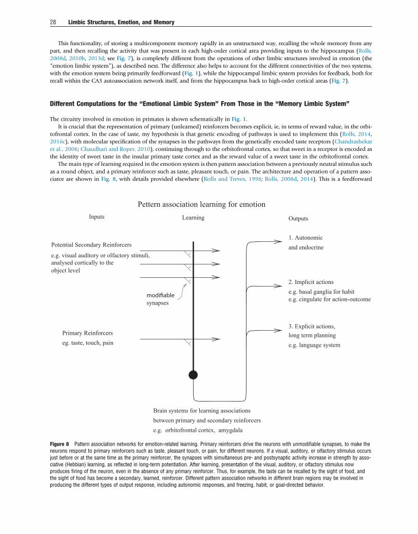
This functionality, of storing a multicomponent memory rapidly in an unstructured way, recalling the whole memory from anypart, and then recalling the activity that was present in each high-order cortical area providing inputs to the hippocampus (Rolls,2008d, 2010b, 2013d; see Fig. 7), is completely different from the operations of other limbic structures involved in emotion (the“emotion limbic system”), as described next. The difference also helps to account for the different connectivities of the two systems,with the emotion system being primarily feedforward (Fig. 1), while the hippocampal limbic system provides for feedback, both forrecall within the CA3 autoassociation network itself, and from the hippocampus back to high-order cortical areas (Fig. 7).
Different Computations for the “Emotional Limbic System” From Those in the “Memory Limbic System”
The circuitry involved in emotion in primates is shown schematically in Fig. 1.It is crucial that the representation of primary (unlearned) reinforcers becomes explicit, ie, in terms of reward value, in the orbi-
tofrontal cortex. In the case of taste, my hypothesis is that genetic encoding of pathways is used to implement this (Rolls, 2014,2016c), with molecular specification of the synapses in the pathways from the genetically encoded taste receptors (Chandrashekaret al., 2006; Chaudhari and Roper, 2010), continuing through to the orbitofrontal cortex, so that sweet in a receptor is encoded asthe identity of sweet taste in the insular primary taste cortex and as the reward value of a sweet taste in the orbitofrontal cortex.
The main type of learning required in the emotion system is then pattern association between a previously neutral stimulus suchas a round object, and a primary reinforcer such as taste, pleasant touch, or pain. The architecture and operation of a pattern asso-ciator are shown in Fig. 8, with details provided elsewhere (Rolls and Treves, 1998; Rolls, 2008d, 2014). This is a feedforward
Brain systems for learning associationsbetween primary and secondary reinforcerse.g. orbitofrontal cortex, amygdala
e.g. language systemlong term planning3. Explicit actions,
e.g. basal ganglia for habite.g. cingulate for action-outcome
2. Implicit actions
and endocrine1. Autonomic
OutputsLearningInputs
Pettern association learning for emotion
Potential Secondary Reinforcers
e.g. visual auditory or olfactory stimuli,analysed cortically to theobject level
Primary Reinforcerseg. taste, touch, pain
synapses
Figure 8 Pattern association networks for emotion-related learning. Primary reinforcers drive the neurons with unmodifiable synapses, to make theneurons respond to primary reinforcers such as taste, pleasant touch, or pain, for different neurons. If a visual, auditory, or olfactory stimulus occursjust before or at the same time as the primary reinforcer, the synapses with simultaneous pre- and postsynaptic activity increase in strength by asso-ciative (Hebbian) learning, as reflected in long-term potentiation. After learning, presentation of the visual, auditory, or olfactory stimulus nowproduces firing of the neuron, even in the absence of any primary reinforcer. Thus, for example, the taste can be recalled by the sight of food, andthe sight of food has become a secondary, learned, reinforcer. Different pattern association networks in different brain regions may be involved inproducing the different types of output response, including autonomic responses, and freezing, habit, or goal-directed behavior.
28 Limbic Structures, Emotion, and Memory
operation. The conditioned stimulus, eg, the visual stimulus, becomes associated with the US (eg, the taste). However, the oppositecannot occur, the visual stimulus cannot be retrieved from the taste. This is a feedforward operation. It is completely different fromthe completion of a memory enabled by the autoassociation network in the hippocampus (Rolls, 2013d, 2016c). The pattern asso-ciation operation enables expected value to be computed from outcome value (Rolls, 2008d, 2014).
In computing emotion-related value representations, inputs about the properties of primary reinforcers and objects are required,not inputs about space. That is why the inputs to the emotional system are from sensors such as those involved in taste, touch andpain, and are from the ventral visual system where objects that can have these properties are represented. This dominance of theventral (“what”) visual system as providing the inputs for emotion and value decoding is strongly to be contrasted with the episodicmemory system, in which where, and when, events happened is important, with input therefore required from the parietal spatial/dorsal visual systems. The orbitofrontal cortex is in a sense one end of the “what” processing systems and does not know about spaceor actions (Rolls, 2014). As we have seen the amygdala is in primates to some extent a secondary player to the orbitofrontal cortex,though involved in at least some classically conditioned responses to stimuli that produce emotions.
The orbitofrontal cortex (but not the amygdala) goes even beyond though the pattern associator computation illustrated inFig. 8 and can involve a one-trial, rule-based change of reward-related behavior. For example, if one visual stimulus suddenlybecomes no longer associated with reward, the orbitofrontal cortex system can immediately switch to another visual stimulus,even though that stimulus has previously been associated with punishment (Thorpe et al., 1983). An integrate-and-fire neuronalnetwork mechanism that utilizes the positive reward prediction error neurons found in the orbitofrontal cortex has been describedand simulated (Deco and Rolls, 2005a). An attractor network keeps the current rule active and biases appropriately the conditionalreward neurons found in the orbitofrontal cortex (Rolls, 2014). The positive reward prediction error neurons, themselves showingcontinuous firing in an attractor, then quench the rule attractor, so that another rule attractor population of neurons can emergebecause it is less adapted than the recently firing rule neurons. The whole network operates well and simulates and provides anaccount for several types of neuron found in the orbitofrontal cortex (Deco and Rolls, 2005a; Rolls, 2014).
The value representations computed in the orbitofrontal cortex are then transmitted to the ACC so that with outcomes repre-sented now in the ACC, and with connections moving back toward the midcingulate cortex, action-outcome learning can be imple-mented. For action-outcome learning, a representation of an action that has just been made is required, and this is present inmidcingulate cortex areas. The action must be remembered, perhaps for several seconds, until the outcome is received. The memoryof the action, I hypothesize, is provided for by an attractor network in the mid/anterior cingulate cortex. Associations can then belearned by pattern association learning in the mid/anterior cingulate cortex between the actions and the outcomes. Often the wholeprocess may be facilitated by the learning of associations between the conditioned stimulus, the expected value representation, andthe action-outcome pairing represented in the mid/anterior cingulate cortex. The special role in this scenario for the anterior- andmidcingulate cortex is then to bring actions into the processing, and the costs of performing actions, so that appropriate instru-mental, goal-directed, actions can be performed to emotion-provoking stimuli represented in terms of reward value (Rolls,2014, 2016c).
Conclusions and Summary: Separate Limbic Structures or Systems for Emotion and for Memory, but No SingleLimbic System
The concept of a (single) limbic system has been shown to be outmoded, in that anterior limbic and related structures involved inemotion can operate independently, and by different computational principles, from the hippocampal memory system. Instead, theanatomical, neurophysiological, functional neuroimaging, and neuropsychological evidence described shows that anterior limbicand related structures including the orbitofrontal cortex and amygdala are involved in emotion, reward valuation, and reward-related decision-making (but not memory), with the value representations transmitted to the ACC for action-outcome learning.In this “emotion limbic system,” feedforward pattern association networks learn associations between visual, olfactory, and audi-tory stimuli with primary reinforcers such as taste, touch, and pain. In primates including humans, this learning can be very rapidand rule-based, with the orbitofrontal cortex overshadowing the amygdala in this learning important for social and emotionalbehavior. The cortical inputs to this limbic system come in primates from areas that represent “what” object is present, includinginferior temporal cortical areas toward the end of the ventral visual system, and the anterior insular taste cortex and somatosensorycortical areas.
The complementary anatomical, neurophysiological, functional neuroimaging, and neuropsychological evidence describedshows that the hippocampus and limbic structures to which it is connected including the posterior cingulate cortex and thefornix-mammillary body-anterior thalamus-posterior cingulate circuit are involved in episodic or event memory, but not emotion.This “hippocampal system” receives information from neocortical areas about spatial location including parietal cortex areastoward the end of the dorsal visual system, and about objects and faces from the temporal cortical visual areas toward the endof the ventral visual system, and can associate this information together by autoassociation in the CA3 region of the hippocampuswhich involves feedback in the recurrent collateral system. The hippocampal memory system can later recall the whole of this infor-mation in the CA3 region from any component, a feedback process, and can recall that information back to neocortical areas, againa feedback (to neocortex) recall process. The nature of the computation in this hippocampal system is thus very different from thefeedforward pattern association involved in stimulus–reward association learning in the emotion system (Rolls, 2013d). Emotionand reward signals can enter the hippocampal memory system from the orbitofrontal cortex, amygdala, and ACC (Rolls and Xiang,
Limbic Structures, Emotion, and Memory 29

2005; Rolls, 2014) and can be recalled back to the orbitofrontal cortex, amygdala, and ACC during memory recall (Smith et al.,2006), as emotion can provide a component of episodic memory.
Thus, the emotional and hippocampal networks or “limbic systems” operate by different principles and operate independentlyof each other except insofar as emotional state may be part of an episodic memory. The concept of a single limbic system may nolonger appropriately reflect how the brain operates. Instead, separate systems, which might be designated the emotional limbicsystem and the episodic memory hippocampal system, are present in the brain, and the separate and different principles of theiroperation have been described here and elsewhere (Rolls, 2008d, 2010b, 2013d, 2014, 2016c).
Acknowledgments
The author has worked on some of the investigations described here with I. De Araujo, G.C. Baylis, L.L. Baylis, A. Berthoz, A. Bilderbeck, M.C.A.Booth, R. Bowtell, A.D. Browning, H.D. Critchley, J.D. Feigenbaum, S. Francis, L. Franco, P. Georges-Francois, F. Grabenhorst, M.E. Hasselmo, C.Holscher, J. Hornak, M. Kadohisa, R.P. Kesner, M. Kringelbach, C.M. Leonard, C. Margot, C. McCabe, F. McGlone, F. Mora, J. O’Doherty, B.A. Parris,D.I. Perrett, J. Redoute, R.G. Robertson, M.K. Sanghera, T.R. Scott, Z. Sienkiewicz, S.J. Stringer, S.J. Thorpe, A. Treves, J. Tromans, M.I. Velazco, J.V.Verhagen, E.A. Wakeman, F.A.W. Wilson, J.-Z. Xiang, and S. Yaxley, and their collaboration is sincerely acknowledged. Some of the research describedwas supported by the Medical Research Council, Wellcome Trust, Human Frontier Science Program, and James S. McDonnell Foundation.
References
Adolphs, R., Gosselin, F., Buchanan, T.W., Tranel, D., Schyns, P., Damasio, A.R., 2005. A mechanism for impaired fear recognition after amygdala damage. Nature 433, 68–72.Aggleton, J.P., 2012. Multiple anatomical systems embedded within the primate medial temporal lobe: implications for hippocampal function. Neurosci. Biobehav. Rev. 36,
1579–1596.Amaral, D.G., 1987. Memory: anatomical organization of candidate brain regions. In: Mountcastle, V.B. (Ed.), Handbook of Physiology. Section 1, The Nervous System. American
Physiological Society, Washington, DC, pp. 211–294.Amaral, D.G., Price, J.L., Pitkanen, A., Carmichael, S.T., 1992. Anatomical organization of the primate amygdaloid complex. In: Aggleton, J.P. (Ed.), The Amygdala. Wiley-Liss, New
York, pp. 1–66.Amaral, D.G., Bauman, M.D., Capitanio, J.P., Lavenex, P., Mason, W.A., Mauldin-Jourdain, M.L., Mendoza, S.P., 2003. The amygdala: is it an essential component of the neural
network for social cognition? Neuropsychologia 41, 517–522.Amaral, D.G., 2003. The amygdala, social behavior, and danger detection. Ann. N.Y. Acad. Sci. 1000, 337–347.Anderson, A.K., Christoff, K., Stappen, I., Panitz, D., Ghahremani, D.G., Glover, G., Gabrieli, J.D., Sobel, N., 2003. Dissociated neural representations of intensity and valence in
human olfaction. Nat. Neurosci. 6, 196–202.Antoniadis, E.A., Winslow, J.T., Davis, M., Amaral, D.G., 2009. The nonhuman primate amygdala is necessary for the acquisition but not the retention of fear-potentiated startle.
Biol. Psychiatry 65, 241–248.Auger, S.D., Maguire, E.A., 2013. Assessing the mechanism of response in the retrosplenial cortex of good and poor navigators. Cortex 49, 2904–2913.Banta Lavenex, P., Lavenex, P., 2009. Spatial memory and the monkey hippocampus: not all space is created equal. Hippocampus 19, 8–19.Barbas, H., Pandya, D.N., 1989. Architecture and intrinsic connections of the prefrontal cortex in the rhesus monkey. J. Comp. Neurol. 286, 353–375.Barbas, H., 1988. Anatomic organization of basoventral and mediodorsal visual recipient prefrontal regions in the rhesus monkey. J. Comp. Neurol. 276, 313–342.Barbas, H., 1993. Organization of cortical afferent input to the orbitofrontal area in the rhesus monkey. Neuroscience 56, 841–864.Barbas, H., 1995. Anatomic basis of cognitive-emotional interactions in the primate prefrontal cortex. Neurosci. Biobehav. Rev. 19, 499–510.Barbas, H., 2007. Specialized elements of orbitofrontal cortex in primates. Ann. N.Y. Acad. Sci. 1121, 10–32.Baylis, L.L., Rolls, E.T., 1991. Responses of neurons in the primate taste cortex to glutamate. Physiol. Behav. 49, 973–979.Baylis, L.L., Rolls, E.T., Baylis, G.C., 1995. Afferent connections of the orbitofrontal cortex taste area of the primate. Neuroscience 64, 801–812.Bechara, A., Damasio, H., Damasio, A.R., Lee, G.P., 1999. Different contributions of the human amygdala and ventromedial prefrontal cortex to decision-making. J. Neurosci. 19,
5473–5481.Bechara, A., Damasio, H., Damasio, A.R., 2000. Emotion, decision making and the orbitofrontal cortex. Cereb. Cortex 10, 295–307.Bechterew, W., 1900. Demonstration eines gehirns mit Zestörung der vorderenund inneren Theile der Hirnrinde beider Schläfenlappen. Neurol. Cent. 20, 990–991.Berlin, H., Rolls, E.T., Kischka, U., 2004. Impulsivity, time perception, emotion, and reinforcement sensitivity in patients with orbitofrontal cortex lesions. Brain 127, 1108–1126.Berlin, H., Rolls, E.T., Iversen, S.D., 2005. Borderline personality disorder, impulsivity and the orbitofrontal cortex. Am. J. Psychiatry 162, 2360–2373.Bernoulli, J., 1738/1954. Exposition of a new theory on the measurement of risk. Econometrica 22, 23–36.Brand, M., Grabenhorst, F., Starcke, K., Vandekerckhove, M.M., Markowitsch, H.J., 2007. Role of the amygdala in decisions under ambiguity and decisions under risk: evidence
from patients with Urbach-Wiethe disease. Neuropsychologia 45, 1305–1317.Broca, P., 1878. Anatomie comparée des circonvolutions cérébrales: le grand lobe limbique et la scissure limbique dans la série des mammifères. Rev. Anthropol. 1, 385–498.Bromberg-Martin, E.S., Matsumoto, M., Hikosaka, O., 2010. Dopamine in motivational control: rewarding, aversive, and alerting. Neuron 68, 815–834.Brown, S., Schäfer, E.A., 1888. An investigation into the functions of the occipital and temporal lobes of the monkey’s brain. 303–327. Philos. Trans. R. Soc. Lond. B 179,
303–327.Buckley, M.J., Gaffan, D., 2000. The hippocampus, perirhinal cortex, and memory in the monkey. In: Bolhuis, J.J. (Ed.), Brain, Perception, and Memory: Advances in Cognitive
Neuroscience. Oxford University Press, Oxford, pp. 279–298.Buckley, M.J., 2005. The role of the perirhinal cortex and hippocampus in learning, memory, and perception. Q. J. Exp. Psychol. B 58, 246–268.Burgess, N., Maguire, E.A., O’Keefe, J., 2002. The human hippocampus and spatial and episodic memory. Neuron 35, 625–641.Burgess, N., 2008. Spatial cognition and the brain. Ann. N.Y. Acad. Sci. 1124, 77–97.Burton, M.J., Rolls, E.T., Mora, F., 1976. Effects of hunger on the responses of neurones in the lateral hypothalamus to the sight and taste of food. Exp. Neurol. 51, 668–677.Bush, G., Luu, P., Posner, M.I., 2000. Cognitive and emotional influences in anterior cingulate cortex. Trends Cogn. Sci. 4, 215–222.Bush, G., Vogt, B.A., Holmes, J., Dale, A.M., Greve, D., Jenike, M.A., Rosen, B.R., 2002. Dorsal anterior cingulate cortex: a role in reward-based decision making. Proc. Natl. Acad.
Sci. U.S.A. 99, 523–528.Butter, C.M., 1969. Perseveration in extinction and in discrimination reversal tasks following selective prefrontal ablations in Macaca mulatta. Physiol. Behav. 4, 163–171.Cabanac, M., 1992. Pleasure: the common currency. J. Theor. Biol. 155, 173–200.Cannon, W.B., 1927. The James-Lange theory of emotion: a critical examination and an alternative theory. Am. J. Psychol. 39, 106–124.
30 Limbic Structures, Emotion, and Memory
Cardinal, N., Parkinson, J.A., Hall, J., Everitt, B.J., 2002. Emotion and motivation: the role of the amygdala, ventral striatum, and prefrontal cortex. Neurosci. Biobehav. Rev. 26,321–352.
Carmichael, S.T., Price, J.L., 1994. Architectonic subdivision of the orbital and medial prefrontal cortex in the macaque monkey. J. Comp. Neurol. 346, 366–402.Carmichael, S.T., Price, J.L., 1995a. Limbic connections of the orbital and medial prefrontal cortex in macaque monkeys. J. Comp. Neurol. 363, 615–641.Carmichael, S.T., Price, J.L., 1995b. Sensory and premotor connections of the orbital and medial prefrontal cortex of macaque monkeys. J. Comp. Neurol. 363, 642–664.Carmichael, S.T., Price, J.L., 1996. Connectional networks within the orbital and medial prefrontal cortex of macaque monkeys. J. Comp. Neurol. 371, 179–207.Carmichael, S.T., Clugnet, M.C., Price, J.L., 1994. Central olfactory connections in the macaque monkey. J. Comp. Neurol. 346, 403–434.Cassaday, H.J., Rawlins, J.N., 1997. The hippocampus, objects, and their contexts. Behav. Neurosci. 111, 1228–1244.Catani, M., Dell’acqua, F., Thiebaut de Schotten, M., 2013. A revised limbic system model for memory, emotion and behaviour. Neurosci. Biobehav. Rev. 37, 1724–1737.Chadwick, M.J., Hassabis, D., Weiskopf, N., Maguire, E.A., 2010. Decoding individual episodic memory traces in the human hippocampus. Curr. Biol. 20, 544–547.Chandrashekar, J., Hoon, M.A., Ryba, N.J., Zuker, C.S., 2006. The receptors and cells for mammalian taste. Nature 444, 288–294.Chaudhari, N., Roper, S.D., 2010. The cell biology of taste. J. Cell Biol. 190, 285–296.Chaudhari, N., Landin, A.M., Roper, S.D., 2000. A metabotropic glutamate receptor variant functions as a taste receptor. Nat. Neurosci. 3, 113–119.Chib, V.S., Rangel, A., Shimojo, S., O’Doherty, J.P., 2009. Evidence for a common representation of decision values for dissimilar goods in human ventromedial prefrontal cortex.
J. Neurosci. 29, 12315–12320.Corkin, S., 2002. What’s new with the amnesic patient H.M.? Nat. Rev. Neurosci. 3, 153–160.Craig, A.D., 2009. How do you feel – now? The anterior insula and human awareness. Nat. Rev. Neurosci. 10, 59–70.Craig, A.D., 2011. Significance of the insula for the evolution of human awareness of feelings from the body. Ann. N.Y. Acad. Sci. 1225, 72–82.Crane, J., Milner, B., 2005. What went where? Impaired object-location learning in patients with right hippocampal lesions. Hippocampus 15, 216–231.Critchley, H.D., Harrison, N.A., 2013. Visceral influences on brain and behavior. Neuron 77, 624–638.Critchley, H.D., Rolls, E.T., 1996a. Hunger and satiety modify the responses of olfactory and visual neurons in the primate orbitofrontal cortex. J. Neurophysiol. 75, 1673–1686.Critchley, H.D., Rolls, E.T., 1996b. Responses of primate taste cortex neurons to the astringent tastant tannic acid. Chem. Senses 21, 135–145.Critchley, H.D., Rolls, E.T., 1996c. Olfactory neuronal responses in the primate orbitofrontal cortex: analysis in an olfactory discrimination task. J. Neurophysiol. 75, 1659–1672.Critchley, H.D., Wiens, S., Rotshtein, P., Ohman, A., Dolan, R.J., 2004. Neural systems supporting interoceptive awareness. Nat. Neurosci. 7, 189–195.Damasio, A., Damasio, H., Tranel, D., 2013. Persistence of feelings and sentience after bilateral damage of the insula. Cereb. Cortex 23, 833–846.Damasio, A.R., 1994. Descartes’ Error. Putnam, New York.Damasio, A.R., 1996. The somatic marker hypothesis and the possible functions of the prefrontal cortex. Philos. Trans. R. Soc. Lond B Biol. Sci. 351, 1413–1420.Davis, M., 1994. The role of the amygdala in emotional learning. Int. Rev. Neurobiol. 36, 225–266.Davis, M., 2006. Neural systems involved in fear and anxiety measured with fear-potentiated startle. Am. Psychol. 61, 741–756.Day, M., Langston, R., Morris, R.G., 2003. Glutamate-receptor-mediated encoding and retrieval of paired-associate learning. Nature 424, 205–209.de Araujo, I.E.T., Rolls, E.T., 2004. The representation in the human brain of food texture and oral fat. J. Neurosci. 24, 3086–3093.de Araujo, I.E.T., Kringelbach, M.L., Rolls, E.T., Hobden, P., 2003a. The representation of umami taste in the human brain. J. Neurophysiol. 90, 313–319.de Araujo, I.E.T., Kringelbach, M.L., Rolls, E.T., McGlone, F., 2003b. Human cortical responses to water in the mouth, and the effects of thirst. J. Neurophysiol. 90, 1865–1876.de Araujo, I.E.T., Rolls, E.T., Kringelbach, M.L., McGlone, F., Phillips, N., 2003c. Taste-olfactory convergence, and the representation of the pleasantness of flavour, in the human
brain. Eur. J. Neurosci. 18, 2059–2068.de Araujo, I.E.T., Rolls, E.T., Velazco, M.I., Margot, C., Cayeux, I., 2005. Cognitive modulation of olfactory processing. Neuron 46, 671–679.Deco, G., Rolls, E.T., 2005a. Synaptic and spiking dynamics underlying reward reversal in orbitofrontal cortex. Cereb. Cortex 15, 15–30.Deco, G., Rolls, E.T., 2005b. Neurodynamics of biased competition and co-operation for attention: a model with spiking neurons. J. Neurophysiol. 94, 295–313.Deco, G., Rolls, E.T., 2005c. Attention, short-term memory, and action selection: a unifying theory. Prog. Neurobiol. 76, 236–256.Deco, G., Rolls, E.T., 2006. Decision-making and Weber’s Law: a neurophysiological model. Eur. J. Neurosci. 24, 901–916.Deco, G., Rolls, E.T., Romo, R., 2009. Stochastic dynamics as a principle of brain function. Prog. Neurobiol. 88, 1–16.Deco, G., Rolls, E.T., Albantakis, L., Romo, R., 2013. Brain mechanisms for perceptual and reward-related decision-making. Prog. Neurobiol. 103, 194–213.Delatour, B., Witter, M.P., 2002. Projections from the parahippocampal region to the prefrontal cortex in the rat: evidence of multiple pathways. Eur. J. Neurosci. 15, 1400–1407.Dere, E., Easton, A., Nadel, L., Huston, J.P. (Eds.), 2008. Handbook of Episodic Memory. Elsevier, Amsterdam.Desimone, R., Duncan, J., 1995. Neural mechanisms of selective visual attention. Annu. Rev. Neurosci. 18, 193–222.Devinsky, O., Morrell, M.J., Vogt, B.A., 1995. Contributions of anterior cingulate cortex to behaviour. Brain 118, 279–306.Eichenbaum, H., 1997. Declarative memory: insights from cognitive neurobiology. Annu. Rev. Psychol. 48, 547–572.Everitt, B.J., Robbins, T.W., 2013. From the ventral to the dorsal striatum: devolving views of their roles in drug addiction. Neurosci. Biobehav. Rev. 37, 1946–1954.Everitt, B.J., Cardinal, R.N., Parkinson, J.A., Robbins, T.W., 2003. Appetitive behavior: impact of amygdala-dependent mechanisms of emotional learning. Ann. N.Y. Acad. Sci. 985,
233–250.Feigenbaum, J.D., Rolls, E.T., 1991. Allocentric and egocentric spatial information processing in the hippocampal formation of the behaving primate. Psychobiology 19, 21–40.Fellows, L.K., Farah, M.J., 2003. Ventromedial frontal cortex mediates affective shifting in humans: evidence from a reversal learning paradigm. Brain 126, 1830–1837.Fellows, L.K., 2007. The role of orbitofrontal cortex in decision making: a component process account. Ann. N.Y. Acad. Sci. 1121, 421–430.Fellows, L.K., 2011. Orbitofrontal contributions to value-based decision making: evidence from humans with frontal lobe damage. Ann. N.Y. Acad. Sci. 1239, 51–58.Ferry, A.T., Ongur, D., An, X., Price, J.L., 2000. Prefrontal cortical projections to the striatum in macaque monkeys: evidence for an organization related to prefrontal networks.
J. Comp. Neurol. 425, 447–470.Francis, S., Rolls, E.T., Bowtell, R., McGlone, F., O’Doherty, J., Browning, A., Clare, S., Smith, E., 1999. The representation of pleasant touch in the brain and its relationship with
taste and olfactory areas. Neuroreport 10, 453–459.Freese, J.L., Amaral, D.G., 2009. Neuroanatomy of the primate amygdala. In: Whalen, P.J., Phelps, E.A. (Eds.), The Human Amygdala. Guilford, New York, pp. 3–42.Frey, S., Petrides, M., 2002. Orbitofrontal cortex and memory formation. Neuron 36, 171–176.Frey, S., Petrides, M., 2003. Greater orbitofrontal activity predicts better memory for faces. Eur. J. Neurosci. 17, 2755–2758.Friston, K.J., Buechel, C., Fink, G.R., Morris, J., Rolls, E.T., Dolan, R.J., 1997. Psychophysiological and modulatory interactions in neuroimaging. Neuroimage 6, 218–229.Fyhn, M., Molden, S., Witter, M.P., Moser, E.I., Moser, M.B., 2004. Spatial representation in the entorhinal cortex. Science 305, 1258–1264.Gaffan, D., Gaffan, E.A., 1991. Amnesia in man following transection of the fornix. A review. Brain 114, 2611–2618.Gaffan, D., Harrison, S., 1989a. A comparison of the effects of fornix section and sulcus principalis ablation upon spatial learning by monkeys. Behav. Brain Res. 31, 207–220.Gaffan, D., Harrison, S., 1989b. Place memory and scene memory: effects of fornix transection in the monkey. Exp. Brain Res. 74, 202–212.Gaffan, D., Saunders, R.C., 1985. Running recognition of configural stimuli by fornix transected monkeys. Q. J. Exp. Psychol. 37B, 61–71.Gaffan, D., 1994. Scene-specific memory for objects: a model of episodic memory impairment in monkeys with fornix transection. J. Cogn. Neurosci. 6, 305–320.Gallagher, M., Holland, P.C., 1994. The amygdala complex: multiple roles in associative learning and attention. Proc. Natl. Acad. Sci. U.S.A. 91, 11771–11776.Ge, T., Feng, J., Grabenhorst, F., Rolls, E.T., 2012. Componential Granger causality, and its application to identifying the source and mechanisms of the top-down biased activation
that controls attention to affective vs sensory processing. Neuroimage 59, 1846–1858.Gelbard-Sagiv, H., Mukamel, R., Harel, M., Malach, R., Fried, I., 2008. Internally generated reactivation of single neurons in human hippocampus during free recall. Science 322,
96–101.
Limbic Structures, Emotion, and Memory 31
Georges-François, P., Rolls, E.T., Robertson, R.G., 1999. Spatial view cells in the primate hippocampus: allocentric view not head direction or eye position or place. Cereb. Cortex 9,197–212.
Ghashghaei, H.T., Barbas, H., 2002. Pathways for emotion: interactions of prefrontal and anterior temporal pathways in the amygdala of the rhesus monkey. Neuroscience 115,1261–1279.
Giocomo, L.M., Hasselmo, M.E., 2007. Neuromodulation by glutamate and acetylcholine can change circuit dynamics by regulating the relative influence of afferent input andexcitatory feedback. Mol. Neurobiol. 36, 184–200.
Glimcher, P.W., Fehr, E. (Eds.), 2013. Neuroeconomics: Decision-Making and the Brain, second ed. Academic Press, New York.Glimcher, P.W., Camerer, C.F., Fehr, E., Poldrack, R.A. (Eds.), 2009. Neuroeconomics: Decision-Making and the Brain. Academic Press, London.Glimcher, P.W., 2011. Foundations of Neuroeconomic Analysis. Oxford University Press, Oxford.Gothard, K.M., Battaglia, F.P., Erickson, C.A., Spitler, K.M., Amaral, D.G., 2007. Neural responses to facial expression and face identity in the monkey amygdala. J. Neurophysiol.
97, 1671–1683.Gottfried, J.A., Small, D.M., Zald, D.H., 2006. The chemical senses. In: Zald, D.H., Rauch, S.L. (Eds.), The Orbitofrontal Cortex. Oxford University Press, Oxford, pp. 125–171.Grabenhorst, F., Rolls, E.T., 2008. Selective attention to affective value alters how the brain processes taste stimuli. Eur. J. Neurosci. 27, 723–729.Grabenhorst, F., Rolls, E.T., 2009. Different representations of relative and absolute value in the human brain. Neuroimage 48, 258–268.Grabenhorst, F., Rolls, E.T., 2010. Attentional modulation of affective vs sensory processing: functional connectivity and a top-down biased activation theory of selective attention.
J. Neurophysiol. 104, 1649–1660.Grabenhorst, F., Rolls, E.T., 2011. Value, pleasure, and choice in the ventral prefrontal cortex. Trends Cogn. Sci. 15, 56–67.Grabenhorst, F., Rolls, E.T., Margot, C., da Silva, M.A.A.P., Velazco, M.I., 2007. How pleasant and unpleasant stimuli combine in different brain regions: odor mixtures. J. Neurosci.
27, 13532–13540.Grabenhorst, F., Rolls, E.T., Parris, B.A., 2008a. From affective value to decision-making in the prefrontal cortex. Eur. J. Neurosci. 28, 1930–1939.Grabenhorst, F., Rolls, E.T., Bilderbeck, A., 2008b. How cognition modulates affective responses to taste and flavor: top down influences on the orbitofrontal and pregenual cingulate
cortices. Cereb. Cortex 18, 1549–1559.Grabenhorst, F., Rolls, E.T., Parris, B.A., D’Souza, A., 2010a. How the brain represents the reward value of fat in the mouth. Cereb. Cortex 20, 1082–1091.Grabenhorst, F., D’Souza, A., Parris, B.A., Rolls, E.T., Passingham, R.E., 2010b. A common neural scale for the subjective pleasantness of different primary rewards. Neuroimage
51, 1265–1274.Grabenhorst, F., Rolls, E.T., Margot, C., 2011. A hedonically complex odor mixture captures the brain’s attention. Neuroimage 55, 832–843.Gray, J.A., 1975. Elements of a Two-Process Theory of Learning. Academic Press, London.Guest, S., Grabenhorst, F., Essick, G., Chen, Y., Young, M., McGlone, F., de Araujo, I., Rolls, E.T., 2007. Human cortical representation of oral temperature. Physiol. Behav. 92,
975–984.Haber, S.N., Kim, K.S., Mailly, P., Calzavara, R., 2006. Reward-related cortical inputs define a large striatal region in primates that interface with associative cortical connections,
providing a substrate for incentive-based learning. J. Neurosci. 26, 8368–8376.Hafting, T., Fyhn, M., Molden, S., Moser, M.B., Moser, E.I., 2005. Microstructure of a spatial map in the entorhinal cortex. Nature 436, 801–806.Hare, T.A., O’Doherty, J., Camerer, C.F., Schultz, W., Rangel, A., 2008. Dissociating the role of the orbitofrontal cortex and the striatum in the computation of goal values and
prediction errors. J. Neurosci. 28, 5623–5630.Hassabis, D., Chu, C., Rees, G., Weiskopf, N., Molyneux, P.D., Maguire, E.A., 2009. Decoding neuronal ensembles in the human hippocampus. Curr. Biol. 19, 546–554.Hasselmo, M.E., Rolls, E.T., Baylis, G.C., 1989. The role of expression and identity in the face-selective responses of neurons in the temporal visual cortex of the monkey. Behav.
Brain Res. 32, 203–218.Hertz, J., Krogh, A., Palmer, R.G., 1991. An Introduction to the Theory of Neural Computation. Addison-Wesley, Wokingham.Holland, P.C., Gallagher, M., 1999. Amygdala circuitry in attentional and representational processes. Trends Cogn. Sci. 3, 65–73.Hölscher, C., Jacob, W., Mallot, H.A., 2003a. Reward modulates neuronal activity in the hippocampus of the rat. Behav. Brain Res. 142, 181–191.Hölscher, C., Rolls, E.T., Xiang, J.-Z., 2003b. Perirhinal cortex neuronal activity related to long-term familiarity memory in the macaque. Eur. J. Neurosci. 18, 2037–2046.Hopfield, J.J., 1982. Neural networks and physical systems with emergent collective computational abilities. Proc. Natl. Acad. Sci. U.S.A. 79, 2554–2558.Hornak, J., Rolls, E.T., Wade, D., 1996. Face and voice expression identification in patients with emotional and behavioural changes following ventral frontal lobe damage.
Neuropsychologia 34, 247–261.Hornak, J., Bramham, J., Rolls, E.T., Morris, R.G., O’Doherty, J., Bullock, P.R., Polkey, C.E., 2003. Changes in emotion after circumscribed surgical lesions of the orbitofrontal and
cingulate cortices. Brain 126, 1691–1712.Hornak, J., O’Doherty, J., Bramham, J., Rolls, E.T., Morris, R.G., Bullock, P.R., Polkey, C.E., 2004. Reward-related reversal learning after surgical excisions in orbitofrontal and
dorsolateral prefrontal cortex in humans. J. Cogn. Neurosci. 16, 463–478.Insausti, R., Amaral, D.G., Cowan, W.M., 1987. The entorhinal cortex of the monkey. II. Cortical afferents. J. Comp. Neurol. 264, 356–395.Isaacson, R.L., 1982. The Limbic System, second ed. Plenum, New York.Itskov, P.M., Vinnik, E., Diamond, M.E., 2011. Hippocampal representation of touch-guided behavior in rats: persistent and independent traces of stimulus and reward location.
PLoS One 6, e16462.Iversen, S.D., Mishkin, M., 1970. Perseverative interference in monkeys following selective lesions of the inferior prefrontal convexity. Exp. Brain Res. 11, 376–386.Izquierdo, A., Murray, E.A., 2004. Combined unilateral lesions of the amygdala and orbital prefrontal cortex impair affective processing in rhesus monkeys. J. Neurophysiol. 91,
2023–2039.Izquierdo, A., Suda, R.K., Murray, E.A., 2004. Bilateral orbital prefrontal cortex lesions in rhesus monkeys disrupt choices guided by both reward value and reward contingency.
J. Neurosci. 24, 7540–7548.Jacobs, J., Weidemann, C.T., Miller, J.F., Solway, A., Burke, J.F., Wei, X.X., Suthana, N., Sperling, M.R., Sharan, A.D., Fried, I., Kahana, M.J., 2013. Direct recordings of grid-like
neuronal activity in human spatial navigation. Nat. Neurosci. 16, 1188–1190.Jarrard, E.L., 1993. On the role of the hippocampus in learning and memory in the rat. Behav. Neural Biol. 60, 9–26.Jeffery, K.J., Hayman, R., 2004. Plasticity of the hippocampal place cell representation. Rev. Neurosci. 15, 309–331.Jeffery, K.J., Anderson, M.I., Hayman, R., Chakraborty, S., 2004. A proposed architecture for the neural representation of spatial context. Neurosci. Biobehav. Rev. 28, 201–218.Johnson, T.N., Rosvold, H.E., Mishkin, M., 1968. Projections from behaviorally defined sectors of the prefrontal cortex to the basal ganglia, septum and diencephalon of the monkey.
Exp. Neurol. 21, 20–34.Jones, B., Mishkin, M., 1972. Limbic lesions and the problem of stimulus-reinforcement associations. Exp. Neurol. 36, 362–377.Kable, J.W., Glimcher, P.W., 2009. The neurobiology of decision: consensus and controversy. Neuron 63, 733–745.Kadohisa, M., Rolls, E.T., Verhagen, J.V., 2004. Orbitofrontal cortex neuronal representation of temperature and capsaicin in the mouth. Neuroscience 127, 207–221.Kadohisa, M., Rolls, E.T., Verhagen, J.V., 2005a. The primate amygdala: neuronal representations of the viscosity, fat texture, temperature, grittiness and taste of foods.
Neuroscience 132, 33–48.Kadohisa, M., Rolls, E.T., Verhagen, J.V., 2005b. Neuronal representations of stimuli in the mouth: the primate insular taste cortex, orbitofrontal cortex, and amygdala. Chem.
Senses 30, 401–419.Kemp, J.M., Powell, T.P.S., 1970. The cortico-striate projections in the monkey. Brain 93, 525–546.Kesner, R.P., Rolls, E.T., 2001. Role of long term synaptic modification in short term memory. Hippocampus 11, 240–250.
32 Limbic Structures, Emotion, and Memory
Kesner, R.P., Rolls, E.T., 2015. A computational theory of hippocampal function, and tests of the theory: new developments. Neurosci. Biobehav. Rev. 48, 92–147.Kesner, R.P., Lee, I., Gilbert, P., 2004. A behavioral assessment of hippocampal function based on a subregional analysis. Rev. Neurosci. 15, 333–351.Kesner, R.P., Morris, A.M., Weeden, C.S.S., 2012. Spatial, temporal, and associative behavioral functions associated with different subregions of the hippocampus. In: Zentall, T.R.,
Wasserman, E.A. (Eds.), Oxford Handbook of Comparative Cognition. Oxford University Press, Oxford, pp. 322–346.Kesner, R.P., 1998. Neural mediation of memory for time: role of hippocampus and medial prefrontal cortex. Psychol. Bull. Rev. 5, 585–596.Killian, N.J., Jutras, M.J., Buffalo, E.A., 2012. A map of visual space in the primate entorhinal cortex. Nature 491, 761–764.Kluver, H., Bucy, P.C., 1939. Preliminary analysis of functions of the temporal lobes in monkeys. Arch. Neurol. Psychiatry 42, 979–1000.Knutson, B., Rick, S., Wimmer, G.E., Prelec, D., Loewenstein, G., 2007. Neural predictors of purchases. Neuron 53, 147–156.Kobayashi, S., Pinto de Carvalho, O., Schultz, W., 2010. Adaptation of reward sensitivity in orbitofrontal neurons. J. Neurosci. 30, 534–544.Kolb, B., Whishaw, I.Q., 2003. Fundamentals of Human Neuropsychology, fifth ed. Worth, New York.Kringelbach, M.L., Rolls, E.T., 2003. Neural correlates of rapid reversal learning in a simple model of human social interaction. Neuroimage 20, 1371–1383.Kringelbach, M.L., Rolls, E.T., 2004. The functional neuroanatomy of the human orbitofrontal cortex: evidence from neuroimaging and neuropsychology. Prog. Neurobiol. 72,
341–372.Kringelbach, M.L., O’Doherty, J., Rolls, E.T., Andrews, C., 2003. Activation of the human orbitofrontal cortex to a liquid food stimulus is correlated with its subjective pleasantness.
Cereb. Cortex 13, 1064–1071.Krolak-Salmon, P., Henaff, M.A., Isnard, J., Tallon-Baudry, C., Guenot, M., Vighetto, A., Bertrand, O., Mauguiere, F., 2003. An attention modulated response to disgust in human
ventral anterior insula. Ann. Neurol. 53, 446–453.Kropff, E., Treves, A., 2008. The emergence of grid cells: intelligent design or just adaptation? Hippocampus 18, 1256–1269.Lavenex, P., Suzuki, W.A., Amaral, D.G., 2004. Perirhinal and parahippocampal cortices of the macaque monkey: intrinsic projections and interconnections. J. Comp. Neurol. 472,
371–394.LeDoux, J.E., 1995. Emotion: clues from the brain. Annu. Rev. Psychol. 46, 209–235.LeDoux, J.E., 2000a. Emotion circuits in the brain. Annu. Rev. Neurosci. 23, 155–184.LeDoux, J.E., 2000b. The amygdala, fear conditioning and emotion. In: Aggleton, J.P. (Ed.), The Amygdala: A Functional Analysis. Oxford University Press, Oxford.Leonard, C.M., Rolls, E.T., Wilson, F.A.W., Baylis, G.C., 1985. Neurons in the amygdala of the monkey with responses selective for faces. Behav. Brain Res. 15, 159–176.Luo, Q., Ge, T., Grabenhorst, F., Feng, J., Rolls, E.T., 2013. Attention-dependent modulation of cortical taste circuits revealed by Granger causality with signal-dependent noise.
PLoS Comput. Biol. 9, e1003265.MacLean, P.D., 1949. Psychosomatic disease and the ‘visceral brain’: recent developments bearing on the Papez theory of emotion. Psychosom. Med. 11, 338–353.MacLean, P.D., 1952. Some psychiatric implications of physiological studies on frontotemporal portion of limbic system (visceral brain). Electroencephalogr. Clin. Neurophysiol. 4,
407–418.Maia, T.V., McClelland, J.L., 2004. A reexamination of the evidence for the somatic marker hypothesis: what participants really know in the Iowa gambling task. Proc. Natl. Acad.
Sci. U.S.A. 101, 16075–16080.Malkova, L., Mishkin, M., 2003. One-trial memory for object-place associations after separate lesions of hippocampus and posterior parahippocampal region in the monkey.
J. Neurosci. 23, 1956–1965.Martin, S.J., Grimwood, P.D., Morris, R.G., 2000. Synaptic plasticity and memory: an evaluation of the hypothesis. Annu. Rev. Neurosci. 23, 649–711.Maruyama, Y., Pereira, E., Margolskee, R.F., Chaudhari, N., Roper, S.D., 2006. Umami responses in mouse taste cells indicate more than one receptor. J. Neurosci. 26,
2227–2234.Matsumoto, M., Hikosaka, O., 2009. Two types of dopamine neuron distinctly convey positive and negative motivational signals. Nature 459, 837–841.Matsumoto, M., Matsumoto, K., Abe, H., Tanaka, K., 2007. Medial prefrontal selectivity signalling prediction errors of action values. Nat. Neurosci. 10, 647–656.McCabe, C., Rolls, E.T., 2007. Umami: a delicious flavor formed by convergence of taste and olfactory pathways in the human brain. Eur. J. Neurosci. 25, 1855–1864.McCabe, C., Rolls, E.T., Bilderbeck, A., McGlone, F., 2008. Cognitive influences on the affective representation of touch and the sight of touch in the human brain. Social. Cogn.
Affect. Neurosci. 3, 97–108.McFarland, D.J., Sibly, R.M., 1975. The behavioural final common path. Philos. Trans. R. Soc. Lond B Biol. Sci. 270, 265–293.McNaughton, B.L., Barnes, C.A., O’Keefe, J., 1983. The contributions of position, direction, and velocity to single unit activity in the hippocampus of freely-moving rats. Exp. Brain
Res. 52, 41–49.McNaughton, B.L., Battaglia, F.P., Jensen, O., Moser, E.I., Moser, M.B., 2006. Path integration and the neural basis of the ‘cognitive map’. Nat. Rev. Neurosci. 7, 663–678.Medina, L., Bupesh, M., Abellan, A., 2011. Contribution of genoarchitecture to understanding forebrain evolution and development, with particular emphasis on the amygdala. Brain
Behav. Evol. 78, 216–236.Menon, V., Uddin, L.Q., 2010. Saliency, switching, attention and control: a network model of insula function. Brain Struct. Funct. 214, 655–667.Mesulam, M.M., 2000. Behavioral neuroanatomy: large-scale networks, association cortex, frontal syndromes, the limbic system, and hemispheric specializations. In:
Mesulam, M.M. (Ed.), Principles of Behavioral and Cognitive Neurology, second ed. Oxford University Press, Oxford, pp. 1–120.Moll, J., Krueger, F., Zahn, R., Pardini, M., de Oliveira-Souza, R., Grafman, J., 2006. Human fronto-mesolimbic networks guide decisions about charitable donation. Proc. Natl.
Acad. Sci. U.S.A. 103, 15623–15628.Montague, P.R., Berns, G.S., 2002. Neural economics and the biological substrates of valuation. Neuron 36, 265–284.Mora, F., Avrith, D.B., Phillips, A.G., Rolls, E.T., 1979. Effects of satiety on self-stimulation of the orbitofrontal cortex in the monkey. Neurosci. Lett. 13, 141–145.Mora, F., Avrith, D.B., Rolls, E.T., 1980. An electrophysiological and behavioural study of self-stimulation in the orbitofrontal cortex of the rhesus monkey. Brain Res. Bull. 5,
111–115.Morecraft, R.J., Tanji, J., 2009. Cingulofrontal interaction and the cingulate motor areas. In: Vogt, B.A. (Ed.), Cingulate Neurobiology and Disease. Oxford Univesrity Press, Oxford,
pp. 113–144.Morecraft, R.J., Geula, C., Mesulam, M.-M., 1992. Cytoarchitecture and neural afferents of orbitofrontal cortex in the brain of the monkey. J. Comp. Neurol. 232, 341–358.Morris, J.S., Dolan, R.J., 2001. Involvement of human amygdala and orbitofrontal cortex in hunger-enhanced memory for food stimuli. J. Neurosci. 21, 5304–5310.Mufson, E.J., Mesulam, M.-M., 1982. Insula of the old world monkey: II: afferent cortical input and comments on the claustrum. J. Comp. Neurol. 212, 23–37.Muller, R.U., Kubie, J.L., Bostock, E.M., Taube, J.S., Quirk, G.J., 1991. Spatial firing correlates of neurons in the hippocampal formation of freely moving rats. In: Paillard, J. (Ed.),
Brain and Space. Oxford University Press, Oxford, pp. 296–333.Murray, E.A., Izquierdo, A., 2007. Orbitofrontal cortex and amygdala contributions to affect and action in primates. Ann. N.Y. Acad. Sci. 1121, 273–296.Murray, E.A., Baxter, M.G., Gaffan, D., 1998. Monkeys with rhinal cortex damage or neurotoxic hippocampal lesions are impaired on spatial scene learning and object reversals.
Behav. Neurosci. 112, 1291–1303.Nauta, W.J.H., 1964. Some efferent connections of the prefrontal cortex in the monkey. In: Warren, J.M., Akert, K. (Eds.), The Frontal Granular Cortex and Behavior. McGraw Hill,
New York, pp. 397–407.Niki, H., Watanabe, M., 1979. Prefrontal and cingulate unit activity during timing behavior in the monkey. Brain Res. 171, 213–224.Norgren, R., 1984. Central neural mechanisms of taste. In: Darien-Smith, I. (Ed.), Handbook of Physiology – The Nervous System III. Sensory Processes 1. American Physiological
Society, Washington, DC, pp. 1087–1128.O’Doherty, J., Rolls, E.T., Francis, S., Bowtell, R., McGlone, F., Kobal, G., Renner, B., Ahne, G., 2000. Sensory-specific satiety related olfactory activation of the human orbitofrontal
cortex. Neuroreport 11, 893–897.
Limbic Structures, Emotion, and Memory 33
O’Doherty, J., Rolls, E.T., Francis, S., Bowtell, R., McGlone, F., 2001a. The representation of pleasant and aversive taste in the human brain. J. Neurophysiol. 85, 1315–1321.O’Doherty, J., Kringelbach, M.L., Rolls, E.T., Hornak, J., Andrews, C., 2001b. Abstract reward and punishment representations in the human orbitofrontal cortex. Nat. Neurosci. 4,
95–102.O’Doherty, J.P., Deichmann, R., Critchley, H.D., Dolan, R.J., 2002. Neural responses during anticipation of a primary taste reward. Neuron 33, 815–826.O’Doherty, J., Winston, J., Critchley, H., Perrett, D., Burt, D.M., Dolan, R.J., 2003. Beauty in a smile: the role of medial orbitofrontal cortex in facial attractiveness. Neuropsychologia
41, 147–155.O’Keefe, J., Nadel, L., 1978. The Hippocampus as a Cognitive Map. Clarendon Press, Oxford.O’Keefe, J., 1984. Spatial memory within and without the hippocampal system. In: Seifert, W. (Ed.), Neurobiology of the Hippocampus. Academic Press, London,
pp. 375–403.Olausson, H., Lamarre, Y., Backlund, H., Morin, C., Wallin, B.G., Starck, G., Ekholm, S., Strigo, I., Worsley, K., Vallbo, A.B., Bushnell, M.C., 2002. Unmyelinated tactile afferents
signal touch and project to insular cortex. Nat. Neurosci. 5, 900–904.Öngür, D., Price, J.L., 2000. The organisation of networks within the orbital and medial prefrontal cortex of rats, monkeys and humans. Cereb. Cortex 10, 206–219.Öngür, D., Ferry, A.T., Price, J.L., 2003. Architectonic division of the human orbital and medial prefrontal cortex. J. Comp. Neurol. 460, 425–449.Padoa-Schioppa, C., Assad, J.A., 2006. Neurons in the orbitofrontal cortex encode economic value. Nature 441, 223–226.Padoa-Schioppa, C., Assad, J.A., 2008. The representation of economic value in the orbitofrontal cortex is invariant for changes of menu. Nat. Neurosci. 11, 95–102.Padoa-Schioppa, C., 2009. Range-adapting representation of economic value in the orbitofrontal cortex. J. Neurosci. 29, 14004–14014.Padoa-Schioppa, C., 2011. Neurobiology of economic choice: a good-based model. Annu. Rev. Neurosci. 34, 333–359.Palomero-Gallagher, N., Zilles, K., 2004. Isocortex. In: Paxinos, G. (Ed.), The Rat Nervous System. Elsevier Academic Press, San Diego, CA, pp. 729–757.Pandya, D.N., Yeterian, E.H., 1996. Comparison of prefrontal architecture and connections. Philos. Trans. R. Soc. Lond. B 351, 1423–1431.Papez, J.W., 1937. A proposed mechanism for emotion. Arch. Neurol. Psychiatry 38, 725–743.Parkinson, J.K., Murray, E.A., Mishkin, M., 1988. A selective mnemonic role for the hippocampus in monkeys: memory for the location of objects. J. Neurosci. 8, 4059–4167.Passingham, R.E.P., Wise, S.P., 2012. The Neurobiology of the Prefrontal Cortex. Oxford University Press, Oxford.Paton, J.J., Belova, M.A., Morrison, S.E., Salzman, C.D., 2006. The primate amygdala represents the positive and negative value of visual stimuli during learning. Nature 439,
865–870.Petrides, M., Pandya, D.N., 1995. Comparative architectonic analysis of the human and macaque frontal cortex. In: Boller, F., Grafman, J. (Eds.), Handbook of Neuropsychology.
Elsevier Science, Amsterdam, pp. 17–58.Petrides, M., 1985. Deficits on conditional associative-learning tasks after frontal- and temporal-lobe lesions in man. Neuropsychologia 23, 601–614.Petrides, M., 2007. The orbitofrontal cortex: novelty, deviation from expectation, and memory. Ann. N.Y. Acad. Sci. 1121, 33–53.Phelps, E.A., LeDoux, J.E., 2005. Contributions of the amygdala to emotion processing: from animal models to human behavior. Neuron 48, 175–187.Phillips, M.L., Young, A.W., Scott, S.K., Calder, A.J., Andrew, C., Giampietro, V., Williams, S.C., Bullmore, E.T., Brammer, M., Gray, J.A., 1998. Neural responses to facial and vocal
expressions of fear and disgust. Proc. R. Soc. Lond B Biol. Sci. 265, 1809–1817.Phillips, M.L., Williams, L.M., Heining, M., Herba, C.M., Russell, T., Andrew, C., Bullmore, E.T., Brammer, M.J., Williams, S.C., Morgan, M., Young, A.W., Gray, J.A., 2004.
Differential neural responses to overt and covert presentations of facial expressions of fear and disgust. Neuroimage 21, 1484–1496.Pitkanen, A., Kelly, J.L., Amaral, D.G., 2002. Projections from the lateral, basal, and accessory basal nuclei of the amygdala to the entorhinal cortex in the macaque monkey.
Hippocampus 12, 186–205.Preuss, T.M., 1995. Do rats have prefrontal cortex? The Rose-Woolsey-Akert program reconsidered. J. Cogn. Neurosci. 7, 1–24.Price, J.L., Carmichael, S.T., Carnes, K.M., Clugnet, M.-C., Kuroda, M., Ray, J.P., 1991. Olfactory input to the prefrontal cortex. In: Davis, J.L., Eichenbaum, H. (Eds.), Olfaction: A
Model System for Computational Neuroscience. MIT Press, Cambridge, Mass, pp. 101–120.Price, J.L., 2006. Connections of orbital cortex. In: Zald, D.H., Rauch, S.L. (Eds.), The Orbitofrontal Cortex. Oxford University Press, Oxford, pp. 39–55.Price, J.L., 2007. Definition of the orbital cortex in relation to specific connections with limbic and visceral structures and other cortical regions. Ann. N.Y. Acad. Sci. 1121, 54–71.Pritchard, T.C., Hamilton, R.B., Morse, J.R., Norgren, R., 1986. Projections of thalamic gustatory and lingual areas in the monkey, Macaca fascicularis. J. Comp. Neurol. 244,
213–228.Pritchard, T.C., Edwards, E.M., Smith, C.A., Hilgert, K.G., Gavlick, A.M., Maryniak, T.D., Schwartz, G.J., Scott, T.R., 2005. Gustatory neural responses in the medial orbitofrontal
cortex of the old world monkey. J. Neurosci. 25, 6047–6056.Rawlins, J.N.P., 1985. Associations across time: the hippocampus as a temporary memory store. Behav. Brain Sci. 8, 479–496.Robertson, R.G., Rolls, E.T., Georges-François, P., 1998. Spatial view cells in the primate hippocampus: effects of removal of view details. J. Neurophysiol. 79, 1145–1156.Rolls, E.T., Baylis, G.C., 1986. Size and contrast have only small effects on the responses to faces of neurons in the cortex of the superior temporal sulcus of the monkey. Exp. Brain
Res. 65, 38–48.Rolls, E.T., Baylis, L.L., 1994. Gustatory, olfactory, and visual convergence within the primate orbitofrontal cortex. J. Neurosci. 14, 5437–5452.Rolls, E.T., Deco, G., 2002. Computational Neuroscience of Vision. Oxford University Press, Oxford.Rolls, E.T., Deco, G., 2010. The Noisy Brain: Stochastic Dynamics as a Principle of Brain Function. Oxford University Press, Oxford.Rolls, E.T., Grabenhorst, F., 2008. The orbitofrontal cortex and beyond: from affect to decision-making. Prog. Neurobiol. 86, 216–244.Rolls, E.T., Kesner, R.P., 2006. A computational theory of hippocampal function, and empirical tests of the theory. Prog. Neurobiol. 79, 1–48.Rolls, E.T., McCabe, C., 2007. Enhanced affective brain representations of chocolate in cravers vs non-cravers. Eur. J. Neurosci. 26, 1067–1076.Rolls, E.T., Rolls, J.H., 1997. Olfactory sensory-specific satiety in humans. Physiol. Behav. 61, 461–473.Rolls, E.T., Scott, T.R., 2003. Central taste anatomy and neurophysiology. In: Doty, R.L. (Ed.), Handbook of Olfaction and Gustation, second ed. Dekker, New York, pp. 679–705.Rolls, E.T., Treves, A., 1998. Neural Networks and Brain Function. Oxford University Press, Oxford.Rolls, E.T., Treves, A., 2011. The neuronal encoding of information in the brain. Prog. Neurobiol. 95, 448–490.Rolls, E.T., Xiang, J.-Z., 2005. Reward-spatial view representations and learning in the hippocampus. J. Neurosci. 25, 6167–6174.Rolls, E.T., Xiang, J.-Z., 2006. Spatial view cells in the primate hippocampus, and memory recall. Rev. Neurosci. 17, 175–200.Rolls, E.T., Burton, M.J., Mora, F., 1976. Hypothalamic neuronal responses associated with the sight of food. Brain Res. 111, 53–66.Rolls, E.T., Judge, S.J., Sanghera, M., 1977. Activity of neurones in the inferotemporal cortex of the alert monkey. Brain Res. 130, 229–238.Rolls, E.T., Burton, M.J., Mora, F., 1980. Neurophysiological analysis of brain-stimulation reward in the monkey. Brain Res. 194, 339–357.Rolls, B.J., Rolls, E.T., Rowe, E.A., Sweeney, K., 1981a. Sensory specific satiety in man. Physiol. Behav. 27, 137–142.Rolls, B.J., Rolls, E.T., Rowe, E.A., Sweeney, K., 1981b. How sensory properties of foods affect human feeding behaviour. Physiol. Behav. 29, 409–417.Rolls, E.T., Scott, T.R., Sienkiewicz, Z.J., Yaxley, S., 1988. The responsiveness of neurones in the frontal opercular gustatory cortex of the macaque monkey is independent of
hunger. J. Physiol. 397, 1–12.Rolls, E.T., Sienkiewicz, Z.J., Yaxley, S., 1989a. Hunger modulates the responses to gustatory stimuli of single neurons in the caudolateral orbitofrontal cortex of the macaque
monkey. Eur. J. Neurosci. 1, 53–60.Rolls, E.T., Miyashita, Y., Cahusac, P.M.B., Kesner, R.P., Niki, H., Feigenbaum, J., Bach, L., 1989b. Hippocampal neurons in the monkey with activity related to the place in which
a stimulus is shown. J. Neurosci. 9, 1835–1845.Rolls, E.T., Yaxley, S., Sienkiewicz, Z.J., 1990. Gustatory responses of single neurons in the caudolateral orbitofrontal cortex of the macaque monkey. J. Neurophysiol. 64,
1055–1066.
34 Limbic Structures, Emotion, and Memory
Rolls, E.T., Hornak, J., Wade, D., McGrath, J., 1994. Emotion-related learning in patients with social and emotional changes associated with frontal lobe damage. J. Neurol.Neurosurg. Psychiatry 57, 1518–1524.
Rolls, E.T., Critchley, H.D., Mason, R., Wakeman, E.A., 1996a. Orbitofrontal cortex neurons: role in olfactory and visual association learning. J. Neurophysiol. 75, 1970–1981.Rolls, E.T., Critchley, H., Wakeman, E.A., Mason, R., 1996b. Responses of neurons in the primate taste cortex to the glutamate ion and to inosine 50-monophosphate. Physiol.
Behav. 59, 991–1000.Rolls, E.T., Robertson, R.G., Georges-François, P., 1997a. Spatial view cells in the primate hippocampus. Eur. J. Neurosci. 9, 1789–1794.Rolls, E.T., Treves, A., Foster, D., Perez-Vicente, C., 1997b. Simulation studies of the CA3 hippocampal subfield modelled as an attractor neural network. Neural Netw. 10,
1559–1569.Rolls, E.T., Critchley, H.D., Browning, A., Hernadi, I., 1998a. The neurophysiology of taste and olfaction in primates, and umami flavor. Ann. N.Y. Acad. Sci. 855, 426–437.Rolls, E.T., Treves, A., Robertson, R.G., Georges-François, P., Panzeri, S., 1998b. Information about spatial view in an ensemble of primate hippocampal cells. J. Neurophysiol. 79,
1797–1813.Rolls, E.T., Critchley, H.D., Browning, A.S., Hernadi, A., Lenard, L., 1999. Responses to the sensory properties of fat of neurons in the primate orbitofrontal cortex. J. Neurosci. 19,
1532–1540.Rolls, E.T., Kringelbach, M.L., de Araujo, I.E.T., 2003a. Different representations of pleasant and unpleasant odors in the human brain. Eur. J. Neurosci. 18, 695–703.Rolls, E.T., Verhagen, J.V., Kadohisa, M., 2003b. Representations of the texture of food in the primate orbitofrontal cortex: neurons responding to viscosity, grittiness and capsaicin.
J. Neurophysiol. 90, 3711–3724.Rolls, E.T., O’Doherty, J., Kringelbach, M.L., Francis, S., Bowtell, R., McGlone, F., 2003c. Representations of pleasant and painful touch in the human orbitofrontal and cingulate
cortices. Cereb. Cortex 13, 308–317.Rolls, E.T., Xiang, J.-Z., Franco, L., 2005a. Object, space and object-space representations in the primate hippocampus. J. Neurophysiol. 94, 833–844.Rolls, E.T., Franco, L., Stringer, S.M., 2005b. The perirhinal cortex and long-term familiarity memory. Q. J. Exp. Psychol. B 58, 234–245.Rolls, E.T., Browning, A.S., Inoue, K., Hernadi, S., 2005c. Novel visual stimuli activate a population of neurons in the primate orbitofrontal cortex. Neurobiol. Learn Mem. 84,
111–123.Rolls, E.T., Stringer, S.M., Elliot, T., 2006a. Entorhinal cortex grid cells can map to hippocampal place cells by competitive learning. Netw. Comput. Neural Syst. 17, 447–465.Rolls, E.T., Critchley, H.D., Browning, A.S., Inoue, K., 2006b. Face-selective and auditory neurons in the primate orbitofrontal cortex. Exp. Brain Res. 170, 74–87.Rolls, E.T., McCabe, C., Redoute, J., 2008a. Expected value, reward outcome, and temporal difference error representations in a probabilistic decision task. Cereb. Cortex 18,
652–663.Rolls, E.T., Grabenhorst, F., Parris, B.A., 2008b. Warm pleasant feelings in the brain. Neuroimage 41, 1504–1513.Rolls, E.T., Tromans, J., Stringer, S.M., 2008c. Spatial scene representations formed by self-organizing learning in a hippocampal extension of the ventral visual system. Eur. J.
Neurosci. 28, 2116–2127.Rolls, E.T., Grabenhorst, F., Margot, C., da Silva, M.A.A.P., Velazco, M.I., 2008d. Selective attention to affective value alters how the brain processes olfactory stimuli. J. Cogn.
Neurosci. 20, 1815–1826.Rolls, E.T., Grabenhorst, F., Deco, G., 2010a. Decision-making, errors, and confidence in the brain. J. Neurophysiol. 104, 2359–2374.Rolls, E.T., Grabenhorst, F., Deco, G., 2010b. Choice, difficulty, and confidence in the brain. Neuroimage 53, 694–706.Rolls, E.T., Grabenhorst, F., Parris, B.A., 2010c. Neural systems underlying decisions about affective odors. J. Cogn. Neurosci. 22, 1069–1082.Rolls, E.T., Critchley, H.D., Verhagen, J.V., Kadohisa, M., 2010d. The representation of information about taste and odor in the orbitofrontal cortex. Chemosens. Percept. 3, 16–33.Rolls, E.T., 1984. Neurons in the cortex of the temporal lobe and in the amygdala of the monkey with responses selective for faces. Hum. Neurobiol. 3, 209–222.Rolls, E.T., 1987. Information representation, processing and storage in the brain: analysis at the single neuron level. In: Changeux, J.-P., Konishi, M. (Eds.), The Neural and
Molecular Bases of Learning. Wiley, Chichester, pp. 503–540.Rolls, E.T., 1989a. Functions of neuronal networks in the hippocampus and cerebral cortex in memory. In: Cotterill, R.M.J. (Ed.), Models of Brain Function. Cambridge University
Press, Cambridge, pp. 15–33.Rolls, E.T., 1989b. Functions of neuronal networks in the hippocampus and neocortex in memory. In: Byrne, J.H., Berry, W.O. (Eds.), Neural Models of Plasticity: Experimental and
Theoretical Approaches. Academic Press, San Diego, pp. 240–265.Rolls, E.T., 1989c. The representation and storage of information in neuronal networks in the primate cerebral cortex and hippocampus. In: Durbin, R., Miall, C., Mitchison, G. (Eds.),
The Computing Neuron. Addison-Wesley, Wokingham, England, pp. 125–159.Rolls, E.T., 1990a. Theoretical and neurophysiological analysis of the functions of the primate hippocampus in memory. Cold Spring Harb. Symp. Quant. Biol. 55, 995–1006.Rolls, E.T., 1990b. Functions of the primate hippocampus in spatial processing and memory. In: Olton, D.S., Kesner, R.P. (Eds.), Neurobiology of Comparative Cognition. L.Erlbaum,
Hillsdale, NJ, pp. 339–362.Rolls, E.T., 1991. Functions of the primate hippocampus in spatial and non-spatial memory. Hippocampus 1, 258–261.Rolls, E.T., 1992a. Neurophysiology and functions of the primate amygdala. In: Aggleton, J.P. (Ed.), The Amygdala. Wiley-Liss, New York, pp. 143–165.Rolls, E.T., 1992b. Neurophysiological mechanisms underlying face processing within and beyond the temporal cortical visual areas. Philos. Trans. R. Soc. Lond. B 335, 11–21.Rolls, E.T., 1995a. Central taste anatomy and neurophysiology. Ch. 24. In: Doty, R.L. (Ed.), Handbook of Olfaction and Gustation. Dekker, New York, pp. 549–573.Rolls, E.T., 1995b. A model of the operation of the hippocampus and entorhinal cortex in memory. Int. J. Neural Syst. 6, 51–70.Rolls, E.T., 1996a. The orbitofrontal cortex. Philos. Trans. R. Soc. Lond. B 351, 1433–1444.Rolls, E.T., 1996b. A theory of hippocampal function in memory. Hippocampus 6, 601–620.Rolls, E.T., 1997. Taste and olfactory processing in the brain and its relation to the control of eating. Crit. Rev. Neurobiol. 11, 263–287.Rolls, E.T., 1999a. The Brain and Emotion. Oxford University Press, Oxford.Rolls, E.T., 1999b. Spatial view cells and the representation of place in the primate hippocampus. Hippocampus 9, 467–480.Rolls, E.T., 1999c. The functions of the orbitofrontal cortex. Neurocase 5, 301–312.Rolls, E.T., 2000a. Taste, olfactory, visual and somatosensory representations of the sensory properties of foods in the brain, and their relation to the control of food intake. In:
Berthoud, H.-R., Seeley, R.J. (Eds.), Neural and Metabolic Control of Macronutrient Intake. CRC Press, Boca-Raton, Florida, pp. 247–262.Rolls, E.T., 2000b. Functions of the primate temporal lobe cortical visual areas in invariant visual object and face recognition. Neuron 27, 205–218.Rolls, E.T., 2000c. Neurophysiology and functions of the primate amygdala, and the neural basis of emotion. In: Aggleton, J.P. (Ed.), The Amygdala: A Functional Analysis, second
ed. Oxford University Press, Oxford, pp. 447–478.Rolls, E.T., 2000d. The representation of umami taste in the taste cortex. J. Nutr. 130, S960–S965.Rolls, E.T., 2004. The functions of the orbitofrontal cortex. Brain Cogn. 55, 11–29.Rolls, E.T., 2005. Emotion Explained. Oxford University Press, Oxford.Rolls, E.T., 2007a. An attractor network in the hippocampus: theory and neurophysiology. Learn Mem. 14, 714–731.Rolls, E.T., 2007b. The representation of information about faces in the temporal and frontal lobes. Neuropsychologia 45, 125–143.Rolls, E.T., 2007c. Understanding the mechanisms of food intake and obesity. Obes. Rev. 8, 67–72.Rolls, E.T., 2007d. Sensory processing in the brain related to the control of food intake. Proc. Nutr. Soc. 66, 96–112.Rolls, E.T., 2008a. Face processing in different brain areas, and critical band masking. J. Neuropsychol. 2, 325–360.Rolls, E.T., 2008b. Functions of the orbitofrontal and pregenual cingulate cortex in taste, olfaction, appetite and emotion. Acta Physiol. Hung. 95, 131–164.Rolls, E.T., 2008c. Top-down control of visual perception: attention in natural vision. Perception 37, 333–354.
Limbic Structures, Emotion, and Memory 35
Rolls, E.T., 2008d. Memory, Attention, and Decision-Making: A Unifying Computational Neuroscience Approach. Oxford University Press, Oxford.Rolls, E.T., 2009a. From reward value to decision-making: neuronal and computational principles. In: Dreher, J.-C., Tremblay, L. (Eds.), Handbook of Reward and Decision-Making.
Academic Press, New York, pp. 95–130.Rolls, E.T., 2009b. The anterior and midcingulate cortices and reward. In: Vogt, B.A. (Ed.), Cingulate Neurobiology and Disease. Oxford University Press, Oxford, pp. 191–206.Rolls, E.T., 2009c. Functional neuroimaging of umami taste: what makes umami pleasant. Am. J. Clin. Nutr. 90, 803S–814S.Rolls, E.T., 2009d. The neurophysiology and computational mechanisms of object representation. In: Dickinson, S., Tarr, M., Leonardis, A., Schiele, B. (Eds.), Object Categorization:
Computer and Human Vision Perspectives. Cambridge University Press, Cambridge, pp. 257–287.Rolls, E.T., 2010a. Taste, olfactory and food texture processing in the brain and the control of appetite. In: Dube, L., Bechara, A., Dagher, A., Drewnowski, A., LeBel, J., James, P.,
Yada, R.Y. (Eds.), Obesity Prevention. Academic Press, London, pp. 41–56.Rolls, E.T., 2010b. A computational theory of episodic memory formation in the hippocampus. Behav. Brain Res. 215, 180–196.Rolls, E.T., 2010c. The affective and cognitive processing of touch, oral texture, and temperature in the brain. Neurosci. Biobehav. Rev. 34, 237–245.Rolls, E.T., 2011a. Face neurons. In: Calder, A.J., Rhodes, G., Johnson, M.H., Haxby, J.V. (Eds.), The Oxford Handbook of Face Perception. Oxford University Press, Oxford,
pp. 51–75.Rolls, E.T., 2011b. Taste, olfactory, and food texture reward processing in the brain and obesity. Int. J. Obes. 35, 550–561.Rolls, E.T., 2011c. The neural representation of oral texture including fat texture. J. Texture Stud. 42, 137–156.Rolls, E.T., 2012a. Invariant visual object and face recognition: neural and computational bases, and a model, VisNet. Front. Comput. Neurosci. 6 (35), 1–70.Rolls, E.T., 2012b. Taste, olfactory, and food texture reward processing in the brain and the control of appetite. Proc. Nutr. Soc. 71, 488–501.Rolls, E.T., 2013a. What are emotional states, and why do we have them? Emot. Rev. 5, 241–247.Rolls, E.T., 2013b. A biased activation theory of the cognitive and attentional modulation of emotion. Front. Hum. Neurosci. 7, 74.Rolls, E.T., 2013c. A quantitative theory of the functions of the hippocampal CA3 network in memory. Front. Cell Neurosci. 7, 98.Rolls, E.T., 2013d. The mechanisms for pattern completion and pattern separation in the hippocampus. Front. Syst. Neurosci. 7, 74.Rolls, E.T., 2014. Emotion and Decision-Making Explained. Oxford University Press, Oxford.Rolls, E.T., 2015a. Taste, olfactory, and food reward value processing in the brain. Prog. Neurobiol. 127–128, 64–90.Rolls, E.T., 2015b. Limbic systems for emotion and for memory, but no single limbic system. Cortex 62, 119–157.Rolls, E.T., 2015c. Functions of the anterior insula in taste, autonomic, and related functions. Brain Cogn. http://dx.doi.org/10.1016/j.bandc.2015.1007.1002.Rolls, E.T., 2016a. A non-reward attractor theory of depression. Neurosci. Biobehav. Rev. 68, 47–58.Rolls, E.T., 2016b. Reward systems in the brain and nutrition. Annu. Rev. Nutr. 36, 14.11–14.36.Rolls, E.T., 2016c. Cerebral Cortex: Principles of Operation. Oxford University Press, Oxford.Rolls, E.T., 2016d. Pattern separation, completion, and categorisation in the hippocampus and neocortex. Neurobiol. Learn Mem. 129, 4–28.Romanski, L.M., Goldman-Rakic, P.S., 2001. An auditory domain in primate prefrontal cortex. Nat. Neurosci. 5, 15–16.Romanski, L.M., Tian, B., Fritz, J., Mishkin, M., Goldman-Rakic, P.S., Rauschecker, J.P., 1999. Dual streams of auditory afferents target multiple domains in the primate
orbitofrontal cortex. Nat. Neurosci. 2, 1131–1136.Rudebeck, P.H., Murray, E.A., 2011. Dissociable effects of subtotal lesions within the macaque orbital prefrontal cortex on reward-guided behavior. J. Neurosci. 31, 10569–10578.Rupniak, N.M.J., Gaffan, D., 1987. Monkey hippocampus and learning about spatially directed movements. J. Neurosci. 7, 2331–2337.Rushworth, M.F., Behrens, T.E., 2008. Choice, uncertainty and value in prefrontal and cingulate cortex. Nat. Neurosci. 11, 389–397.Rushworth, M.F., Walton, M.E., Kennerley, S.W., Bannerman, D.M., 2004. Action sets and decisions in the medial frontal cortex. Trends Cogn. Sci. 8, 410–417.Rushworth, M.F., Behrens, T.E., Rudebeck, P.H., Walton, M.E., 2007a. Contrasting roles for cingulate and orbitofrontal cortex in decisions and social behaviour. Trends Cogn. Sci.
11, 168–176.Rushworth, M.F., Buckley, M.J., Behrens, T.E., Walton, M.E., Bannerman, D.M., 2007b. Functional organization of the medial frontal cortex. Curr. Opin. Neurobiol. 17, 220–227.Rushworth, M.F., Noonan, M.P., Boorman, E.D., Walton, M.E., Behrens, T.E., 2011. Frontal cortex and reward-guided learning and decision-making. Neuron 70, 1054–1069.Sanfey, A.G., Rilling, J.K., Aronson, J.A., Nystrom, L.E., Cohen, J.D., 2003. The neural basis of economic decision-making in the Ultimatum Game. Science 300, 1755–1758.Sanghera, M.K., Rolls, E.T., Roper-Hall, A., 1979. Visual responses of neurons in the dorsolateral amygdala of the alert monkey. Exp. Neurol. 63, 610–626.Schoenbaum, G., Roesch, M.R., Stalnaker, T.A., Takahashi, Y.K., 2009. A new perspective on the role of the orbitofrontal cortex in adaptive behaviour. Nat. Rev. Neurosci. 10,
885–892.Schultz, W., Tremblay, L., Hollerman, J.R., 2000. Reward processing in primate orbitofrontal cortex and basal ganglia. Cereb. Cortex 10, 272–284.Schultz, W., 2004. Neural coding of basic reward terms of animal learning theory, game theory, microeconomics and behavioural ecology. Curr. Opin. Neurobiol. 14, 139–147.Schultz, W., 2006. Behavioral theories and the neurophysiology of reward. Annu. Rev. Psychol. 57, 87–115.Schultz, W., 2013. Updating dopamine reward signals. Curr. Opin. Neurobiol. 23, 229–238.Scott, T.R., Yaxley, S., Sienkiewicz, Z.J., Rolls, E.T., 1986. Gustatory responses in the frontal opercular cortex of the alert cynomolgus monkey. J. Neurophysiol. 56, 876–890.Seltzer, B., Pandya, D.N., 1989. Intrinsic connections and architectonics of the superior temporal sulcus in the rhesus monkey. J. Comp. Neurol. 290, 451–471.Seymour, B., Dolan, R., 2008. Emotion, decision making, and the amygdala. Neuron 58, 662–671.Shibata, H., Yukie, M., 2009. Thalamocingulate connections in the monkey. In: Vogt, B.A. (Ed.), Cingulate Neurobiology and Disease. Oxford University Press, Oxford, pp. 95–111.Shima, K., Tanji, J., 1998. Role for cingulate motor area cells in voluntary movement selection based on reward. Science 282, 1335–1338.Simmons, J.M., Minamimoto, T., Murray, E.A., Richmond, B.J., 2010. Selective ablations reveal that orbital and lateral prefrontal cortex play different roles in estimating predicted
reward value. J. Neurosci. 30, 15878–15887.Small, D.M., Scott, T.R., 2009. Symposium overview: what happens to the pontine processing? Repercussions of interspecies differences in pontine taste representation for tasting
and feeding. Ann. N.Y. Acad. Sci. 1170, 343–346.Small, D.M., Zatorre, R.J., Dagher, A., Evans, A.C., Jones-Gotman, M., 2001. Changes in brain activity related to eating chocolate: from pleasure to aversion. Brain 124,
1720–1733.Small, D.M., Gregory, M.D., Mak, Y.E., Gitelman, D., Mesulam, M.M., Parrish, T., 2003. Dissociation of neural representation of intensity and affective valuation in human gustation.
Neuron 39, 701–711.Small, D.M., Gerber, J.C., Mak, Y.E., Hummel, T., 2005. Differential neural responses evoked by orthonasal versus retronasal odorant perception in humans. Neuron 47, 593–605.Smith, M.L., Milner, B., 1981. The role of the right hippocampus in the recall of spatial location. Neuropsychologia 19, 781–793.Smith, A.P., Stephan, K.E., Rugg, M.D., Dolan, R.J., 2006. Task and content modulate amygdala-hippocampal connectivity in emotional retrieval. Neuron 49, 631–638.Spezio, M.L., Huang, P.Y., Castelli, F., Adolphs, R., 2007. Amygdala damage impairs eye contact during conversations with real people. J. Neurosci. 27, 3994–3997.Spitzer, M., Fischbacher, U., Herrnberger, B., Gron, G., Fehr, E., 2007. The neural signature of social norm compliance. Neuron 56, 185–196.Squire, L.R., Wixted, J.T., 2011. The cognitive neuroscience of human memory since H.M. Annu. Rev. Neurosci. 34, 259–288.Stefanacci, L., Suzuki, W.A., Amaral, D.G., 1996. Organization of connections between the amygdaloid complex and the perirhinal and parahippocampal cortices in macaque
monkeys. J. Comp. Neurol. 375, 552–582.Suzuki, W.A., Amaral, D.G., 1994a. Topographic organization of the reciprocal connections between the monkey entorhinal cortex and the perirhinal and parahippocampal cortices.
J. Neurosci. 14, 1856–1877.Suzuki, W.A., Amaral, D.G., 1994b. Perirhinal and parahippocampal cortices of the macaque monkey – cortical afferents. J. Comp. Neurol. 350, 497–533.
36 Limbic Structures, Emotion, and Memory
Tabuchi, E., Mulder, A.B., Wiener, S.I., 2003. Reward value invariant place responses and reward site associated activity in hippocampal neurons of behaving rats. Hippocampus 13,117–132.
Thorpe, S.J., Rolls, E.T., Maddison, S., 1983. Neuronal activity in the orbitofrontal cortex of the behaving monkey. Exp. Brain Res. 49, 93–115.Tremblay, L., Schultz, W., 1999. Relative reward preference in primate orbitofrontal cortex. Nature 398, 704–708.Treves, A., Rolls, E.T., 1991. What determines the capacity of autoassociative memories in the brain? Network 2, 371–397.Treves, A., Rolls, E.T., 1992. Computational constraints suggest the need for two distinct input systems to the hippocampal CA3 network. Hippocampus 2, 189–199.Treves, A., Rolls, E.T., 1994. A computational analysis of the role of the hippocampus in memory. Hippocampus 4, 374–391.van Haeften, T., Baks-te-Bulte, L., Goede, P.H., Wouterlood, F.G., Witter, M.P., 2003. Morphological and numerical analysis of synaptic interactions between neurons in deep and
superficial layers of the entorhinal cortex of the rat. Hippocampus 13, 943–952.Van Hoesen, G.W., 1981. The differential distribution, diversity and sprouting of cortical projections to the amygdala in the rhesus monkey. In: Ben-Ari, Y. (Ed.), The Amygdaloid
Complex. Elsevier, Amsterdam, pp. 77–90.Van Hoesen, G.W., 1982. The parahippocampal gyrus. New observations regarding its cortical connections in the monkey. Trends Neurosci. 5, 345–350.Verhagen, J.V., Rolls, E.T., Kadohisa, M., 2003. Neurons in the primate orbitofrontal cortex respond to fat texture independently of viscosity. J. Neurophysiol. 90, 1514–1525.Verhagen, J.V., Kadohisa, M., Rolls, E.T., 2004. The primate insular/opercular taste cortex: neuronal representations of the viscosity, fat texture, grittiness, temperature and taste of
foods. J. Neurophysiol. 92, 1685–1699.Vogt, B.A., Derbyshire, S.W.G., 2009. Visceral circuits and cingulate-mediated autonomic functions. In: Vogt, B.A. (Ed.), Cingulate Neurobiology and Disease. Oxford University
Press, Oxford, pp. 219–235.Vogt, B.A., Pandya, D.N., 1987. Cingulate cortex of the rhesus monkey: II. Cortical afferents. J. Comp. Neurol. 262, 271–289.Vogt, B.A. (Ed.), 2009. Cingulate Neurobiology and Disease. Oxford University Press, Oxford.Wang, X.J., 2002. Probabilistic decision making by slow reverberation in cortical circuits. Neuron 36, 955–968.Wang, X.J., 2008. Decision making in recurrent neuronal circuits. Neuron 60, 215–234.Weiskrantz, L., 1956. Behavioral changes associated with ablation of the amygdaloid complex in monkeys. J. Comp. Physiol. Psychol. 49, 381–391.Weiskrantz, L., 1968. Emotion. In: Weiskrantz, L. (Ed.), Analysis of Behavioural Change. Harper and Row, New York and London, pp. 50–90.Whalen, P.J., Phelps, E.A. (Eds.), 2009. The Human Amygdala. Guilford, New York.Willis, T., 1664. Cerebri Anatome. Martyn & Allestry, London.Wilson, F.A.W., Rolls, E.T., 2005. The primate amygdala and reinforcement: a dissociation between rule-based and associatively-mediated memory revealed in amygdala neuronal
activity. Neuroscience 133, 1061–1072.Wise, S.P., 2008. Forward frontal fields: phylogeny and fundamental function. Trends Neurosci. 31, 599–608.Witter, M.P., Wouterlood, F.G., Naber, P.A., Van Haeften, T., 2000. Anatomical organization of the parahippocampal-hippocampal network. Ann. N.Y. Acad. Sci. 911, 1–24.Witter, M.P., 1993. Organization of the entorhinal-hippocampal system: a review of current anatomical data. Hippocampus 3, 33–44.Wood, E.R., Dudchenko, P.A., Eichenbaum, H., 1999. The global record of memory in hippocampal neuronal activity. Nature 397, 613–616.Yakovlev, P.J., 1948. Motility, behavior and the brain; stereodynamic organization and neural coordinates of behavior. J. Nerv. Ment. Disord. 107, 313–335.Yan, J., Scott, T.R., 1996. The effect of satiety on responses of gustatory neurons in the amygdala of alert cynomolgus macaques. Brain Res. 740, 193–200.Yaxley, S., Rolls, E.T., Sienkiewicz, Z.J., 1988. The responsiveness of neurons in the insular gustatory cortex of the macaque monkey is independent of hunger. Physiol. Behav. 42,
223–229.Yaxley, S., Rolls, E.T., Sienkiewicz, Z.J., 1990. Gustatory responses of single neurons in the insula of the macaque monkey. J. Neurophysiol. 63, 689–700.Yukie, M., Shibata, H., 2009. Temporocingulate interactions in the monkey. In: Vogt, B.A. (Ed.), Cingulate Neurobiology and Disease. Oxford University Press, Oxford, pp. 145–162.Zald, D.H., Rauch, S.L. (Eds.), 2006. The Orbitofrontal Cortex. Oxford University Press, Oxford.Zhao, G.Q., Zhang, Y., Hoon, M.A., Chandrashekar, J., Erlenbach, I., Ryba, N.J., Zuker, C.S., 2003. The receptors for mammalian sweet and umami taste. Cell 115, 255–266.
Limbic Structures, Emotion, and Memory 37
Top Related