


Languages
Pages
Legal


Leucocytes
Neutrophil Eosinophil Basophil
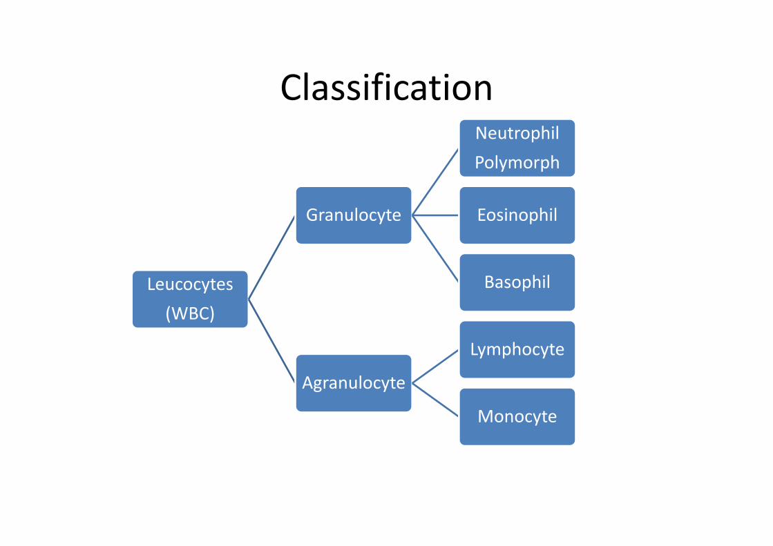
Classification
Leucocytes(WBC)
Granulocyte
NeutrophilPolymorph
Eosinophil
Basophil
Agranulocyte
Lymphocyte
Monocyte
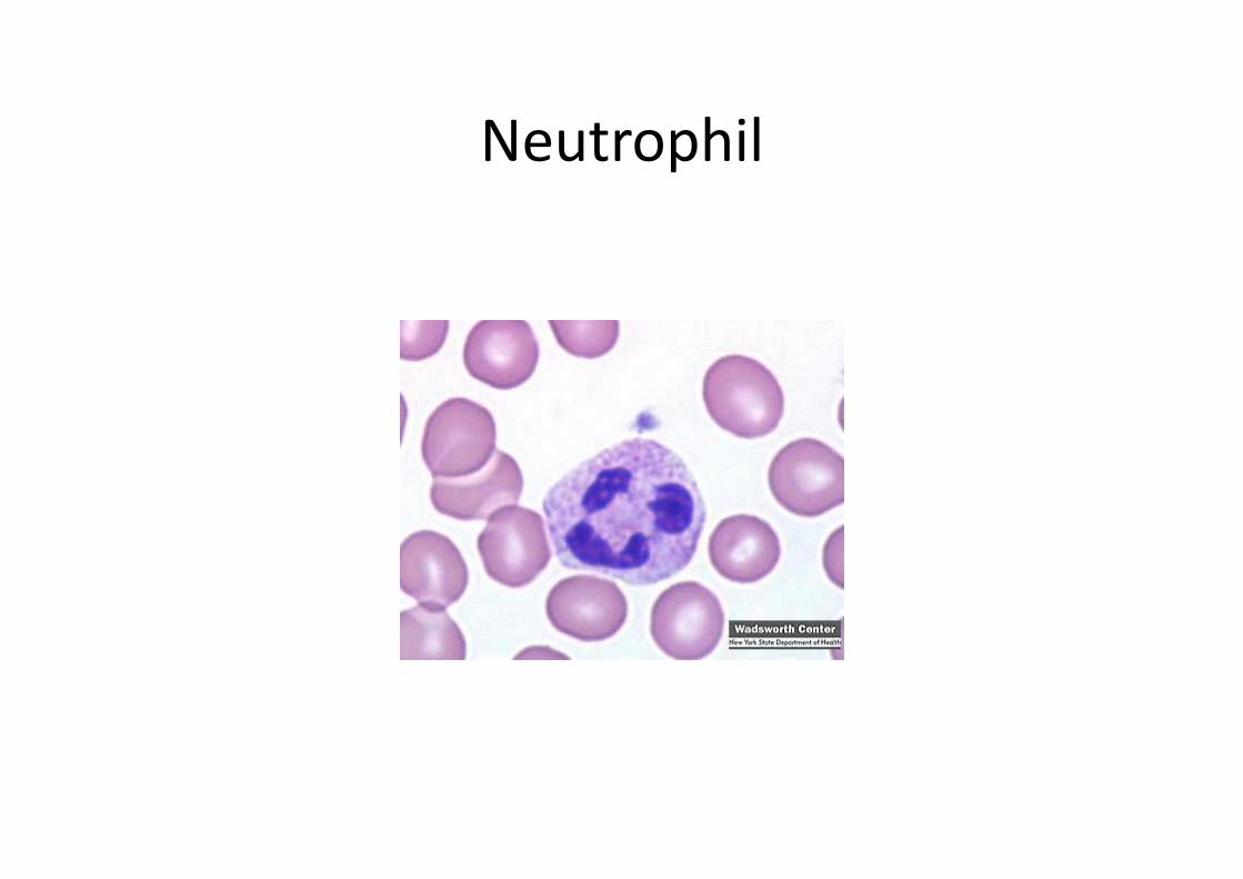
Neutrophil
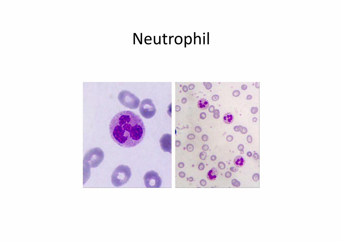
Neutrophil
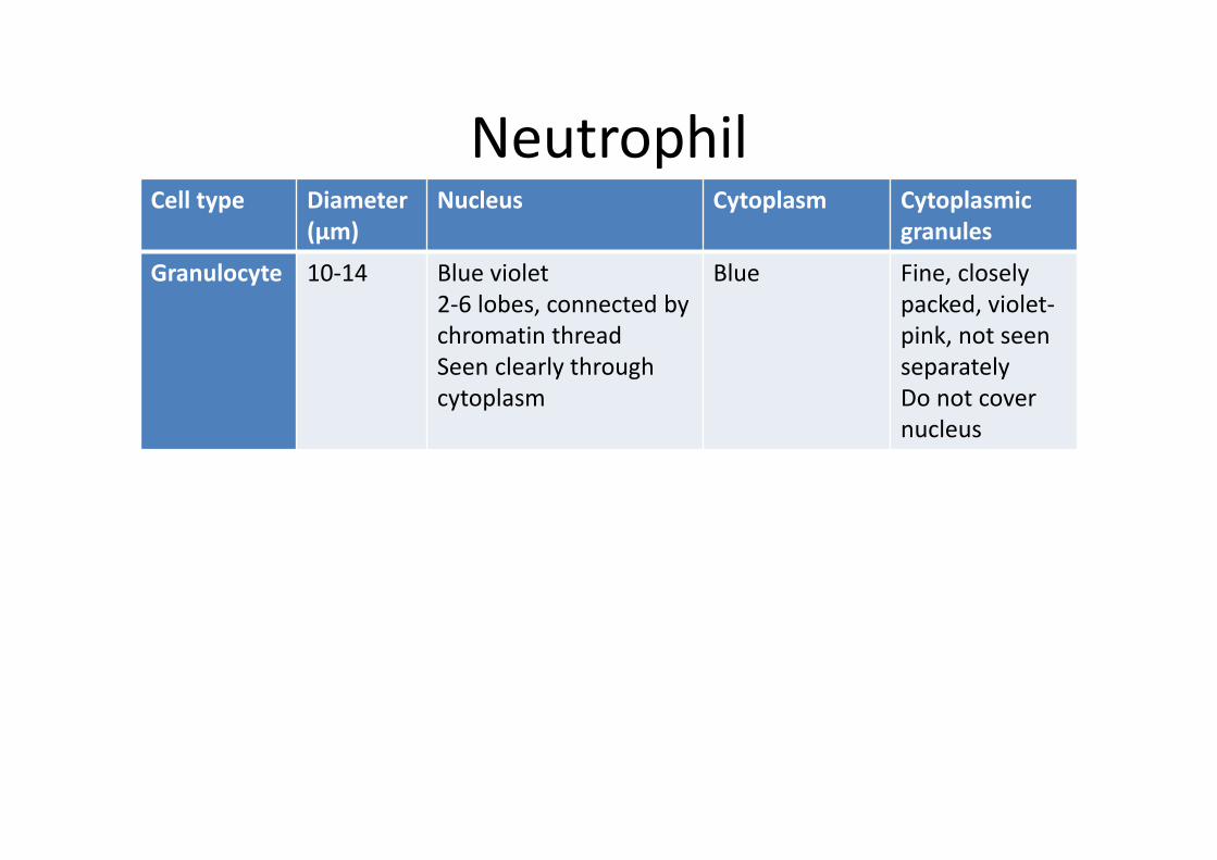
NeutrophilCell type Diameter
(µm)Nucleus Cytoplasm Cytoplasmic
granules
Granulocyte 10‐14 Blue violet2‐6 lobes, connected by chromatin threadSeen clearly through cytoplasm
Blue Fine, closely packed, violet‐pink, not seen separatelyDo not cover nucleus
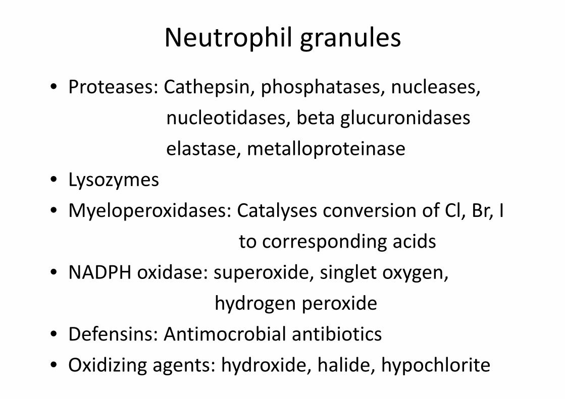
Neutrophil granules
• Proteases: Cathepsin, phosphatases, nucleases,nucleotidases, beta glucuronidaseselastase, metalloproteinase
• Lysozymes• Myeloperoxidases: Catalyses conversion of Cl, Br, I
to corresponding acids• NADPH oxidase: superoxide, singlet oxygen,
hydrogen peroxide• Defensins: Antimocrobial antibiotics• Oxidizing agents: hydroxide, halide, hypochlorite
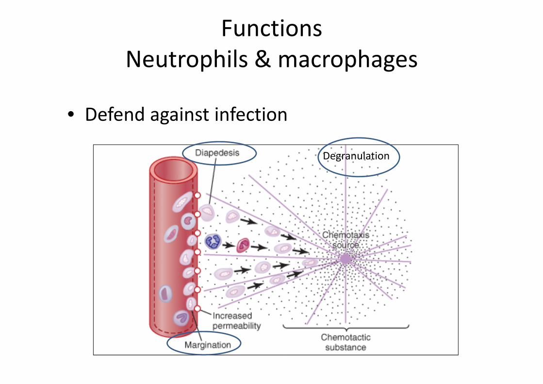
FunctionsNeutrophils & macrophages
• Defend against infection
Degranulation

Chemotactic substances• Bacterial or viral toxins• Degenerative products of inflammed tissues• Reaction products of the complement complex
Phagocytosis
Selection of a substance for phagocytosis• Rough surface• Absence of protein coat• OpsonizationSteps in phagocytosis• Attachment to the particle• Projecting pseudopodia all around it• Pseudopodia on opposite side fuse• Phagocytic vesicle / phagosome formed

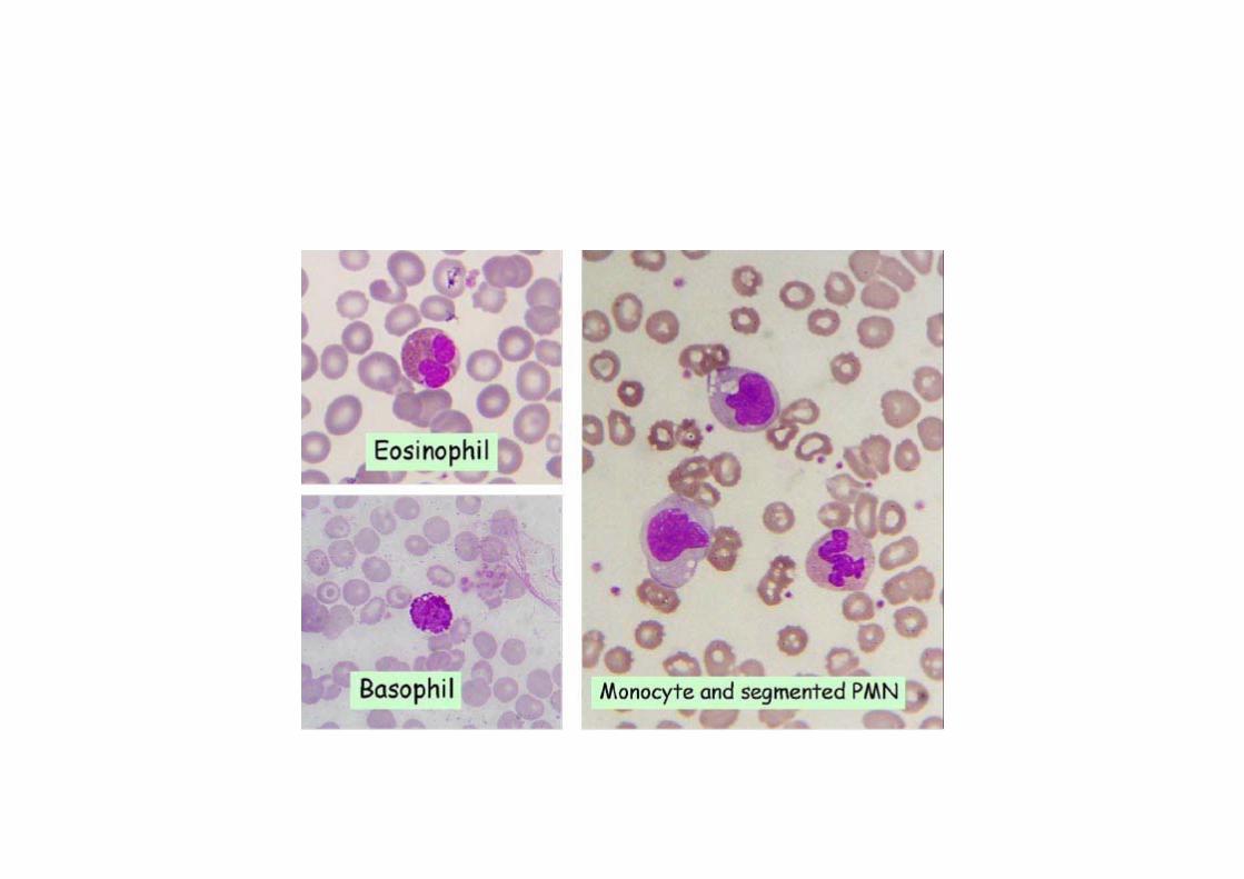
EosinophilCell type Diameter
(µm)Nucleus Cytoplasm Cytoplasmic
granules
Granulocyte
10‐15 Blue –violet2 lobesLobes connected by thick chromatin strandSeen clearly through cytoplasm
Light pink‐red
Large, coarseUniform sizedBrick red –orangeSeen separately Do not cover nucleus

Composition of Eosinophil granules• MBP: Major basic protein: Disrupts the membrane of parasites&
• induces histamine release from • Basophils& mast cells• ECP: Eosinophilic cationic protein: Binds to heparin & neutralises its
• anticoagulant activity• EDN: Eosinophil derived neurotoxin: Neurotoxin acting on myelinated nerves
• EPO: Eosinophil peroxidase: A sticky protein : adheres to host cells & mast cells

Functions of Eosinophils
• Specialise in dealing with parasites which are too large to be phagocytosed
• Chemicals released by degranulation are toxic to larvae of parasites
• Also degrade histamine & inhibit mast cell degranulation. Thus they decrease the intensity of allergic reactions
• Show phagocytosis but are less efficient than neutrophils
• Especially abundant in the mucosa of GIT, respiratory & urinary tracts

Eosinophil and Basophil

BasophilCell type
Diameter(µm)
Nucleus Cytoplasm Cytoplasmicgranules
Granulocyte
10‐15 Blue‐violetS shapedNot clearly seen because overlaid with granules
Bluish Large Very coarseVariable sizedDeep purpleSeen separatelyCompletely fill the center & cover the nucleus
Granules contain•Heparin•Histamine, bradykinin, serotonin, eosinophil chemotactic factorslow reacting substance
Functions of Basophils• Essential for immediate type hypersensitivity reactions eg. Urticaria, rhinitis, anaphylactic shock
• IgE mediated allergies.They have receptors for the constant region of IgEmoleculesThe allergen & the IgE molecule possibly form a complex with basophil which leads to allergic manifestations
• Protection from some parasitic infections eg Scabies• Heparin release by basophils after a meal may facilitate post absorptive metabolism of dietary triglycerides by activating lipoprotein lipase

Basophil

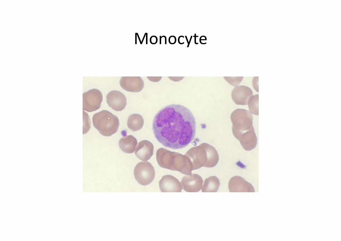
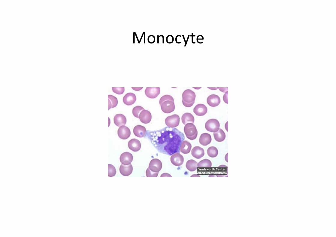
MonocyteCell type Diameter
(µm)Nucleus Cytoplasm Cytoplasmic
granules
Agranulocyte 12‐20 Pale blue violetLarge singleIndented or horse shoe or kidney shaped
AbundantFrostySlate blueAmount may be larger than that of nucleus
No visible granules
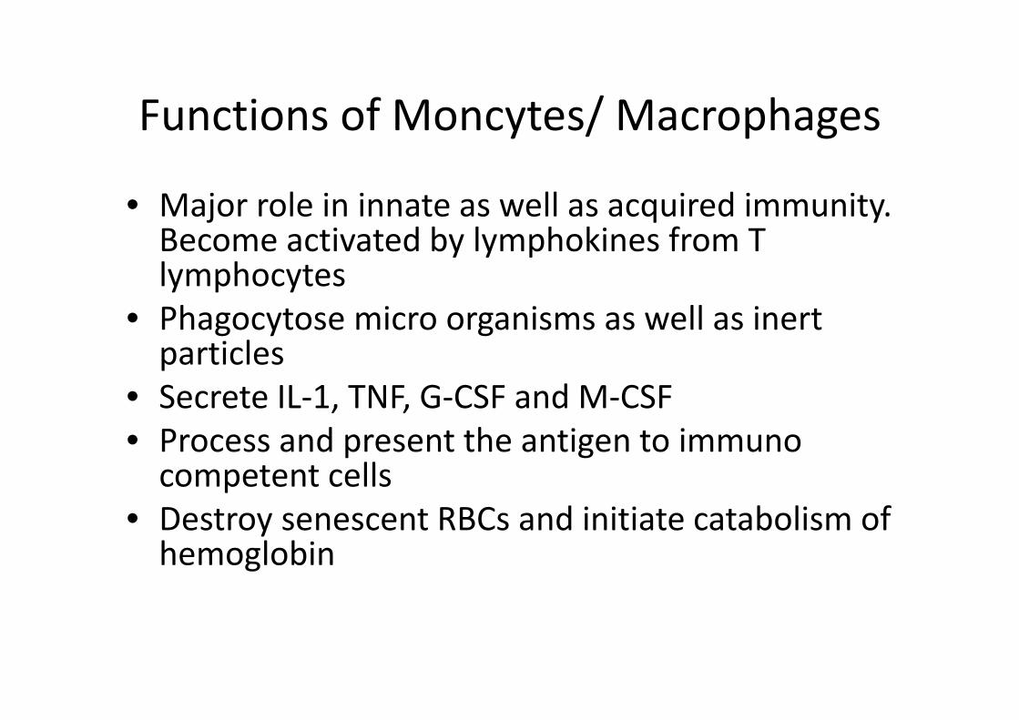
Functions of Moncytes/ Macrophages
• Major role in innate as well as acquired immunity. Become activated by lymphokines from T lymphocytes
• Phagocytose micro organisms as well as inert particles
• Secrete IL‐1, TNF, G‐CSF and M‐CSF• Process and present the antigen to immunocompetent cells
• Destroy senescent RBCs and initiate catabolism of hemoglobin
Monocyte
Monocyte
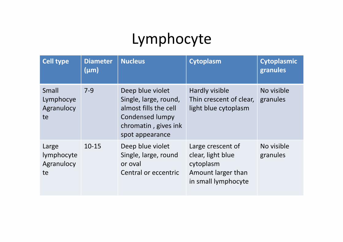
LymphocyteCell type Diameter
(µm)Nucleus Cytoplasm Cytoplasmic
granules
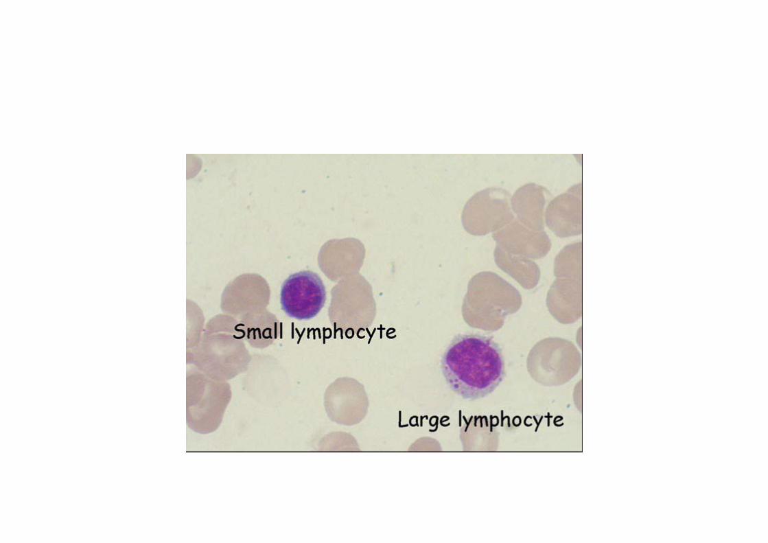
SmallLymphocyeAgranulocyte
7‐9 Deep blue violetSingle, large, round, almost fills the cellCondensed lumpy chromatin , gives ink spot appearance
Hardly visibleThin crescent of clear, light blue cytoplasm
No visible granules
Large lymphocyteAgranulocyte
10‐15 Deep blue violetSingle, large, round or ovalCentral or eccentric
Large crescent of clear, light blue cytoplasmAmount larger than in small lymphocyte
No visible granules
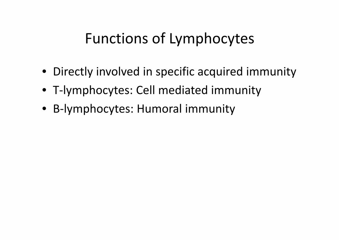
Functions of Lymphocytes
• Directly involved in specific acquired immunity• T‐lymphocytes: Cell mediated immunity• B‐lymphocytes: Humoral immunity
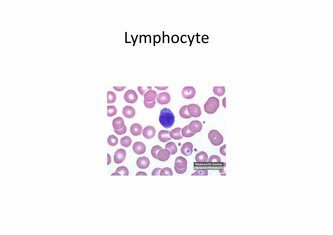
Lymphocyte
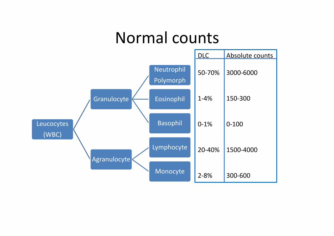
Normal counts
Leucocytes(WBC)
Granulocyte
NeutrophilPolymorph
Eosinophil
Basophil
Agranulocyte
Lymphocyte
Monocyte
DLC Absolute counts
50‐70% 3000‐6000
1‐4% 150‐300
0‐1% 0‐100
20‐40% 1500‐4000
2‐8% 300‐600
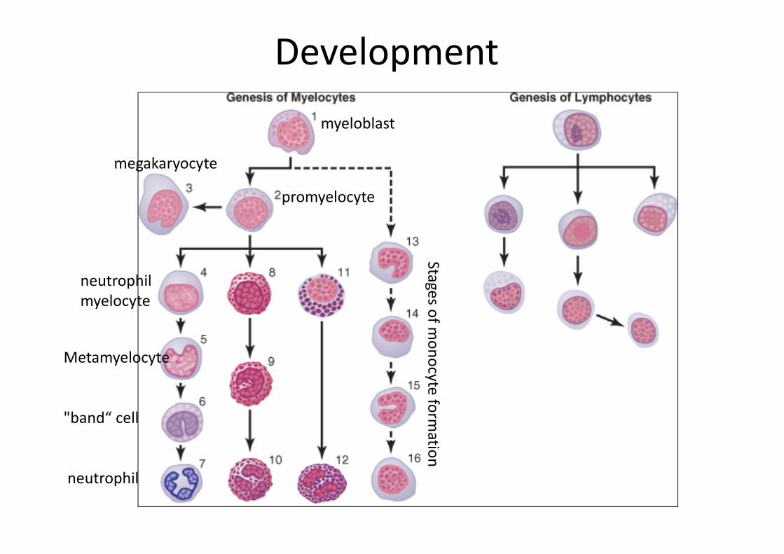
Developmentmyeloblast
Stages of monocyte
formation
promyelocyte
megakaryocyte
neutrophilmyelocyte
Metamyelocyte
"band“ cell
neutrophil
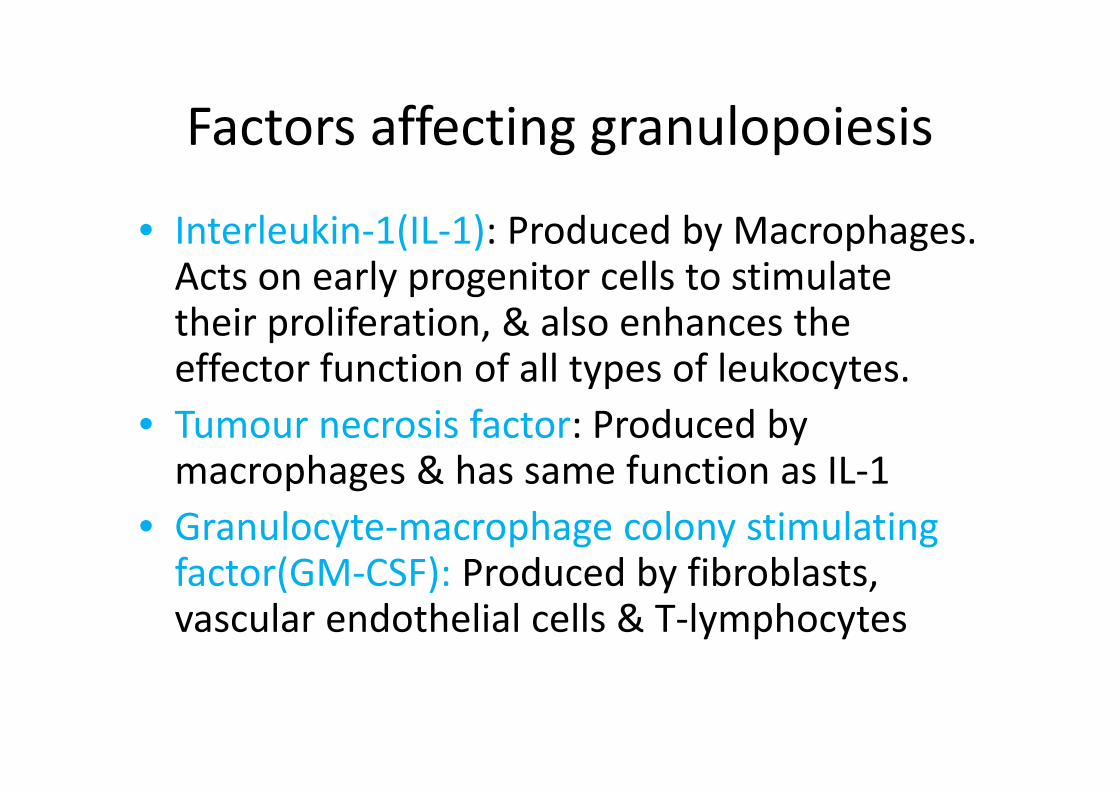
Factors affecting granulopoiesis
• Interleukin‐1(IL‐1): Produced by Macrophages. Acts on early progenitor cells to stimulate their proliferation, & also enhances the effector function of all types of leukocytes.
• Tumour necrosis factor: Produced by macrophages & has same function as IL‐1
• Granulocyte‐macrophage colony stimulating factor(GM‐CSF): Produced by fibroblasts, vascular endothelial cells & T‐lymphocytes
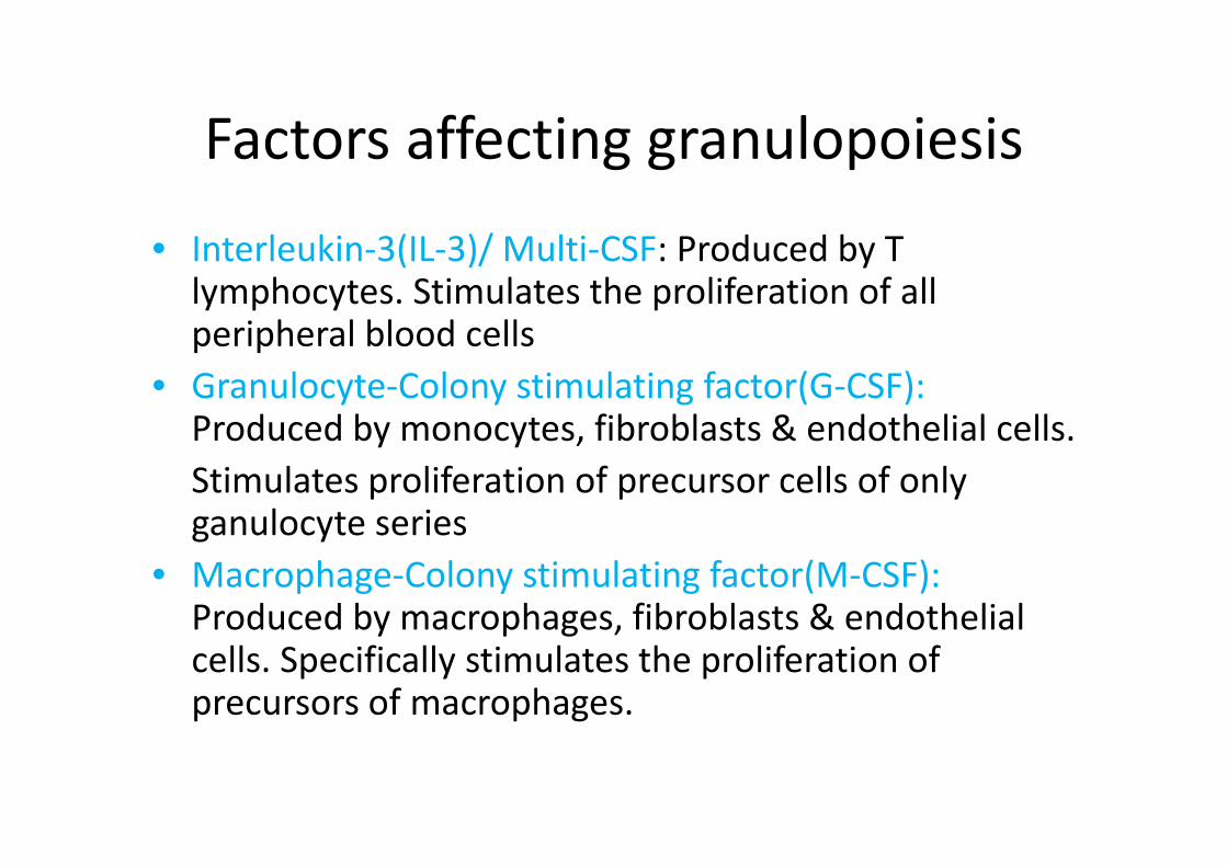
Factors affecting granulopoiesis
• Interleukin‐3(IL‐3)/ Multi‐CSF: Produced by T lymphocytes. Stimulates the proliferation of all peripheral blood cells
• Granulocyte‐Colony stimulating factor(G‐CSF): Produced by monocytes, fibroblasts & endothelial cells.Stimulates proliferation of precursor cells of only ganulocyte series
• Macrophage‐Colony stimulating factor(M‐CSF): Produced by macrophages, fibroblasts & endothelial cells. Specifically stimulates the proliferation of precursors of macrophages.
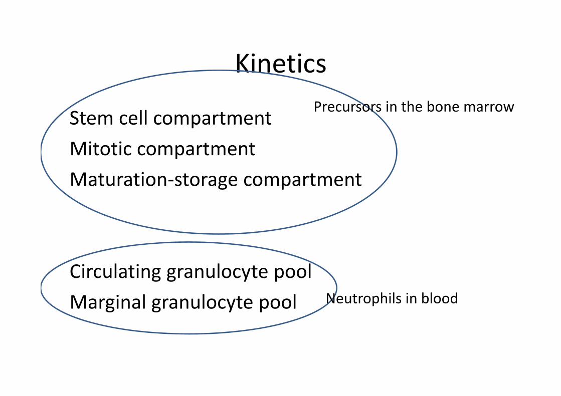
Kinetics
Stem cell compartmentMitotic compartmentMaturation‐storage compartment
Circulating granulocyte poolMarginal granulocyte pool
Precursors in the bone marrow
Neutrophils in blood
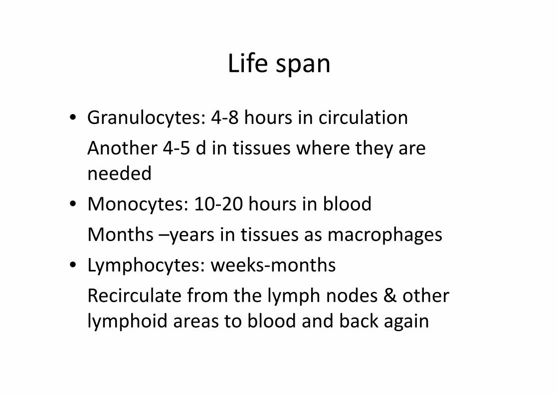
Life span
• Granulocytes: 4‐8 hours in circulationAnother 4‐5 d in tissues where they are needed
• Monocytes: 10‐20 hours in bloodMonths –years in tissues as macrophages
• Lymphocytes: weeks‐monthsRecirculate from the lymph nodes & other lymphoid areas to blood and back again
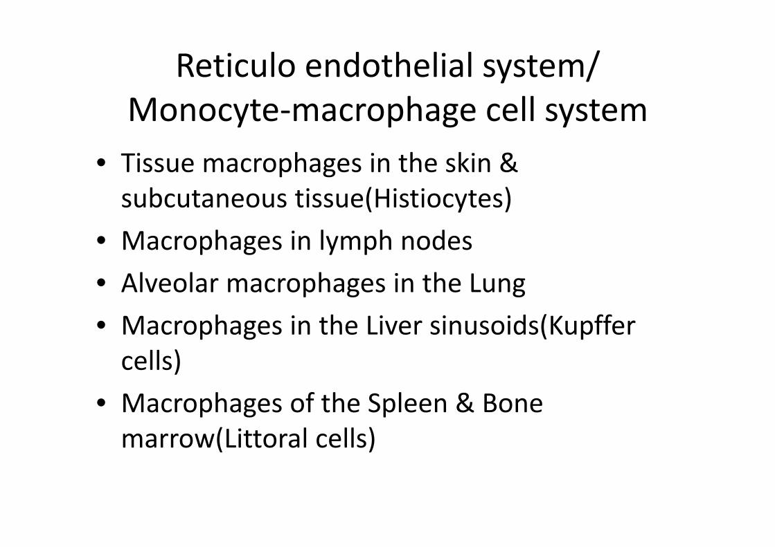
Reticulo endothelial system/ Monocyte‐macrophage cell system
• Tissue macrophages in the skin & subcutaneous tissue(Histiocytes)
• Macrophages in lymph nodes• Alveolar macrophages in the Lung• Macrophages in the Liver sinusoids(Kupffercells)
• Macrophages of the Spleen & Bone marrow(Littoral cells)

Function of RES
The RES acts as a physiological unity• Ingest & destroy RBCs and form & release blilrubin
• Also destroy leukocytes & platelets• Ingest bacteria• Ingest & process antigen which then stimulates antibody formation in the plasma cells

Inflammation: Role of Neutrophil & Macrophages
• First line of defence: Tissue macrophage, within first hour or so
• Second line of defence: Neutrophil invasion, within few hours & increase in the number of neutrophils in blood
• Third line of defence:Secondary macrophage invasion, within days
• Fourth line of defence: Increased production of granulocytes & monocytes

Feedback control of Macrophage & Neutrophil response
• TNF α• IL‐1• GM‐CSF• G‐CSF• M‐CSF

Leukocytosis
Physiological causes of Leukocytosis
• Newborn infants• Food intake• Exercise• Pregnancy• Sun exposure

Neutrophilia
• Acute infections: Especially localised infections due to Cocci(Streptococci, Staphylococci)
• Burns, acute hemorrhage, hemolysis, trauma, surgery
• Tissue necrosis: Myocardial infarction, pulmonary infarction
• Drugs: glucocorticoids• Physiological causes: Muscular exercise, stress, after meals, pregnancy
Eosinophilia
• Allergic conditions: Asthma, urticaria, eczema, hay fever, food sensitivity
• Parasitic infections: hook worm, tape worm• Drugs: Aspirin• Tropical pulmonary eosinophilia
Basophilia• Viral infections: Influenza• Alllergic diseases
Monocytosis
• Malaria, kala azar, Rheumatoid arthritis, Leukemia
Lymphocytosis• Infants &children, whooping cough, viral infections, autoimmune diseases
• Chronic infections: TB, Hepatitis, chronic cholecystitis, chronic pancreatitis
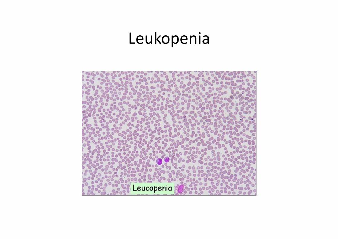
Leukopenia
Physiological causes of leukopenia
• Exposure to extreme cold
Neutropenia
• Typhoid & Paratyphoid infections• Viral: Influenza• AIDS• Kala azar• Bone marrow depression: Chloremphenicol, aspirin & radiations
• Autoimmune disease

Eosinopenia
• Acute stressful illness• ACTH & glucocorticoid treatment• Acute pyogenic conditions
Basopenia• Acute pyogenic infections• Glucocorticoid treatment
Monocytopenia
• Bone marrow depression
Lymphocytopenia• Steroid treatment
Leukemia
Uncontrolled production of WBCs caused by cancerous mutation of a myelogenous or lymphogenous cell
Types• Lymphocytic• Myelogenous
Effects of leukemia on the body
• Metastatic growth of leukemic cells in abnormal areas of the body
Eg bone invasion leading to pain, easy fractures• Spread to spleen, lymph nodes, liver & other vascular regions
• Infections• Severe anemia• Bleeding tendency
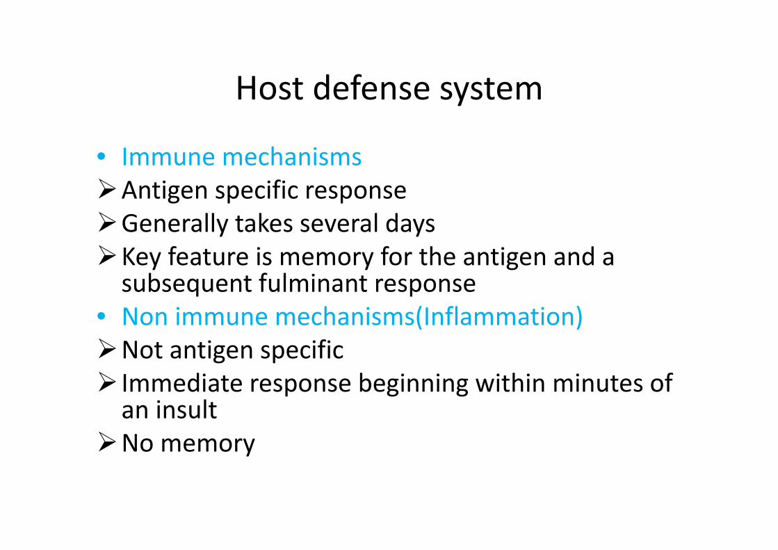
Host defense system
• Immune mechanismsAntigen specific responseGenerally takes several daysKey feature is memory for the antigen and a subsequent fulminant response
• Non immune mechanisms(Inflammation)Not antigen specific Immediate response beginning within minutes of an insult
No memory
Immunity
• Innate immunity• Acquired( adaptive) immunity
B cell mediated (Humoral)immunityT cell mediated(Cellular) immunity
Innate immunity
• Barriers• Phagocytosis• Activation of complement system• Release of interferons• Production of antibacterial peptides• Other proteolytic cascades

Complement system
• Consists of about 20 proteins• Activation via 3 different pathways• Classical pathway: Triggered by immune complexes
• Mannose binding lectin pathway: Triggered when lectin binds mannose group in the bacteria
• Alternative/ Properdin pathway: Triggered by various viruses, bacteria, fungi & tumour cells

Complement system
Causes microbial destruction by 3 mechanismsForming a coating over microorganisms which can then be easily phagocytosed since the phagocytes have receptors for the same complement components which coat the microorganisms
Release of histamine by mast cells & basophilgranules
Membrane attack complex which punches holes (perforins) in the microbial wall
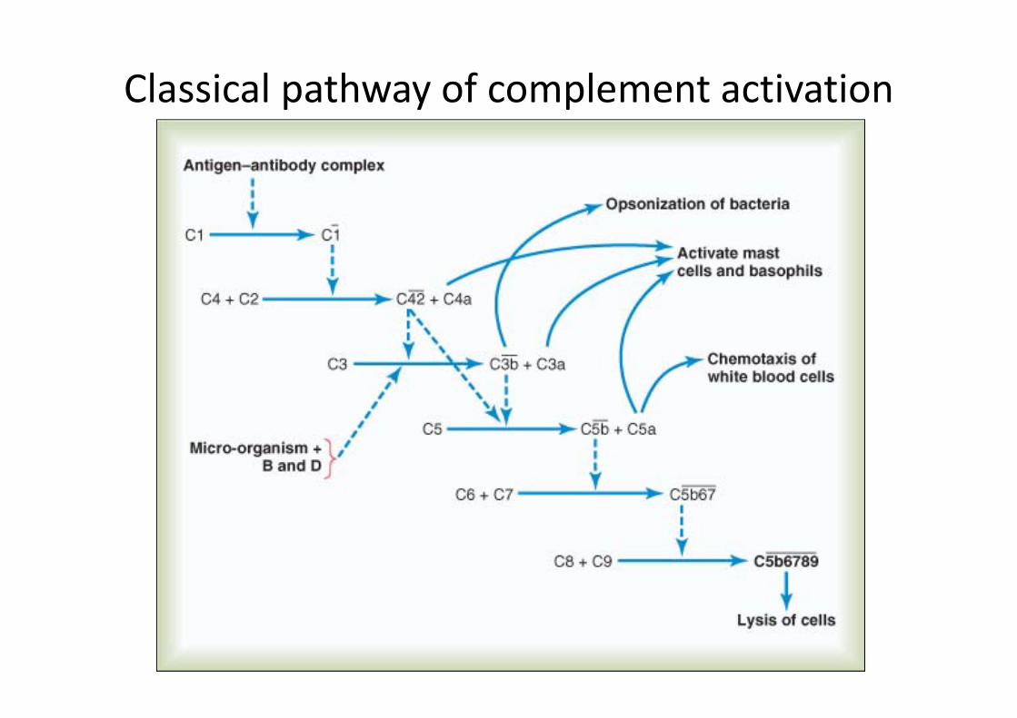
Classical pathway of complement activation

Interferons
• Released by virally infected cellsFunctionsForm a protective ring of uninfected cells thereby limiting the spread of infectionInhibit protein synthesis by interfering with translation and degradation of mRNA
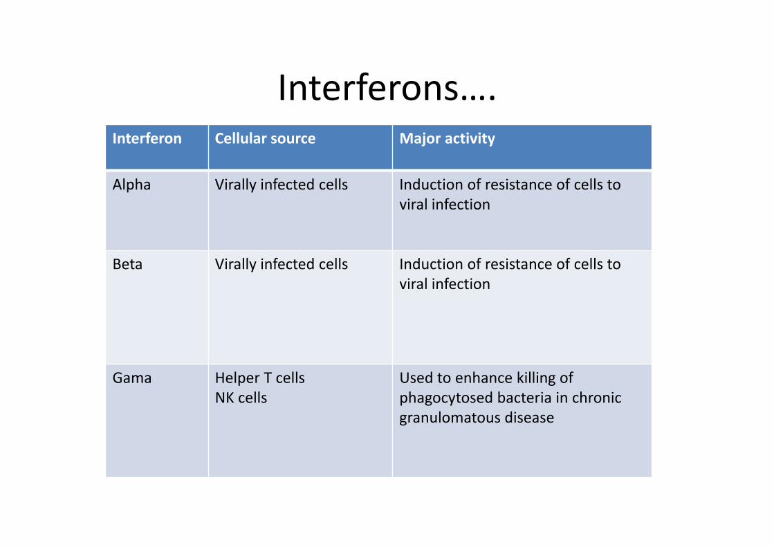
Interferons….Interferon Cellular source Major activity
Alpha Virally infected cells Induction of resistance of cells to viral infection
Beta Virally infected cells Induction of resistance of cells to viral infection
Gama Helper T cellsNK cells
Used to enhance killing of phagocytosed bacteria in chronic granulomatous disease

Cells mediating innate immunity
• Neutrophils• Macrophages• Natural killer cells(NK cells): Large lymphocytes that are not T cells but are cytotoxic
Natural killer cells
• Kill virally infected and tumour cells• They have receptors for certain glycoproteinswhich appear on the surface of some virally infected cells
• Surface receptor KIR (Killer Inhibitory Receptor) interacts with HLA class I molecules on the surface of normal cells.
• Cancerous cells have modified ligands which do not bind properly with the receptor therefore the NK cells kill them
Major histocomptibility complex(MHC)
MHC gene is situated in a cluster of loci on chromosome 6
Its products the cell surface molecules are of 2 classes I &II
Term MHC is given because antigenic determinants on these 2 classes determine whether the organ or tissue transplants are recognised as self or foreign and thus are accepted or rejected
Human leukocyte antigen(HLA)
• In humans the MHC is referred to as HLA because it was first recognized by analyzing patterns of reactions of peripheral blood lymphocytes with sera containing antibodies to leukocytes
• HLA class I: Comprises A, B, C loci• HLA class II: Comprises DR, DQ, DP loci• HLA class III: Comprises complement region containing genes for complement components C2 & C4 of classical pathway, properdin factor B of alternate pathway & TNF alpha & beta

Acquired immunity
• Cytokines released from the cells mediating innate immunity activate cells of the acquired immune system
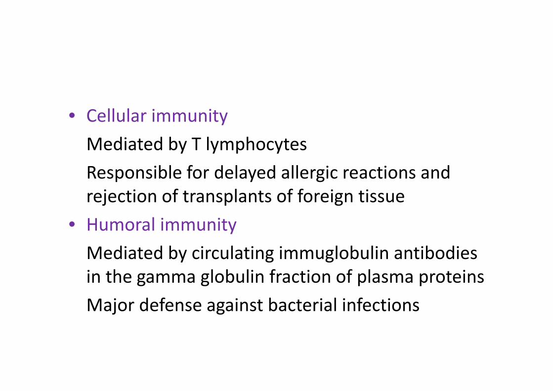
• Cellular immunityMediated by T lymphocytesResponsible for delayed allergic reactions and rejection of transplants of foreign tissue
• Humoral immunityMediated by circulating immuglobulin antibodies in the gamma globulin fraction of plasma proteinsMajor defense against bacterial infections
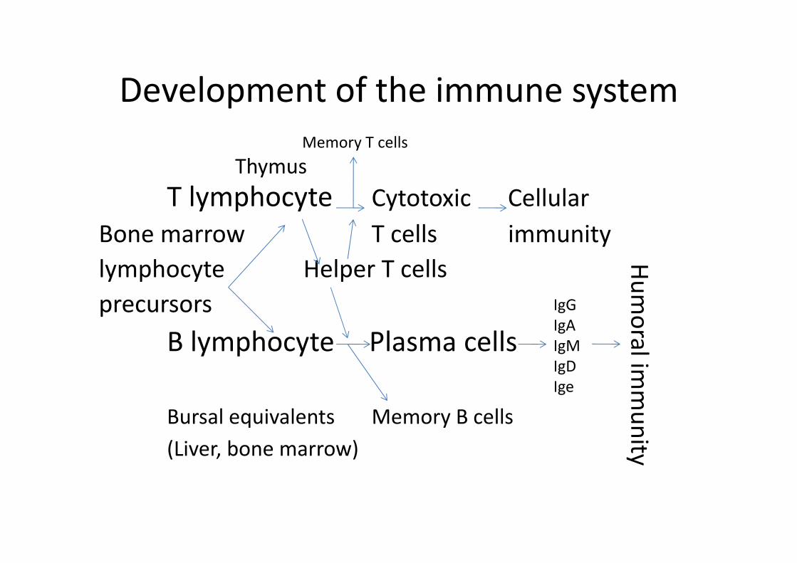
Development of the immune system
ThymusT lymphocyte Cytotoxic Cellular
Bone marrow T cells immunitylymphocyte Helper T cellsprecursors
B lymphocyte Plasma cells
Bursal equivalents Memory B cells(Liver, bone marrow)
IgGIgAIgMIgDIge
Humoralim
munity
Memory T cells
“Self recognition process” ‐ Burnet
• All potentially antigenic material encountered in fetal life whether ‘self’ or ‘not self’ elicits no response either then or subsequently.

Clonal selection theory‐ Burnet
• It proposes that certain lymphocytes known as immunologically competent are genetically endowed with the capacity to respond to one or very few molecules of specific antigenic pattern by making antibodies against that pattern
• Antigenic stimulation thus produces a “clone” of cells , all of similar specific reactivity
Immune tolerance
• Ehrlich: First to emphasize that animals do not usually make any immunological response to their own plasma proteins or tissue cells although they are excellent antigens in other species
• Burnet & Fenner: Proposed an explanationIn utero during the period of immunological immaturity all the potential antigens that the lymphocytes come in contact are recognized as “self”
T lymphocyte : Ontogeny & functions
T cell precursors(Bone marrow)Extreme high rate of division(Thymic Cortex)Early thymocyte (Majority die within thymiccortex)Minority migrate to medulla of thymus & undergo differentiationPossess a T cell surface antigen receptor whichhas following recognition properties
T lymphocyte : Ontogeny & functions…
Properties of T cell surface antigen• Distinguishes between foreign MHC proteins & self MHC proteins reacting with the former and tolerating the latter
• The receptor does not react with a self MHC protein in the absence of antigen but it recognizes & reacts with a complex made up of an antigen derived peptide bound to a self MHC protein
• The receptor of each T cell will be clonotypic, i.e. will recognize only a single specific immunological determinant (epitope) of an antigen derived peptide bound to an MHC protein
Antigen recognition & T cell activation
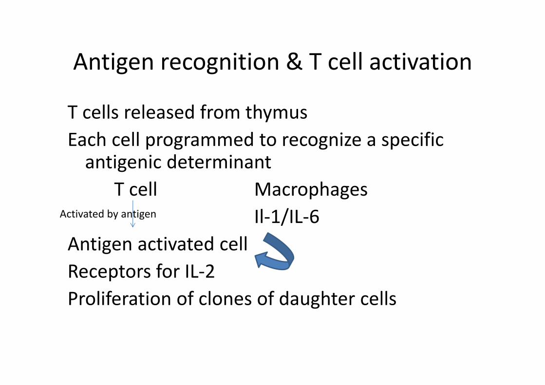
T cells released from thymusEach cell programmed to recognize a specific antigenic determinant
T cell MacrophagesIl‐1/IL‐6
Antigen activated cellReceptors for IL‐2Proliferation of clones of daughter cells
Activated by antigen
T lymphocyte : Ontogeny & functions…
Effector functions• Helper T cells help B cells to secrete antibody• Helper T cells mediate delayed hypersensitivity reactions
• Suppressor T cells keep immune responses from escaping control by damping the proliferation & differentiation of antibody producing B cells & cytotoxic T cells
• Activated T cells secrete lymphokines

Types and functions of T lymphocytes
• Cytotoxic T cells: Destroy transplanted & other foreign cells
• Helper T cells: Help in the development of the former TH1 cells: secrete IL‐2 & gamma interferon, concerned primarily with cellular immunity
TH2 cells: Secrete IL‐4 & IL‐5, interact primarily with B cells in relation to humoral immunity
• Memory T cells: Produce an accelerated response to a second exposure of antigen
• Suppressor T cells: Help in immune tolerance

Activation of T cells requires interaction of T‐cell receptors with an antigen (foreign protein) that is transported to the surface of the antigen‐presenting cell by a major histocompatibility complex (MHC) protein. Cell‐
to‐cell adhesion proteins enable the T cell to bind to the antigen‐presenting cell long enough to become activated.
MHC restriction
• T cells respond to antigen on the macrophages & other cells only when they are presented along with the self MHC antigen
• CD4 positive lymphocyte(Helper T cells) recognize class II antigen
• CD 8 positive lymphocytes(Cytotoxic T cells) recognize class I antigen
HLA antigens and their role• HLA class IFound on the surface of virtually all nucleated cellsPrincipal antigens responsible for graft rejection & cell mediated cytolysis
• HLA class IIFound only on cells of immune system‐macrophages, dendritic cells, activated T cells & B cellsResponsible for graft vs host response & immediate leukocyte reactions
• HLA class IIIHeterogenous
HLA antigens and their role…
• The MHC system was originally identified in the context of transplantation which is an artificial event
• In the natural state they serve asCell surface markers: that help infected cells to signal cytotoxic & helper T cells
Enormous polymorphism of MHC gene helps maximize protection against mirobial infections
Increase the specificity of the self antigen thus it prevents microbes with similar genetic makeup to sneak past by molecular mimicry
B cell ontogeny & functionsB cell differentiation from precursor to plasma cell occurs in 2 phases
Non antigen driven phase: Occurs within central lymphoid tissue
Very rapid cell division & rapid cell death also• Rearranement of a functional gene for the heavy chain(IgM)
• Synthesis of a heavy chain of IgM• Rearrangement of a gene for light chain synthesis• Synthesis of light chain• Next a second class of heavy chain (IgG) is also made

B cell ontogeny & functions…
Antigen dependent phaseOccurs in peripheral lymphoid tissueProgrammed to recognize antigen in the presence of cytokines from activated T cells and macrophages
This leads to formation of• IgM secreting cell• Another B cell whose surface IgM &IgD are replaced with surface IgG, IgA or IgE
• B cell memory pool

Regulation of the immune system, emphasizing a pivotal role of the helper T cells
Il‐2IL‐3IL‐4IL‐5IL‐6GM‐CSFIF‐Gamma

Direct destruction of an invading cell by sensitized lymphocytes (cytotoxic T cells).
Types and functions of B lymphocytes
• Plasma cells which form antibodies• Memory B cells

Time course of the antibody response in the circulating blood to a primary injection of antigen and to a secondary injection
several months later.
Nature of antibodies
• Gamma globulins• Molecular weight: 160,000 – 970,000• Constitute 20% of all plasma proteins

Structure of antibody

Structure of immunoglobulins
• Monomer:Basic unit : 2light+2 heavy chains• Polymers: 2‐5 basic units + J chain that holds
the units together• Amino acids of L and H chains form loops known as domains
• Variable region : NH2 terminal• Hypervariable region: amino acid sequences unique to that polypeptide chain
• Constant portion: carboxy terminal
Structure of immunoglobulins….
• Two types of light chains: kappa and lambda• Five types of heavy chains• Ig G: Subtypes γ 1, γ2, γ3, γ4
• Ig A: Subtypes α1, α2• Ig M: Subtypes µ1, µ2• Ig D: δ• Ig E: ε
Structure of antibody
Constant portion determines:• Diffusivity of the antibody• Adherence to specific structures within tissues• Attachment to complement complex• Passage through membranesVariable portion determines:• Specificity to a particular type of antigen
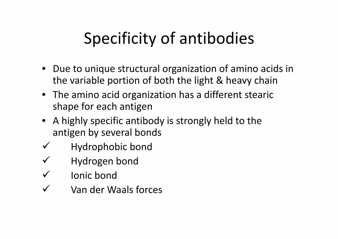
Specificity of antibodies
• Due to unique structural organization of amino acids in the variable portion of both the light & heavy chain
• The amino acid organization has a different stearicshape for each antigen
• A highly specific antibody is strongly held to the antigen by several bonds
Hydrophobic bond Hydrogen bond Ionic bond Van der Waals forces
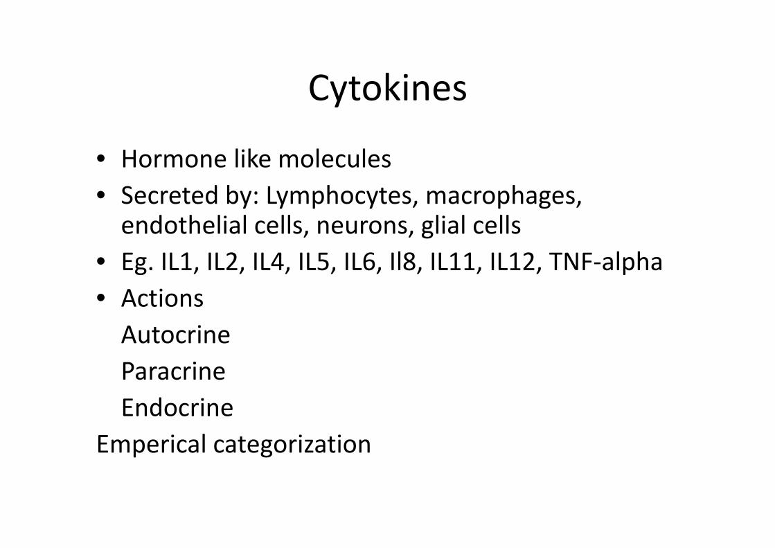
Cytokines
• Hormone like molecules• Secreted by: Lymphocytes, macrophages, endothelial cells, neurons, glial cells
• Eg. IL1, IL2, IL4, IL5, IL6, Il8, IL11, IL12, TNF‐alpha• ActionsAutocrineParacrineEndocrine
Emperical categorization

Emperical categorization
• Immunoregulatory cytokines: Involved in activation growth & differentiation of Lymphocytes & MonocytesEg. IL‐2, IL‐4, TGF‐beta
• Proinflammatory cytokines: Produced predominantly by mononuclear phagocytes in response to infectious agentsEg. IL‐1, TNF alpha, IL‐6, IL‐8
• Regulating immature leukocyte growth & differentiationEg. IL‐3, IL‐7, GM‐CSF
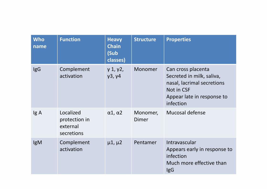
Who name
Function HeavyChain (Sub classes)
Structure Properties
IgG Complement activation
γ 1, γ2, γ3, γ4
Monomer Can cross placentaSecreted in milk, saliva, nasal, lacrimal secretionsNot in CSFAppear late in response to infection
Ig A Localizedprotection in external secretions
α1, α2 Monomer, Dimer
Mucosal defense
IgM Complement activation
µ1, µ2 Pentamer IntravascularAppears early in response to infectionMuch more effective than IgG

Who name
Function Heavychain
Structure Properties
Ig D Antigen recognition by B cells
δ Monomer Found in trace amounts in plasmaFunction unknownActs as antigen recognition site on the surface of B cells
Ig E Reagin activityRelease histamine from basophils & mast cells
ε Monomer Secreted in helminthicinfectionsAtopic allergyCan invoke mast cell triggered immediate hypersensitivity reactions

Mechanism of action of antibodies
Direct action of antibody on invading agent
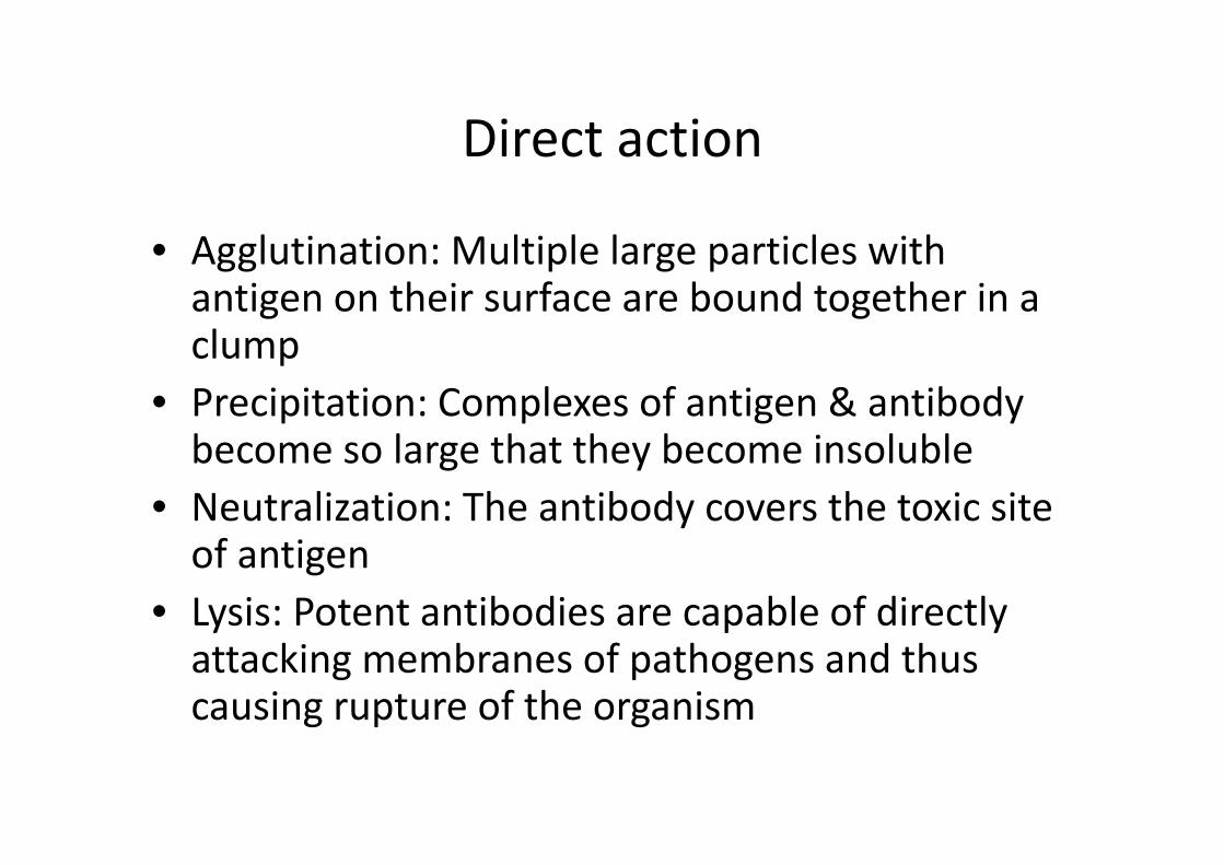
Direct action
• Agglutination: Multiple large particles with antigen on their surface are bound together in a clump
• Precipitation: Complexes of antigen & antibody become so large that they become insoluble
• Neutralization: The antibody covers the toxic site of antigen
• Lysis: Potent antibodies are capable of directly attacking membranes of pathogens and thus causing rupture of the organism
Complement system for antibody action
Classical pathway effects• Opsonization & phagocytosis• Lysis• Agglutination• Neutralization• Chemotaxis• Activation of mast cells & basophils• Inflammatory effects
Antigen presenting cells(APC)
• Specialized cells in the lymph nodes, spleen & Langerhan’s cells in the skin
• In APC the products of antigen digestion are complexedto products of MHC (Major Histocompatibility Gene) and presented on the surface of the cell
• MHC gene – chromosome 6• Its product is a glycoprotein • Class I antigen is found in all nucleated cells• Class II antigen is found on APC, B & T lymphocytes
Lymphoid tissueEmbryological developmentLymphopoiesis first appears at 3 months of IUL in the fetal
liver & thymus At birth Thymus weighs 10‐12 gCell mediated immunity is well developed so that graft
rejection can occurPeripheral lymphoid tissue( lymph nodes, spleen, gut
associated lymphoid tissue) is very slightly developed IgA & IgE: TracesIgM: Formed before birth & present in significant amountsIgG: High amount but is maternally derived
Lymphoid tissue…
Central lymphoid tissue: Thymus & bone marrowPeripheral lymphoid tissue• Lymph nodes• Spleen• Ring of tonsillar tissue in oropharynx• Submucosal accumulation of lymphocytes in the respiratory tract, urinary tract & gut(Peyer’spatches)
Small lymphocytes: Quiescent lymphocytes of the peripheral lymphoid tissue
Large lymphocytes: After exposure to antigen


Lymphatic system
Proteins exert colloidal osmotic pressure of 25 mmHg
Lymphatic system
Function• To return excess tissue fluid and protein to the intravascular compartment by pinocytosis
• As a transport mechanism to remove RBCs, bacteria• Filtering system in the lymph nodesEndothelium of the lymphatic capillaries is similar to that of blood capillaries with little or no basement membrane
Diameter variable depending on state of function and the organ
Lymph pump (for anterograde flow)
• Arteriolar vasocinstriction• Arteriolar pulsation• Muscle contraction• Retrograde flow is prevented by lymph valves
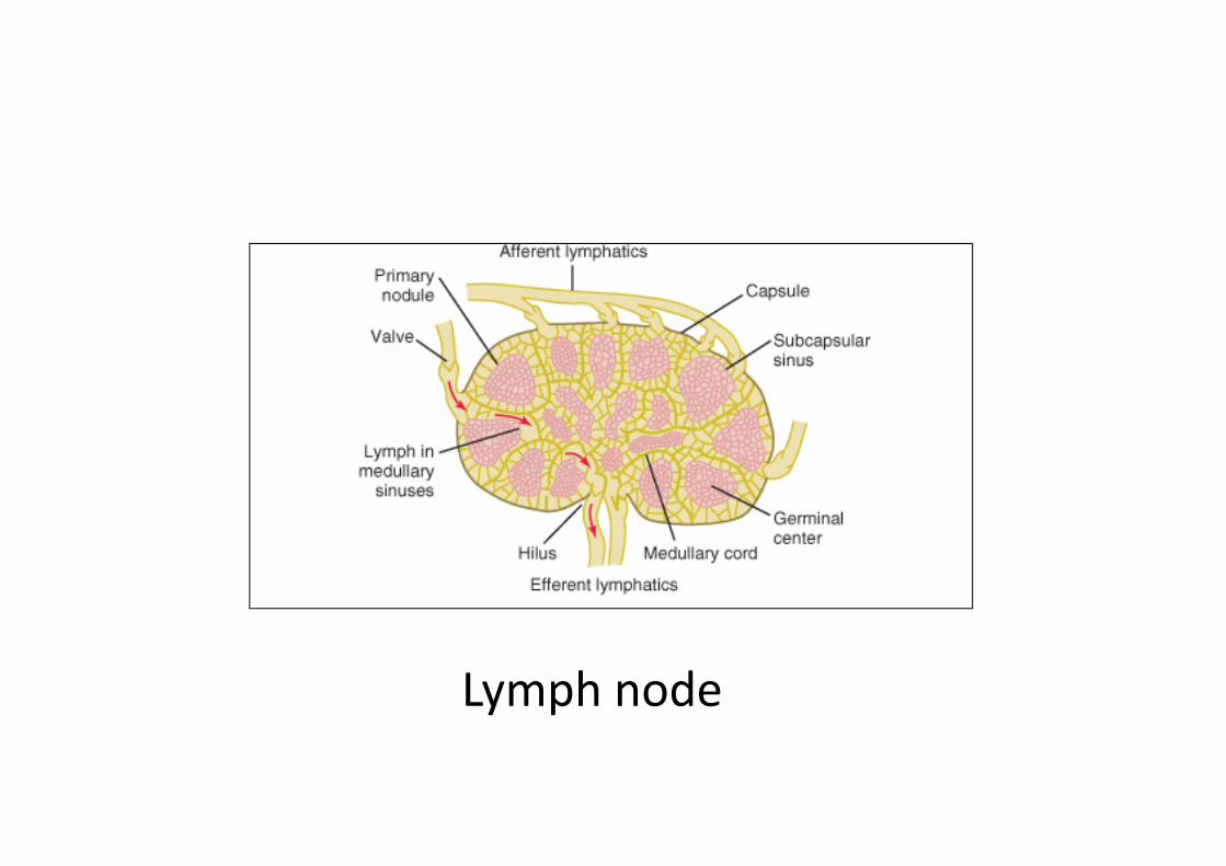
Lymph node

Location of T & B lymphocytes in adults
T cells B cellsPeripheral blood 60‐80% 20‐30%Thoracic duct 85‐90% 10‐15%
Lymph nodes Paracortical SubcapsularGerminal centerMedullary cords
Spleen Periarteriolar Germinal centerssheath Red pulp
Around periarteriolar sheath

• Lymphatics are most abundant on the undersurface of the diaphragm
• Lymphatic system exists in all organd exceptCNSCornea
Composition of lymph
• Protein concentration lower than plasma• All clotting factors present , some synthesised by the liver are in higher concentration in the Hepatic lymph
• Antibodies• Electrolyte concentration : Positive ions are slightly less and negative ions are slightly more
• Lipids: Chylomicrons will be higher after a fatty meal• Cells: Lymphocytes, monoctye/macrophage, granulocyte
• Clonal anergy/ immunological silence: When B and T lymphocytes in fetal life are exposed to potentially antigenic materials in the tissues, they are subsequently unable to make a specific immune response to these material i.e. B and T lymphocytes enter a prolonged hyporesponsive phase
• Clonal abortion/ negative selectionMany of the not/anti self lymphocytes are eliminated in the thymus during their early development. Suppressor T cells keep the development of such not self antibodies in check
Autoimmunization
• It can occur when new antigenic material are formed after the period of immunological immaturityEg spermatozoa
• Breakdown of the barrier which anatomically segregates the potential antigen from the immunocompetent cellEg Brain constituents, heart, pancreas, thyroglobulin
• Breakdown of acquired tolerance and appearance of immune reactive cells “forbidden clones” in the blood
• Deficiency of suppressor T cells

Criteria for the presence of autoimmune disease
• Mere presence of auto antibodies or even of sensitization does not establish the autoimmune nature of disease
• It is necessary to show in addition that the administration of serum antibodies or immunologically potent cells from the lymph node to normal animals can reproduce the disease
Autoimmune diseases
They can be B/ T cell mediated and organ specific/ tissue specific
Eg• Acquired hemolytic anemia: Antibody against RBC
• Diabetes mellitus(Type I): Pancreatic B cell antibody
• Myasthenia gravis: Antibody against nicotinic cholinergic receptor
• Grave’s disease: Antibody against TSH receptor
Autoimmune diseases…
• Molecular mimicryRheumatic fever following Streptococcal throat infection
A portion of the cardiac myosin resembles Streptococcal M protein
Active vs passive immunity
• Active immunity: Ones own body develops either antibodies or activated T cells in response to invasion of the body by foreign antigen
• Passive immunity: Develops by infusing preformed antibodies or activated T cells or both
Immunization by injections

Hypersensitivity reactions
• Anaphylactic type (Immediate)• Cytotoxic type • Complex mediated• Cell mediated (delayed type)

TYPE I Hypersensitivity Classic allergy
• Mediated by IgE attached to Mast cells.
• The symptoms resulting from allergic responses are known as anaphylaxis.
• Includes: Hay fever, asthma, eczema, bee stings, food allergies.
Allergens
• Allergens are nonparasite antigens that can stimulate a type I hypersensitivity response.
• Allergens bind to IgE and trigger degranulation of chemical mediators.

Allergens

Characteristics of allergens
• Small 15-40,000 MW proteins. • Specific protein components
– Often enzymes.• Low dose of allergen • Mucosal exposure
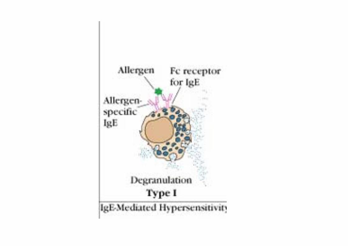

Mechanisms of allergic response
Sensitization• The IgE can attach to Mast cells by Fc
receptor, which increases the life span of the IgE.
• Half-life of IgE in serum is days whereas attached to FcR it is increased to months.
Immediate hypersensitivity Anaphylactic type
• Introduction of specific antigens in previously sensitized persons can produce immediate reactions in skin and mucous membranes
• Skin: wheal & flare• Nasal mucosa: swelling & irritation leading to sneezing(Hay fever, allergic rhinitis)
• Bronchi & bronchioles: mucosal swelling & increased smooth muscle tone producing bronchial asthma characterized by great difficulty in breathing & signs of hypoxia

Immediate hypersensitivity Anaphylactic type
• IgE immunoglobulins strongly attach to mast cells/ basophils
• On combining with antigen the mast cells/ basophils rupture and release
HistamineProteasesSlow reacting substance of anaphylaxisEosinophil chemotactic factorHeparinPlatelet activating factor

Immediate hypersensitivity: Anaphylactic type…Influence of cAMP
• Increased cAMP reduces all chemical mediators of immediate hypersensitivity reaction
• Drugs which raise cAMP levels antagonize effects of antigen‐IgE interactions eg. Isoprenaline, salbutamol, theophylline


Cytotoxic type hypersensitivity
In this type antibodies bind to antigen on a cell surface
This promote contact with phagocyte byReducing electrical charge on the surfaceOpsonic adherenceImmune adherence in which C3 component of complement promotes phagocytosis
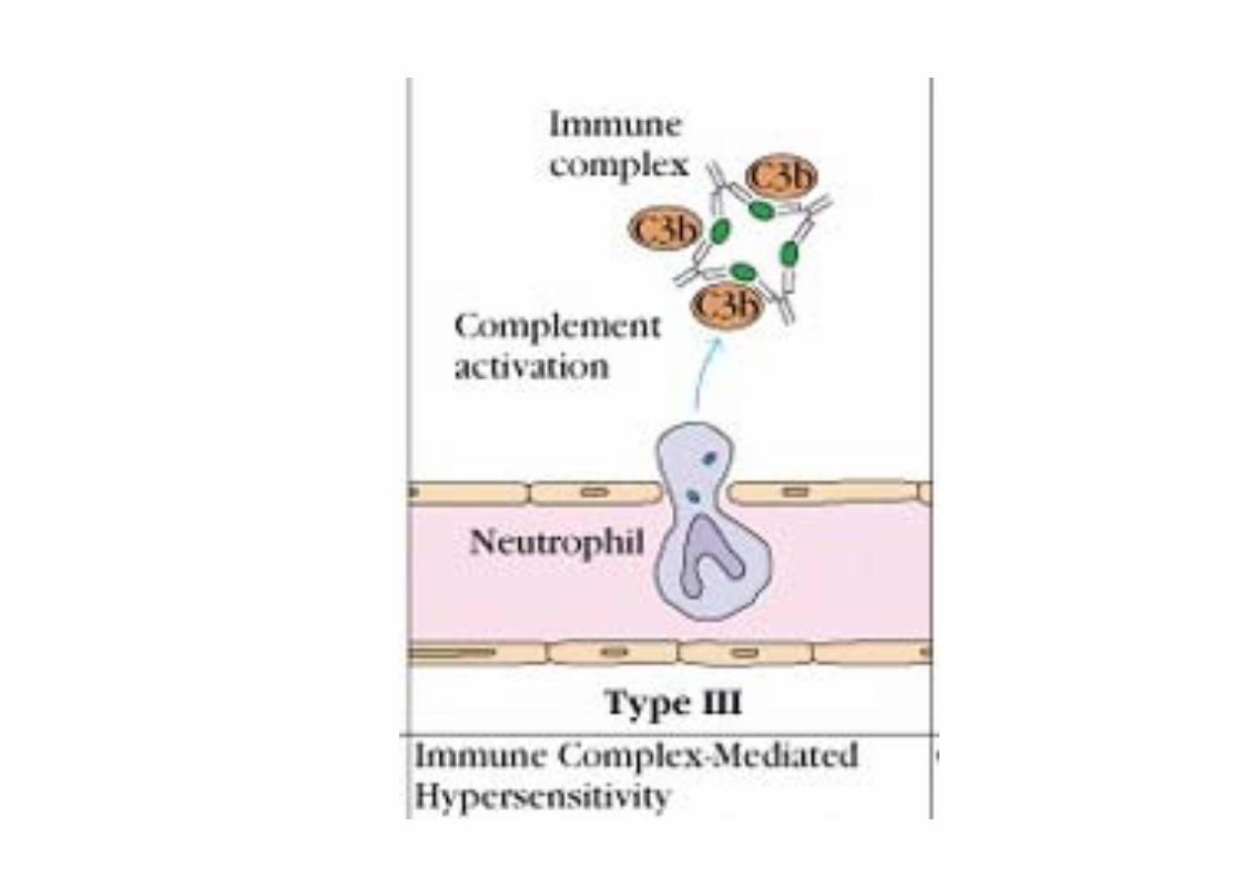
TYPE III Antigen antibody immune complexes.
IgG mediated
Immune Complex Disease• Large amount of antigen and antibodies form
complexes in blood.
• If not eliminated can deposit in capillaries or joints and trigger inflammation.

TYPE III Immune Complexes
• PMNs and macrophages bind to immune complexes via FcR and phagocytize the complexes.
BUT• If unable to phagocytize the immune complexes can
cause inflammation via C’ activation ---> C3a C4a, C5a and "frustrated phagocytes".
TYPE III Immune Complex Disease
"Frustrated Phagocytes"
• If neutrophils and macrophages are unable to phagocytize the immune complexes these cells will degranulate in the area of immune complex deposition and trigger inflammation.
• Unable to eat -------try to digest outside cell.

TYPE III Immune Complex Disease
Localized disease
• Deposited in joints causing local inflammation = arthritis.
• Deposited in kidneys = glomerulonephritis.

TYPE III Immune Complex Disease
• Serum sickness from large amounts of antigen such as injection of foreign serum.
• Serum sickness is usually transient immune complex disease with removal of antigen source.
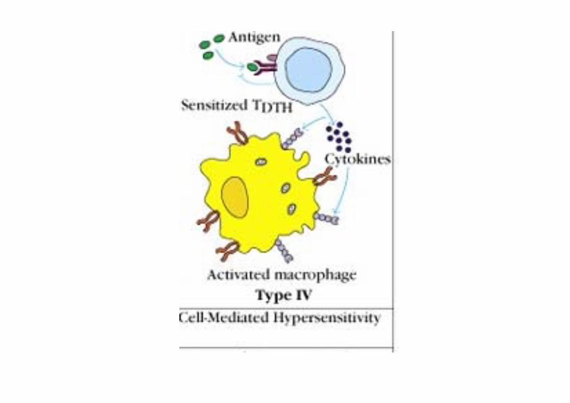
Delayed type hypersensitivity Th1 cells and macrophages
• DTH response is from:– Th1 cells release cytokines to activate macrophages
causing inflammation and tissue damage. – Continued macrophage activation can cause chronic
inflammation resulting in tissue lesions, scarring, and granuloma formation.
• Delayed is relative because DTH response arise 24-72 hours after exposure rather than within minutes.

Stages of Type IV DTHSensitization stage
• Memory Th1 cells against DTH antigens are generated by dendritic cells during the sensitization stage.
• These Th1 cells can activate macrophages and trigger inflammatory response.
Stages of Type IV DTHEffector stage
• Secondary contact yields what we call DTH. • Th1 memory cells are activated and produce
cytokines. – IFN-, TNF-and TNF- which cause tissue
destruction, inflammation. – IL-2 that activates T cells and CTLs.– Chemokines- for macrophage recruitment. – IL-3, GM-CSF for increased monocyte/macrophage

Stages of Type IV DTHEffector stage
Secondary exposure to antigen• Inflamed area becomes red and fluid filled can
form lesion. – From tissue damage there is activation of clotting
cascades and tissue repair. • Continued exposure to antigen can cause chronic
inflammation and result in granuloma formation.
Type IV DTHContact dermatitis
• The response to poison oak is a classic Type IV. – Small molecules act as haptens and complex with skin
proteins to be taken up by APCs and presented to Th1 cells to get sensitization.
– During secondary exposure Th1 memory cells become activated to cause DTH.

Contact dermatitis

Delayed type hypersensitivity (DTH)
DTH is a type of immune response classified by Th1 and macrophage activation that results in tissue damage.
DTH can be the result ofChronic infection or Exposure to some antigens.
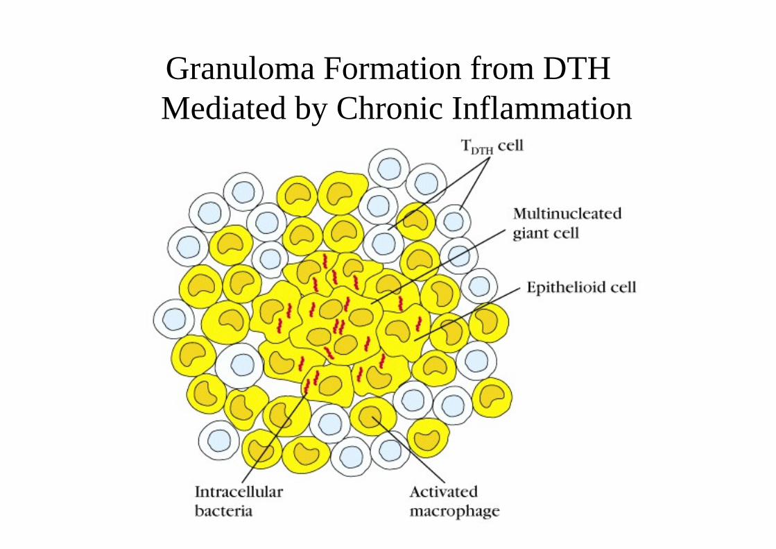
Granuloma Formation from DTHMediated by Chronic Inflammation
Human Immunodefficiency Virus(HIV)
• A retrovirus• Virus binds to CD4 cells• Decrease in CD 4 cells• Failure of proliferation of CD8 & B lymphocytes• Death from infections due to normally non pathogenic bacteria
• Normal ratio of CD4 to CD 8 cells is 1.2‐3.0• Permanent & progressive decrease in AIDS
Delayed hypersensitivity…(Examples)
• Tuberculin hypersensitivity: Classical reaction of skin induced by tuberculinSkin erythema and induration develops after several hours and is maximal at 24‐ 48.Histologically there is initial perivascular cuffing with mononuclear cells , then a more extensive exudation of monocytes and polymorphs within the dermis. The latter soon move away from the lesion leaving behind monocytes and lymphocytes
• Contact hypersensitivity: Extracts of plant poisons, Ivy

Tissue transplantation
• Allograft: Graft from one person to another• Autograft: Skin from one part of a person to other
• Xenograft: An organ/ tissue transplant from one animal species to other
• Syngenic: Same genetic constitution • Allogenic: Different genetic constitution

Allograft rejection
3 stagesStimulus: Passenger leukocytes derived from the donor that leave the graft & reach regional lymph nodes
Afferent stage: Release of antigen from graftCentral stage: Activation of immune responseEfferent stage: Generation & release of humoral& CMI effectors that bring about destruction of graft

Allograft reaction
• An immunological response produced by the recipient against the transplant antigen of the graft
• For a few days: Allo & auto graft are normal• After a week: Circulation to allograft decreases• 2‐3 weeks: Necrosis & sloughing offBiological significance: Ability to reject occasional mutant cells formed during the normal course of cell division