


Languages
Pages
Legal

Seediscussions,stats,andauthorprofilesforthispublicationat:https://www.researchgate.net/publication/299419180
LaboratoryToolstoQuantifyBiogenicDissolutionofRocksandMinerals:AModelRockBiofilmGrowinginPercolationColumns
ArticleinFrontiersinEarthSciences·April2016
DOI:10.3389/feart.2016.00031
READS
124
5authors,including:
FranzSeiffert
BundesanstaltfürMaterialforschungund-…
3PUBLICATIONS6CITATIONS
SEEPROFILE
NicoleBandow
BundesanstaltfürMaterialforschungund-…
20PUBLICATIONS228CITATIONS
SEEPROFILE
UteKalbe
BundesanstaltfürMaterialforschungund-…
37PUBLICATIONS238CITATIONS
SEEPROFILE
AnnaAGorbushina
FreieUniversitätBerlin
102PUBLICATIONS2,393CITATIONS
SEEPROFILE
Allin-textreferencesunderlinedinbluearelinkedtopublicationsonResearchGate,
lettingyouaccessandreadthemimmediately.
Availablefrom:FranzSeiffert
Retrievedon:23August2016

ORIGINAL RESEARCHpublished: 21 April 2016
doi: 10.3389/feart.2016.00031
Frontiers in Earth Science | www.frontiersin.org 1 April 2016 | Volume 4 | Article 31
Edited by:
Dina M. Bower,
NASA Goddard Space Flight
Center/University of Maryland, USA
Reviewed by:
Eric D. Van Hullebusch,
University Paris-Est, France
Alan W. Decho,
University of South Carolina, USA
*Correspondence:
Anna A. Gorbushina
Specialty section:
This article was submitted to
Biogeoscience,
a section of the journal
Frontiers in Earth Science
Received: 03 August 2015
Accepted: 15 March 2016
Published: 21 April 2016
Citation:
Seiffert F, Bandow N, Kalbe U, Milke R
and Gorbushina AA (2016) Laboratory
Tools to Quantify Biogenic Dissolution
of Rocks and Minerals: A Model Rock
Biofilm Growing in Percolation
Columns. Front. Earth Sci. 4:31.
doi: 10.3389/feart.2016.00031
Laboratory Tools to QuantifyBiogenic Dissolution of Rocks andMinerals: A Model Rock BiofilmGrowing in Percolation Columns
Franz Seiffert 1, Nicole Bandow 1, Ute Kalbe 1, Ralf Milke 2 and Anna A. Gorbushina 1, 2*
1Department of Materials and Environment, Federal Institute for Materials Research and Testing, Berlin, Germany,2Department of Earth Sciences, Freie Universität Berlin, Berlin, Germany
Sub-aerial biofilms (SAB) are ubiquitous, self-sufficient microbial ecosystems found on
mineral surfaces at all altitudes and latitudes. SABs, which are the principal causes
of weathering on exposed terrestrial surfaces, are characterized by patchy growth
dominated by associations of algae, cyanobacteria, fungi and heterotrophic bacteria.
A recently developed in vitro system to study colonization of rocks exposed to air
included two key SAB participants - the rock-inhabiting ascomycete Knufia petricola
(CBS 123872) and the phototrophic cyanobacterium Nostoc punctiforme ATCC29133.
Both partners are genetically tractable and we used them here to study weathering of
granite, K-feldspar and plagioclase. Small fragments of the various rocks or minerals
(1–6 mm) were packed into flow-through columns and incubated with 0.1% glucose
and 10 µM thiamine-hydrochloride (90 µL min−1) to compare weathering with and
without biofilms. Dissolution of the minerals was followed by: (i) analysing the degradation
products in the effluent from the columns via Inductively Coupled Plasma Spectroscopy
and (ii) by studying polished sections of the incubated mineral fragments/grains using
scanning electron microscopy, transmission electron microscopy and energy dispersive
X-ray analyses. K. petricola/N. punctiforme stimulated release of Ca, Na, Mg and Mn.
Analyses of the polished sections confirmed depletion of Ca, Na and K near the surface
of the fragments. The abrupt decrease in Ca concentration observed in peripheral areas
of plagioclase fragments favored a dissolution-reprecipitation mechanism. Percolation
columns in combination with a model biofilm can thus be used to study weathering
in closed systems. Columns can easily be filled with different minerals and biofilms,
the effluent as well as grains can be collected after long-term exposure under axenic
conditions and easily analyzed.
Keywords: biotic weathering, flow-through columns, plagioclase, K-feldspar, granite
INTRODUCTION
As life began to spread onto land at least 1.2 Ga ago, the first settlers were oxygenic cyanobacteriaalong with various organotrophic microorganisms. Present day sub-aerial (bare) rock surfacesare inhabited by similar microbial communities. Sub-aerial biofilms (SAB) are ubiquitous,self-sufficient, miniature microbial ecosystems that are found on mineral surfaces at all altitudes
Seiffert et al. Rock Biofilms in Percolation Columns
and latitudes. Patchy growth dominated by associations of algae,cyanobacteria, fungi and heterotrophic bacteria are amongst theprincipal characteristics of SABs. In addition to being one ofthe principal causes of weathering, SABs also accelerate thedecay of cultural heritages (Dornieden et al., 2000a,b; Warscheidand Braams, 2000; de los Ríos and Ascaso, 2005). Microbialcolonization of architectural monuments and works of artalso causes discoloring as the SAB inhabitants often producehighly colored pigments including carotenoids, chlorophylls andmelanins (Gorbushina et al., 1993; Diakumaku et al., 1995;Urzì and Realini, 1998; Gorbushina and Broughton, 2009).Furthermore some organisms induce acidolytic and oxido-reductive corrosion processes, excrete geochemically activesubstances as siderophores and low molecular weight organicacids, form detrimental crusts and therefore destroy solidmaterials (Warscheid and Braams, 2000). Limiting or preventingbiodeterioration is therefore critical to conserving objects ofcultural value.
Various microbes enhance the release of different elementsfrom rocks (Davis et al., 2007; Abdulla, 2009; Bonneville et al.,2011; Brunner et al., 2011; Lapanje et al., 2012; Olsson-Franciset al., 2012). Excreted organic acids and respiration-derived CO2
are often active in these processes (Silverman and Munoz, 1970;Delatorre et al., 1993; Gómez-Alarcón et al., 1994; Machill et al.,1997; Landeweert et al., 2001; Abdulla, 2009; Weber et al., 2011),although fungal hyphae can directly penetrate and widen fissuresin rocks (Sterflinger, 2000; Gorbushina et al., 2003; Chertovet al., 2004). The extreme microbial diversity of soil organisms(Jongmans et al., 1997; Landeweert et al., 2001; Schöll et al.,2008; Abdulla, 2009; Bonneville et al., 2009; Rosling et al., 2009;Taylor et al., 2009) and accompanying macroscopic vegetationresults in high rates of weathering. As weathering of rocks isthe first step in soil formation (Chadwick et al., 1990), surfaceweathering is an essential component of biogeomorphologicalprocesses. Nevertheless, the morphologically simpler microbialecosystems that prevail on bare rocks also have multiple effectson element cycles (Gorbushina, 2007). The relative simplicity ofSAB communities as compared to microbes in soils means thatthey are ideal in the study of biotic impact of microbes on thedissolution of minerals. Due to the extreme conditions on barerock surfaces, only certain stress tolerant microorganisms thatcan withstand extreme dryness, wide temperature fluctuations,intense solar irradiation and low nutrient availability are ableto survive. Often phototrophic cyanobacteria and oligotrophicmicrocolonial fungi (MCF) initially dominate this ecologicalniche (Staley et al., 1982; Sterflinger, 1998; Tamaru et al., 2005;Gorbushina, 2007; Knowles and Castenholz, 2008; Ozturk andAslim, 2010). Both types of organisms are extremely tolerant ofmany kinds of stress conditions and are therefore involved inbiodeterioration of monuments (Gorbushina et al., 1993, 2003;Diakumaku et al., 1995; Ortega-Calvo et al., 1995; Büdel et al.,2004; Olsson-Francis et al., 2012).
Various systems have been proposed to study the dissolutionof minerals under different environmental conditions but flow-through systems result in high aeration and dissolutionrates, which are important for cultivation of aerobicmicroorganisms and ideal for modeling SAB growth on
rocks and minerals. Flow-through percolation columns allowfor long-term microbiologically and geochemically stableexperiments. Furthermore, quantification of dissolution canbe performed on the liquid phase by measuring releasedelements via spectrometric methods like ICP-MS and ICP-OES(inductively coupled plasma mass spectrometry/optical emissionspectrometry) (Olesik, 1991). Changes in the mineral phasecan be analyzed via spectroscopic methods including EDX(energy dispersive X-ray spectroscopy), SEM (scanning electronmicroscope) or TEM (transmission electron microscope)(Eggert, 2005). Additionally, the solid mineral phase can bechemically digested and the contents measured by ICP-MS/OES.Degradation of solid phases is often described by “weatheringindices” which are the ratios of mobile and immobile elements(Price and Velbel, 2003; Takahashi and Shimaoka, 2012). Tobe useful, weathering indices must take into account a certainspectrum of mobile elements in the particular mineral and beapplicable to as broader range of different rock-types as possible.Two widely accepted indices are the Weathering Index of Parker(WIP) and the Chemical Index of Alteration (CIA), calculatedaccording to Equations (1) and (2). WIP values decrease andCIA values increase with rising degrees of weathering.
WIP = 100∗[(2Na2O/0.35)+ (MgO/0.9)+ (2K2O/0.25)
+(CaO/0.7)] (1)
CIA = 100∗[Al2O3/(Al2O3 + CaO+Na2O+ K2O)] (2)
Equally, the types of rocks used in model weathering studiesshould be of global relevance and contain components thatweather rapidly. Granite, being among the most abundant rocktypes within the Earth crust and crucially important in theconstruction of buildings andmonuments was an obvious choice.Mica and feldspar are typical components of granite, which haverelatively high dissolution rates and are sensitive to microbialdegradation (Lasaga et al., 1994; Bonneville et al., 2011).
Finally, reproducible studies of microbial effects on theweathering of rocks are only possible if the number of variablesis limited (i.e., simplified laboratory models are a prerequisite)and under well-controlled, laboratory conditions. In a previousstudy, a laboratory rock-inhabiting biofilm consisting of theheterotrophic MCF Knufia petricola and the nitrogen-fixingcyanobacteriumNostoc punctiformewas established (Gorbushinaand Broughton, 2009). Here we used this biofilm to study thebiological impact on weathering of granite and related mineralsin a new setting of a geomicrobiologically-modified percolationcolumn.
MATERIALS AND METHODS
Model Biofilm—Starting Cultures andInoculum PreparationNostoc punctiforme ATCC29133 was kindly supplied by J.M.Meeks (University of California, Davis, CA, USA) (Ekman et al.,2013). Cultures used for the experiments were grown in BG11medium (Stanier et al., 1971) at pH 7.5 25◦C and 90 µM photonsof photosynthetically active light m2 s−1 for 24 h d−1 and shaking
Frontiers in Earth Science | www.frontiersin.org 2 April 2016 | Volume 4 | Article 31
Seiffert et al. Rock Biofilms in Percolation Columns
(160 rpm). Knufia petricola (CBS 123872) was isolated from aweathered marble monument in Athens (Greece) (Nai et al.,2013). Cultures used for the experiments were grown in 2%malt extract broth [MEB: 2% (w/v) malt extract; 0.1% (w/v)casein-digested peptone; 2% (w/v) D-(+)-glucose] under shaking(100 rpm) at 25◦C. Both organisms were transferred weekly bydiluting 1:100 into fresh media. Seven to 14 d old cultures wereused for the preparation of inocula for rock/mineral dissolutionexperiments.
To prepare inocula, biomass of both organisms was dispersedin a ball-mill, centrifuged (6603 RCF for 15 min), the pellets werewashed three times and finally re-suspended in 5mMglucose and10 µM thiamine-hydrochloride for the rock/mineral dissolutionexperiments. Dispersion of clumped cells was performed instainless steel beakers containing eight stainless steel beads (ca. 5mm diameter) for 10 min at 30 Hz (for K. petricola) or with fourglass beads for 5 min at 25 Hz (forN. punctiforme) in a mixer mill(MM400, Retsch GmbH, Haan, Germany).
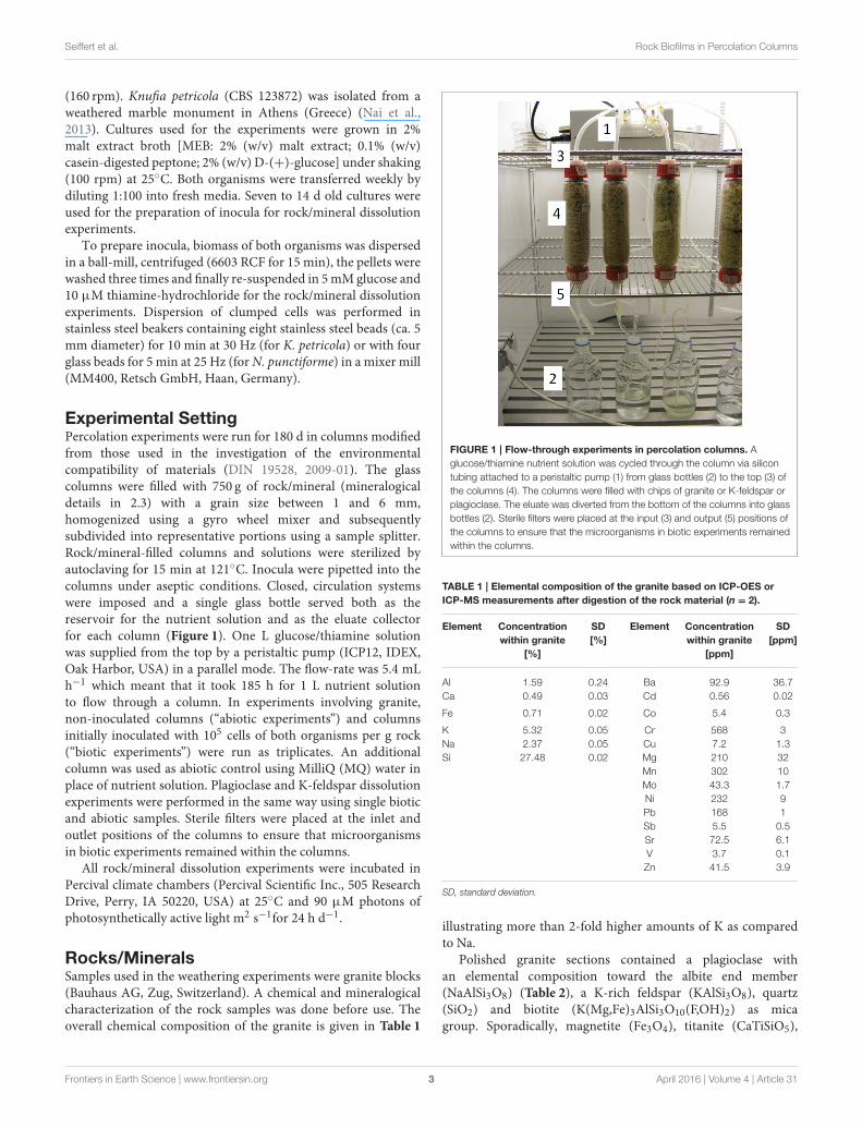
Experimental SettingPercolation experiments were run for 180 d in columns modifiedfrom those used in the investigation of the environmentalcompatibility of materials (DIN 19528, 2009-01). The glasscolumns were filled with 750 g of rock/mineral (mineralogicaldetails in 2.3) with a grain size between 1 and 6 mm,homogenized using a gyro wheel mixer and subsequentlysubdivided into representative portions using a sample splitter.Rock/mineral-filled columns and solutions were sterilized byautoclaving for 15 min at 121◦C. Inocula were pipetted into thecolumns under aseptic conditions. Closed, circulation systemswere imposed and a single glass bottle served both as thereservoir for the nutrient solution and as the eluate collectorfor each column (Figure 1). One L glucose/thiamine solutionwas supplied from the top by a peristaltic pump (ICP12, IDEX,Oak Harbor, USA) in a parallel mode. The flow-rate was 5.4 mLh−1 which meant that it took 185 h for 1 L nutrient solutionto flow through a column. In experiments involving granite,non-inoculated columns (“abiotic experiments”) and columnsinitially inoculated with 105 cells of both organisms per g rock(“biotic experiments”) were run as triplicates. An additionalcolumn was used as abiotic control using MilliQ (MQ) water inplace of nutrient solution. Plagioclase and K-feldspar dissolutionexperiments were performed in the same way using single bioticand abiotic samples. Sterile filters were placed at the inlet andoutlet positions of the columns to ensure that microorganismsin biotic experiments remained within the columns.
All rock/mineral dissolution experiments were incubated inPercival climate chambers (Percival Scientific Inc., 505 ResearchDrive, Perry, IA 50220, USA) at 25◦C and 90 µM photons ofphotosynthetically active light m2 s−1for 24 h d−1.
Rocks/MineralsSamples used in the weathering experiments were granite blocks(Bauhaus AG, Zug, Switzerland). A chemical and mineralogicalcharacterization of the rock samples was done before use. Theoverall chemical composition of the granite is given in Table 1
FIGURE 1 | Flow-through experiments in percolation columns. A
glucose/thiamine nutrient solution was cycled through the column via silicon
tubing attached to a peristaltic pump (1) from glass bottles (2) to the top (3) of
the columns (4). The columns were filled with chips of granite or K-feldspar or
plagioclase. The eluate was diverted from the bottom of the columns into glass
bottles (2). Sterile filters were placed at the input (3) and output (5) positions of
the columns to ensure that the microorganisms in biotic experiments remained
within the columns.
TABLE 1 | Elemental composition of the granite based on ICP-OES or
ICP-MS measurements after digestion of the rock material (n = 2).
Element Concentration
within granite
[%]
SD
[%]
Element Concentration
within granite
[ppm]
SD
[ppm]
Al
Ca
1.59
0.49
0.24
0.03
Ba
Cd
92.9
0.56
36.7
0.02
Fe 0.71 0.02 Co 5.4 0.3
K
Na
Si
5.32
2.37
27.48
0.05
0.05
0.02
Cr
Cu
Mg
Mn
Mo
Ni
Pb
Sb
Sr
V
Zn
568
7.2
210
302
43.3
232
168
5.5
72.5
3.7
41.5
3
1.3
32
10
1.7
9
1
0.5
6.1
0.1
3.9
SD, standard deviation.
illustrating more than 2-fold higher amounts of K as comparedto Na.
Polished granite sections contained a plagioclase withan elemental composition toward the albite end member(NaAlSi3O8) (Table 2), a K-rich feldspar (KAlSi3O8), quartz(SiO2) and biotite (K(Mg,Fe)3AlSi3O10(F,OH)2) as micagroup. Sporadically, magnetite (Fe3O4), titanite (CaTiSiO5),
Frontiers in Earth Science | www.frontiersin.org 3 April 2016 | Volume 4 | Article 31
Seiffert et al. Rock Biofilms in Percolation Columns
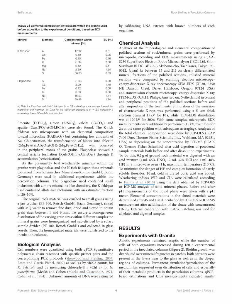
TABLE 2 | Elemental composition of feldspars within the granite used
before exposition to the experimental conditions, based on EDX
measurements.
Mineral Element Concentration within
mineral [%]
SD [%]
K-feldspar Al
Ca
Fe
K
Mg
Na
Si
17.92
0.10
0.15
21.94
0.12
2.89
56.83
0.31
0.05
0.16
2.36
0.41
1.60
0.83
Plagioclase Al
Ca
Fe
K
Na
Si
21.03
2.99
0.12
0.83
15.03
59.98
0.88
1.48
0.08
0.56
1.14
1.74
(a) Data for the observed K-rich feldspar (n = 13) indicating a mineralogy toward the
microcline end member. (b) Data for the observed plagioclase (n = 21) indicating a
mineralogy toward the albite end member.
ilmenite (FeTiO3), zircon (ZrSiO4), calcite (CaCO3) andapatite (Ca10(PO4)6(OH,F,Cl)2) were also found. The K-richfeldspar was microporous with an elemental compositiontoward microcline (KAlSi3O8) but containing low amounts ofNa. Chloritization, the transformation of biotite into chlorite((Mg,Fe)3(Si,Al)4O10(OH)2(Mg,Fe)3(OH)6), was observedin the peripheral zones of the grains. Plagioclase showed acentral sericite formation (KAl2(OH,F)2AlSi3O10) through Kaccumulation (sericitization).
As the presumably best weatherable minerals within thegranite were plagioclase and the K-rich feldspar, these minerals(obtained from Rheinisches Mineralien-Kontor GmbH, Bonn,Germany) were used in additional experiments within thepercolation columns. The plagioclase used contained smallinclusions with a more microcline-like chemistry, the K-feldsparused contained albite-like inclusions with an estimated fractionof 20–30%.
The original rock material was crushed to small grains usinga jaw crusher (BB 300, Retsch GmbH, Haan, Germany), rinsedwith MQ water to remove fine dust, dried and sieved to obtaingrain sizes between 1 and 6 mm. To ensure a homogeneousdistribution of the varying grain sizes within different samples themineral grains were homogenized and sub-divided by a rotarysample divider (PT 100, Retsch GmbH) and collected in glassvessels. Then, the homogenized materials were transferred to theincubation columns.
Biological AnalysesCell numbers were quantified using both qPCR (quantitativepolymerase chain reaction) with specific primer pairs and thecorresponding PCR protocols (Sherwood and Presting, 2007;Bates and Garcia-Pichel, 2009) as well as by viable counts forK. petricola and by measuring chlorophyll a (Chl a) for N.punctiforme [Meeks and Cohen (Meeks and Castenholz, 1971;Cohen et al., 1994)]. Unknown amounts of DNA were estimated
by calibrating DNA extracts with known numbers of eachorganism.
Chemical AnalysisAnalyses of the mineralogical and elemental composition ofpolished sections of rock/mineral grains were performed bymicroprobe recording and EDX measurements using a JXA-8230 SuperProbe Electron ProbeMicroanalyzer (JEOL Ltd, Shin-Suzuharu BLDG. 3F 2-8-3 Akebono-cho, Tachikawa, Tokyo 190-0012, Japan) (n between 13 and 21) on clearly differentiatedmineral fractions of the polished sections. Polished mineralsections were compared by scanning electron microscopy-energy-dispersive X-ray spectroscopy SEM-EDX (XL30, 5350NE Dawson Creek Drive, Hillsboro, Oregon 97124 USA)and transmission electron microscopy- energy-dispersive X-ray(TEM-EDX)(CM12, Philips, Amsterdam, Netherlands) in centraland peripheral positions of the polished sections before andafter imposition of the treatments. Stimulation of the emissionof characteristic X-rays was performed using a 5 µm thickelectron beam at 15 kV for 10 s, while TEM-EDX stimulationwas at 120 kV for 300 s. With some samples, microprobe-EDXmeasurements were additionally performed (15 kV, five times for2 s at the same position with subsequent averaging). Analyses ofthe total chemical composition were done by ICP-OES (ICAP7400 Duo, Thermo Fisher Scientific Inc., Waltham, MA 02451,USA) or depending on the concentration by ICP-MS (ICAP-Q, Thermo Fisher Scientific) after acid digestion of powderedsample materials both before and after dissolution experiments.To do this, 0.2 g of ground rock material was digested with anacid mixture (4mL 65% HNO3; 2.mL 32% HCl and 1mL 48%HF) in a microwave oven (1 h, maximum temperature 210◦C).To minimize the danger of HF and complex formation of barelysoluble fluorides, 10mL cold saturated boric acid was added.Weathering indices WIP and CIA were calculated accordingto Fiantis et al. (2010) using the data obtained by ICP-OESor ICP-MS analysis of solid mineral phases. Before and afterpH measurements of the liquid phase were taken with a pHmeter. Elemental concentrations in the eluted materials weredetermined after 45 and 180 d incubation by ICP-OES or ICP-MSmeasurement after acidification of the eluate with concentratedHNO3. External calibration with matrix matching was used forall eluted and digested samples.
RESULTS
Experiments with GraniteAbiotic experiments remained aseptic while the number ofcells of both organisms increased during 180 d experimentalperiod in the inoculated columns (Figure 2). Biofilm growth wasdistributed overmineral fragments in patches, both partners werepresent in the layers near to the glass as well as in the deeperregions of columns. Permanent circulation/percolation of themedium has supported even distribution of cells and especiallyof their metabolic products in the percolation columns. qPCR-based estimations and Chla measurements indicated similar
Frontiers in Earth Science | www.frontiersin.org 4 April 2016 | Volume 4 | Article 31
Seiffert et al. Rock Biofilms in Percolation Columns
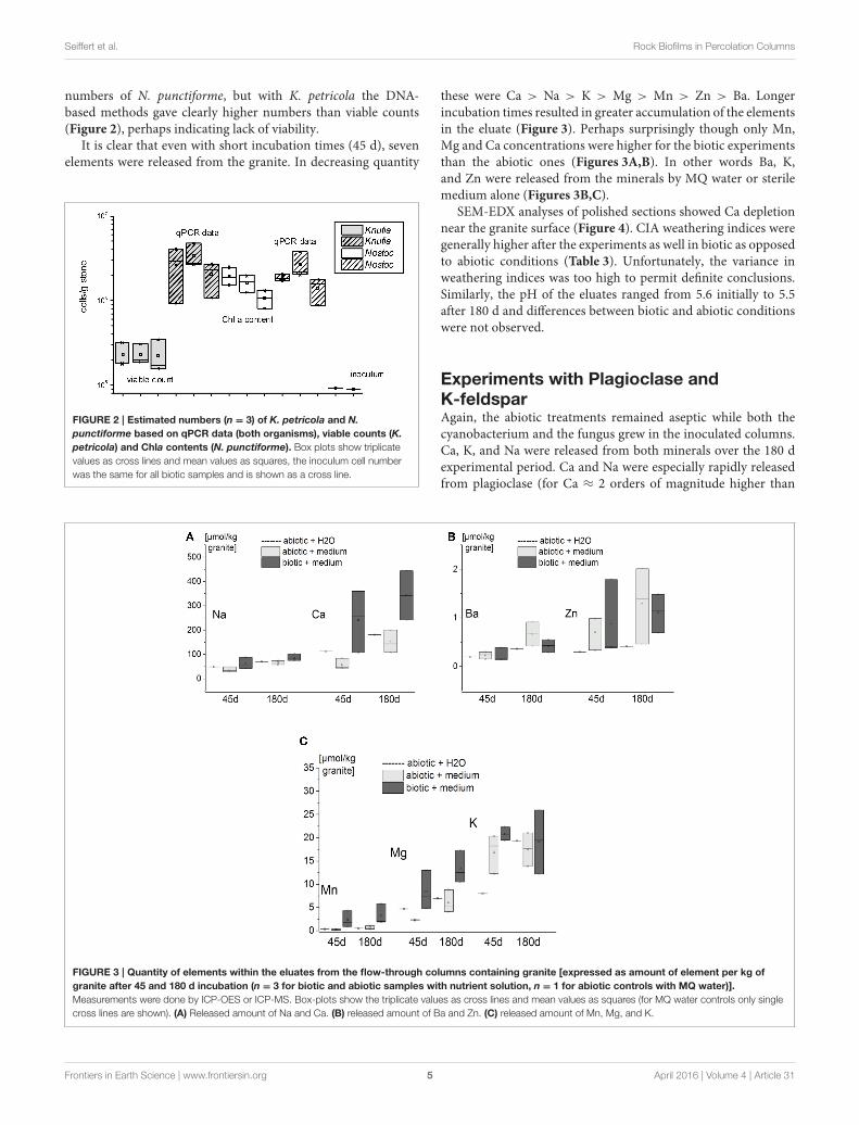
numbers of N. punctiforme, but with K. petricola the DNA-based methods gave clearly higher numbers than viable counts(Figure 2), perhaps indicating lack of viability.
It is clear that even with short incubation times (45 d), sevenelements were released from the granite. In decreasing quantity
FIGURE 2 | Estimated numbers (n = 3) of K. petricola and N.
punctiforme based on qPCR data (both organisms), viable counts (K.
petricola) and Chla contents (N. punctiforme). Box plots show triplicate
values as cross lines and mean values as squares, the inoculum cell number
was the same for all biotic samples and is shown as a cross line.
these were Ca > Na > K > Mg > Mn > Zn > Ba. Longerincubation times resulted in greater accumulation of the elementsin the eluate (Figure 3). Perhaps surprisingly though only Mn,Mg and Ca concentrations were higher for the biotic experimentsthan the abiotic ones (Figures 3A,B). In other words Ba, K,and Zn were released from the minerals by MQ water or sterilemedium alone (Figures 3B,C).
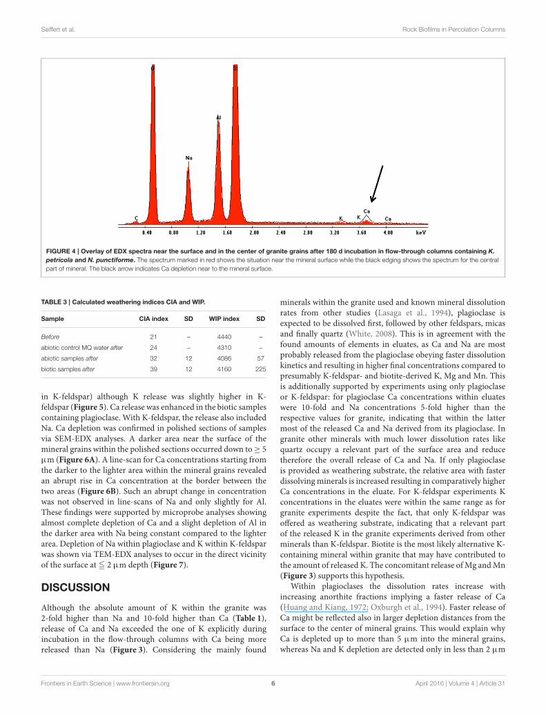
SEM-EDX analyses of polished sections showed Ca depletionnear the granite surface (Figure 4). CIA weathering indices weregenerally higher after the experiments as well in biotic as opposedto abiotic conditions (Table 3). Unfortunately, the variance inweathering indices was too high to permit definite conclusions.Similarly, the pH of the eluates ranged from 5.6 initially to 5.5after 180 d and differences between biotic and abiotic conditionswere not observed.
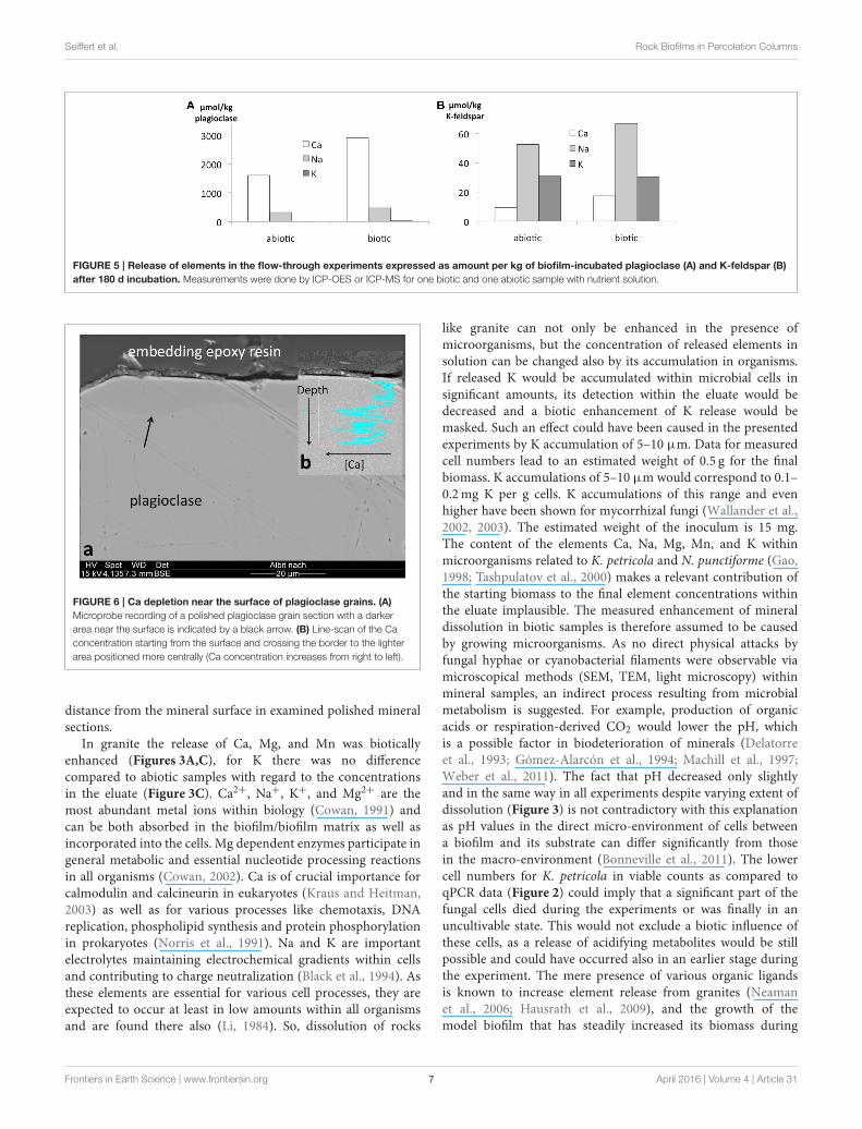
Experiments with Plagioclase andK-feldsparAgain, the abiotic treatments remained aseptic while both thecyanobacterium and the fungus grew in the inoculated columns.Ca, K, and Na were released from both minerals over the 180 dexperimental period. Ca and Na were especially rapidly releasedfrom plagioclase (for Ca ≈ 2 orders of magnitude higher than
FIGURE 3 | Quantity of elements within the eluates from the flow-through columns containing granite [expressed as amount of element per kg of
granite after 45 and 180 d incubation (n = 3 for biotic and abiotic samples with nutrient solution, n = 1 for abiotic controls with MQ water)].
Measurements were done by ICP-OES or ICP-MS. Box-plots show the triplicate values as cross lines and mean values as squares (for MQ water controls only single
cross lines are shown). (A) Released amount of Na and Ca. (B) released amount of Ba and Zn. (C) released amount of Mn, Mg, and K.
Frontiers in Earth Science | www.frontiersin.org 5 April 2016 | Volume 4 | Article 31
Seiffert et al. Rock Biofilms in Percolation Columns
FIGURE 4 | Overlay of EDX spectra near the surface and in the center of granite grains after 180 d incubation in flow-through columns containing K.
petricola and N. punctiforme. The spectrum marked in red shows the situation near the mineral surface while the black edging shows the spectrum for the central
part of mineral. The black arrow indicates Ca depletion near to the mineral surface.
TABLE 3 | Calculated weathering indices CIA and WIP.
Sample CIA index SD WIP index SD
Before 21 – 4440 –
abiotic control MQ water after 24 – 4310 –
abiotic samples after 32 12 4086 57
biotic samples after 39 12 4160 225
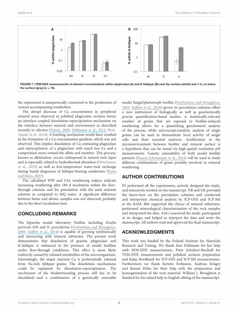
in K-feldspar) although K release was slightly higher in K-feldspar (Figure 5). Ca release was enhanced in the biotic samplescontaining plagioclase. With K-feldspar, the release also includedNa. Ca depletion was confirmed in polished sections of samplesvia SEM-EDX analyses. A darker area near the surface of themineral grains within the polished sections occurred down to≥ 5µm (Figure 6A). A line-scan for Ca concentrations starting fromthe darker to the lighter area within the mineral grains revealedan abrupt rise in Ca concentration at the border between thetwo areas (Figure 6B). Such an abrupt change in concentrationwas not observed in line-scans of Na and only slightly for Al.These findings were supported by microprobe analyses showingalmost complete depletion of Ca and a slight depletion of Al inthe darker area with Na being constant compared to the lighterarea. Depletion of Na within plagioclase and K within K-feldsparwas shown via TEM-EDX analyses to occur in the direct vicinityof the surface at ≦ 2 µm depth (Figure 7).
DISCUSSION
Although the absolute amount of K within the granite was2-fold higher than Na and 10-fold higher than Ca (Table 1),release of Ca and Na exceeded the one of K explicitly duringincubation in the flow-through columns with Ca being morereleased than Na (Figure 3). Considering the mainly found
minerals within the granite used and known mineral dissolutionrates from other studies (Lasaga et al., 1994), plagioclase isexpected to be dissolved first, followed by other feldspars, micasand finally quartz (White, 2008). This is in agreement with thefound amounts of elements in eluates, as Ca and Na are mostprobably released from the plagioclase obeying faster dissolutionkinetics and resulting in higher final concentrations compared topresumably K-feldspar- and biotite-derived K, Mg and Mn. Thisis additionally supported by experiments using only plagioclaseor K-feldspar: for plagioclase Ca concentrations within eluateswere 10-fold and Na concentrations 5-fold higher than therespective values for granite, indicating that within the lattermost of the released Ca and Na derived from its plagioclase. Ingranite other minerals with much lower dissolution rates likequartz occupy a relevant part of the surface area and reducetherefore the overall release of Ca and Na. If only plagioclaseis provided as weathering substrate, the relative area with fasterdissolvingminerals is increased resulting in comparatively higherCa concentrations in the eluate. For K-feldspar experiments Kconcentrations in the eluates were within the same range as forgranite experiments despite the fact, that only K-feldspar wasoffered as weathering substrate, indicating that a relevant partof the released K in the granite experiments derived from otherminerals than K-feldspar. Biotite is the most likely alternative K-containing mineral within granite that may have contributed tothe amount of released K. The concomitant release ofMg andMn(Figure 3) supports this hypothesis.
Within plagioclases the dissolution rates increase withincreasing anorthite fractions implying a faster release of Ca(Huang and Kiang, 1972; Oxburgh et al., 1994). Faster release ofCa might be reflected also in larger depletion distances from thesurface to the center of mineral grains. This would explain whyCa is depleted up to more than 5 µm into the mineral grains,whereas Na and K depletion are detected only in less than 2 µm
Frontiers in Earth Science | www.frontiersin.org 6 April 2016 | Volume 4 | Article 31
Seiffert et al. Rock Biofilms in Percolation Columns
FIGURE 5 | Release of elements in the flow-through experiments expressed as amount per kg of biofilm-incubated plagioclase (A) and K-feldspar (B)
after 180 d incubation. Measurements were done by ICP-OES or ICP-MS for one biotic and one abiotic sample with nutrient solution.
FIGURE 6 | Ca depletion near the surface of plagioclase grains. (A)
Microprobe recording of a polished plagioclase grain section with a darker
area near the surface is indicated by a black arrow. (B) Line-scan of the Ca
concentration starting from the surface and crossing the border to the lighter
area positioned more centrally (Ca concentration increases from right to left).
distance from the mineral surface in examined polished mineralsections.
In granite the release of Ca, Mg, and Mn was bioticallyenhanced (Figures 3A,C), for K there was no differencecompared to abiotic samples with regard to the concentrationsin the eluate (Figure 3C). Ca2+, Na+, K+, and Mg2+ are themost abundant metal ions within biology (Cowan, 1991) andcan be both absorbed in the biofilm/biofilm matrix as well asincorporated into the cells. Mg dependent enzymes participate ingeneral metabolic and essential nucleotide processing reactionsin all organisms (Cowan, 2002). Ca is of crucial importance forcalmodulin and calcineurin in eukaryotes (Kraus and Heitman,2003) as well as for various processes like chemotaxis, DNAreplication, phospholipid synthesis and protein phosphorylationin prokaryotes (Norris et al., 1991). Na and K are importantelectrolytes maintaining electrochemical gradients within cellsand contributing to charge neutralization (Black et al., 1994). Asthese elements are essential for various cell processes, they areexpected to occur at least in low amounts within all organismsand are found there also (Li, 1984). So, dissolution of rocks
like granite can not only be enhanced in the presence ofmicroorganisms, but the concentration of released elements insolution can be changed also by its accumulation in organisms.If released K would be accumulated within microbial cells insignificant amounts, its detection within the eluate would bedecreased and a biotic enhancement of K release would bemasked. Such an effect could have been caused in the presentedexperiments by K accumulation of 5–10 µm. Data for measuredcell numbers lead to an estimated weight of 0.5 g for the finalbiomass. K accumulations of 5–10 µmwould correspond to 0.1–0.2mg K per g cells. K accumulations of this range and evenhigher have been shown for mycorrhizal fungi (Wallander et al.,2002, 2003). The estimated weight of the inoculum is 15 mg.The content of the elements Ca, Na, Mg, Mn, and K withinmicroorganisms related to K. petricola and N. punctiforme (Gao,1998; Tashpulatov et al., 2000) makes a relevant contribution ofthe starting biomass to the final element concentrations withinthe eluate implausible. The measured enhancement of mineraldissolution in biotic samples is therefore assumed to be causedby growing microorganisms. As no direct physical attacks byfungal hyphae or cyanobacterial filaments were observable viamicroscopical methods (SEM, TEM, light microscopy) withinmineral samples, an indirect process resulting from microbialmetabolism is suggested. For example, production of organicacids or respiration-derived CO2 would lower the pH, whichis a possible factor in biodeterioration of minerals (Delatorreet al., 1993; Gómez-Alarcón et al., 1994; Machill et al., 1997;Weber et al., 2011). The fact that pH decreased only slightlyand in the same way in all experiments despite varying extent ofdissolution (Figure 3) is not contradictory with this explanationas pH values in the direct micro-environment of cells betweena biofilm and its substrate can differ significantly from thosein the macro-environment (Bonneville et al., 2011). The lowercell numbers for K. petricola in viable counts as compared toqPCR data (Figure 2) could imply that a significant part of thefungal cells died during the experiments or was finally in anuncultivable state. This would not exclude a biotic influence ofthese cells, as a release of acidifying metabolites would be stillpossible and could have occurred also in an earlier stage duringthe experiment. The mere presence of various organic ligandsis known to increase element release from granites (Neamanet al., 2006; Hausrath et al., 2009), and the growth of themodel biofilm that has steadily increased its biomass during
Frontiers in Earth Science | www.frontiersin.org 7 April 2016 | Volume 4 | Article 31
Seiffert et al. Rock Biofilms in Percolation Columns
FIGURE 7 | TEM-EDX measurements of element concentrations within plagioclase (A) and K-feldspar (B) near the surface (white) and 1–2 µm below
the surface (gray) (n = 10).
the experiment is unequivocally connected to the production ofvarious accompanying metabolites.
The abrupt decrease of Ca concentration in peripheralmineral areas observed in polished plagioclase sections favorsan interface-coupled dissolution-reprecipitation mechanism onthe interface between mineral and environment as describedrecently in silicates (Putnis, 2009; Hellmann et al., 2012; Ruiz-Agudo et al., 2014). A leaching mechanism would have resultedin the formation of a Ca concentration gradient, which was notobserved. This implies dissolution of Ca-containing plagioclaseand reprecipitation of a plagioclase with much less Ca and acomposition more toward the albite end member. This process,known as albitisation, occurs widespread in natural rock typesand is especially related to hydrothermal alteration (Hövelmannet al., 2010) as well as low-temperature water-rock exchangeduring burial diagenesis of feldspar-bearing sandstones (Perezand Boles, 2005).
The calculated WIP and CIA weathering indices indicateincreasing weathering after 180 d incubation within the flow-through columns and for percolation with the used nutrientsolution as compared to MQ water. A significant differencebetween biotic and abiotic samples was not observed, probablydue to the short incubation time.
CONCLUDING REMARKS
The bipartite model laboratory biofilm including Knufiapetricola A95 and N. punctiforme (Gorbushina and Broughton,2009; Seiffert et al., 2014) is capable of growing symbioticallyand interacting with mineral substrates. The present workdemonstrates that dissolution of granite, plagioclase andK-feldspar is enhanced in the presence of model biofilmsunder flow-through conditions. This effect is most likelyindirectly caused by released metabolites of the microorganisms.Interestingly, the major nutrient Ca is preferentially releasedfrom Na-rich feldspar grains. The dissolution mechanismscould be explained by dissolution-reprecipitation. Themechanism of the biodeteriorating process still has to beelucidated—and a combination of a genetically amenable
model fungal/phototroph biofilm (Gorbushina and Broughton,
2009; Seiffert et al., 2014) grown in percolation columns offersa new instrument of biologically as well as geochemicallyprecise quantification-based studies. A statistically-relevantnumber of grains that are exposed to biofilm-inducedweathering allows for a quantifying geochemical analysisof the process, while microscopic/analytic analysis of singlegrains can be used to demonstrate local activity of singlecells and their excreted matrices. Acidification in themicroenvironment between biofilm and mineral surface isa hypothesis that can be tested via high-spatial resolution pHmeasurements. Genetic amenability of both model biofilmpartners (Noack-Schönmann et al., 2014) will be used to studydifferent combinations of genes possibly involved in mineralweathering.
AUTHOR CONTRIBUTIONS
FS performed all the experiments, actively designed the study,and intensively worked on the manuscript. NB and UK providedthe know-how on the percolation columns and conductedand interpreted chemical analysis by ICP-OES and ICP-MSat the BAM. RM supported the choice of mineral substrates,performed mineralogical characterization of the rock samplesand interpreted the data. AAG conceived the study, participatedin its design, and helped to interpret the data and write themanuscript. All authors read and approved the final manuscript.
ACKNOWLEDGMENTS
This work was funded by the Federal Institute for MaterialsResearch and Testing. We thank Ines Feldmann for her helpwith SEM-EDX measurements, Peter Schubert-Bischoff forTEM-EDX measurements and polished sections preparationand Katja Nordhauß for ICP-OES and ICP-MS measurements.Furthermore we thank Kerstin Erdmann, Andreas Krügerand Renate Helm for their help with the preparation andhomogenization of the rock material. William J. Broughton isthanked for his valued help in English editing of the manuscript.
Frontiers in Earth Science | www.frontiersin.org 8 April 2016 | Volume 4 | Article 31
Seiffert et al. Rock Biofilms in Percolation Columns
REFERENCES
Abdulla, H. (2009). Bioweathering and biotransformation of granitic rockminerals
by actinomycetes.Microb. Ecol. 58, 753–761. doi: 10.1007/s00248-009-9549-1
Bates, S. T., and Garcia-Pichel, F. (2009). A culture-independent study of free-
living fungi in biological soil crusts of the Colorado Plateau: their diversity and
relative contribution to microbial biomass. Environ. Microbiol. 11, 56–67. doi:
10.1111/j.1462-2920.2008.01738.x
Black, C. B., Huang, H. W., and Cowan, J. A. (1994). Biological coordination
chemistry of magnesium, sodium, and potassium-ions - protein and
nucleotide-binding sites. Coord. Chem. Rev. 135, 165–202. doi: 10.1016/0010-
8545(94)80068-5
Bonneville, S., Morgan, D. J., Schmalenberger, A., Bray, A., Brown, A.,
Banwart, S. A., et al. (2011). Tree-mycorrhiza symbiosis accelerate
mineral weathering: evidences from nanometer-scale elemental fluxes at
the hypha-mineral interface. Geochim. Cosmochim. Acta 75, 6988–7005. doi:
10.1016/j.gca.2011.08.041
Bonneville, S., Smits, M. M., Brown, A., Harrington, J., Leake, J. R., Brydson, R.,
et al. (2009). Plant-driven fungal weathering: early stages of mineral alteration
at the nanometer scale. Geology 37, 615–618. doi: 10.1130/G25699A.1
Brunner, I., Plötze, M., Rieder, S., Zumsteg, A., Furrer, G., and Frey, B. (2011).
Pioneering fungi from the Damma glacier forefield in the Swiss Alps can
promote granite weathering. Geobiology 9, 266–279. doi: 10.1111/j.1472-
4669.2011.00274.x
Büdel, B., Weber, B., Kühl, M., Pfanz, H., Sültemeyer, D., and Wessels, D.
(2004). Reshaping of sandstone surfaces by cryptoendolithic cyanobacteria:
bioalkalization causes chemical weathering in arid landscapes. Geobiology 2,
261–268. doi: 10.1111/j.1472-4677.2004.00040.x
Chadwick, O. A., Brimhall, G. H., and Hendricks, D. M. (1990). From a black to
a gray box - a mass balance interpretation of pedogenesis. Geomorphology 3,
369–390. doi: 10.1016/0169-555X(90)90012-F
Chertov, O., Gorbushina, A., and Deventer, B. (2004). A model for
microcolonial fungi growth on rock surfaces. Ecol. Model. 177, 415–426.
doi: 10.1016/j.ecolmodel.2004.02.011
Cohen, M. F., Wallis, J. G., Campbell, E. L., and Meeks, J. C. (1994). Transposon
mutagenesis of Nostoc sp. strain ATCC 29133, a filamentous cyanobacterium
with multiple cellular differentiation alternatives. Microbiology 140(Pt 12),
3233–3240. doi: 10.1099/13500872-140-12-3233
Cowan, J. A. (1991). Metallobiochemistry of magnesium - coordination-complexes
with biological substrates - site specificity, kinetics and thermodynamics
of binding, and implications for activity. Inorg. Chem. 30, 2740–2747. doi:
10.1021/ic00013a008
Cowan, J. A. (2002). Structural and catalytic chemistry of magnesium-dependent
enzymes. Biometals 15, 225–235. doi: 10.1023/A:1016022730880
Davis, K. J., Nealson, K. H., and Luttge, A. (2007). Calcite and dolomite dissolution
rates in the context of microbe-mineral surface interactions. Geobiology 5,
191–205. doi: 10.1111/j.1472-4669.2007.00112.x
Delatorre, M. A., Gómez-Alarcón, G., Vizcaino, C., and Garcia, M. T. (1993).
Biochemical-mechanisms of stone alteration carried out by filamentous
fungi living in monuments. Biogeochemistry 19, 129–147. doi: 10.1007/BF00
000875
de los Ríos, A., and Ascaso, C. (2005). Contributions of in situ microscopy
to the current understanding of stone biodeterioration. Int. Microbiol.
8, 181–188. Available online at: http://scielo.isciii.es/scielo.php?pid=S1139-
67092005000300005&script=sci_arttext&tlng=pt
DIN 19528 (2009-01). Leaching of Solid Materials - Percolation Method for the Joint
Examination of the Leaching Behaviour of Organic and Inorganic Substances
for Materials with a Particle Size upto 32 mm - Basic Characterization Using
a Comprehensive Column Test and Compliance Test Using a Quick Column
Test. Deutsches Institut für Normung, German Standardization Organization,
Berlin.
Diakumaku, E., Gorbushina, A. A., Krumbein, W. E., Panina, L., and
Soukharjevski, S. (1995). Black fungi in marble and limestones - an aesthetical,
chemical and physical problem for the conservation of monuments. Sci. Total
Environ. 167, 295–304. doi: 10.1016/0048-9697(95)04590-W
Dornieden, T., Gorbushina, A. A., and Krumbein, W. E. (2000a). Biodecay of
cultural heritage as a space/time-related ecological situation - an evaluation of
a series of studies. Int. Biodeter. Biodegrad. 46, 261–270. doi: 10.1016/S0964-
8305(00)00107-4
Dornieden, T., Gorbushina, A. A., and Krumbein,W. E. (2000b). “Patina – physical
and chemical interactions of sub-aerial biofilms with objects of art,” in Of
Microbes and Art: The Role of Microbial Communities in the Degradation and
Protection of Cultural Heritage, eds O. Ciferri, P. Tiano, andG.Mastromei (New
York, NY; Boston, MA; Dordrecht; London; Moscow: Kluwer), 105–119.
Eggert, F. (2005). Standardfreie Elektronenstrahl-Mikroanalyse. Books on Demand.
Ekman, M., Picossi, S., Campbell, E. L., Meeks, J. C., and Flores, E. (2013). A
Nostoc punctiforme sugar transporter necessary to establish a Cyanobacterium-
plant symbiosis. Plant Physiol. 161, 1984–1992. doi: 10.1104/pp.112.
213116
Fiantis, D., Nelson, M., Shamshuddin, J., Goh, T. B., and Van Ranst, E. (2010).
Determination of the geochemical weathering indices and trace elements
content of New Volcanic ash deposits from Mt. Talang (West Sumatra)
Indonesia. Eur. Soil Sci. 43, 1477–1485. doi: 10.1134/S1064229310130077
Gao, K. (1998). Chinese studies on the edible blue-green alga, Nostoc flagelliforme:
a review. J. Appl. Phycol. 10, 37–49. doi: 10.1023/A:1008014424247
Gómez-Alarcón, G., Munoz, M. L., and Flores, M. (1994). Excretion of organic-
acids by fungal strains isolated from decayed sandstone. Int. Biodeter. Biodegr.
34, 169–180. doi: 10.1016/0964-8305(94)90006-X
Gorbushina, A. A. (2007). Life on the rocks. Environ. Microbiol. 9, 1613–1631. doi :
10.1111/j.1462-2920.2007.01301.x
Gorbushina, A. A., and Broughton, W. J. (2009). Microbiology of the atmosphere-
rock interface: how biological interactions and physical stresses modulate a
sophisticated microbial ecosystem. Annu. Rev. Microbiol. 63, 431–450. doi:
10.1146/annurev.micro.091208.073349
Gorbushina, A. A., Krumbein, W. E., Hamman, C. H., Panina, L., Soukharjevski,
S., and Wollenzien, U. (1993). Role of black fungi in colour change and
biodeterioration of antique marbles. Geomicrobiol. J. 11, 205–221. doi:
10.1080/01490459309377952
Gorbushina, A. A., Krumbein,W. E., Rullkötter, J., and Volkmann,M. (2003). How
and why do rocks turn black - A history of surface biogeochemistry. Geochim.
Cosmochim. Acta 67:A123.
Hausrath, E. M., Neaman, A., and Brantley, S. L. (2009). Elemental release rates
from dissolving basalts and granite with and without organic ligands. Am. J.
Sci. 309, 633–660. doi: 10.2475/08.2009.01
Hellmann, R., Wirth, R., Daval, D., Barnes, J. P., Penisson, J. M., Tisserand,
D., et al. (2012). Unifying natural and laboratory chemical weathering with
interfacial dissolution–reprecipitation: a study based on the nanometer-scale
chemistry of fluid–silicate interfaces. Chem. Geol. 294–295, 203–216. doi:
10.1016/j.chemgeo.2011.12.002
Hövelmann, J., Putnis, A., Geisler, T., Schmidt, B. C., and Golla-Schindler,
U. (2010). The replacement of plagioclase feldspars by albite: observations
from hydrothermal experiments. Contribut. Mineral. Petrol. 159, 43–59. doi:
10.1007/s00410-009-0415-4
Huang, W. H., and Kiang, W. C. (1972). Laboratory dissolution of plagioclase
feldspars in water and organic-acids at room-temperature. Am. Mineral. 57,
1849–1859.
Jongmans, A. G., van Breemen, N., Lundström, U. P.,van Hees, A. W., Finlay,
R. D., and Olsson, M. (1997). Rock-eating fungi. Nature 389, 682–683. doi:
10.1038/39493
Knowles, E. J., and Castenholz, R. W. (2008). Effect of exogenous extracellular
polysaccharides on the desiccation and freezing tolerance of rock-inhabiting
phototrophic microorganisms. Fems Microbiol. Ecol. 66, 261–270. doi:
10.1111/j.1574-6941.2008.00568.x
Kraus, P. R., and Heitman, J. (2003). Coping with stress: calmodulin and
calcineurin in model and pathogenic fungi. Biochem. Biophys. Res. Commun.
311, 1151–1157. doi: 10.1016/S0006-291X(03)01528-6
Landeweert, R., Hoffland, E., Finlay, R. D., Kuyper, T. W., and van Breemen, N.
(2001). Linking plants to rocks: ectomycorrhizal fungi mobilize nutrients from
minerals. Trends Ecol. Evol. 16, 248–254. doi: 10.1016/S0169-5347(01)02122-X
Lapanje, A., Wimmersberger, C., Furrer, G., Brunner, I., and Frey, B. (2012).
Pattern of elemental release during the granite dissolution can be changed by
aerobic heterotrophic bacterial strains isolated from damma glacier (central
alps) deglaciated granite sand. Microb. Ecol. 63, 865–882. doi: 10.1007/s00248-
011-9976-7
Frontiers in Earth Science | www.frontiersin.org 9 April 2016 | Volume 4 | Article 31
Seiffert et al. Rock Biofilms in Percolation Columns
Lasaga, A. C., Soler, J. M., Ganor, J., Burch, T. E., and Nagy, K. L. (1994). Chemical-
weathering rate laws and global geochemical cycles.Geochim. Cosmochim. Acta
58, 2361–2386. doi: 10.1016/0016-7037(94)90016-7
Li, Y.-H. (1984). Why are the chemical compositions of living organisms
so similar? Schweizerische Zeitschrift Hydrologie 46, 177–184. doi:
10.1007/bf02538059
Machill, S., Althaus, K., Krumbein, W. E., and Steger, W. E. (1997). Identification
of organic compounds extracted from black weathered surfaces of Saxonean
sandstones, correlation with atmospheric input and rock inhabiting
microflora. Organ. Geochem. 27, 79–97. doi: 10.1016/S0146-6380(97)
00041-7
Meeks, J. C., and Castenholz, R. W. (1971). Growth and photosynthesis in an
extreme thermophile, Synechoccoccus lividus (Cyanophyta). Arch. Mikrobiol.
78, 25. doi: 10.1007/BF00409086
Nai, C.,Wong, H. Y., Pannenbecker, A., Broughton,W. J., Benoit, I., de Vries, R. P.,
et al. (2013). Nutritional physiology of a rock-inhabiting, model micro-colonial
fungus from an ancestral lineage of the Chaetothyriales (Ascomycetes). Fungal
Genet. Biol. 56, 54–66. doi: 10.1016/j.fgb.2013.04.001
Neaman, A., Chorover, J., and Brantley, S. L. (2006). Effects of organic ligands on
granite dissolution in batch experiments at pH 6. Am. J. Sci. 306, 451–473. doi:
10.2475/06.2006.03
Noack-Schönmann, S., Bus, T., Banasiak, R., Knabe, N., Broughton, W. J., Den
Dulk-Ras, H., et al. (2014). Genetic transformation of Knufia petricola A95
- a model organism for biofilm-material interactions. AMB Express 4:80. doi:
10.1186/s13568-014-0080-5
Norris, V., Chen, M., Goldberg, M., Voskuil, J., McGurk, G., and Holland, I. B.
(1991). Calcium in bacteria: a solution to which problem? Mol. Microbiol. 5,
775–778. doi: 10.1111/j.1365-2958.1991.tb00748.x
Olesik, J. W. (1991). Elemental analysis using ICP-OES and ICP/MS. Anal. Chem.
63, 12A–21A. doi: 10.1021/ac00001a711
Olsson-Francis, K., Simpson, A. E., Wolff-Boenisch, D., and Cockell, C. S.
(2012). The effect of rock composition on cyanobacterial weathering of
crystalline basalt and rhyolite. Geobiology 10, 434–444. doi: 10.1111/j.1472-
4669.2012.00333.x
Ortega-Calvo, J. J., Arino, X., Hernandez-Marine, M., and Saiz-Jimenez, C.
(1995). Factors affecting the weathering and colonization of monuments
by phototrophic microorganisms. Sci. Total Environ. 167, 329–341. doi:
10.1016/0048-9697(95)04593-P
Oxburgh, R., Drever, J. I., and Sun, T. (1994). Mechanism of plagioclase dissolution
in acid solution at 25◦C. Geochim. Cosmochim. Acta 58, 661–669. doi:
10.1016/0016-7037(94)90496-0
Ozturk, S., and Aslim, B. (2010). Modification of exopolysaccharide composition
and production by three cyanobacterial isolates under salt stress. Environ. Sci.
Poll. Res. 17, 595–602. doi: 10.1007/s11356-009-0233-2
Perez, R., and Boles, A. R. (2005). An empirically derived kinetic model
for albitization of detritic plagioclase. Am. J. Sci. 305, 312–343. doi:
10.2475/ajs.305.4.312
Price, J. R., and Velbel, M. A. (2003). Chemical weathering indices applied to
weathering profiles developed on heterogeneous felsic metamorphic parent
rocks. Chem. Geol. 202, 397–416. doi: 10.1016/j.chemgeo.2002.11.001
Putnis, A. (2009). Mineral replacement reactions. Rev. Mineral. Geochem. 70,
87–124. doi: 10.2138/rmg.2009.70.3
Rosling, A., Roose, T., Hermann, A. M., Davidson, F. A., Finlay, R. D., and Gadd,
G. M. (2009). Approaches to modelling mineral weathering by fungi. Fungal
Biol. Rev. 23, 138–144. doi: 10.1016/j.fbr.2009.09.003
Ruiz-Agudo, E., Putnis, C., and Putnis, A. (2014). Coupled dissolution and
precipitation at mineral–fluid interfaces. Chem. Geol. 383, 132–146. doi:
10.1016/j.chemgeo.2014.06.007
Schöll, L., Kuyper, T. W., Smits, M. M., Landeweert, R., Hoffland, E., and van
Breemen, N. (2008). Rock-eating mycorrhizas: their role in plant nutrition and
biogeochemical cycles. Plant Soil 303, 35–47. doi: 10.1007/s11104-007-9513-0
Seiffert, F., Bouchez, J., von Blanckenburg, F., and Gorbushina, A.
A. (2014). Microbial colonization of bare rocks: laboratory biofilm
enhances mineral weathering. Proc. Earth Plan. Sci. 12, 123–129. doi:
10.1016/j.proeps.2014.08.042
Sherwood, A. R., and Presting, G. G. (2007). Universal primers amplify a 23S rDNA
plastid marker in eukaryotic algae and cyanobacteria. J. Phycol. 43, 605–608.
doi: 10.1111/j.1529-8817.2007.00341.x
Silverman, M. P., and Munoz, E. F. (1970). Fungal attack on rock - solubilization
and altered infrared spectra. Science 169, 985. doi: 10.1126/science.169.3949.985
Staley, J. T., Palmer, F., and Adams, J. B. (1982). Microcolonial
fungi-common inhabitants on desert rocks. Science 215, 1093–1095. doi:
10.1126/science.215.4536.1093
Stanier, R. Y., Kunisawa, R., Mandel, M., and Cohen-Bazire, G. (1971). Purification
and properties of unicellular blue-green algae (order Cchroococcales).
Bacteriol. Rev. 35, 171.
Sterflinger, K. (1998). Temperature and NaCl- tolerance of rock-inhabiting
meristematic fungi. Anton. Van Leeuwenhoek Int. J. Gen. Mol. Microbiol. 74,
271–281. doi: 10.1023/A:1001753131034
Sterflinger, K. (2000). Fungi as geologic agents. Geomicrobiol. J. 17, 97–124. doi:
10.1080/01490450050023791
Takahashi, F., and Shimaoka, T. (2012). The weathering of municipal solid waste
incineration bottom ash evaluated by some weathering indices for natural rock.
Waste Manag. 32, 2294–2305. doi: 10.1016/j.wasman.2012.06.009
Tamaru, Y., Takani, Y., Yoshida, T., and Sakamoto, T. (2005). Crucial role
of extracellular polysaccharides in desiccation and freezing tolerance in the
terrestrial cyanobacterium Nostoc commune. Appl. Environ. Microbiol. 71,
7327–7333. doi: 10.1128/AEM.71.11.7327-7333.2005
Tashpulatov, Z., Baibaev, B. G., and Shul’man, T. S. (2000). Chemical composition
of mycelium of the thermotolerant fungus penicillium atrovenetum. Chem. Nat.
Comp. 36, 518–520. doi: 10.1023/A:1002851826381
Taylor, L. L., Leake, J. R., Quirk, J., Hardy, K., Banwart, S. A., and Beerling, D.
J. (2009). Biological weathering and the long-term carbon cycle: integrating
mycorrhizal evolution and function into the current paradigm. Geobiology 7,
171–191. doi: 10.1111/j.1472-4669.2009.00194.x
Urzì, C., and Realini, M. (1998). Colour changes of Noto’s calcareous sandstone as
related to its colonisation by microorganisms. Int. Biodeter. Biodegr. 42, 45–54.
doi: 10.1016/S0964-8305(98)00045-6
Wallander, H., Johansson, L., and Pallon, J. (2002). PIXE analysis to estimate
the elemental composition of ectomycorrhizal rhizomorphs grown in contact
with different minerals in forest soil. FEMS Microbiol. Ecol. 39, 147–156. doi:
10.1111/j.1574-6941.2002.tb00916.x
Wallander, H., Mahmood, S., Hagerberg, D., Johansson, L., and Pallon, J. (2003).
Elemental composition of ectomycorrhizal mycelia identified by PCR–RFLP
analysis and grown in contact with apatite or wood ash in forest soil. FEMS
Microbiol. Ecol. 44, 57–65. doi: 10.1016/s0168-6496(02)00456-7
Warscheid, T., and Braams, J. (2000). Biodeterioration of stone: a review. Int.
Biodeter. Biodegr. 46, 343–368. doi: 10.1016/S0964-8305(00)00109-8
Weber, B., Scherr, C., Bicker, F., Friedl, T., and Büdel, B. (2011). Respiration-
induced weathering patterns of two endolithically growing lichens. Geobiology
9, 34–43. doi: 10.1111/j.1472-4669.2010.00256.x
White, A. F. (2008). “Quantitative approaches to characterizing natural chemical
weathering rates,” in Kinetics of Water-Rock Interaction, eds S. L. Brantley, J. D.
Kubicki, and A. F. White (New York, NY: Springer), 469–543.
Conflict of Interest Statement: The authors declare that the research was
conducted in the absence of any commercial or financial relationships that could
be construed as a potential conflict of interest.
Copyright © 2016 Seiffert, Bandow, Kalbe, Milke and Gorbushina. This is an open-
access article distributed under the terms of the Creative Commons Attribution
License (CC BY). The use, distribution or reproduction in other forums is permitted,
provided the original author(s) or licensor are credited and that the original
publication in this journal is cited, in accordance with accepted academic practice.
No use, distribution or reproduction is permitted which does not comply with these
terms.
Frontiers in Earth Science | www.frontiersin.org 10 April 2016 | Volume 4 | Article 31
Top Related