


Languages
Pages
Legal

Ligament Injuries to the Knee
C. Benjamin Ma, M.D.Associate Professor
Chief, UCSF Sports Medicine and ShoulderDepartment of Orthopaedic Surgery
University of California, San Francisco
Knee Ligaments
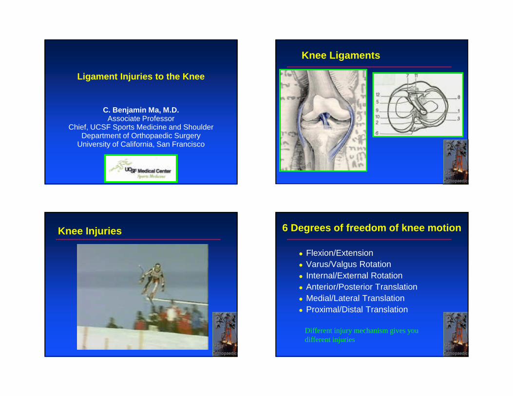
Knee Injuries 6 Degrees of freedom of knee motion
� Flexion/Extension� Varus/Valgus Rotation� Internal/External Rotation� Anterior/Posterior Translation� Medial/Lateral Translation� Proximal/Distal Translation
Different injury mechanism gives you different injuries
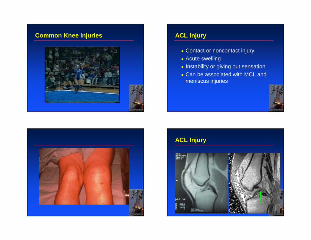
Common Knee Injuries ACL injury
� Contact or noncontact injury� Acute swelling� Instability or giving out sensation� Can be associated with MCL and
meniscus injuries
ACL Injury
� Add nml and inj MRI
ACL tear
� Lachman test� Anterior drawer� Pivot shift test
ACL tear
� Operative� Young active individual� Instability� Protect menisci
� Non-operative� Older individual� No cutting sports� Hamstring strengthening /
reeducation
PCL Injury PCL Injury
� Mechanisms:
� Direct anterior blow to proximal tibia� falling directly on knee/dashboard
injury
� Hyperflexion� fall on flexed knee with foot plantar-
flexed
� Hyperextension� Knee Dislocation (other ligaments
involved)
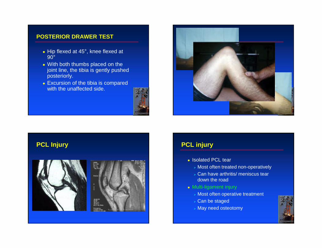
POSTERIOR DRAWER TEST
� Hip flexed at 45°, knee flexed at 90°
� With both thumbs placed on the joint line, the tibia is gently pushed posteriorly.
� Excursion of the tibia is compared with the unaffected side.
PCL Injury PCL injury
� Isolated PCL tear� Most often treated non-operatively� Can have arthritis/ meniscus tear
down the road
� Multi-ligament injury� Most often operative treatment� Can be staged� May need osteotomy
PosteroLateral Corner Injury
� Lateral collateral ligament, popliteustendon, popliteofibular ligament
Mechanisms� Isolated injury rare
� usually injury occurs with PCL or multiligament injury
� Knee hyperextension with varus stress� posterolateral force to knee
� Severe varus stress or ext. rotation of tibia
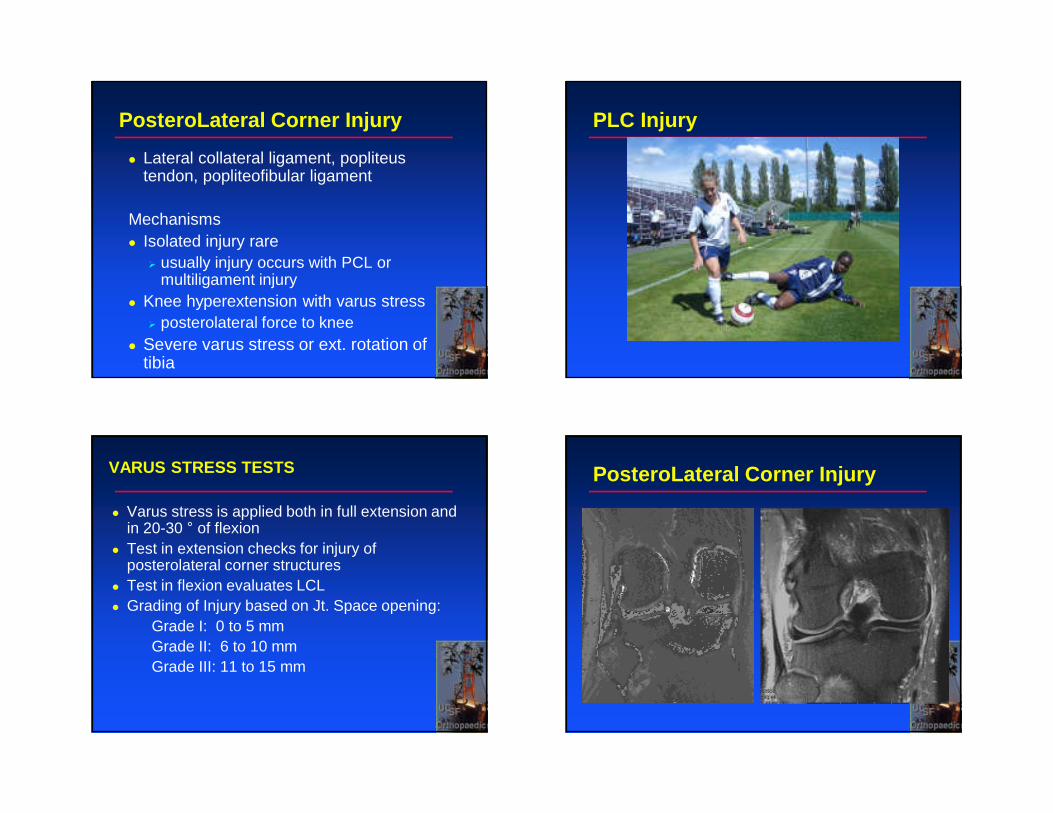
PLC Injury
VARUS STRESS TESTS
� Varus stress is applied both in full extension and in 20-30 ° of flexion
� Test in extension checks for injury of posterolateral corner structures
� Test in flexion evaluates LCL� Grading of Injury based on Jt. Space opening:
Grade I: 0 to 5 mmGrade II: 6 to 10 mmGrade III: 11 to 15 mm
PosteroLateral Corner Injury
Posterolateral Corner Injury
� For acute complete rupture� Want to treat this operatively within
3 weeks of injury
� Repair is better than reconstruction
� Reconstruction for more chronic injuries (>3 weeks) or more servere injury
Don’t want to miss this one!
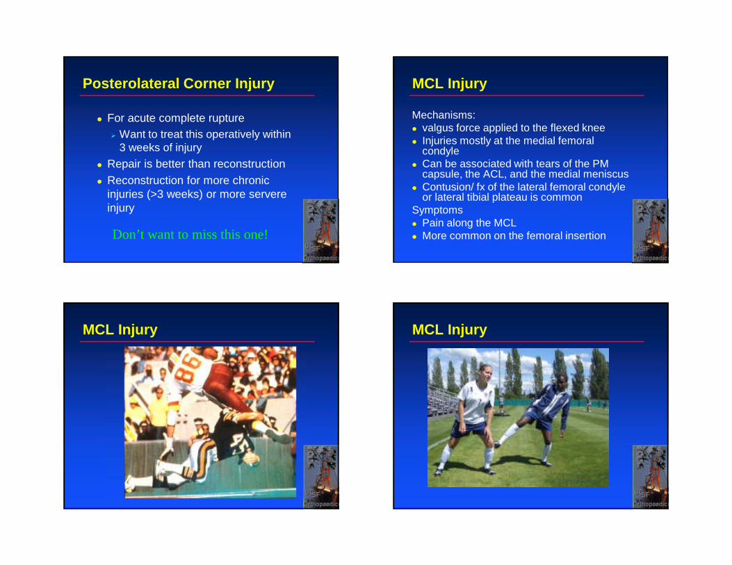
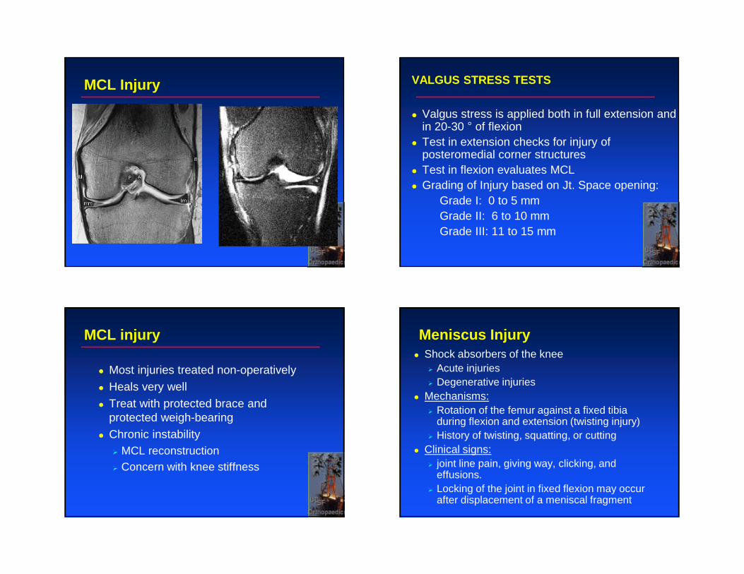
MCL Injury
Mechanisms:� valgus force applied to the flexed knee� Injuries mostly at the medial femoral
condyle� Can be associated with tears of the PM
capsule, the ACL, and the medial meniscus � Contusion/ fx of the lateral femoral condyle
or lateral tibial plateau is common Symptoms� Pain along the MCL � More common on the femoral insertion
MCL Injury MCL Injury
MCL Injury VALGUS STRESS TESTS
� Valgus stress is applied both in full extension and in 20-30 ° of flexion
� Test in extension checks for injury of posteromedial corner structures
� Test in flexion evaluates MCL� Grading of Injury based on Jt. Space opening:
Grade I: 0 to 5 mmGrade II: 6 to 10 mmGrade III: 11 to 15 mm
MCL injury
� Most injuries treated non-operatively
� Heals very well
� Treat with protected brace and protected weigh-bearing
� Chronic instability� MCL reconstruction� Concern with knee stiffness
Meniscus Injury� Shock absorbers of the knee
� Acute injuries� Degenerative injuries
� Mechanisms:� Rotation of the femur against a fixed tibia
during flexion and extension (twisting injury)� History of twisting, squatting, or cutting
� Clinical signs:� joint line pain, giving way, clicking, and
effusions. � Locking of the joint in fixed flexion may occur
after displacement of a meniscal fragment
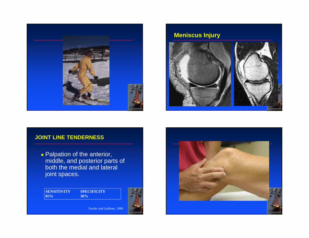
Meniscus Injury
JOINT LINE TENDERNESS
� Palpation of the anterior, middle, and posterior parts of both the medial and lateral joint spaces.
SENSITIVITY SPECIFICITY85% 30%
Fowler and Lubliner, 1989
MCMURRAY’S TEST
� Knee is flexed and placed in external rotation� Examiner applies a valgus or varus force� Knee is then extended. � (+) = Pain and/or a popping/ snapping sensation.
SENSITIVITY SPECIFICITY29% 96%
Fowler and Lubliner, 1989
MCMURRAY’S TEST
McMurray TP: The Semilunar Cartilages.
Br J Surg 29: 407-414, 1942
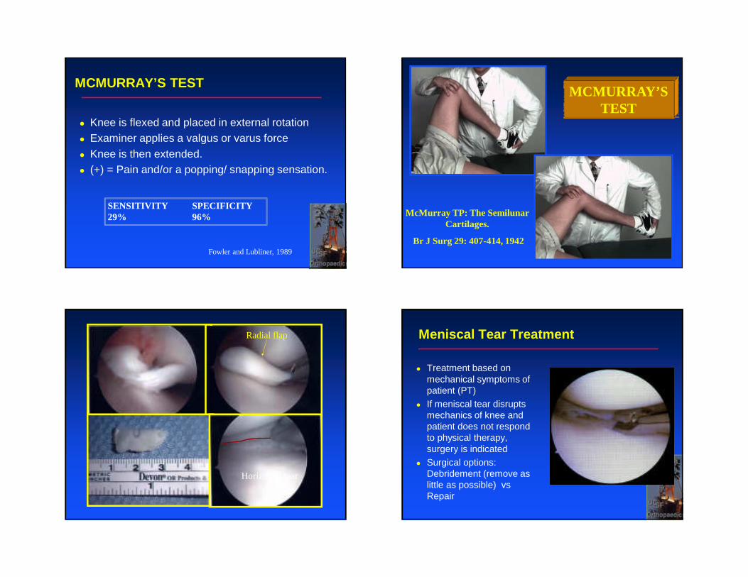
Radial flap
Horizontal tear
Meniscal Tear Treatment
� Treatment based on mechanical symptoms of patient (PT)
� If meniscal tear disrupts mechanics of knee and patient does not respond to physical therapy, surgery is indicated
� Surgical options: Debridement (remove as little as possible) vsRepair
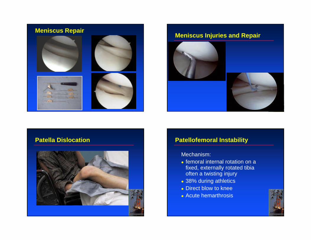
Meniscus RepairMeniscus Injuries and Repair
Patella Dislocation Patellofemoral Instability
Mechanism:� femoral internal rotation on a
fixed, externally rotated tibia often a twisting injury� 38% during athletics� Direct blow to knee� Acute hemarthrosis
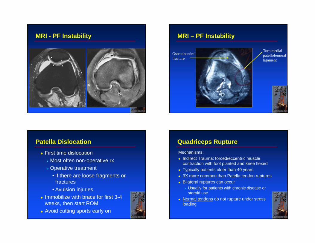
MRI - PF Instability MRI – PF Instability
Torn medialpatellofemoral ligament
Osteochondralfracture
Patella Dislocation
� First time dislocation
� Most often non-operative rx
� Operative treatment• If there are loose fragments or
fractures • Avulsion injuries
� Immobilize with brace for first 3-4 weeks, then start ROM
� Avoid cutting sports early on
Quadriceps Rupture
Mechanisms:� Indirect Trauma: forced/eccentric muscle
contraction with foot planted and knee flexed� Typically patients older than 40 years� 3X more common than Patella tendon ruptures� Bilateral ruptures can occur
� Usually for patients with chronic disease or steroid use
� Normal tendons do not rupture under stress loading
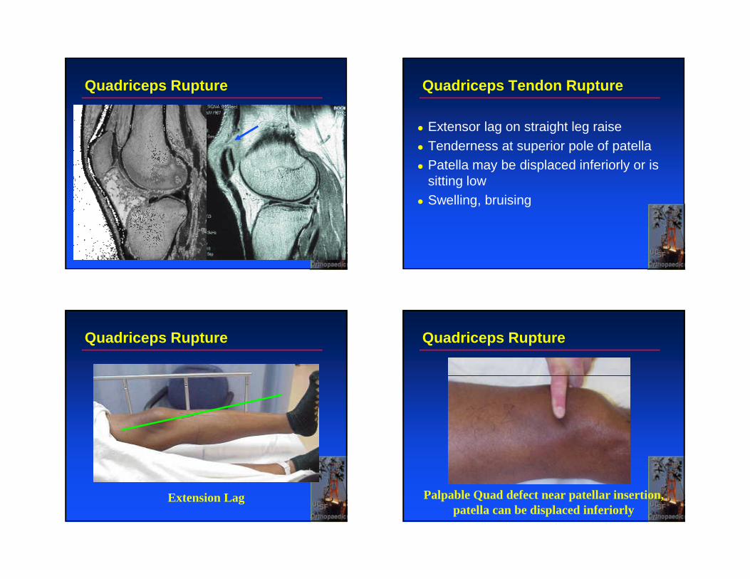
Quadriceps Rupture Quadriceps Tendon Rupture
� Extensor lag on straight leg raise� Tenderness at superior pole of patella� Patella may be displaced inferiorly or is
sitting low� Swelling, bruising
Quadriceps Rupture
Extension Lag
Quadriceps Rupture
Palpable Quad defect near patellar insertion, patella can be displaced inferiorly
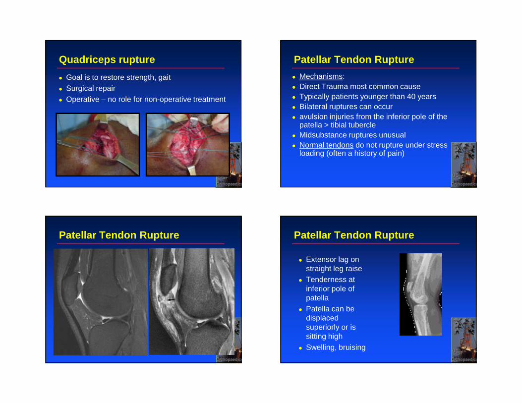
Quadriceps rupture
� Goal is to restore strength, gait� Surgical repair� Operative – no role for non-operative treatment
Patellar Tendon Rupture
� Mechanisms:� Direct Trauma most common cause� Typically patients younger than 40 years� Bilateral ruptures can occur� avulsion injuries from the inferior pole of the
patella > tibial tubercle� Midsubstance ruptures unusual � Normal tendons do not rupture under stress
loading (often a history of pain)
Patellar Tendon Rupture Patellar Tendon Rupture
� Extensor lag on straight leg raise
� Tenderness at inferior pole of patella
� Patella can be displaced superiorly or is sitting high
� Swelling, bruising
Patella Tendon Rupture
� No role of non-operative treatment� Acute loss of extensor function
� Operative intervention
� Brace for 8-10 weeks� Rehabilitation
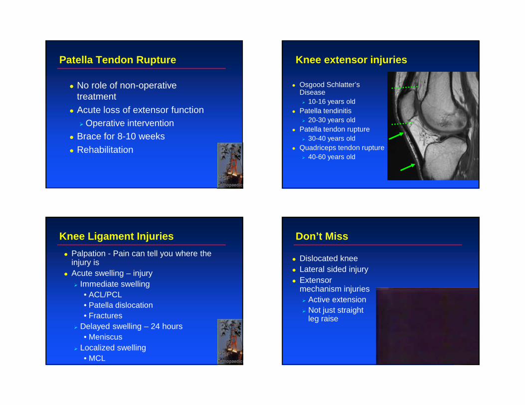
Knee extensor injuries
� Osgood Schlatter’s Disease� 10-16 years old
� Patella tendinitis� 20-30 years old
� Patella tendon rupture� 30-40 years old
� Quadriceps tendon rupture� 40-60 years old
Knee Ligament Injuries
� Palpation - Pain can tell you where the injury is
� Acute swelling – injury� Immediate swelling
• ACL/PCL• Patella dislocation• Fractures
� Delayed swelling – 24 hours• Meniscus
� Localized swelling• MCL
Don’t Miss
� Dislocated knee� Lateral sided injury� Extensor
mechanism injuries� Active extension� Not just straight
leg raise
C. Benjamin [email protected]
Top Related