


Languages
Pages
Legal


ISOLATION AND SCREENING OF ENDOSULFAN-
DEGRADING FUNGI FROM SOIL
Jericho Victor Lagasca Mercado, Josephine Borja*, and Susan Gallardo
Chemical Engineering Department, Gokongwei College of Engineering, De La Salle University-Manila
Manila, Philippines, Tel: (632) 536 0257, *e-mail: [email protected]
Received Date: September 10, 2012
Abstract
Fungal colonies capable of tolerating and degrading endosulfan were enriched and isolated from soil with
history of endosulfan application. The microorganisms were enriched using technical-grade endosulfan
with gradual increase in concentration from 10 to 100 mgl-1
, each enrichment run lasting for 210 h. Four
(4) fungal isolates, labeled as P701, P702, P601 and P801, were obtained by successive plating of enriched colonies. The isolates were screened based on - and -endosulfan degradation in nutrient media with endosulfan as sole-carbon source (NM-SCS) and as sole-sulfur source (NM-SSS). Results indicated that P601 grown in the NM-SSS exhibited the highest endosulfan degradation after 14 d (93.85% for α-
endosulfan and 90.29% for β-endosulfan). Isolate P601 was identified as Candida tropicalis using the API C AUX yeast identification system.
Keywords: Bioremediation, Endosulfan, Enrichment process, Fungal isolate
Introduction
Endosulfan, a cyclodiene organochlorine pesticide, was the most widely used pesticide
worldwide for the last three decades before it was banned or regulated due to its persistence and
toxicity. Most of the endosulfan produced in the past decades now persists in soils where it was
applied. Studies have proven that endosulfan applied in soils transports to waterways thru soil
run-off [1,2]. This phenomenon endangers not only the aquatic ecosystem but also those that
depend on aquatic life as well.
Several studies have been conducted from which the metabolic processes undergone by
microbial communities in degrading endosulfan were understood. Different microbial isolates,
whether bacteria or fungi were used to degrade endosulfan.
In the 1980s, considerable numbers of bacterial strains which possess the ability to degrade
chloroaromatic compounds were isolated [3]. These are Stenotrophomonas maltophilia and
Rhodococcus erythropolis [4], Bacillus sp. [3,5], Mycobacterium sp. [6], and Klebsiella
pneumoniae KE-1 [7]. Likewise, a number of fungal species were also isolated: Phanerochaete
chrysosporium [8], Trichoderma harzianum [9], Chaetosartorya stromatoides, Aspergillus
terricola, Aspergillus terreus [10], and Aspergillus niger [11]. Most of the endosulfan-degrading
microorganisms were isolated from endosulfan-contaminated soils. But Verma [12] was able to
isolate apparently for the first time an endosulfan-degrading bacterium from the gut microflora
of an Indian earthworm Metaphire posthuma. The microflora was enriched with endosulfan with a
concentration of 80 µg ml-1
. The isolate, a Rhodococcus strain, was able to degrade endosulfan without the formation of endosulfan sulfate. An increase in free chloride ion concentration was
observed in the course of degradation.
Invited Paper
ASEAN Engineering Journal Part B, Vol 1 No 1 (2012), ISSN 2286-7694 p.5

In this study, fungal colonies capable of tolerating and degrading endosulfan were enriched
and isolated from soil with history of endosulfan application.
Experimental
Reagents and Media
Technical-grade endosulfan (73% α-endosulfan and 27% β-endosulfan) was used in this study.
The chemicals and solvents used such as n-heptane, ethyl acetate and n-hexane were of analytical
grade.
Two (2) nutrient media were used in this study, one with endosulfan as sole-carbon source
(NM-SCS) and the other with endosulfan as the sole-sulfur source (NM-SSS). The compositions
of the media adapted from Kumar et al. [4] and Gunam [13] were modified. NMCS is composed
of (in gl-1
): KH2PO4, 2.44; Na2HPO4, 5.77; (NH4)2SO4, 4.94; MgSO4, 0.15 and 10 ml l-1
of a mineral solution. On the other hand, NM-SSS is composed of (in gl
-1): KH2PO4, 2.44;
Na2HPO4, 5.77; (NH4)2PO4, 5.57; glucose, 10 and 10 ml l-1
of a mineral solution. The mineral solution amended to both media is composed of 100 mM MgCl2·6H2O, 50 mM CaCl2, 10 mM
FeCl3, 100 uM CuCl2·2H2O and 500 uM MnCl2·4H2O.
Enrichment of Microorganisms
Soil with previous endosulfan application was obtained from a low land rice field. Four (4)
parallel enrichment runs were conducted with different final endosulfan concentrations as
illustrated in Figure 1.
Figure 1. Endosulfan concentration during enrichment runs
For enrichment stage 1, twenty (20) grams of the soil sample was mixed in 250-ml conical
flasks with 100 ml of sterilized potato dextrose broth containing 10 mgl-1
technical-grade endosulfan and shaken at 160 rpm for 18 h at ambient temperature. Five (5) milliliters of the
supernatant liquid were re-suspended into 100 ml potato dextrose broth containing 10 mgl-1
technical-grade endosulfan and 0.5 gl-1
chloramphenicol for enrichment stage 2. The suspension was shaken at 160 rpm and at ambient temperature for 48 h. The inoculum was re-suspended 3
more times, each flask shaken for 48 h for the last three enrichment stages.
ASEAN Engineering Journal Part B, Vol 1 No 1 (2012), ISSN 2286-7694 p.6

Isolation of Fungi
Eight (8) potato dextrose agar plates with 10 mgl-1
endosulfan and 0.5 gl-1
chloramphenicol were initially inoculated for the isolation procedure. Four (4) plates labeled P101, P102, P103 and P104 were plated with the inocula obtained from the 5th stage of enrichment after 24 h of suspension. The culture grown in the plates were successively re-plated to purify isolates (Figure 2). A total of 29 plates were used for the isolation from which 4 isolates were obtained. The isolates were constantly maintained by streak plating into fresh agar media.
Figure 2. Isolation tree diagram
Screening of Isolates
The inocula for the screening experiments were prepared by aseptically cutting 1 cm x 1 cm agar
blocks per isolate where luxurious growth was observed and placing the agar blocks in flasks
containing 50 ml potato dextrose broth with 50 mgl-1
technical-grade endosulfan and 0.5 gl-1
chloramphenicol.
The cultures were allowed to sporulate for 28 h in an orbital shaker oscillating at 160 rpm
under ambient temperature. Approximately 10 ml of the suspensions were collected from each of
ASEAN Engineering Journal Part B, Vol 1 No 1 (2012), ISSN 2286-7694 p.7

the flasks. The collected samples were centrifuged. The supernatant liquid was discarded and the
residues were washed twice with sterilized distilled and deionized water. The mycelia were re-
suspended again in NM-SCS and NM-SSS. Sixty (60) milligrams of the wet mycelia of each
isolate were suspended in 100 ml of the two media. Technical-grade endosulfan was added to
both media to make a concentration of 50 mgl-1
. The suspensions were shaken at 160 rpm for 14 d at ambient temperature. The shake-flasks were shielded from light to remove the effect of light
on endosulfan degradation and fungal growth.
Gas Chromatographic Analyses
Five (5) milliliters of the culture broth in the screening experiments were collected and the
samples were placed in 15-ml vials to which 20 ul of 250 ppm trans-chlordane were added as
surrogate standard. Anhydrous sodium sulfate and Tween 80 were added to prevent formation of
emulsion and increase the recovery of the analyte. Five (5) milliliters of ethyl acetate/n-heptane
(1:1, v/v) were added to the mixtures. The contents of the vials were mixed in a vortex mixer and
then sonicated for 30 minutes at 30-35°C. The sonicated mixtures were then centrifuged and the
ethyl acetate/n-heptane layers were collected. The solvent was evaporated and the residue was
re-dissolved in 1 ml n-hexane.
The concentrations of -endosulfan, -endosulfan and endosulfan sulfate in the samples
were determined using Gas Chromatography equipped with Electron Capture Detector (Hewlett-
Packard 5890 Dual ECD) with columns RTx-CL Pesticides (30 m, 0.32 mm ID, 0.5 um) and Stx-
CL Pesticides (30 m, 0.32 mm ID, 0.25 um).
Results and Discussion
Fungal Isolates
Four (4) isolates were obtained from the enrichment and isolation processes. Two (2) of the
isolates are molds labeled as isolates P701 and P702. The other 2 isolates are yeasts labeled as
P601 and P801.
Degradation of Endosulfan Isomers
The degradation of -endosulfan was found to be greater than that of -endosulfan for all
isolates and media (Figure 3). Isolate P601 exhibited the highest total endosulfan degradation of
84.56% (10349.70 ugl-1
) in 14 d while isolate P702 exhibited the lowest total endosulfan degradation at 43.18% (2299.52 ugl
-1) in 14 d. On the other hand, the screening flasks with
nutrient medium containing endosulfan as the sole-sulfur source (NM-SSS) showed higher total
endosulfan degradation at 73.40% (6077.23 ugl-1
) in 14 d than the nutrient medium with endosulfan as the sole-carbon source (NM-SCS) at 45.21% (2365.36 ugl
-1) in 14 d.
ASEAN Engineering Journal Part B, Vol 1 No 1 (2012), ISSN 2286-7694 p.8

Figure 3. Main effects plots for endosulfan degradation
The responses were also analyzed in a prediction profiler using JMP (version 8, SAS Institute
Inc.) The desirabilities were set to vary linearly with -endosulfan and -endosulfan degradation.
The profiler gives the optimum setting as P601 for isolate and NM-SSS for medium when the
desirabilities were set to maximum (obtained as 0.95). The optimum responses were given
as 93.85 % for -endosulfan degradation, 90.29 % for -endosulfan degradation and 91.35 %
for total endosulfan degradation. The interaction effects plot for endosulfan degradation
illustrates the interactions between the isolates and the media used (Figure 4). The top right box
shows the variation of total endosulfan degradation with medium using different isolates. It
was observed that isolates P702, P601 and P801 prefers NM-SSS while isolate P701 prefers
NM-SCS. It was also observed that isolate P601 exhibited the highest endosulfan degradation
for both media (the lines for P701, P702 and P801 are below the line for P601).
The box at the lower left of Figure 4 shows the variation of total endosulfan degradation with
isolate at different media. It clearly shows that NM-SSS significantly enhances the degradation
capability of P601 among the rest of the isolates.
ASEAN Engineering Journal Part B, Vol 1 No 1 (2012), ISSN 2286-7694 p.9

Figure 4. Interaction effects plot for endosulfan degradation
Degradation of Endosulfan Sulfate
The variation of endosulfan sulfate degradation with nutrient medium for each isolate shows that
only isolate P601 favors endosulfan sulfate degradation with endosulfan as the sole sulfur source
more than as the sole-carbon source as shown in Figure 5. However, isolate P601 exhibited
minimal variation in endosulfan sulfate degradation with the change in nutrient medium. On the
other hand, isolates P702 and P801 utilizing endosulfan as the sole-sulfur source exhibited net
endosulfan sulfate formation rather than degradation at 49.79% and 62.80% increase in
endosulfan sulfate concentration respectively. The isolates utilizing endosulfan as the sole sulfur
source show wider variation in endosulfan sulfate degradation than that with endosulfan as the
sole-carbon source.
Increase in Biomass
The main effects plots for increase in biomass show that the increase in biomass with respect to
the isolates and medium has a similar profile with endosulfan degradation (Figure 6). Isolate
P601 exhibited the highest increase in biomass at 1956.40 mg l-1
(dry weight) while isolate P801
exhibited the lowest increase in biomass at 83.35 mg l-1
(dry weight). The similarity in the main
ASEAN Engineering Journal Part B, Vol 1 No 1 (2012), ISSN 2286-7694 p.10
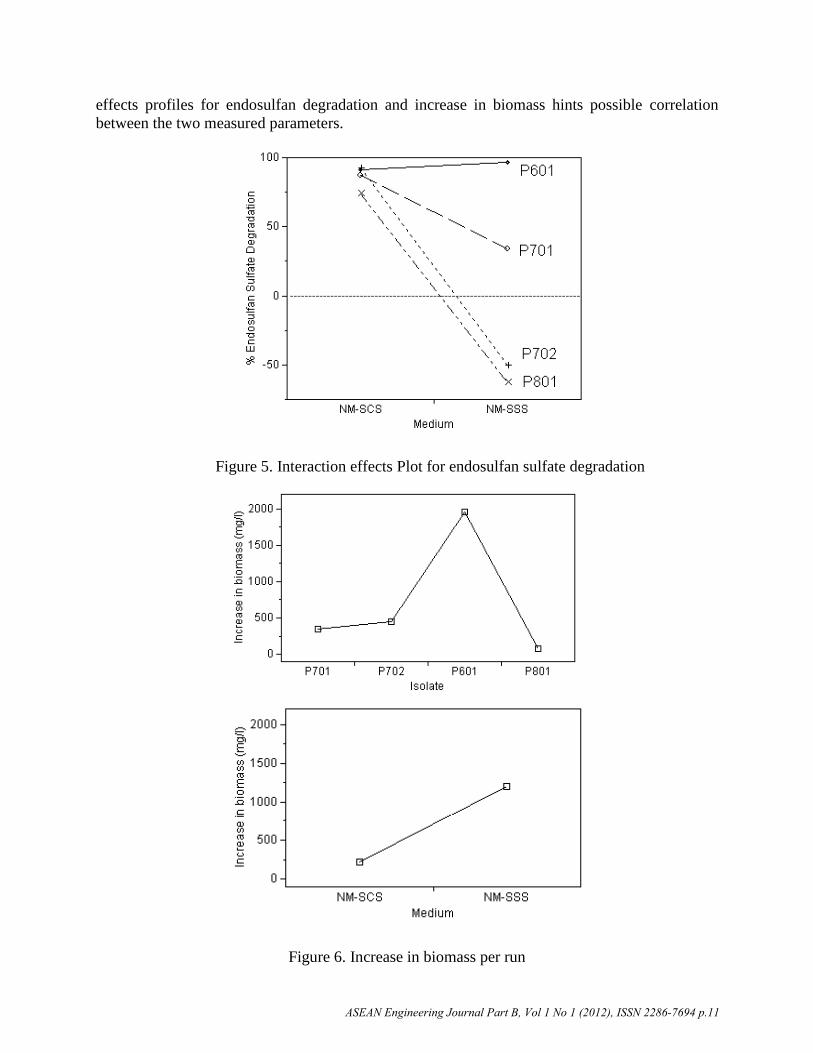
effects profiles for endosulfan degradation and increase in biomass hints possible correlation
between the two measured parameters.
Figure 5. Interaction effects Plot for endosulfan sulfate degradation
Figure 6. Increase in biomass per run
ASEAN Engineering Journal Part B, Vol 1 No 1 (2012), ISSN 2286-7694 p.11

Identification of Yeast Isolate
Isolate P601 was identified as Candida tropicalis using the API C AUX Identification System
(Good identification at 95.8%) by the Philippine National Collection of Microorganisms
(PNCM) of the University of the Philippines National Institute of Molecular Biology and
Biotechnology (U.P. BIOTECH).
Conclusions
This study investigated the biodegradation of - and -endosulfan in soil using a fungal isolate
identified as Candida tropicalis. Indigenous microorganisms present in a soil sample with
history of endosulfan application were enriched using Potato Dextrose broth amended with
technical-grade endosulfan with concentrations gradually increased from 10 to 100 mgl-1
. Luxurious growth was observed on all flasks after 210 h of enrichment. The colonies from the
flask with the highest concentration of endosulfan (100 mgl-1
) were inoculated into plates for fungal isolation. Four (4) fungal isolates (2 mold species, labeled as P701 and P702, and 2 yeast
species labeled as P601 and P801) were obtained after a total of 29 plates were used for isolation
and purification.
The isolates and the nutrient medium whether endosulfan is the sole-carbon source or the
sole-sulfur source was screened based on - and -endosulfan degradation. Isolate P601 showed
the highest endosulfan degradation at 84.56% total endosulfan degradation along with the
nutrient medium with endosulfan as the sole-sulfur source (NM-SSS) at 73.40% total endosulfan
degradation. Combination of the two yields 91.35% total endosulfan degradation (93.85% for
alpha-endosulfan and 90.29% for beta-endosulfan). The results obtained from this study may be
applied to future soil biodegradation studies.
Acknowledgement
The authors are grateful for the financial support given by the Engineering Research and
Development for Technology (ERDT) through the Department of Science and Technology–
Science Education Institute (DOST-SEI), Philippines.
References
[1] S.M. Peterson, and G.E. Batley, “The fate of endosulfan in aquatic ecosystems,”Environmental Pollution, Vol. 82, No. 2, pp. 143-152, 1993.
[2] I.O.D. El Beit, D.E. Cotton, and J.V. Wheelock, “Factors involved in the dynamics of
pesticides in soils: The effect of pesticide concentration on leachability and adsorption,”International Journal of Environmental Studies, Vol. 16, No. 3-4, pp. 181-187, 1981.
[3] H.M. Shivaramaiah, and I.R. Kennedy, “Biodegradation of endosulfan by a soil Bacterium,”Journal of Environmental Science and Health Part B, Vol. 41, No. 6, pp. 895-905, 2006.
[4] K. Kumar, S.S. Devi, K. Krishnamurthi, G.S. Kanade, and T. Chakrabarti, “Enrichment and
isolation of endosulfan degrading and detoxifying bacteria,” Chemosphere, Vol. 68, No. 2,
pp. 317-322, 2007.
[5] N. Awasthi, N. Manickam, and A. Kumar, “Biodegradation of a bacterial coculture,”Bulletin of Environmental Contamination and Toxicology, Vol. 59, No. 11, pp. 928-934,
1997.
ASEAN Engineering Journal Part B, Vol 1 No 1 (2012), ISSN 2286-7694 p.12
[6] T.D. Sutherland, I. Horne, R.L. Harcourt, R.J. Russell, and J.G. Oakeshott, “Isolation and
characterization of a Mycobacterium strain that metabolizes the insecticide endosulfan,”Journal of Applied Microbiology, Vol. 93, No. 3, pp. 380-389, 2002.
[7] G.S. Kwon, J.E. Kim, T.K. Kim, Y.H. Sohn, S.C. Koh, K.S. Shin, and D.G. Kim,
“Klebsiella pneumoniae KE-1 degrades endosulfan without formation of the toxic metabolite,
endosulfan sulfate,” FEMS Microbiology Letters, Vol. 215, No. 2, pp. 255-259, 2002.
[8] S.W. Kullman, and F. Matsumura, “Metabolic pathways utilized by Phanerochaete
chrysosporium for degradation of the cyclodiene pesticide endosulfan,” Applied
and Environmental Microbiology, Vol. 62, No. 2, pp. 593-600, 1996.
[9] A. Katayama, and F. Matsumura, “Degradation of organochlorine pesticides, particularly
endosulfan, by Trichoderma harzianum,” Environmental Toxicology and Chemistry, Vol.
12, No. 6, pp. 1059-1065, 1993.
[10] S. Hussain, M. Arshad, M. Saleem, and Z.A. Zahir, “Screening of soil fungi for in
vitro degradation of endosulfan,” World Journal of Microbiology and Biotechnology, Vol.
23, No. 7, pp. 939-945, 2007.
[11] T. S. Bhalerao, and P. R. Puranik, “Biodegradation of organochlorine pesticide,
endosulfan, by a fungal soil isolate, Aspergillus niger,” International Biodeterioration &Biodegradation, Vol. 59, No. 4, pp. 315-321, 2007.
[12] K. Verma, N. Agrawal, M. Farooq, R.B. Misra, and R.K. Hans, “Endosulfan
degradation by a Rhodococcus strain isolated from earthworm gut,” Ecotoxicology and
Environmental Safety, Vol. 64, No. 3, pp. 377-381, 2006 .
[13] I.B.W. Gunam, Y. Yaku, M. Hirano, K. Yamamura, F. Tomita, T. Sone, and K.
Asano, “Biodesulfurization of alkylated forms of dibenzothiophene and benzothiophene
by Sphingomonas subarctica T7b,” Journal of Bioscience and Bioengineering, Vol. 101,
No. 4, pp. 322-327, 2006.
ASEAN Engineering Journal Part B, Vol 1 No 1 (2012), ISSN 2286-7694 p.13
Top Related