


Languages
Pages
Legal


Leioa, 2018ko Maiatzaren 16a / Leioa, 16 de Mayo de 2018
Gradu Amaierako Lana / Trabajo Fin de Grado
Medikuntzako Gradua / Grado en Medicina
IS THERE AN IDEAL TREATMENT FOR THE
ACUTE RENAL COLIC?
Egilea/Autor:
ANA CRUZ ALONSO DE ARMIÑO RIAÑO
Zuzendaria/Director/a:
ANTONIO ARRUZA ECHEVARRIA
GRADU AMAIERAKO LANA / TRABAJO FIN DE GRADO
© ANA CRUZ ALONSO DE ARMIÑO RIAÑO

INDEX
Pages 1-9 INTRODUCTION
Page 10 HYPOTHESIS AND OBJECTIVES
Page 11-12 MATERIALS AND METHODS
Pages 13-18 RESULTS
Pages 19-22 DISCUSSION
Pages 13-25 CONCLUSIONS
Page 26 RECOMMENDATIONS
Pages 27-33 BIBLIOGRAPHY

1
1. INTRODUCTION
Urolithiasis and renal colics are very common around the world. In fact, we´ve often
seen this suffering among family members, friends or close acquaintances and all of
them used similar words to describe its pain: a severe, unbearable or excruciating
pain in the flank. Every time I was consulted, I found myself unable to specify which
medical treatment stands out from the rest at fighting against the acute renal colic.
So honestly, a bit of ignorance and a lot of curiosity about fairly demonstrated data
on the treatment of the acute renal colic is what encouraged me to research on this
subject for my end-of-career project. Through the shaping of the project, other
important matters came along, such as the economic impact of this illness or the
standard procedures for its diagnosis. In the following lines, I´ll present what I
consider the highlights about the renal colic, in other words, what I consider
everyone must have in mind, fresh-baked, before diving further into this project.
1.1. DEFINITION & PHYSIOPATHOLOGY:
Renal calculi consist of crystal aggregates that deposit in the collecting ducts.
Urinary stones arise because of the breakdown of a delicate balance between
two opposing conditions: solubility and precipitation of salts, as kidneys must
conserve water, but must excrete materials that have low solubility (1, 2).
Additionally, urine contains substances such as citrate, pyrophosphate and
glycoproteins that act as a protective mechanism, inhibiting crystallization.
However, when urine becomes supersaturated with insoluble materials,
because excretion rate is excessive and/or because water conservation is
extreme, crystals form and may grow and aggregate to form a stone (2).
So, nephrolithiasis is a disease that presents an increased urinary concentration
of stone-forming salts and urine volume is a major determinant of the
concentration of this lithogenic factors. Fluid intake is the main determinant of
urine volume and therefore, as several observational studies (3-5) and a
randomized controlled trial (6) have demonstrated, higher fluid intake reduces
the risk of stone formation. Also, a prospective trial affirms that increased
water intake also prolongs the average interval between recurrences (7).

2
1.2. PREVALENCE:
Kidney stone disease is a common malady, affecting nearly 1 in 11 individuals
in the US at some point in their lives and there is evidence that the number of
those who have had a stone is rising (8), with at least 50% of individuals
experiencing another stone within 10 years of the first occurrence (9).
Historically, kidney stones have occurred more commonly in men than in
women, although the gender gap in stone disease is closing (10-12).
The reasons for the observed rise in stone disease among women are not
certain, but the impact of obesity, a known risk factor for kidney stones, was
found to be greater in women than in men (13). In fact, obesity is an
independent risk factor for urinary calculi, particularly in women (14), reason
why weight loss is desirable in these patients. Additionally, the beneficial
effect of dietary moderation in reducing the risk of recurrent stones was
demonstrated by Hoskings and co-workers, who found a reduction in stone
recurrence rate among 108 idiopathic calcium oxalate stone formers who were
encouraged to maintain a high fluid intake and avoid “dietary excess” (15).
1.3. ECONOMIC IMPACT:
Symptomatic urolithiasis manifests robustly in the practice of the Emergency
Department (ED) physician, with a significant economic impact that echoes
beyond the ED visit. It has been reported that over 8´8% of the United States
population will be affected by this malady, and when treatment costs, sick days
from work and third-party payments are considered, costs around $2.1 billion
per year to the US economy alone (8, 11, 16, 17). Sadly, no article was found
on the economic impact of the renal colic in this country, not even in the Intern
resident book from the Spanish Association of Urology (18).
1.4. SYMPTOMATOLOGY:
Renal colic from an obstructing calculus presents classically with sudden-
onset, severe and sharp pain localized to the flank, which increases over the
following 20-60 minutes, with radiation to the lower abdomen, groin or
genitals. It is often accompanied by nausea and vomiting. Urinary symptoms,

3
most commonly frequency and urgency with low voided volumes, are common
in distal ureteral stones (19). Other possible symptoms are pain on micturition,
strangury and/or interruption of urine flow (1, 2). However, not all patients
presenting with flank pain have urinary calculi, so an important aspect of the
initial evaluation is to search for other potential diagnoses (Table 1) (20).
Table 1. Differential Diagnosis for Urinary Calculi.
CLINICAL CLUES SUGGESTED DIAGNOSIS
Anorexia, nausea, vomiting Obstructing urinary calculi, bowel disease
Dysuria UTI, urinary calculi, interstitial cystitis
Fever, chills Viral o bacterial illness
Hematuria (microscopic or gross) Urinary calculi, urothelial tumor, UTI, BPH, renal mass
Hemodynamic instability Nonspecific findings of shock (including possible sepsis)
Inability to get comfortable Urinary calculi, peritonitis
Pain and tenderness
Abdominal pain
Flank pain (sharp, extreme pain with sudden onset)
Flank tenderness
Groin pain (scrotal, labial)
Penile or pelvic pain
Suprapubic tenderness Tachycardia Urinary frequency
Small renal calculi, nonurologic etiology (GI origin) Urinary calculi, musculoskeletal spasm Urinary calculi, musculoskeletal inflammation, pyelonephritis Ureteral calculi, hernia, testicular mass Ureteral calculi, urethritis, prostatitis UTI, interstitial cystitis, prostatitis, urinary calculi, peritonitis Nonspecific response to pain UTI, ureteral calculi, BPH
UTI = Urinary Tract Infection; BPH = Benign Prostatic Hyperplasia.
1.5. DIAGNOSIS:
A typical work-up includes a thorough history and physical examination,
serum chemistry and complete blood count, urinalysis, and an imaging study.
Typical laboratory findings are presented in Table 2 (20).

4
Table 2. Clinical Clues to the Diagnosis of Urinary Calculi.
EVALUATION POSSIBLE FINDINGS
Laboratory evaluations
Complete blood cell count Leukocytosis with struvite calculi
Serum chemistry Elevation in creatinine levels with obstructing calculi; hypokalemia and hyperchloremia with Renal Tubular Acidosis; elevated serum calcium levels with parathyroid disease
Serum parathyroid hormone levels
Elevated in hyperparathyroidism
Urinalysis Microscopic or gross hematuria; acidic urine; alkaline urine (with struvite calculi); pyuria; crystals from involved calculi
24-hour analysis Elevated urinary calcium, oxalate, and sodium levels; decreased urinary volume and citrate levels
Most patients have remediable metabolic disorders that cause stones, so the
composition of kidney stones should be determined when possible, because
treatment depends on stone type (Table 3) (1). Stone composition of uric acid,
cystine or struvite implicates specific metabolic or genetic abnormalities and
knowledge of stone composition may also help direct preventive measures (21,
22). Regarding the metabolic testing, there are conflicting opinions in the
literature about the adequacy of a single 24-hour urine in reliably identifying
urinary abnormalities (23-27). In the absence of clear consensus, either one or
two 24-hour urines may be obtained, although two collections are preferred.
Table 3. Major Causes of Renal Stones.
STONE TYPE PERCENT of all stonesª
PERCENT occurrence of specific causesª
CALCIUM STONES: 75-85% Idiopathic calciuria _______________________ Hypocitraturia ___________________________ Dietary hyperoxaluria _____________________ Hyperuricosuria __________________________ Idiopathic stone disease ___________________
50-55% 20-40% 10-30% 20% 20%

5
a Values are percentages of patients who form a particular type of stone and who display each specific
cause of stones.
Regarding imaging studies, non-contrast computed tomography (CT) has
emerged as the most sensitive and specific modality for detecting ureteral
calculi. Consequently, CT is frequently used in the initial diagnosis of ureteral
calculous disease (28) and in the follow-up of known ureteral calculi before
and after treatment. Additionally, conventional radiography and ultrasound are
endorsed for monitoring the passage of most radiopaque stones (29).
Ultrasound is has a relatively low sensitivity, although it is often used as the
initial imaging test in pregnant patients with flank pain (30). Typical
radiographic findings are presented in Table 4 (20).
Table 4. Clinical Clues to the Diagnosis of Urinary Calculi.
EVALUATION POSSIBLE FINDINGS
Radiographic evaluations
Abdominal, kidney and upper bladder radiography
Urinary calculi larger than 2 mm may be visible
CT (stone protocol) Nearly all calculi are visible on CT. Evaluates renal parenchyma, hydronephrotic changes and surrounding organs
Intravenous pyelography Calculi visible on scout film. Delay in contrast excretion if obstruction is present. Calculi may appear as filling defect
MRI Conventional MRI is not useful for imaging calculi
Ultrasonorography Calculi appear as hyperechoic lesions that cast acoustic shadows. Not reliable for ureteral calculi. May demonstrate dilation of collecting system
CT = computed tomography; MRI = magnetic resonance imaging.
URIC ACID STONES: 5-15% Metabolic syndrome ______________________ Gout ___________________________________ Idiopathic _______________________________
30% 30% 30%
STRUVITE STONES: 5%
CYSTINE STONES: 1%

6
1.6. TREATMENT:
All patients should be counseled to avoid dehydration and drink copious
amounts of water (1). An important study confirmed that increasing urine
volume to 2.5 L per day resulted in a 50% reduction of stone recurrence
compared with the control group (6).
Firstly, stones not causing obstruction may be managed conservatively. We
should provide immediate pain relief, offering a non-steroidal anti-
inflammatory drug (NSAID) as the first drug of choice (30). NSAIDs decrease
the production of arachidonic acid metabolites, mediators of the pain response
released by the stretch of the renal capsule due to downstream obstruction. In
addition, they cause contraction of the afferent arterioles to the glomerulus,
thereby decreasing hydrostatic pressure by reducing glomerular filtration rate.
They are safer than opioids, which could cause depression of level of
consciousness and respiratory drive. However, caution should be exercised in
the elderly and those with renal impairment (19).
Literature suggests that a combination approach with NSAIDs and opioids may
be the most effective method to manage renal colic in the ED (31). This
combination has been studied in numerous comparisons. A systematic review
recommends parenteral NSAIDs be used as first-line therapy for patients with
renal colic, with the use of narcotics as adjuvant or breakthrough analgesia
(32). Paracetamol has also proved to be an effective analgesic for acute colic.
A recent trial showed no significant difference between degree of pain control
and time to relief between iv paracetamol and iv morphine, with fewer adverse
effects in the paracetamol group (33).
Moreover, Medical Expulsive Therapy (MET) may be used if needed, as it
increases luminal diameter and inhibits smooth muscle tone in the ureter and
ureterovesical junction. These effects have been accomplished with the use of
steroids and NSAIDs to limit inflammation-induced narrowing of the ureteral
caliber. This combination has commonly been used in concert with agents
intended to relax ureteral smooth muscle to decrease painful spasm and to
potentially dilate the upper urinary tract to decrease pain (19).

7
The presence of adrenergic receptors in the human ureter (with increasing
density in the distal ureter), the preponderance of the α-1 subtype and the
ability of α-blocking medication to decrease ureteral contractility are well
established (34-36). Calcium channel blockers (CCB) have also shown relaxing
effects on ureteral smooth muscle (37).
Multiple studies suggest that these agents augment the stone expulsion rate
when compared with standard therapy (38-49), although taking a closer look at
the reliability of this studies, the efficacy of tamsulosin and MET in improving
the spontaneous passage of kidney stones is unclear. However, given that this
therapy is generally well tolerated and may improve the rate of expulsion of
kidney stones, a trial of tamsulosin or nifedipine may be considered at
discharge. Based on the literature, α1-adrenergic blockers are preferred to
CCB, due to shorter time to stone passage and fewer adverse effects (31).
Furthermore, if the stone passes into the ureter and does cause obstruction, it
becomes a complicated renal colic which can reduce both glomerular filtration
rate and renal blood flow. The indications for urgent intervention in urinary
tract obstruction are: presence of infection or urosepsis, intractable pain and/or
vomiting, impending acute renal failure, obstruction with a solitary or
transplanted kidney and bilateral obstructing stones (50).
At the Emergency Department of Cruces Universitary Hospital, urine
derivation is practiced when urgent intervention is required, mostly with a
single J (single end single loop) or a double J (both end single loop) catheter. If
insufficient, a nephrostomy is carried out, with or without the infusion of an
alkalizing treatment such as sodium bicarbonate or alkaline citrate. A
randomized controlled trial found that ureteral catheters, ureteral stents, and
percutaneous nephrostomy tubes are equally effective for decompressing the
urinary tract (51). Lastly, flexible or rigid ureterorenoscopy plus intracorporeal
lithotripsy can be used as first or second line treatment and once or more.
Secondly, the interventional approach of nephrolithiasis usually takes place as
second-line treatment. Therefore, I´ll simply mention it, as the bottom-line of
this project is the acute treatment for the renal colic. Truly, advances in

8
urologic technology have rendered open surgery for stones a rare event, leaving
three alternatives: extracorporeal shockwave lithotripsy (SWL), percutaneous
nephrolithotomy (PNL) and ureterorenoscopy (URS) (Figures 1 and 2). In
general terms, the main contraindications for SWL and PNL are pregnancy,
untreated urinary tract infections, bleeding diatheses, anticoagulant therapy,
severe obesity or skeletal malformations (only SWL)... On the other hand, URS
has no specific contraindications, apart from those related to the general
anesthesia and the untreated urinary tract infections (30).
Figure 1. Treatment algorithm for renal calculi (30).
*The term "Endourology" encompasses all PNL and URS interventions.
PNL=percutaneous nephrolithotomy; RIRS=retrograde renal surgery; SWL=shockwave lithotripsy;
URS=ureterorenoscopy.

9
Figure 2. Treatment algorithm for ureteral calculi (GR: A*) (30).
SWL=shockwave lithotripsy; URS=ureterorenoscopy.
As you read through the introduction, you can notice the wide range of
possibilities available for the treatment of the kidney stones. In fact, while
working on this research, I found myself diving in a vast ocean of information
about how renal colic´s acute pain and kidney stones in general should be
treated. As a result, I decided to search for information available and compared
the effectiveness of two of the main approaches of this illness: the European
protocols and the protocols followed in the USA. Finally, I´ll be focusing
particularly on analyzing the protocol that we follow at Cruces University
Hospital, hoping to reach some conclusions regarding its high, intermediate, or
low effectiveness in the acute kidney stone treatment.

10
2. HYPOTHESIS / OBJECTIVES
To frame and give shape to the end-of-career project is essential to define its
objectives. The following are the hypothesis that´ll be the bottom line of this project:
2.1 Searching for significant similarities and/or differences in the protocols stated
by Europe and United States of America on the treatment of urolithiasis. For
this, I´ll be comparing the official European Urology Association (EAU)
guidelines on urolithiasis and the official American Urological Association
(AUA) guidelines on medical and surgical management of kidney stones.
2.2 Searching for valid, significant and current comparative studies between the
effectiveness of either the diagnosis and the medical or surgical treatment for
nephrolithiasis in Europe and USA. For this, I´ll be using globally recognized
databases on health sciences.
2.3 Analyzing the protocol that we follow at Cruces University Hospital for the
diagnosis and treatment of acute kidney stone in the emergency room, and
looking for similarities or differences with the European and the American
guidelines on urolithiasis or other reliable sources of information.
2.4 Reaching some conclusions regarding the most effective treatment for the renal
colic in the emergency room, built upon high level of evidence data if possible.

11
3. MATERIAL AND METHODS
This project is a Literature Review for which the main source of information was
Cruces University Hospital´s online library service for Health sciences. This library
allows access to a wide range of worldwide recognized databases such as Ovid,
Clinical Access, Cochrane, Fisterra, UpToDate, Clinical Key, Micromedex, etc.
Precisely, this investigation has been constructed through a search in Ovid database,
which contains publications on JBI/Joanna Briggs Institute, EMBASE, MEDLINE,
DIF/Drug Information Full text, IPA/International Pharmaceutical Abstracts,
PsycINFO and LWW books and journals. Also, with articles from PubMed and
information obtained from Harrison´s principles of internal medicine and Oxford
handbook of clinical medicine. The online information was searched using
combinations of the following key words:
3.1 „acute‟ +„renal‟ + „colic‟ + „treatment‟
3.2 „renal‟ + „colic‟ + „emergency‟+ „protocols‟
3.3 „urolithiasis‟ + „emergency‟ + „department‟
3.4 „nephrolithiasis‟ + „emergency‟ + „department‟
3.5 „effectiveness‟ + „protocols‟ + „calculus‟
3.6 „management‟ + „nephrolithiasis‟ + „emergency‟ + „department‟
3.7 „renal‟ + „colic‟ + „medical‟+ „expulsive‟+ „therapy‟
3.8 „kidney‟ + „stones‟ + „medical‟ + „expulsive‟ + „therapy‟
3.9 „urgent‟ + „decompression‟ + „ureteral‟ +„calculi‟
3.10 „water‟ + „urinary‟ + „volume‟ + „nephrolithiasis‟
3.11 „urgent‟ + „decompression‟ + „ureteral‟ + „calculi‟
The studies included were published from 1972 to 2017 and recent articles were
given highest priority because they represent the current state-of-the-art treatment.
Meanwhile, older studies were included selectively if historically relevant or if they
addressed issues more adequately than the more recent literature. The paper search
was limited to the English language, although a reference from a Spanish source can
also be found. The European Association of Urology (EAU) and American
Urological Association (AUA) guidelines were used in order to assemble appropriate
evidence-based reference literature.

12
The topics of these guidelines were selected based on the level of evidence A or B, as
described by the Oxford Centre for Evidence-Based Medicine Levels. In this source,
when sufficient evidence exists, the body of evidence for a particular clinical action
is assigned a strength rating of A (high), B (moderate) or C (low). Additionally, the
AUA nomenclature system links strong, moderate or conditional recommendation to
the A, B or C levels of evidence. Also, in the absence of sufficient evidence they
provide additional information as Clinical Principles and Expert Opinions.

13
4. RESULTS
The main results of this bibliographic review are obtained from two of the chief
sources of information used: the guidelines on urolithiasis from the EAU and the
AUA. As we mentioned before, we´ll extract only the principles that are supported
by A or B levels of evidence. These principles have been arranged in two columns,
one for each source, so that we can easily compare the principles that describe same
topic. Some data with lower level of evidence has also been included (grade C,
Clinical Principles -CP- and Expert Opinions -EO), only because its comparison with
the equivalent topic from the other source is considered relevant. For the reader´s
convenience, this low evidence data is marked with an orange background.
4.1 DIAGNOSIS: Imaging.
European Association of Urology (EAU) (30)
GR American Urological Association (AUA) (52)
GR
Following initial ultrasound assessment, use
non-contrast-enhanced CT to confirm stone
diagnosis in patients with acute flank pain, as
it is superior to intravenous urography.
A Non-contrast CT (NCCT) is the preferred
initial imaging study for the adult patient..
The diagnostic accuracy of non-contrast CT
in identifying ureteral calculi is the following:
sensitivity 98% and specificity 97%.
A
In children, use ultrasound as first-line
imaging modality when a stone is suspected.
Perform a KUB X-ray (or low-dose non-
contrast-enhanced CT) as an alternative.
B Renal ultrasonorography is the preferred
initial imaging study for pediatric patients
(<14).
C
Use ultrasound as the preferred method of
imaging in pregnant women.
A Renal ultrasonorography is the preferred
initial imaging study for pregnant patients.
C
4.2 DIAGNOSIS: Additional testing.
European Association of Urology (EAU) (30)
GR American Urological Association (AUA) (53)
GR
Perform stone analysis in first-time formers
using a validated procedure.
A When a stone is available, clinicians should
obtain a stone analysis at least once.
CP
Repeat stone analysis in patients with
recurrent stones despite drug therapy, early
recurrence after stone clearance or late
recurrence after a long stone-free period.
B Clinicians should repeat a stone analysis,
when available, especially in patients not
responding to treatment.
EO
Only high-risk stone formers (all children,
recurrent stone formers, transplanted
kidneys...) require specific metabolic
evaluation after stone removal.
A Clinicians should perform additional
metabolic testing in high-risk or interested
first-time stone formers and recurrent stone
formers.
B
14
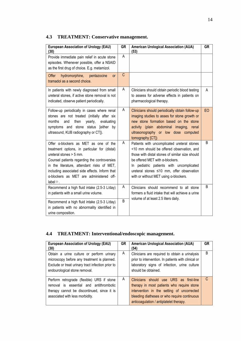
4.3 TREATMENT: Conservative management.
European Association of Urology (EAU) (30)
GR American Urological Association (AUA) (53)
GR
Provide immediate pain relief in acute stone
episodes. Whenever possible, offer a NSAID
as the first drug of choice. E.g. metamizol.
A
Offer hydromorphine, pentazocine or
tramadol as a second choice.
C
In patients with newly diagnosed from small
ureteral stones, if active stone removal is not
indicated, observe patient periodically.
A Clinicians should obtain periodic blood testing
to assess for adverse effects in patients on
pharmacological therapy.
A
Follow-up periodically in cases where renal
stones are not treated (initially after six
months and then yearly, evaluating
symptoms and stone status [either by
ultrasound, KUB radiography or CT]).
A Clinicians should periodically obtain follow-up
imaging studies to asses for stone growth or
new stone formation based on the stone
activity (plain abdominal imaging, renal
ultrasonography or low dose computed
tomography [CT])
EO
Offer α-blockers as MET as one of the
treatment options, in particular for (distal)
ureteral stones > 5 mm.
Counsel patients regarding the controversies
in the literature, attendant risks of MET,
including associated side effects. Inform that
α-blockers as MET are administered off-
label.
A Patients with uncomplicated ureteral stones
<10 mm should be offered observation, and
those with distal stones of similar size should
be offered MET with α-blockers.
In pediatric patients with uncomplicated
ureteral stones ≤10 mm, offer observation
with or without MET using α-blockers.
B
Recommend a high fluid intake (2.5-3 L/day)
in patients with a small urine volume.
A Clinicians should recommend to all stone
formers a fluid intake that will achieve a urine
volume of at least 2.5 liters daily.
B
Recommend a high fluid intake (2.5-3 L/day)
in patients with no abnormality identified in
urine composition.
B
4.4 TREATMENT: Interventional/endoscopic management.
European Association of Urology (EAU) (30)
GR American Urological Association (AUA) (54)
GR
Obtain a urine culture or perform urinary
microscopy before any treatment is planned.
Exclude or treat urinary tract infection prior to
endourological stone removal.
A Clinicians are required to obtain a urinalysis
prior to intervention. In patients with clinical or
laboratory signs of infection, urine culture
should be obtained.
B
Perform retrograde (flexible) URS if stone
removal is essential and antithrombotic
therapy cannot be discontinued, since it is
associated with less morbidity.
A Clinicians should use URS as first-line
therapy in most patients who require stone
intervention in the setting of uncorrected
bleeding diatheses or who require continuous
anticoagulation / antiplatelet therapy.
C

15
Offer perioperative antibiotic prophylaxis to all
patients undergoing endourological
treatment.
A Antimicrobial prophylaxis should be
administered prior to stone intervention and is
based primarily on prior urine culture results,
the local antibiogram, and in consultation with
the current BPS on Antibiotic Prophylaxis.
CP
Treatment algorithm for RENAL CALCULI (if
indicated for active stone removal):
I. Kidney stone:
‐ >20mm: 1º PNL and 2º
URS/SWL.
‐ 10-20mm: SWL/URS/PNL
‐ <10mm: 1º SWL/URS and 2º
PNL.
II. Lower pole stone:
‐ 10-20mm: 1º SWL (if possible)
and 2º URS/PNL.
In complex stone cases, use open or
laparoscopic approaches as an alternative
B In patients who fail or are unlikely to have
successful results with SWL and/or URS,
clinicians may offer PNL, laparoscopic, open,
or robotic assisted stone removal.
C
Treatment algorithm for URETERAL
CALCULI (if indicated for active stone
removal):
III. Proximal ureteral stone:
‐ >10mm: 1º URS and 2º SWL.
‐ <10mm: SWL or URS.
IV. Distal ureteral stone:
‐ >10mm: 1º URS and 2º SWL.
‐ <10mm: SWL or URS.
A Clinicians should inform patients that SWL is
the procedure with the least morbidity and
lowest complication rate, but URS has a
greater stone-free rate in a single procedure.
In patients with mid or distal ureteral stones
who require intervention (who were not
candidates for or who failed MET),
recommend URS as first-line therapy. For
patients who decline URS, clinicians should
offer SWL.
B
After SWL and URS, and in the presence of
residual fragments, offer MET using an alpha-
blocker to improve fragment clearance.
A Clinicians may prescribe α-blockers to
facilitate stone fragment passage after SWL
and add antimuscarinic therapy for stent
discomfort.
B
Treat all uncomplicated cases of urolithiasis
in pregnancy conservatively (except those
that have clinical indications for intervention).
A In pregnant patients with ureteral stones and
well controlled symptoms, clinicians should
offer observation as first-line therapy.
B
Moving on to the results of other influential articles included in this project, the
Review Article named "Renal colic: current protocols for emergency presentations"
contains quite the same information found in the EAU and the AUA guidelines
regarding diagnosis of urolithiasis. However, in terms of its medical treatment in the
Emergency Department (ED) it sheds some light on the guidelines that should be
followed. This article mentions several other sources of information, some of which
were considered relevant. We found the original articles through the bibliography
and summarized the results in the following table (19).

16
SOURCES RESULTS
"Systematic review of the relative efficacy of nonsteroidal anti-inflammatory drugs and opioids in the treatment of acute renal colic" (32).
‐ NSAIDs achieve slightly greater reductions in pain scores than opioids in patients suffering from renal colic.
‐ These patients are less likely to need rescue analgesia if treated with NSAIDs.
‐ Opioids are associated with a higher rate of vomiting and other adverse effects.
"Intravenous paracetamol versus morphine for renal colic in the emergency department: a randomized double-blind controlled trial" (33).
‐ IV paracetamol is an effective analgesic for acute renal colic.
‐ There was no significant difference between the degree of pain control and time to relief between IV paracetamol and IV morphine.
‐ There were fewer adverse effects in the paracetamol group.
"Renal colic: current protocols for emergency presentations" (19).
‐ Active warming of the lower back to 42°C and acupuncture have also shown benefit for pain reduction.
‐ Significant improvement in appropriate analgesia usage by simply providing the patient with written (instead of just verbal) instructions before discharge (from 40% to 71% of patients), resulting in increased patient satisfaction.
Meta-analysis by European Association of Urology (EAU) and American Urology Association (AUA) to form the joint guidelines on urolithiasis in 2007 (55, 56).
‐ Nifedipine was associated with a nonstatistically significant 9% increase in stone passage rates, whereas α-blockers were associated with a significant 29% increase in stone passage, up to a size of 10mm.
“Medical expulsive therapy in adults with ureteral colic: a multicentre, randomized, placebo-controlled trial”. The SUSPEND trial in 2015 (57).
‐ Comparing nifedipine, tamsulosin and placebo in patients with a CT confirmed ureteral stone, no difference was observed in terms of stone expulsion rates or reduction in analgesia requirements irrespective of stone site or size.
Next, we will be analyzing the protocol followed at Cruces University Hospital for
the diagnosis and treatment of acute kidney stone in the emergency room. This
protocol was recently carried out by the Radiology Department in order to
standardize the use of imaging studies for patients in the Emergency Department
(ED). If imaging study becomes mandatory, then non-contrast-enhanced ADB-PV
CT scan is the appropriate study to be carried out. This protocol contains the
definition of an uncomplicated renal colic as well as the alarm signs that indicate
that´s become complicated (high fever ≥38'5ºC or septic shock/systemic infection
signs). The rest is divided in three sections (Annex 1):

17
I. Modulating factors:
I.I. There´s no modulating factors.
I.II. Solitary kidney or transplanted kidney.
I.III. Previous renal impairment or acute renal failure.
I.IV. Clinical suspicion of urinary tract obstruction (oliguria/anuria).
I.V. Intractable pain after 1h.
I.VI. Abrupt pain recurrence after effective initial analgesia (readmission in <48h).
I.VII. Persistent nausea and vomiting.
I.VIII. Need for differential diagnosis: men >60 years old with left flank pain (acute
aortic syndrome must be discarded).
Each of these modulating factors can be clicked if present. The instructions to follow
can be found next to this list, which are the following: in case there´s no modulating
factors: do not request X-Rays or do approach Radiologist. In case of ≥1 modulating
factor: request non-contrast-enhanced ABD-PV CT. In case previous radiopaque
calculi history: request ABD X-Rays +/- Ultrasound. Lastly, in children 14 to 18
years old or pregnant patients: request ABD Ultrasound (Annex 1).
II. Recommended analgesia for uncomplicated renal colic:
II.I. Begin treatment with IV dexketoprofen (50mg) and IV paracetamol (1g).
II.II. Provide IV morphine (3mg) in case there´s no improvement.
II.III. If nausea or vomiting, combine with primperam or yatrox.
II.IV. Consider individual changes (if allergies, possible interactions with active
medical treatments).

18
III. Recommendation after patient discharge:
III.I. Regular analgesia at the treating physician´s discretion: NSAIDs + 1st and
2nd step analgesics + weak opioid as rescue medication.
III.II. Without modulating factors: follow up by family physician, who will decide
further outpatient clinic checkups and supplementary tests.
III.III. With modulating factors, refer patient to the urologist.

19
5. DISCUSSION
5.1 GENERAL ASPECTS
While comparing the EAU and the AUA guidelines on urolithiasis, there´s one
disparity that quickly catches the reader´s eye and that is the big difference
between the low evidence data among the principles included in the document.
As you can see, there´s eight low evidence based principles in the AUA, versus
only one in the EAU. In other words, the EAU generally holds best
evidence supported principles (16 A-s, 6 B-s and only 1 C), while the AUA
includes a big amount of low evidence based data (3 A-s, 8 B-s, 4 C-s, 2
Clinical Principles and 2 Expert Opinions) (30, 52-54).
5.2 DIAGNOSIS: IMAGING STUDIES
As for the diagnosis, the non-contrast-enhanced CT (NCCT) is the
preferred initial imaging study for the adult patient with acute flank pain.
This is one of the most important principles included in the guidelines, which
quickly calls our attention also because it happens to be one of the only two
principles that share evidence level A in both EAU and AUA guidelines. On
top of that, the AUA supports by an A level of evidence the diagnostic
accuracy of this imaging study, which are 98% sensitivity and 97% specificity.
On the other hand, the ultrasound should be used as the preferred first-line
imaging modality in children and pregnant women suffering from a renal
colic. However, this is supported by strong evidence in the EAU (B and A
respectively) while is supported by weak evidence in the AUA (C for both).
The reason why these analogous principles present such uneven levels of
evidence remains unknown.
5.3 ADDITIONAL TESTING AND MEDICAL TREATMENT
Either with or without strong recommendation, the guidelines affirm that a
stone analysis should be carried out in first-time stone formers, repeating it
depending on particular circumstances. This should always be done, as the
final composition of the stone will be shedding light on the steps to follow for
its management and definitive treatment.

20
Additionally, the guidelines state that the metabolic evaluation is particularly
required in high-risk stone formers, supported by strong evidence. Taking a
closer look at this, one can wonder what being a high-risk stone former means.
In fact, we believe that the stone analysis and the following metabolic
testing are actually the resources that will give physicians enough
information to categorize a patient as a high or low risk stone-former,
reason why they should both be mandatory in all first-time stone formers.
For instance, if we found the most common type of stones, Calcium stones, we
will be doing further metabolic testing to search for idiopathic calciuria,
hypocitraturia… among others. On the other hand, if we found uric acid stones,
then we would be studying the patient´s cardiovascular risk factors, presence of
gout… (see Table 3).
Regarding the use of MET, the mentioned SUSPEND trial was the most
robust, well-powered study to date and it dismissed the role of tamsulosin
and nifedipine as part of this treatment in 2015 (57). Such was the relevance
of this study, that the following year, "Medical Expulsive Therapy for
Ureterolithiasis: The EAU Recommendations in 2016" was published. In it, the
EAU explained how lots of publications over the years had supported the use
of α-blockers as MET, although several recently published high quality, large,
placebo-controlled randomized trials raised serious doubts about the
effectiveness of α-blockers (58).
Moreover, in the latest update of the EAU guidelines on urolithiasis (2017),
strong evidence still supports the use of MET in distal ureteral stones and also
to facilitate passage of residual fragments after SWL and URS. However, to
this first recommendation, the EAU guidelines add a couple of principles that
seem cautious and ambiguous at the same time. They state that patients should
be counseled regarding controversies in the literature, associated side
effects (hypotension being the most frequent) as well as informing them that
α-blockers as MET are administered off-label. This couple of principles
reflect, again, the EAU´s rectification after the SUSPEND trial came to light.

21
Furthermore, attached to these ambiguous principles, two precise footnotes can
be found: one, that it is not known if tamsulosin harms the human fetus or if it
is found in breast milk and two, that MET using α-blockers in children and
during pregnancy cannot be recommended due to the limited data in this
specific population. On the other hand, the AUA states that in pediatric
patients with uncomplicated ureteral stones ≤10 mm, clinicians should offer
observation with or without MET using α-blockers. This is a big disparity
between the two sources.
So not only in adult patients but there´s also great controversy about the use of
MET in children, which added to the controversy raised up by the SUSPEND
trial about the inefficacy of MET (57), generates doubt about the truthfulness
of the original studies that supported the launching of this medication into
market. In fact, all of this leads us to think that substantial commercial
interests from influential pharmaceutical companies could probably be
underneath the overcome MET fever.
Moving on, the perioperative antibiotic prophylaxis in all patients
undergoing endourological treatment is supported with strong evidence by
the EAU (A level of evidence), while is supported with weak evidence by the
AUA (Clinical Principle). The reason why these analogous principles present
such uneven level of evidence remains unknown. However, this prophylaxis
should be considered a must, as it is generally known that the stones have
quite a high probability of infection and administrating the antibiotics by
protocol could save time and money to the system, as well as potential
complications to the patient.
5.4 CRUCES UNIVERSITARY HOSPITAL´S PROTOCOL FOR THE
TREATMENT OF THE RENAL COLIC IN THE EMERGENCY
DEPARTMENT
Overall, this protocol follows quite strictly the established European and
American guidelines on the treatment of urolithiasis. Firstly, it declares the
non-contrast-enhanced ABD-PV CT scan as the standardized imaging
study. Secondly, this protocol also declares the ABD ultrasound as the

22
preferred imaging study for children and pregnant women, which meets all
the criteria from the mentioned sources.
Thirdly, the modulating factors included in the protocol are the so called
standard indications for urgent intervention, which we have seen earlier in the
“Management of kidney stones” article: presence of infection or urosepsis,
intractable pain and/or vomiting, impending acute renal failure, obstruction
with a solitary or transplanted kidney and bilateral obstructing stones (50). This
protocol goes even slightly further, as it includes another indication: the need
for a differential diagnosis in case of men over 60 years old with left flank
pain, as we must discard an acute aortic syndrome.
Regarding the medical treatment of the renal colic, the analgesics included in
the protocol are IV NSAID (dexketoprofen) as the first drug of choice and
iv paracetamol, same as stated by the EAU with an A level of evidence. As
a second choice analgesic, the protocol includes IV morphine, even though the
EAU supports this with a C level of evidence. Surprisingly, this protocol does
not mention the use of α-blockers as MET, which is recommended by the
EAU in particular for distal ureteral stones >5 mm with an A level of evidence.
Again, this could be due to the existing controversy about the efficacy of MET,
reflected in the SUSPEND trial as we mentioned earlier.

23
6. CONCLUSIONS
6.1 GENERAL ASPECTS
While completing the introduction of this project, the first conclusion we
reached was that there´s a national lack of investigation on this topic.
Regardless of how common the renal colic is among the population, no
article could be found on the prevalence or the economic impact of this
disease in the country. Economical data from the US was used to complete
this project, as the country enjoys widely supported researching resources.
However, we should again highlight the fact that the European guidelines on
urolithiasis generally hold best evidence supported principles, while the
AUA includes a big amount of low evidence based data, even though the
reason for such uneven levels of evidence remains unknown.
6.2 DIAGNOSIS: IMAGING STUDIES
As we mentioned before, the non-contrast-enhanced CT scan has proved to
be the most worthwhile imaging study for the diagnosis of urolithiasis,
although it must be used rationally. Also, the ultrasound should be used as the
preferred first-line imaging study in children and pregnant women
suffering from a renal colic.
6.3 ADDITIONAL TESTING AND MEDICAL TREATMENT
The stone analysis and the metabolic testing of the patients with urolithiasis
is a determining procedure for a more precise treatment in stone-formers. We
realized that there is no standardized protocol for this in Cruces
University Hospital, resulting in a random chance of performing the tests
depending on the attending physician. We encourage the hospital to develop
one, as this will provide a rising and spreading awareness to perform both the
stone analysis and metabolic testing, especially between the most
inexperienced physicians. Eventually, this would lead to a more precise
treatment/intervention/prevention program, which would also lead to lower
chances of recurrent renal colic, which would at the same time turn into lower
spending rates for the system.

24
Regarding the medical treatment of the renal colic, it´s safe to affirm that we
have found the answer to our most primitive question, the question that
originated this project. Yes, there is an ideal treatment for the acute renal
colic, built in fact, upon high level of evidence data. This is based on IV
NSAIDs plus IV paracetamol, to which IV opioids such as morphine can
be added up in case the pain is still uncontrolled. On the other hand, we
discovered that highly accurate and reliable sources have proved MET with α-
blockers to be ineffective in the treatment of urolithiasis.
As we have already mentioned, there´s strong evidence supporting
perioperative antimicrobial prophylaxis to all patients undergoing
endourological treatment. We also found that there´s no standardized
protocol for this in Cruces University Hospital, so we encourage them to
develop one in order to diminish the chances of mistake.
6.4 CRUCES UNIVERSITARY HOSPITAL´S PROTOCOL FOR THE
TREATMENT OF THE RENAL COLIC IN THE EMERGENCY
DEPARTMENT
With all the data found and all the comparisons made, we stand in absolute
favor of this protocol, as it is not only based on theoretical high level of
evidence data from reliable sources, but has also proved to be profitable in the
long run for the applied patient´s assistance, despite the initial distrust and
skepticism that caused. In fact, including the non-contrast-enhanced CT
scan in this protocol has provided higher accuracy on the diagnosis of
urolithiasis and on the consequent medical and/or interventionist/surgical
treatment, as well as helping with the early detection of accidental
discoveries such as cancer or metastasis in any abdominal organ.
6.5 INTERVENTIONAL TREATMENT
Finally, we found that SWL is the procedure with the least morbidity and
lowest complication rate, while URS has a greater stone-free rate in a single
procedure. Meanwhile, there´s also strong evidence supporting that PNL
should be the initial interventional treatment of choice for large kidney stones
(>20mm), while smaller kidney stones or lower pole stones should ideally

25
be treated with SWL. Meanwhile, there´s also strong evidence supporting
larger ureteral stones (>10mm, no matter if higher or lower location) should
be initially approached with URS, while smaller stones (<10mm, no matter
if higher or lower location) should be first treated with SWL.

26
7. RECOMMENDATIONS TO THE HEALTH SERVICE
7.1 Research on the outcomes of any intervention should be more encouraged in all
hospital services, as a way to guarantee that the right measures are being put
into practice for the system´s improvement. In our case, we recommend
researching on the effectiveness of Medical Expulsive Therapy (MET)
treatment for urolithiasis in our community.
7.2 Create protocols for the metabolic testing as part of the renal colic´s
general study, as well as for the prophylactic administration of
antimicrobial therapy in patients with urolithiasis that require intervention.
Ideally, after creating and applying the protocols, research should be
encouraged in order to evaluate the outcomes.
7.3 Cruces Universitary Hospital´s protocol for the treatment of the renal
colic in the emergency department has proved to be effective, so its
integration in other health institutes should be considered. For this,
Emergency Departments should be supplied with CT scans in order to
facilitate their use as the everyday diagnostic study.

27
8. BIBLIOGRAPHY
(1) Asplin JR, Coe FL, Favus MJ. Nephrolithiasis. In: Longo DL et al., editors.
Harrison´s principles of internal medicine Vol.2. 18th ed. USA: McGraw-Hill; 2011.
p. 2382-87.
(2) Murray L, Wilkinson I, Turmezei T, Chung CK. Oxford handbook of clinical
medicine. 7th ed. Oxford: Oxford University Press; 2007.
(3) Curhan GC, Willett WC, Rimm EB, Stampfer MJ. A prospective study of dietary
calcium and other nutrients and the risk of symptomatic kidney stones. N Engl J Med
1993; 328(12):833-8.
(4) Curhan GC, Willett WC, Speizer FE, Spiegelman D, Stampfer MJ. Comparison
of dietary calcium with supplemental calcium and other nutrients as factors affecting
the risk for kidney stones in women. Ann Intern Med 1997; 126(7):497-504.
(5) Curhan GC, Willett WC, Knight EL, Stampfer MJ. Dietary factors and the risk of
incident kidney stones in younger women (Nurses' Health Study II). Arch Intern Med
2004; 164(8):885-91.
(6) Borghi L, Meschi T, Amato F, Briganti A, Novarini A, Giannini A. Urinary
volume, water and recurrences in idiopathic calcium nephrolithiasis: a 5-year
randomized prospective study. J Urol 1996; 155(3):839-43.
(7) Qiang W, Ke Z. Water for preventing urinary calculi. Cochrane Database Syst
Rev 2004; (3): CD004292.
(8) Scales CD Jr, Smith AC, Hanley JM, Saigal CS. Prevalence of kidney stones in
the United States. Eur Urol 2012; 62(1):160-5.
(9) Uribarri J, Oh MS, Carroll HJ. The first kidney stones. Ann Intern Med 1989;
111(12):1006-9.
(10) Stamatelou KK, Francis ME, Jones CA, Nyberg LM, Curhan GC. Time trends
in reported prevalence of kidney stones in the United States: 1976-1994. Kidney Int
2003; 63(5):1817-23.

28
(11) Pearle MS, Calhoun EA, Curhan GC. Urologic diseases in America project:
urolithiasis. J Urol 2005; 173(3):848-57.
(12) Scales CD Jr, Curtis LH, Norris RD, Springhart WP, Sur RL, Schulman KA et
al. Changing gender prevalence of stone disease. J Urol 2007; 177(3):979-82.
(13) Taylor EN, Stampfer MJ, Curhan GC. Obesity, weight gain, and the risk of
kidney stones. JAMA 2005; 293(4):455-62.
(14) Curhan GC, Willett WC, Rimm EB, Speizer FE, Stampfer MJ. Body size and
risk of kidney stones. J Am Soc Nephrol 1998; 9(9):1645-52.
(15) Hosking DH, Erickson SB, Van den Berg CJ, Wilson DM, Smith LH. The stone
clinic effect in patients with idiopathic calcium urolithiasis. J Urol 1983; 130(6):
1115-8.
(16) Saigal CS, Joyce G, Timilsina AR. Urologic diseases in America project: Direct
and indirect costs of nephrolithiasis in an employed population: opportunity for
disease management? Kidney Int 2005; 68(4):1808-14.
(17) Brown J. Diagnostic and treatment patterns for renal colic in US emergency
departments. Int Urol Nephrol 2006; 38(1):87–92.
(18) Carballido J, Franco A, Gausa L, Robles JE, Sánchez M, Server G, et al.
Etiología y fisiopatología de la litiasis. In: Castiñeiras J. Libro del Residente de
Urología. 1º ed. Madrid: Grupo ENE Publicidad, S.A.; 2007. p.751-765.
(19) Leveridge M, D'Arcy FT, O'Kane, D, Ischia JJ, Webb DR, Bolton DM et al.
Renal colic: current protocols for emergency presentations. EJEM 2016; 23(1): 2-7.
(20) Pietrow PK, Karellas ME. Medical management of common urinary calculi. Am
Fam Physician 2006; 1; 74(1):86-94.
(21) Pak CY, Poindexter JR, Adams-Huet B, Pearle MS. Predictive value of kidney
stone composition in the detection of metabolic abnormalities. Am J Med 2004;
115(1): 26-32.

29
(22) Kourambas J, Aslan P, Teh CL, Mathias BJ, Preminger GM. Role of stone
analysis in metabolic evaluation and medical treatment of nephrolithiasis. J Endourol
2001; 15(2):181-6.
(23) Castle SM, Cooperberg MR, Sadetsky N, Eisner BH, Stoller ML. Adequacy of a
single 24-hour urine collection for metabolic evaluation of recurrent nephrolithiasis.
J Urol 2010; 184(2): 579-83.
(24) Pak CY, Peterson R, Poindexter JR. Adequacy of a single stone risk analysis in
the medical evaluation of urolithiasis. J Urol 2001; 165(2): 378-81.
(25) Parks JH, Goldfisher E, Asplin J, Coe FL. A single 24-hour urine collection is
inadequate for the medical evaluation of nephrolithiasis. J Urol 2002; 167(4):1607-
12.
(26) Healy KA, Hubosky SG, Bagley DH. 24-Hour Urine collection in the metabolic
evaluation of stone formers: Is one study adequate? J Endourol 2013; 27(3):374-8.
(27) Nayan M, Elkoushy MA, Andonian S. Variations between two 24-hour urine
collections in patients presenting to a tertiary stone clinic. Can Urol Assoc J 2012;
6(1):30-3.
(28) Hyams ES, Korley FK, Pham JC, Matlaga BR. Trends in imaging use during the
emergency department evaluation of flank pain. J Urol 2011; 186(6): 2270-4.
(29) Fulgham PF, Assimos DG, Pearle MS, Preminger GM. Clinical Effectiveness
Protocols for Imaging in the Management of Ureteral Calculous Disease: AUA
Technology Assessment. J Urol 2013; 189:1203-1213.
(30) Turk C, Neisius A, Petrik A, Seitz C, Skolarikos A, Tepeler A, et al. [Internet].
Guidelines on Urolithiasis. The Netherlands: European Association of Urology; 2017
[access on February the 2nd, 2018]. Available at:
http://uroweb.org/guideline/urolithiasis/

30
(31) Graham A, Luber S, Wolfson AB. Urolithiasis in the Emergency Department.
Emerg Med Clin North Am. 2011; 29(3):519-538.
(32) Holdgate A, Pollock T. Systematic review of the relative efficacy of
nonsteroidal anti-inflammatory drugs and opioids in the treatment of acute renal
colic. BMJ 2004; 328(7453):1401.
(33) Serinken M, Eken C, Turkcuer I, Elicabuk H, Uyanik E, Schultz CH.
Intravenous paracetamol versus morphine for renal colic in the emergency
department: a randomised double-blind controlled trial. Emerg Med J 2012;
29(11):902–5.
(34) Kubacz GJ, Catchpole BN. The role of adrenergic blockade in the treatment of
ureteral colic. J Urol 1972; 107(2):949–51.
(35) Park HK, Choi EY, Jeong BC, Kim HH, Kim BK. Localizations and
expressions of alpha-1A, alpha-1B and alpha-1D adrenoceptors in human ureter.
Urol Res 2007; 35(6):325–9.
(36) Nakada SY. Tamsulosin: ureteric motility. BJU Int 2008; 101(9):1061–2.
(37) Floyd RV, Borisova L, Bakran A, Hart CA, Wray S, Burdyga TV. Morphology,
calcium signaling and mechanical activity in human ureter. J Urol 2008; 180(1):398–
405.
(38) Preminger GM. Editorial comment. The value of intensive medical management
of distal ureteral calculi in an effort to facilitate spontaneous stone passage. Urology
2000; 56(1):582-3.
(39) Porpiglia F, Destefanis P, Fiori C, Fontana D. Effectiveness of nifedipine and
deflazacort in the management of distal ureter stones. Urology 2000; 56(4):579-82.
(40) Dellabella M, Milanese G, Muzzonigro G. Efficacy of tamsulosin in the medical
management of juxtavesical ureteral stones. J Urol. 2003; 170(6 Pt 1):2202-5.

31
(41) Saita A, Bonaccorsi A, Marchese F, Condorelli SV, Motta M. Our experience
with nifedipine and prednisolone as expulsive therapy for ureteral stones. Urol Int.
2004; 1(72):43-5.
(42) Dellabella M, Milanese G, Muzzonigro G. Medical-expulsive therapy for distal
Ureterolithiasis: randomized prospective study on role of corticosteroids used in
combination with tamsulosin-simplified treatment regimen and health-related quality
of life. Urology 2005; 66(4):712-5.
(43) Dellabella M, Milanese G, Muzzonigro G. Randomized trial of the efficacy of
tamsulosin, nifedipine and phloroglucinol in medical expulsive therapy for distal
ureteral calculi. J Urol 2005; 174(1):167-72.
(44) Porpiglia F, Ghignone G, Fiori C, et al. Nifedipine versus tamsulosin for the
management of lower ureteral stones. J Urol 2004; 172(2):568-71.
(45) Hollingsworth JM, Rogers MA, Kaufman SR, Bradford TJ, Saint S, Wei JT et
al. Medical therapy to facilitate urinary stone passage: a meta-analysis. Lancet 2006;
368(9542):1171-9.
(46) Yilmaz E, Batislam E, Basar MM, Tuglu D, Ferhat M, Basar H. The comparison
and efficacy of 3 different alpha1-adrenergic blockers for distal ureteral stones. J
Urol 2005; 173(6):2010-2.
(47) Parsons JL, Hergan LA, Sakamoto K, Lakin C. Efficacy of alpha-blockers for
the treatment of ureteral stones. J Urol 2007; 177(3):983-7.
(48) Agrawal M, Gupta M, Gupta A, Agrawal A, Sarkari A, Lavania P. Prospective
and randomized trial comparing efficacy of alfuzosin and tamsulosin in management
of lower ureteral stones. Urology 2009; 73(4):706-9.
(49) Ferre RM, Wasielewski JM, Strout TD, Perron AD. Tamsulosin for ureteral
stones in the emergency department: a randomized, controlled trial. Ann Emerg Med
2009; 54(3):432-9.

32
(50) Miller NL, Lingeman JE. Management of kidney stones. BMJ. 2007; 334(7591):
468-72.
(51) Pearle MS, Pierce HL, Miller GL, Summa JA, Mutz JM, Petty BA et al. Optimal
method of urgent decompression of the collecting system for obstruction and
infection due to ureteral calculi. J Urol. 1998; 160(4):1260-4.
(52) Fulgham PF, Assimos DG, Pearle MS, Preminger GM. [Internet]. Clinical
Effectiveness Protocols For Imaging In The Management Of Ureteral Calculous
Disease: AUA Technology Assessment. Maryland: American Urological
Association; 2012 [access on February the 2nd, 2018]. Available at:
http://www.auanet.org/guidelines/imaging-for-ureteral-calculous-disease
(53) Pearle MS, Goldfarb DS, Assimos DG, Curhan G, Denu-Ciocca CJ, Matlaga
BR, et al. [Internet]. Medical Management of Kidney Stones. Maryland: American
Urological Association; 2014 [access on February the 2nd, 2018]. Available at:
http://www.auanet.org/guidelines/medical-management-of-kidney-stones-(2014)
(54) Assimos DG, Krambeck A, Miller NL, Monga M, Murad MH, Nelson CP, et al.
[Internet]. Surgical Management of Stones: AUA/Endourology Society Guideline.
Maryland: American Urological Association; 2016 [access on February the 2nd,
2018]. Available at:
http://www.auanet.org/guidelines/surgical-management-of-stones-
(aua/endourological-society-guideline-2016)
(55) European Association of Urology. European Association of Urology: stone
guidelines 2007: European Association of Urology. Available at:
http://uroweb.org/wp-content/uploads/22-Urolithiasis_LR.pdf.
(56) American Urological Association. American Urological Association guidelines:
ureteral stones 2007: American Urological Association. Available at:
https://www.auanet.org/education/guidelines/ureteral-calculi.cfm.

33
(57) Pickard R, Starr K, MacLennan G, Lam T, Thomas R, Burr J et al. Medical
expulsive therapy in adults with ureteric colic: a multicentre, randomised, placebo-
controlled trial. Lancet 2015; 386:341–9.
(58) Türk C, Knoll T, Seitz C, Skolarikos A, Chapple C, McClinton S. Medical
Expulsive Therapy for Ureterolithiasis: The EAU Recommendations in 2016. Eur
Urol. 2017; 71(4): 504-7.

ANNEXES
ANNEX 1
Top Related