


![International Journal of Pharmaceutics · or untransfected cells)×100] ± SD (n=2 independent treatments ∗ V.V. Ambardekar et al. International Journal of Pharmaceutics 543 (2018)](https://static.fdocuments.net/doc/165x107/5e6f9c5d0e5c8d6b5e6892d4/international-journal-of-pharmaceutics-or-untransfected-cells100-sd-n2.jpg)
Languages
Pages
Legal

Tf
AYa
b
c
a
ARR2AA
KpTRNPD
1
ttsecoa2
cti
0h
International Journal of Pharmaceutics 441 (2013) 30– 39
Contents lists available at SciVerse ScienceDirect
International Journal of Pharmaceutics
jo ur n al homep age: www.elsev ier .com/ locate / i jpharm
emperature- and pH-responsive nanoparticles of biocompatible polyurethanesor doxorubicin delivery
nning Wanga, Hui Gaoa,∗ , Yanfang Suna, Yu-long Sunb, Ying-Wei Yangb, Guolin Wuc, Yinong Wangc,unge Fanc, Jianbiao Maa,∗
School of Chemistry and Chemical Engineering, Tianjin University of Technology, Tianjin 300384, ChinaState Key Laboratory of Supramolecular Structure and Materials, College of Chemistry, Jilin University, Changchun 130012, ChinaKey Laboratory of Functional Polymer Materials (Ministry of Education), Institute of Polymer Chemistry, Nankai University, Tianjin 300071, China
r t i c l e i n f o
rticle history:eceived 18 October 2012eceived in revised form0 November 2012ccepted 12 December 2012vailable online xxx
eywords:Hemperatureesponsiveanoparticleolyurethaneoxorubicin
a b s t r a c t
A series of temperature- and pH-responsive polyurethanes based on hexamethylene diisocyanate (HDI)and 4,4′-diphenylmethane diisocyanate (MDI) were synthesized by a coupling reaction with bis-1,4-(hydroxyethyl) piperazine (HEP), N-methyldiethanolamine (MDEA) and N-butyldiethanolamine (BDEA),respectively. The chemical structure, molecular weight, thermal property and crystallization proper-ties were characterized by Fourier transform infrared (FT-IR) spectroscopy, nuclear magnetic resonance(NMR) spectroscopy, gel permeation chromatography (GPC), differential scanning calorimetry (DSC) andX-ray diffraction (XRD) spectroscopy. The resulting polyurethanes were then used to prepare nanopar-ticles either by direct dispersion method or dialysis method. Their pH and temperature responsibilitieswere evaluated by optical transmittance and size measurement in aqueous media. Interestingly, HDI-based and MDI-based polyurethanes exhibited different pH and temperature responsive properties.Nanoparticles based on HDI-HEP and HDI-MDEA were temperature-responsive, while MDI-based bio-materials were not. All of them showed pH-sensitive behavior. The possible responsive mechanism
1
was investigated by H NMR spectroscopy. The cytotoxicity of the polyurethanes was evaluated usingmethylthiazoletetrazolium (MTT) assay in vitro. It was shown that the HDI-based polyurethanes werenon-toxic, and could be applied to doxorubicin (DOX) encapsulation. The experimental results indicatedthat DOX could be efficiently encapsulated into polyurethane nanoparticles and uptaken by Huh-7 cells.The loaded DOX molecules could be released from the drug-loaded polyurethane nanoparticles upon pHand temperature changes, responsively.. Introduction
Intelligent polymers, whose characteristics changes in responseo various external stimuli, such as light, pH, electric poten-ial, magnetic field, temperature, etc., are spotlighted in materialcience, tissue engineering and drug delivery systems (Leet al., 2007). Especially, pH and temperature changes as spe-ial triggers have drawn much attention in recent years,wing to their physiological importance in the human bodynd practical advantages both in vitro and in vivo (Fu et al.,011).
As a class of important smart material, pH-sensitive polymers
an respond to pH changes in the surrounding medium to adjustheir structures and conformations, due to the disequilibrium ofonization–deionization of polymers in aqueous solution caused by∗ Corresponding authors. Tel.: +86 2260214259; fax: +86 2260214251.E-mail addresses: [email protected] (H. Gao), [email protected] (J. Ma).
378-5173/$ – see front matter © 2012 Elsevier B.V. All rights reserved.ttp://dx.doi.org/10.1016/j.ijpharm.2012.12.021
© 2012 Elsevier B.V. All rights reserved.
ionizable functional groups (a weak acid or a weak base) (Filippovet al., 2008). It is well known that different parts of the body havedifferent optimal pH levels (Bae et al., 2005a, 2005b; Zhou et al.,2012). The slightly acidic microenvironment of tumors could beemployed as targeting sites for pH-sensitive drug delivery (Aryalet al., 2010; Engler et al., 2011; Haining et al., 2004; Poon et al.,2011; Wang et al., 2004). On the other hand, temperature is also oneof the most commonly used stimuli due to its easy operation andmany other practical advantages both in vitro and in vivo (He et al.,2008). Temperature-sensitive nanoparticles show great potentialas cancer drug carriers, because the temperature in a specific patho-logical site is usually higher or can be externally manipulated tohigher values (Chen et al., 2007). Taking advantage of this prop-erty, drug delivery can be intelligently triggered by temperaturechanges. Inspired by these, nanoparticles responsive to the fluctu-
ation of both pH and temperature in physiological conditions canbe more attractive as advanced drug carriers for cancer therapy dueto possible synergistic advantages (Akiyoshi et al., 1997; Qiu et al.,1997).
nal of
sepstmsilwbpbs
utece2b22toVoMb(savbspsnEme
2
2
db9CfNrdabfrRr
A. Wang et al. / International Jour
Recently, researchers developed pH/temperature multiple sen-itive polymers for drug delivery in the form of beads (Kimt al., 1994) or hydrogels (Dayananda et al., 2008). However,H/temperature-responsive polymeric nanoparticles are rarelytudied, to the best of our knowledge. Nanoparticles will endowhe polymer specific properties that are quite different from bulk
aterials (Shah et al., 2012). Kang et al. (2003) synthesized aeries of polymers by the copolymerization of methacryloyl poly(N-sopropylacrylamide-co-N,N-dimethylacrylamide) and methacry-oyl sulfamethoxypyridazine telomers. The resulting polymers
ere then used to fabricate nanoparticles loaded with doxoru-icin (DOX), showing a pH and temperature-sensitive releaserofile. However, polymers composed of acylamide were notiodegradable and their cytotoxicities were not considered in theirtudy.
Polyurethanes as important biomaterials have been widelysed in stimuli-responsive drug delivery systems (DDS) for con-rolled drug release, tissue engineering scaffolds, artificial muscles,tc., due to their attractive physical properties and good bio-ompatibility (Chen et al., 2000; Gavini et al., 2009; Huynht al., 2010; Lan et al., 1996; Loh et al., 2008; Zhang et al.,008). Recently, pH-sensitive polyurethane micelles based on car-oxylic groups (Ding et al., 2009), amino groups (Huynh et al.,010) and acidic cleavage hydrazone linkage (Bae et al., 2003,005a, 2005b) have been developed for drug delivery applica-ions. The biologically active drug is released by the disassemblyf micelle in response to external pH change (Sun et al., 2011).ery recently, temperature-responsive polyurethanes were devel-ped by our research groups (Fu et al., 2011). Herein, HDI andDI bearing an alkyl and aromatic chain, as well as three diols,
is-1,4-(hydroxyethyl) piperazine (HEP), N-methyldiethanolamineMDEA) and N-butyldiethanolamine (BDEA), composed of pH-ensitive amino groups bearing different carbon chain lengthnd steric structure were chosen to prepare polyurethanes. Thisariation in structure will influence the hydrophilic–hydrophobicalance of the polymer, and thus pH and temperature sen-itivity. Nanoparticles were successfully fabricated from theseolymers and their pH/temperature-responsive properties wereystematically investigated. In addition, the cytotoxicity of theseanoparticles was evaluated against Human Umbilical Veinndothelial Cells (HUVEC cells), and the potential use of these poly-eric nanoparticles as drug delivery platform was also explored,
mploying DOX as a model anti-cancer drug (Fig. 1).
. Experimental
.1. Materials
Hexamethylene diisocyanate (HDI, 99%), 4,4′-diphenylmethaneiisocyanate (MDI, 99%), N-methyldiethanolamine (MDEA, 99%), N-utyldiethanolamine (BDEA, 99%) and dibutyltin dilaurate (DBTDL,5%) were purchased from Aladdin Reagent Co., Ltd. (Shanghai,hina). Bis-1,4-(hydroxyethyl) piperazine (HEP, 99%) was obtainedrom Nanjing Chemlin Chemical Industry Co., Ltd. (Nanjing, China).-Dimethylformamide (DMF, 99%) was freshly distilled under
educed pressure before subjecting to any reactions. Toluene andichloroethane were distilled over sodium or calcium hydridet ambient pressure, dried over activated 4 A molecular sievesefore use. Doxorubicin hydrochloride (DOX) was purchased
rom Beijing Huafeng United Technology Co., Ltd. All othereagents and organic solvents obtained from Tianjin Chemicaleagent Co. (Tianjin, China) were of reagent grade and used aseceived.Pharmaceutics 441 (2013) 30– 39 31
2.2. Synthesis of polyurethanes
The two series of polyurethanes were synthesized by the con-densation reaction via the coupling reaction between terminalhydroxyl groups of HEP, MDEA and BDEA and isocyanate groupsof HDI and MDI, respectively. As for HDI-based polyurethanes,dibutyltin dilaurate was used as catalyst, while no catalyst wasemployed for MDI-based polyurethanes due to its high reactionactivity. The stoichiometric ratio of OH and NCO groups wasadjusted to NCO/OH = 1.0 (in the case of HDI) or 1.2 (in the caseof MDI). Briefly, HEP (1.76 g, 10 mmol) was added into a dry three-neck round bottom flask equipped with a magnetic stir bar. Theflask was heated at 100 ◦C under vacuum for 10 min, followed bynitrogen replacement for three times. After cooling the flask to80 ◦C, dibutyltin dilaurate (0.5 wt%, with respect to the reactant,in the case of HDI-based polyurethanes) and 60 mL of anhydroustoluene/dichloroethane (50:50) as solvent were injected. Subse-quently, the requisite amount of HDI (1.63 mL, 10 mmol) or MDI(3.02 g, 12 mmol) was added and the reaction mixture was allowedto react at 80 ◦C for 4 h under nitrogen atmosphere. When MDEA(1.17 mL, 10 mmol) or BDEA (1.65 mL, 10 mmol) was used as reac-tant, DMF was employed as reaction solvent. Finally, the resultingproducts were purified twice by precipitating in a 10-fold excessof diethyl ether, before drying under vacuum to constant weight at40 ◦C for 48 h. The yields of all these polymers were over 70%.
2.3. Characterization of polyurethanes
2.3.1. Nuclear magnetic resonance (NMR) spectroscopy andFourier transform infrared (FT-IR) spectroscopy
The resulting polyurethanes were characterized by 1H and 13CNMR spectroscopy. The spectra were recorded at room temperaturein chloroform-d (CDCl3) or (methyl sulfoxide)-d6 (DMSO-d6), usinga bruker-400 MHz NMR spectrometer. FT-IR tests were measuredby Bio-Rod 6000 (Thermo Electron, USA) spectrometer using KBrpellets.
2.3.2. Gel permeation chromatography (GPC)The molecular weights and polydispersity indexes (PDI) of syn-
thesized polymers were determined by GPC (Waters 2414 systemMilford, MA, equipped with a refractive index detector), using DMFor chloroform as eluent at a flow rate of 1.0 mL/min at 35 ◦C. Calibra-tion curves were obtained with nearly monodispersed polystyrene.
2.3.3. Thermal analysisThe thermogravimetry analysis (TGA) of these polyurethanes
was carried out under nitrogen atmosphere with a heating rateat 10 ◦C/min using a thermogravimetric analyzer (Netzsch TG209).DSC was carried out under a nitrogen flow rate of 50 mL/min witha differential scanning calorimeter (Netzsch DSC200). Specimens(3–5 mg) in an aluminum pan was heated from room tempera-ture to 200 ◦C (HDI-based polyurethanes) or 160 ◦C (MDI-basedpolyurethanes); cooled to −60 ◦C rapidly and kept at 60 ◦C for3 min; and heated again to 200 ◦C or 160 ◦C with a heating rate at10 ◦C/min. Heating curves were collected during the second heatingrun.
2.3.4. X-ray diffraction (XRD) spectroscopyCrystallization properties of samples were determined by
ARLX’TRA powder XRD system (Rigaku D/max 2500v/pc, Japan).
Samples were freeze-dried before measurement. X-ray generatorwas equipped with a rotating copper anode and nickel filter. Allthe polyurethanes were scanned at 40 kV, 100 mA using Cu K�1radiation (� = 1.5406 A) at the range of 3–50◦.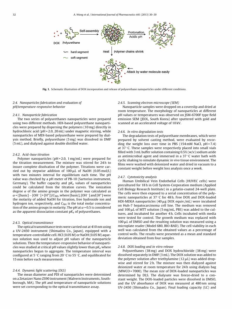
32 A. Wang et al. / International Journal of Pharmaceutics 441 (2013) 30– 39
lease
2p
2
uchny(
2
tirwvGcd˛thta
2
atoscnc1
2
obw
Fig. 1. Schematic illustration of DOX incorporation and re
.4. Nanoparticles fabrication and evaluation ofH/temperature-responsive behavior
.4.1. Nanoparticle fabricationThe two series of polyurethanes nanoparticles were prepared
sing two different methods. HDI-based polyurethane nanoparti-les were prepared by dispersing the polymers (10 mg) directly inydrochloric acid (pH = 2.0, 20 mL) under magnetic stirring, whileanoparticles of MDI-based polyurethane were prepared by dial-sis method. Briefly, polyurethane (5 mg) was dissolved in DMF5 mL), and dialyzed against double distilled water.
.4.2. Acid–base titrationPolymer nanoparticles (pH = 2.0, 1 mg/mL) were prepared for
he titration measurement. The mixture was stirred for 24 h tonsure complete disolvation of the polymer. Titrations were car-ied out by stepwise addition of 100 �L of NaOH (0.05 mol/L)ith two minutes interval for equilibrium each time. The pH
alue was checked by a pH meter of PB-10 (Sartorius instrument,ermany). The buffer capacity and pKa values of nanoparticlesould be calculated from the titration curves. The ionizationegree of the amino groups in the polymer was calculated as
= ([basic] − [OH−] + [H+])/CNH, where [basic], [OH−] and [H+] werehe molarity of added NaOH for titration, free hydroxide ion andydrogen ion, respectively, and CNH is the total molar concentra-ion of the amino groups in molarity. The pH at = 0.5 is considereds the apparent dissociation constant pKa of polyurethanes.
.4.3. Optical transmittanceThe optical transmittance tests were carried out at 410 nm using
UV-2450 instrument (Shimadzu Co., Japan), equipped with aemperature-controllable cell. HCl (0.05 M) or NaOH (0.05 M) aque-us solution was used to adjust pH values of the nanoparticleolutions. Then the temperature-responsive behavior of nanoparti-les was studied at critical pH values slightly lower than pKa whereanoparticles begun to aggregate. The temperature interval wasonfigured at 5 ◦C ranging from 20 ◦C to 55 ◦C, and equilibrated for5 min before each measurement.
.4.4. Dynamic light scattering (DLS)
The mean diameter and PDI of nanoparticles were determinedn a Zetasizer Nano ZS90 instrument (Malvern Instruments, South-orough, MA). The pH and temperature of nanoparticle solutionsere set corresponding to the optical transmittance assay.
of polyurethane nanoparticles under different conditions.
2.4.5. Scanning electron microscope (SEM)Nanoparticle samples were dropped on a coverslip and dried at
room temperature. The morphology of nanoparticles at differentpH values or temperatures was observed on JSM-6700F type fieldemission SEM (JEOL, South Korea) after sputtered with gold andscanned at an accelerated voltage of 10 kV.
2.4.6. In vitro degradation testsThe degradation tests of polyurethane membranes, which were
prepared by solvent casting method, were evaluated by recor-ding the weight loss over time in PBS (154 mM NaCl, pH = 7.4)at 37 ◦C. These samples were respectively placed into small vialsfilled with 3 mL buffer solution containing 0.5% (w/v) sodium azideas antimicrobial agent and immersed in a 37 ◦C water bath withcyclic shaking to simulate dynamic in vivo tissue environment. Thefilms were washed with deionized water and dried in vacuum to aconstant weight before weight loss analysis once a week.
2.4.7. Cytotoxicity analysisHuman Umbilical Vein Endothelial Cells (HUVEC cells) were
precultured for 18 h in Cell System-Corporation medium (AppliedCell Biology Research Institute) in a gelatin-coated 24-well plate.The cells were then exposed to a serial concentration of the poly-meric nanoparticles at 37 ◦C for 48 h. Free DOX and DOX-loadedHDI-MDEA nanoparticles (40 �g DOX equiv./mL) were incubatedon Huh-7 hepatocarcinoma cell line. The medium was removedand 100 �L of MTT solution (5 mg/mL, PBS) was added to the cul-tures, and incubated for another 4 h. Cells incubated with mediawere tested for control. The growth medium was replaced with100 �L of DMSO and the resulting solution was measured using amicroplate reader (Model 680, BIO-RAD). The cell viability in eachwell was calculated from the obtained values as a percentage ofcontrol wells. The results were presented as a mean and standarddeviation obtained from four samples.
2.4.8. DOX loading and in vitro releasePolyurethanes (38 mg) and DOX hydrochloride (38 mg) were
dissolved separately in DMF (3 mL). The DOX solution was added tothe polymer solution after triethylamine (12 �L) was added drop-wise and stirred for 2 h. The mixture was then dialyzed againstdeionized water at room temperature for 24 h using dialysis bag(MWCO = 7000). The mean size of DOX-loaded nanoparticles was
determined by DLS. The dialysate was freeze-dried to a con-stant weight. The DOX-loaded particles were dissolved in DMSO,and the UV absorbance of DOX was measured at 480 nm usingUV-2450 (Shimadzu Co., Japan). Final loading capacity (LC) and
nal of Pharmaceutics 441 (2013) 30– 39 33
ee
L
E
v(pabaAwtbwtar
c
wtn
2
n(TwCsJLtlHu
3
3
sapmcbdHa4ptcmpI
the polyurethane bearing same hard segments, Tg hard was affectedby the mobility of soft segments, where BDEA < MDEA < HEP. Inaddition, ODT caused by micro-phase separation was observed at
Table 1Molecular weights, PDI and DSC data of polyurethanes.
Polyurethanes Mn Mw PDI Tg hard (◦C) ODT (◦C) Tm (◦C)
HDI-HEP 25,100 52,700 2.10 −32.9 30.3HDI-MDEA 23,900 43,500 1.82 −8.5 56.2
A. Wang et al. / International Jour
ncapsulation efficiency (EE) were calculated from the followingquations (Eqs. (1) and (2)):
C (% w/w) = mass of loaded guestmass of nano particles
× 100 (1)
E (%) = final loadinginitial loading
× 100 (2)
In vitro release tests were carried out under different pHalues and temperatures. Lyophilized DOX-loaded nanoparticles2 mg/mL) were dispersed in acetate buffer solution (154 mM NaCl,H = 4.0) or PBS (154 mM NaCl, pH = 7.4), respectively, and dialyzedgainst their corresponding buffer solutions (20 mL) in cappedeakers at 37 ◦C. In vitro release at different temperatures (37 ◦Cnd 50 ◦C) was carried out at pKa of polyurethane nanoparticles.t every designated interval, buffer solutions (5 mL) in the beakerere taken out and fresh buffer solution (5 mL) was replenished
o keep a constant volume. The amount of DOX released into theuffer solution was analyzed using a UV spectrophotometer at aavelength of 480 nm. The concentration of DOX released from
he nanoparticles was expressed as a percentage of the total DOXvailable and plotted as a function of time. The cumulative DOXelease was calculated through equation below (Eq. (3)):
umulative DOX release (%) = Mt
M∞× 100 (3)
here Mt is the amount of drug released from nanoparticles atime t and M∞ is the amount of drug released from the polymericanoparticles at time infinity.
.4.9. Confocal laser scanning microscopy (CLSM)The intracellular distribution of free DOX and drug-loaded
anoparticles were followed with CLSM using Huh-7 cells5 × 104 cells/well) cultured in a 35 mm glass base dish (Iwaki,okyo, Japan) for one day. Then, the complex (40 �g DOX equiv/mL)as added, and cells were cultured for 6, or 24 h in a humidified 5%O2-containing atmosphere. After washing with PBS, the cells weretained with Hoechst 33342 (Dojindo Laboratories, Kumamoto,apan). CLSM images of cells were obtained using a Zeiss LSM510aser Confocal Scanning Microscopy imaging system at the excita-ion wavelengths of 480 nm (Ar laser) for DOX, and 710 nm (MaiTaiaser, 2 photon excitation; Spectra-Physics, Mountain View, CA) foroechst 33342. The cells treated with free DOX (40 �g/mL) weresed as a control.
. Results and discussion
.1. Synthesis and characterization of polyurethanes
Two series of polyurethanes were synthesized by a conden-ation reaction between the isocyanate groups of HDI or MDInd hydroxyl groups of HEP, MDEA or BDEA (Scheme 1). Theolyurethanes were consisted of HDI or MDI as hydrophobic seg-ents, and HEP, MDEA or BDEA as hydrophilic segments. The
hemical structures of the resulting polymers were characterizedy 1H and 13C NMR as well as FT-IR. The molecular weights wereetermined by GPC. Fig. S1 shows 1H and 13C NMR spectra of HDI-EP. The peaks a, b and c at 3.16 ppm, 1.5 ppm and 1.34 ppm weressigned to the methylene protons of HDI. The peaks e, f and g at.19 ppm, 2.65 ppm and 2.59 ppm were assigned to the methylenerotons of HEP. A feeble peak d appeared at 5.08 ppm was attributedo OOCNH . In 13C NMR (Fig. S1b), the characteristic peak of iso-
yanate at 122.9 ppm was not observed, indicating that the HDIonomer was completely consumed during the reaction. Instead,eak d aroused at 156.5 ppm was attributed to OOCNH . The FT-R measurements further confirmed the success of condensation
Scheme 1. Synthesis of two series of polyurethanes based on HDI and MDI.
reaction. Assignments of adsorption peaks for HDI-HEP and MDI-HEP are presented in Fig. S2. The peaks at 3339 cm−1 and 1532 cm−1
correspond to the N H stretching and deformation vibration bandof the urethane, respectively. The C O stretching band belongingto the hard segment appears at 1713 cm−1. In addition, the absenceof peaks at around 2260 cm−1 indicates that no isocyanate groupremains in the obtained polymer. These records confirmed the for-mation of polyurethanes.
The molecular weight and PDI of polyurethanes are givenin Table 1. Their molecular weights were in the rangeof (1.22–4.05) × 104 with PDI of 1.55–2.68, endowing thepolyurethanes appropriate mechanical and processing propertiesfor biomedical applications. As shown in Table 1, the molecularweights of HDI-based polyurethanes are similar, while MDI-basedpolyurethanes exhibit a decrease in molecular weight with the pro-longation of alkyl side chains. The influence of steric hindranceeffect resulted from the alkyl side chains of diol on MDI-bearingaromatic ring was greater than that of HDI-bearing alkyl chain. Theexistence of steric hindrance effect of butyl side chains hinderedMDI and BDEA from polymerization, thus resulting in the lowestmolecular weight for polyurethane MDI-BDEA.
3.2. Thermal analysis of polyurethanes
Thermal properties of the synthesized polyurethanes were stud-ied by DSC (Fig. S3 and Table 1). Endothermic peaks were detectedfor HDI-based polyurethanes, suggesting crystalline region existedin HDI-based polyurethanes, which was confirmed from the anal-ysis of the XRD profiles (Fig. S4). The hydrogen bonding arosefrom HDI series may be attributed to the formation of crystallineregion. Basically, these thermograms presented two characteristictransitions corresponding to hard phase transition of diisocyanate(Tg hard) and order–disorder transition (ODT), respectively. Theglass transition of diol of soft phase (Tg soft) was probably lower thanthe measurement temperature. Tg hard of MDI-based polyurethaneswas higher than that of HDI-based compartment, which may beattributed to the relative rigid conformation of MDI segment. For
HDI-BDEA 22,600 35,000 1.55 −6.0 145 65.5MDI-HEP 40,500 70,900 1.75 3.7 139MDI-MDEA 25,500 68,400 2.68 4.2 82.5MDI-BDEA 12,200 24,700 2.02 4.6 77.1
34 A. Wang et al. / International Journal of Pharmaceutics 441 (2013) 30– 39
s of po
hata
3
spmwdbp6TicMsdtoehtc
3
ppdtccetdwtew
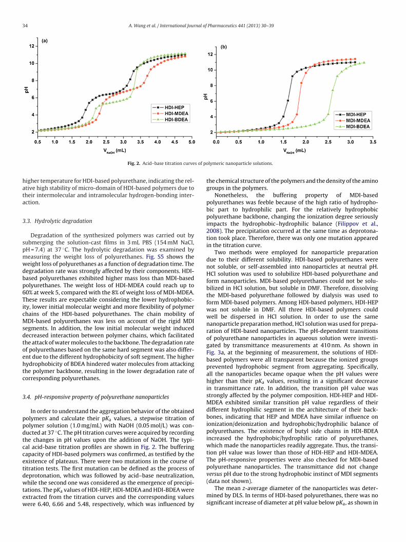
Fig. 2. Acid–base titration curve
igher temperature for HDI-based polyurethane, indicating the rel-tive high stability of micro-domain of HDI-based polymers due toheir intermolecular and intramolecular hydrogen-bonding inter-ction.
.3. Hydrolytic degradation
Degradation of the synthesized polymers was carried out byubmerging the solution-cast films in 3 mL PBS (154 mM NaCl,H = 7.4) at 37 ◦C. The hydrolytic degradation was examined byeasuring the weight loss of polyurethanes. Fig. S5 shows theeight loss of polyurethanes as a function of degradation time. Theegradation rate was strongly affected by their components. HDI-ased polyurethanes exhibited higher mass loss than MDI-basedolyurethanes. The weight loss of HDI-MDEA could reach up to0% at week 5, compared with the 8% of weight loss of MDI-MDEA.hese results are expectable considering the lower hydrophobic-ty, lower initial molecular weight and more flexibility of polymerhains of the HDI-based polyurethanes. The chain mobility ofDI-based polyurethanes was less on account of the rigid MDI
egments. In addition, the low initial molecular weight inducedecreased interaction between polymer chains, which facilitatedhe attack of water molecules to the backbone. The degradation ratef polyurethanes based on the same hard segment was also differ-nt due to the different hydrophobicity of soft segment. The higherydrophobicity of BDEA hindered water molecules from attackinghe polymer backbone, resulting in the lower degradation rate oforresponding polyurethanes.
.4. pH-responsive property of polyurethane nanoparticles
In order to understand the aggregation behavior of the obtainedolymers and calculate their pKa values, a stepwise titration ofolymer solution (1.0 mg/mL) with NaOH (0.05 mol/L) was con-ucted at 37 ◦C. The pH titration curves were acquired by recordinghe changes in pH values upon the addition of NaOH. The typi-al acid-base titration profiles are shown in Fig. 2. The bufferingapacity of HDI-based polymers was confirmed, as testified by thexistence of plateaus. There were two mutations in the course ofitration tests. The first mutation can be defined as the process ofeprotonation, which was followed by acid–base neutralization,
hile the second one was considered as the emergence of precipi-ations. The pKa values of HDI-HEP, HDI-MDEA and HDI-BDEA werextracted from the titration curves and the corresponding valuesere 6.40, 6.66 and 5.48, respectively, which was influenced by
lymeric nanoparticle solutions.
the chemical structure of the polymers and the density of the aminogroups in the polymers.
Nonetheless, the buffering property of MDI-basedpolyurethanes was feeble because of the high ratio of hydropho-bic part to hydrophilic part. For the relatively hydrophobicpolyurethane backbone, changing the ionization degree seriouslyimpacts the hydrophobic–hydrophilic balance (Filippov et al.,2008). The precipitation occurred at the same time as deprotona-tion took place. Therefore, there was only one mutation appearedin the titration curve.
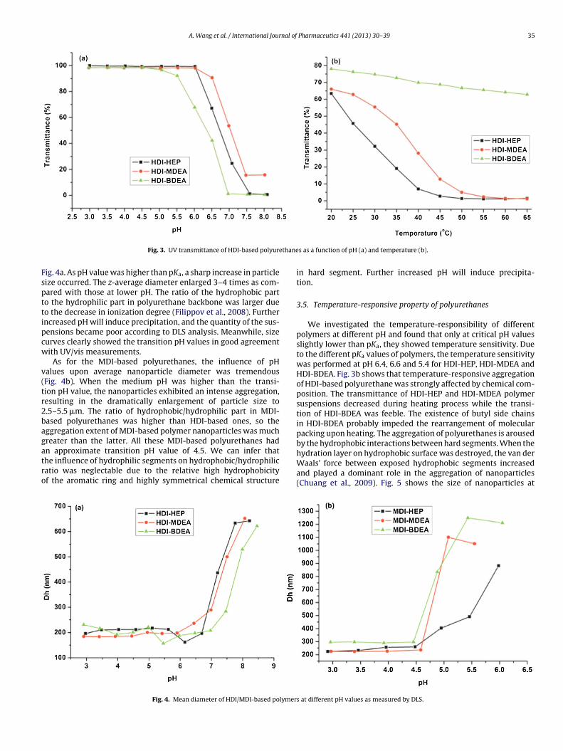
Two methods were employed for nanoparticle preparationdue to their different solubility. HDI-based polyurethanes werenot soluble, or self-assembled into nanoparticles at neutral pH.HCl solution was used to solubilize HDI-based polyurethane andform nanoparticles. MDI-based polyurethanes could not be solu-bilized in HCl solution, but soluble in DMF. Therefore, dissolvingthe MDI-based polyurethane followed by dialysis was used toform MDI-based polymers. Among HDI-based polymers, HDI-HEPwas not soluble in DMF. All three HDI-based polymers couldwell be dispersed in HCl solution. In order to use the samenanoparticle preparation method, HCl solution was used for prepa-ration of HDI-based nanoparticles. The pH-dependent transitionsof polyurethane nanoparticles in aqueous solution were investi-gated by transmittance measurements at 410 nm. As shown inFig. 3a, at the beginning of measurement, the solutions of HDI-based polymers were all transparent because the ionized groupsprevented hydrophobic segment from aggregating. Specifically,all the nanoparticles became opaque when the pH values werehigher than their pKa values, resulting in a significant decreasein transmittance rate. In addition, the transition pH value wasstrongly affected by the polymer composition. HDI-HEP and HDI-MDEA exhibited similar transition pH value regardless of theirdifferent hydrophilic segment in the architecture of their back-bones, indicating that HEP and MDEA have similar influence onionization/deionization and hydrophobic/hydrophilic balance ofpolyurethanes. The existence of butyl side chains in HDI-BDEAincreased the hydrophobic/hydrophilic ratio of polyurethanes,which made the nanoparticles readily aggregate. Thus, the transi-tion pH value was lower than those of HDI-HEP and HDI-MDEA.The pH-responsive properties were also checked for MDI-basedpolyurethane nanoparticles. The transmittance did not changeversus pH due to the strong hydrophobic instinct of MDI segments
(data not shown).The mean z-average diameter of the nanoparticles was deter-mined by DLS. In terms of HDI-based polyurethanes, there was nosignificant increase of diameter at pH value below pKa, as shown in
A. Wang et al. / International Journal of Pharmaceutics 441 (2013) 30– 39 35
thane
Fspttipcw
v(tr2bagatro
Fig. 3. UV transmittance of HDI-based polyure
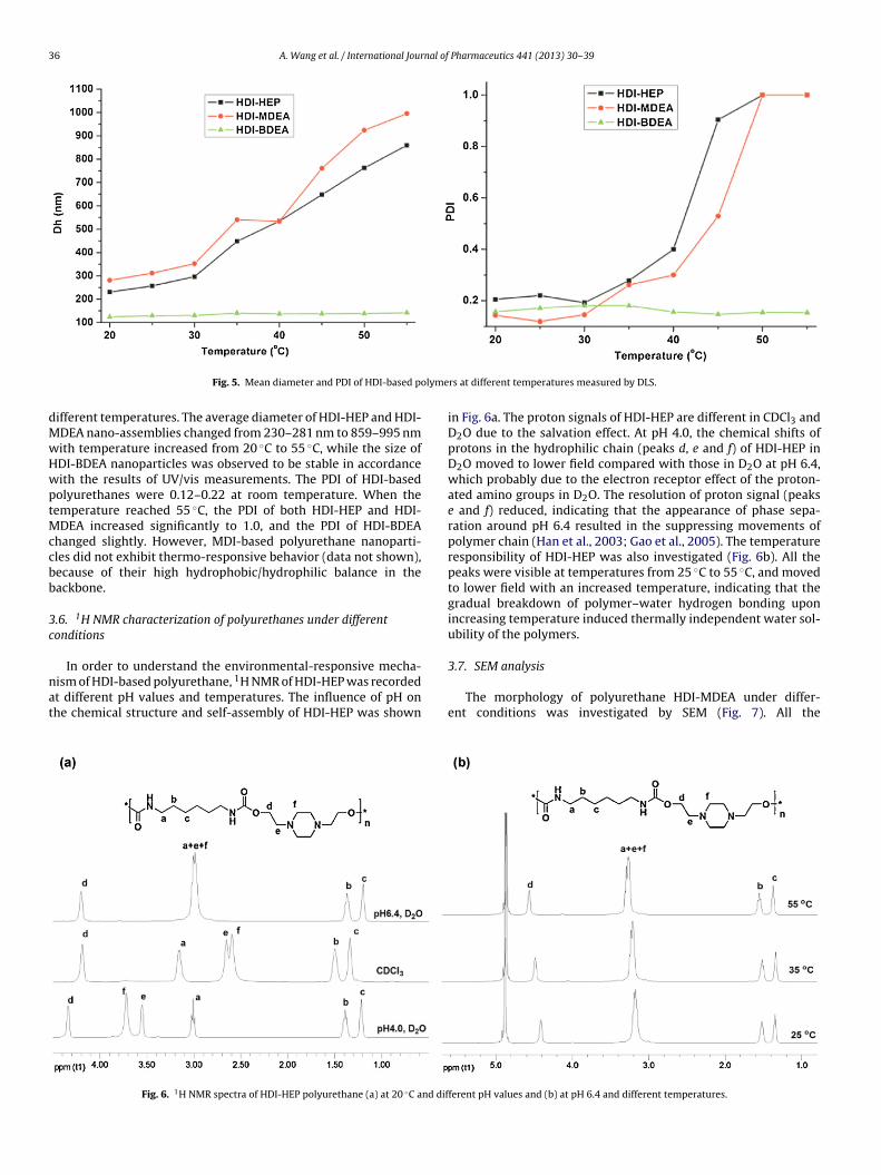
ig. 4a. As pH value was higher than pKa, a sharp increase in particleize occurred. The z-average diameter enlarged 3–4 times as com-ared with those at lower pH. The ratio of the hydrophobic parto the hydrophilic part in polyurethane backbone was larger dueo the decrease in ionization degree (Filippov et al., 2008). Furtherncreased pH will induce precipitation, and the quantity of the sus-ensions became poor according to DLS analysis. Meanwhile, sizeurves clearly showed the transition pH values in good agreementith UV/vis measurements.
As for the MDI-based polyurethanes, the influence of pHalues upon average nanoparticle diameter was tremendousFig. 4b). When the medium pH was higher than the transi-ion pH value, the nanoparticles exhibited an intense aggregation,esulting in the dramatically enlargement of particle size to.5–5.5 �m. The ratio of hydrophobic/hydrophilic part in MDI-ased polyurethanes was higher than HDI-based ones, so theggregation extent of MDI-based polymer nanoparticles was muchreater than the latter. All these MDI-based polyurethanes had
n approximate transition pH value of 4.5. We can infer thathe influence of hydrophilic segments on hydrophobic/hydrophilicatio was neglectable due to the relative high hydrophobicityf the aromatic ring and highly symmetrical chemical structureFig. 4. Mean diameter of HDI/MDI-based polymer
s as a function of pH (a) and temperature (b).
in hard segment. Further increased pH will induce precipita-tion.
3.5. Temperature-responsive property of polyurethanes
We investigated the temperature-responsibility of differentpolymers at different pH and found that only at critical pH valuesslightly lower than pKa, they showed temperature sensitivity. Dueto the different pKa values of polymers, the temperature sensitivitywas performed at pH 6.4, 6.6 and 5.4 for HDI-HEP, HDI-MDEA andHDI-BDEA. Fig. 3b shows that temperature-responsive aggregationof HDI-based polyurethane was strongly affected by chemical com-position. The transmittance of HDI-HEP and HDI-MDEA polymersuspensions decreased during heating process while the transi-tion of HDI-BDEA was feeble. The existence of butyl side chainsin HDI-BDEA probably impeded the rearrangement of molecularpacking upon heating. The aggregation of polyurethanes is arousedby the hydrophobic interactions between hard segments. When the
hydration layer on hydrophobic surface was destroyed, the van derWaals’ force between exposed hydrophobic segments increasedand played a dominant role in the aggregation of nanoparticles(Chuang et al., 2009). Fig. 5 shows the size of nanoparticles ats at different pH values as measured by DLS.
36 A. Wang et al. / International Journal of Pharmaceutics 441 (2013) 30– 39
olyme
dMwHwptMccbb
3c
nat
Fig. 5. Mean diameter and PDI of HDI-based p
ifferent temperatures. The average diameter of HDI-HEP and HDI-DEA nano-assemblies changed from 230–281 nm to 859–995 nmith temperature increased from 20 ◦C to 55 ◦C, while the size ofDI-BDEA nanoparticles was observed to be stable in accordanceith the results of UV/vis measurements. The PDI of HDI-basedolyurethanes were 0.12–0.22 at room temperature. When theemperature reached 55 ◦C, the PDI of both HDI-HEP and HDI-
DEA increased significantly to 1.0, and the PDI of HDI-BDEAhanged slightly. However, MDI-based polyurethane nanoparti-les did not exhibit thermo-responsive behavior (data not shown),ecause of their high hydrophobic/hydrophilic balance in theackbone.
.6. 1H NMR characterization of polyurethanes under differentonditions
In order to understand the environmental-responsive mecha-ism of HDI-based polyurethane, 1H NMR of HDI-HEP was recordedt different pH values and temperatures. The influence of pH onhe chemical structure and self-assembly of HDI-HEP was shown
Fig. 6. 1H NMR spectra of HDI-HEP polyurethane (a) at 20 ◦C and dif
rs at different temperatures measured by DLS.
in Fig. 6a. The proton signals of HDI-HEP are different in CDCl3 andD2O due to the salvation effect. At pH 4.0, the chemical shifts ofprotons in the hydrophilic chain (peaks d, e and f) of HDI-HEP inD2O moved to lower field compared with those in D2O at pH 6.4,which probably due to the electron receptor effect of the proton-ated amino groups in D2O. The resolution of proton signal (peakse and f) reduced, indicating that the appearance of phase sepa-ration around pH 6.4 resulted in the suppressing movements ofpolymer chain (Han et al., 2003; Gao et al., 2005). The temperatureresponsibility of HDI-HEP was also investigated (Fig. 6b). All thepeaks were visible at temperatures from 25 ◦C to 55 ◦C, and movedto lower field with an increased temperature, indicating that thegradual breakdown of polymer–water hydrogen bonding uponincreasing temperature induced thermally independent water sol-ubility of the polymers.
3.7. SEM analysis
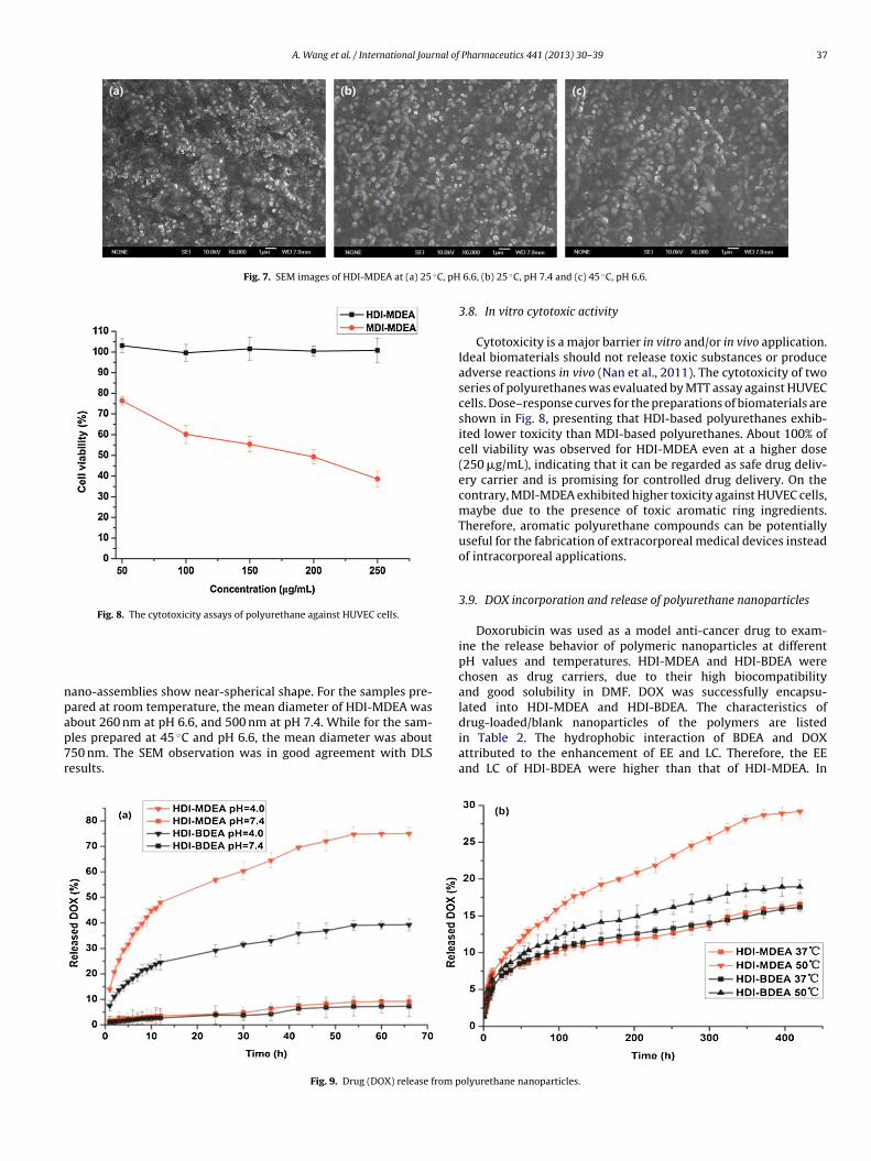
The morphology of polyurethane HDI-MDEA under differ-ent conditions was investigated by SEM (Fig. 7). All the
ferent pH values and (b) at pH 6.4 and different temperatures.
A. Wang et al. / International Journal of Pharmaceutics 441 (2013) 30– 39 37
Fig. 7. SEM images of HDI-MDEA at (a) 25 ◦C, pH
Fig. 8. The cytotoxicity assays of polyurethane against HUVEC cells.
npap7r
drug-loaded/blank nanoparticles of the polymers are listedin Table 2. The hydrophobic interaction of BDEA and DOXattributed to the enhancement of EE and LC. Therefore, the EE
ano-assemblies show near-spherical shape. For the samples pre-ared at room temperature, the mean diameter of HDI-MDEA wasbout 260 nm at pH 6.6, and 500 nm at pH 7.4. While for the sam-les prepared at 45 ◦C and pH 6.6, the mean diameter was about50 nm. The SEM observation was in good agreement with DLS
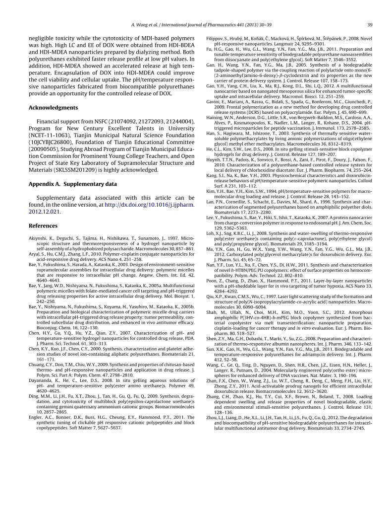
esults.Fig. 9. Drug (DOX) release from p
6.6, (b) 25 ◦C, pH 7.4 and (c) 45 ◦C, pH 6.6.
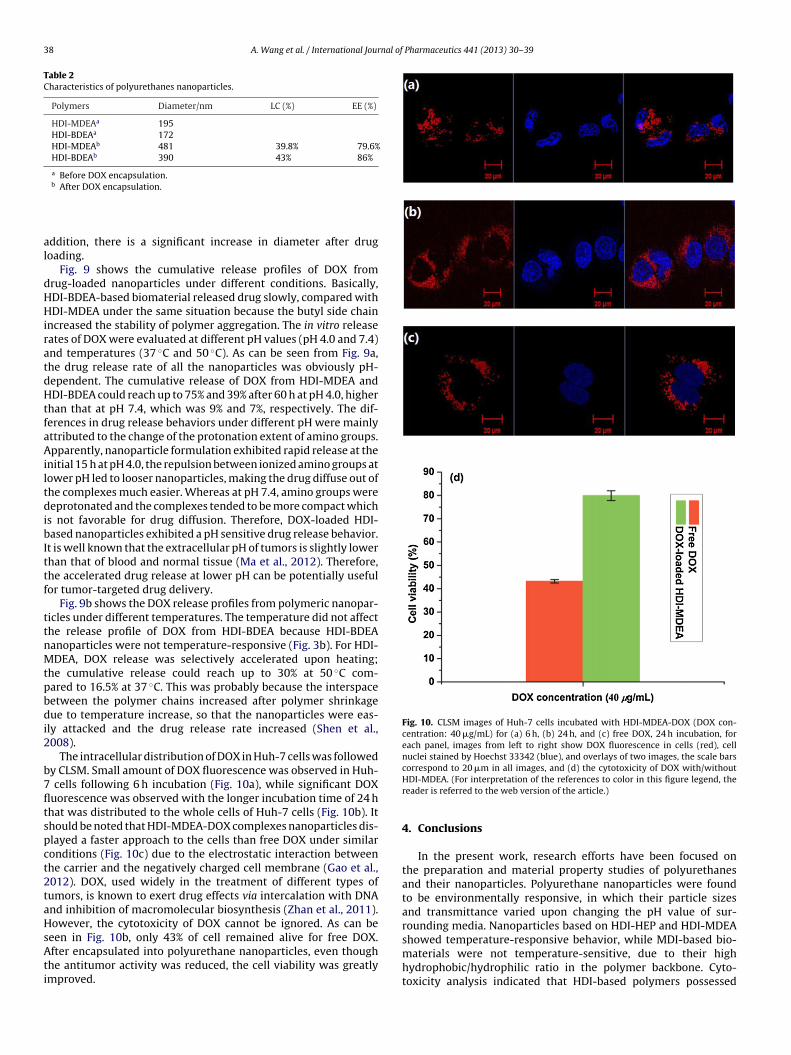
3.8. In vitro cytotoxic activity
Cytotoxicity is a major barrier in vitro and/or in vivo application.Ideal biomaterials should not release toxic substances or produceadverse reactions in vivo (Nan et al., 2011). The cytotoxicity of twoseries of polyurethanes was evaluated by MTT assay against HUVECcells. Dose–response curves for the preparations of biomaterials areshown in Fig. 8, presenting that HDI-based polyurethanes exhib-ited lower toxicity than MDI-based polyurethanes. About 100% ofcell viability was observed for HDI-MDEA even at a higher dose(250 �g/mL), indicating that it can be regarded as safe drug deliv-ery carrier and is promising for controlled drug delivery. On thecontrary, MDI-MDEA exhibited higher toxicity against HUVEC cells,maybe due to the presence of toxic aromatic ring ingredients.Therefore, aromatic polyurethane compounds can be potentiallyuseful for the fabrication of extracorporeal medical devices insteadof intracorporeal applications.
3.9. DOX incorporation and release of polyurethane nanoparticles
Doxorubicin was used as a model anti-cancer drug to exam-ine the release behavior of polymeric nanoparticles at differentpH values and temperatures. HDI-MDEA and HDI-BDEA werechosen as drug carriers, due to their high biocompatibilityand good solubility in DMF. DOX was successfully encapsu-lated into HDI-MDEA and HDI-BDEA. The characteristics of
and LC of HDI-BDEA were higher than that of HDI-MDEA. In
olyurethane nanoparticles.
38 A. Wang et al. / International Journal of Pharmaceutics 441 (2013) 30– 39
Table 2Characteristics of polyurethanes nanoparticles.
Polymers Diameter/nm LC (%) EE (%)
HDI-MDEAa 195HDI-BDEAa 172HDI-MDEAb 481 39.8% 79.6%HDI-BDEAb 390 43% 86%
a Before DOX encapsulation.
al
dHHiratdHtfaAiltdibIttf
ttnMtpbdi2
b7fltspct2taHsAti
Fig. 10. CLSM images of Huh-7 cells incubated with HDI-MDEA-DOX (DOX con-centration: 40 �g/mL) for (a) 6 h, (b) 24 h, and (c) free DOX, 24 h incubation, foreach panel, images from left to right show DOX fluorescence in cells (red), cellnuclei stained by Hoechst 33342 (blue), and overlays of two images, the scale barscorrespond to 20 �m in all images, and (d) the cytotoxicity of DOX with/without
showed temperature-responsive behavior, while MDI-based bio-materials were not temperature-sensitive, due to their high
b After DOX encapsulation.
ddition, there is a significant increase in diameter after drugoading.
Fig. 9 shows the cumulative release profiles of DOX fromrug-loaded nanoparticles under different conditions. Basically,DI-BDEA-based biomaterial released drug slowly, compared withDI-MDEA under the same situation because the butyl side chain
ncreased the stability of polymer aggregation. The in vitro releaseates of DOX were evaluated at different pH values (pH 4.0 and 7.4)nd temperatures (37 ◦C and 50 ◦C). As can be seen from Fig. 9a,he drug release rate of all the nanoparticles was obviously pH-ependent. The cumulative release of DOX from HDI-MDEA andDI-BDEA could reach up to 75% and 39% after 60 h at pH 4.0, higher
han that at pH 7.4, which was 9% and 7%, respectively. The dif-erences in drug release behaviors under different pH were mainlyttributed to the change of the protonation extent of amino groups.pparently, nanoparticle formulation exhibited rapid release at the
nitial 15 h at pH 4.0, the repulsion between ionized amino groups atower pH led to looser nanoparticles, making the drug diffuse out ofhe complexes much easier. Whereas at pH 7.4, amino groups wereeprotonated and the complexes tended to be more compact which
s not favorable for drug diffusion. Therefore, DOX-loaded HDI-ased nanoparticles exhibited a pH sensitive drug release behavior.
t is well known that the extracellular pH of tumors is slightly lowerhan that of blood and normal tissue (Ma et al., 2012). Therefore,he accelerated drug release at lower pH can be potentially usefulor tumor-targeted drug delivery.
Fig. 9b shows the DOX release profiles from polymeric nanopar-icles under different temperatures. The temperature did not affecthe release profile of DOX from HDI-BDEA because HDI-BDEAanoparticles were not temperature-responsive (Fig. 3b). For HDI-DEA, DOX release was selectively accelerated upon heating;
he cumulative release could reach up to 30% at 50 ◦C com-ared to 16.5% at 37 ◦C. This was probably because the interspaceetween the polymer chains increased after polymer shrinkageue to temperature increase, so that the nanoparticles were eas-
ly attacked and the drug release rate increased (Shen et al.,008).
The intracellular distribution of DOX in Huh-7 cells was followedy CLSM. Small amount of DOX fluorescence was observed in Huh-
cells following 6 h incubation (Fig. 10a), while significant DOXuorescence was observed with the longer incubation time of 24 hhat was distributed to the whole cells of Huh-7 cells (Fig. 10b). Ithould be noted that HDI-MDEA-DOX complexes nanoparticles dis-layed a faster approach to the cells than free DOX under similaronditions (Fig. 10c) due to the electrostatic interaction betweenhe carrier and the negatively charged cell membrane (Gao et al.,012). DOX, used widely in the treatment of different types ofumors, is known to exert drug effects via intercalation with DNAnd inhibition of macromolecular biosynthesis (Zhan et al., 2011).owever, the cytotoxicity of DOX cannot be ignored. As can be
een in Fig. 10b, only 43% of cell remained alive for free DOX.fter encapsulated into polyurethane nanoparticles, even though
he antitumor activity was reduced, the cell viability was greatlymproved.
HDI-MDEA. (For interpretation of the references to color in this figure legend, thereader is referred to the web version of the article.)
4. Conclusions
In the present work, research efforts have been focused onthe preparation and material property studies of polyurethanesand their nanoparticles. Polyurethane nanoparticles were foundto be environmentally responsive, in which their particle sizesand transmittance varied upon changing the pH value of sur-rounding media. Nanoparticles based on HDI-HEP and HDI-MDEA
hydrophobic/hydrophilic ratio in the polymer backbone. Cyto-toxicity analysis indicated that HDI-based polymers possessed
nal of
nwapaptsp
A
P(((tPM
A
f2
R
A
A
B
B
B
C
C
C
D
D
E
A. Wang et al. / International Jour
egligible toxicity while the cytotoxicity of MDI-based polymersas high. High LC and EE of DOX were obtained from HDI-BDEA
nd HDI-MDEA nanoparticles prepared by dialyzing method. Botholyurethanes exhibited faster release profile at low pH values. Inddition, HDI-MDEA showed an accelerated release at high tem-erature. Encapsulation of DOX into HDI-MDEA could improvehe cell viability and cellular uptake. The pH/temperature respon-ive nanoparticles fabricated from biocompatible polyurethanesrovide an opportunity for the controlled release of DOX.
cknowledgments
Financial support from NSFC (21074092, 21272093, 21244004),rogram for New Century Excellent Talents in UniversityNCET-11-1063), Tianjin Municipal Natural Science Foundation10JCYBJC26800), Foundation of Tianjin Educational Committee20090505), Studying Abroad Program of Tianjin Municipal Educa-ion Commission for Prominent Young College Teachers, and Openroject of State Key Laboratory of Supramolecular Structure andaterials (SKLSSM201209) is highly acknowledged.
ppendix A. Supplementary data
Supplementary data associated with this article can beound, in the online version, at http://dx.doi.org/10.1016/j.ijpharm.012.12.021.
eferences
kiyoshi, K., Deguchi, S., Tajima, H., Nishikawa, T., Sunamoto, J., 1997. Micro-scopic structure and thermoresponsiveness of a hydrogel nanoparticle byself-assembly of a hydrophobized polysaccharide. Macromolecules 30, 857–861.
ryal, S., Hu, C.M.J., Zhang, L.F., 2010. Polymer-cisplatin conjugate nanoparticles foracid-responsive drug delivery. ACS Nano 4, 251–258.
ae, Y., Fukushima, S., Harada, A., Kataoka, K., 2003. Design of environment-sensitivesupramolecular assemblies for intracellular drug delivery: polymeric micellesthat are responsive to intracellular pH change. Angew. Chem. Int. Ed. 42,4640–4643.
ae, Y., Jang, W.D., Nishiyama, N., Fukushima, S., Kataoka, K., 2005a. Multifunctionalpolymeric micelles with folate-mediated cancer cell targeting and pH-triggereddrug releasing properties for active intracellular drug delivery. Mol. Biosyst. 1,242–250.
ae, Y., Nishiyama, N., Fukushima, S., Koyama, H., Yasuhiro, M., Kataoka, K., 2005b.Preparation and biological characterization of polymeric micelle drug carrierswith intracellular pH-triggered drug release property: tumor permeability, con-trolled subcellular drug distribution, and enhanced in vivo antitumor efficacy.Bioconjug. Chem. 16, 122–130.
hen, H.Y., Gu, Y.Q., Hu, Y.Z., Qian, Z.Y., 2007. Characterization of pH- andtemperature-sensitive hydrogel nanoparticles for controlled drug release, PDA.J. Pharm. Sci. Technol. 61, 303–313.
hen, K.Y., Kuo, J.F., Chen, C.Y., 2000. Synthesis, characterization and platelet adhe-sion studies of novel ion-containing aliphatic polyurethanes. Biomaterials 21,161–171.
huang, C.Y., Don, T.M., Chiu, W.Y., 2009. Synthesis and properties of chitosan-basedthermo- and pH-responsive nanoparticles and application in drug release. J.Polym. Sci. Part A: Polym. Chem. 47, 2798–2810.
ayananda, K., He, C., Lee, D.S., 2008. In situ gelling aqueous solutions ofpH- and temperature-sensitive poly(ester amino urethane)s. Polymer 49,4620–4625.
ing, M.M., Li, J.H., Fu, X.T., Zhou, J., Tan, H., Gu, Q., Fu, Q., 2009. Synthesis, degra-dation, and cytotoxicity of multiblock poly(epsilon-caprolactone urethane)s
containing gemini quaternary ammonium cationic groups. Biomacromolecules10, 2857–2865.ngler, A.C., Bonner, D.K., Buss, H.G., Cheung, E.Y., Hammond, P.T., 2011. Thesynthetic tuning of clickable pH responsive cationic polypeptides and blockcopolypeptides. Soft Matter 7, 5627–5637.
Pharmaceutics 441 (2013) 30– 39 39
Filippov, S., Hruby, M., Konák, C., Macková, H., Spírková, M., Stepánek, P., 2008. NovelpH-responsive nanoparticles. Langmuir 24, 9295–9301.
Fu, H.G., Gao, H., Wu, G.L., Wang, Y.N., Fan, Y.G., Ma, J.B., 2011. Preparation andtunable temperature sensitivity of biodegradable polyurethane nanoassembliesfrom diisocyanate and poly(ethylene glycol). Soft Matter 7, 3546–3552.
Gao, H., Wang, Y.N., Fan, Y.G., Ma, J.B., 2005. Synthesis of a biodegradabletadpole-shaped polymer via the coupling reaction of polylactide onto mono(6-(2-aminoethyl)amino-6-deoxy)-ˇ-cyclodextrin and its properties as the newcarrier of protein delivery system. J. Control. Release 107, 158–173.
Gao, Y.H., Yang, C.H., Liu, X., Ma, R.J., Kong, D.L., Shi, L.Q., 2012. A multifunctionalnanocarrier based on nanogated mesoporous silica for enhanced tumor-specificuptake and intracellular delivery. Macromol. Biosci. 12, 251–259.
Gavini, E., Mariani, A., Rassu, G., Bidali, S., Spada, G., Bonferoni, M.C., Giunchedi, P.,2009. Frontal polymerization as a new method for developing drug controlledrelease systems (DCRS) based on polyacrylamide. Eur. Polym. J. 45, 690–699.
Haining, W.N., Anderson, D.G., Little, S.R., von Bergwelt-Baildon, M.S., Cardoso, A.A.,Alves, P., Kosmatopoulos, K., Nadler, L.M., Langer, R., Kohane, D.S., 2004. pH-triggered microparticles for peptide vaccination. J. Immunol. 173, 2578–2585.
Han, S., Hagiwara, M., Ishizone, T., 2003. Synthesis of thermally sensitive water-soluble polymethacrylates by living anionic polymerizations of oligo(ethyleneglycol) methyl ether methacrylates. Macromolecules 36, 8312–8319.
He, C.L., Kim, S.W., Lee, D.S., 2008. In situ gelling stimuli-sensitive block copolymerhydrogels for drug delivery. J. Control. Release 127, 189–207.
Huynh, T.T.N., Padois, K., Sonvico, F., Rossi, A., Zani, F., Pirot, F., Doury, J., Falson, F.,2010. Characterization of a polyurethane-based controlled release system forlocal delivery of chlorhexidine diacetate. Eur. J. Pharm. Biopharm. 74, 255–264.
Kang, S.I., Na, K., Bae, Y.H., 2003. Physicochemical characteristics and doxorubicin-release behaviors of pH/temperature-sensitive polymeric nanoparticles. ColloidSurf. A 231, 103–112.
Kim, Y.H., Bae, Y.H., Kim, S.W., 1994. pH/temperature-sensitive polymers for macro-molecular drug loading and release. J. Control. Release 28, 143–152.
Lan, P.N., Corneillie, S., Schacht, E., Davies, M., Shard, A., 1996. Synthesis and char-acterization of segmented polyurethanes based on amphiphilic polyether diols.Biomaterials 17, 2273–2280.
Lee, Y., Fukushima, S., Bae, Y., Hiki, S., Ishii, T., Kataoka, K., 2007. A protein nanocarrierfrom charge-conversion polymer in response to endosomal pH. J. Am. Chem. Soc.129, 5362–5363.
Loh, X.J., Sng, K.B.C., Li, J., 2008. Synthesis and water-swelling of thermo-responsivepoly(ester urethane)s containing poly(ε-caprolactone), poly(ethylene glycol)and poly(propylene glycol). Biomaterials 29, 3185–3194.
Ma, Y.N., Gao, H., Gu, W.X., Yang, Y.W., Wang, Y.N., Fan, Y.G., Wu, G.L., Ma, J.B.,2012. Carboxylated poly(glycerol methacrylate)s for doxorubicin delivery. Eur.J. Pharm. Sci. 45, 65–72.
Nan, Y.F., Luo, Y.L., Xu, F., Chen, Y.S., Di, H.W., 2011. Synthesis and characterizationof novel h-HTBN/PEG PU copolymers: effect of surface properties on hemocom-patibility. Polym. Adv. Technol. 22, 802–810.
Poon, Z., Chang, D., Zhao, X., Hammond, P.T., 2011. Layer-by-layer nanoparticleswith a pH-sheddable layer for in vivo targeting of tumor hypoxia. ACS Nano 33,4284–4292.
Qiu, X.P., Kwan, C.M.S., Wu, C., 1997. Laser light scattering study of the formation andstructure of poly(N-isopropylacrylamide-co-acrylic acid) nanoparticles. Macro-molecules 30, 6090–6094.
Shah, M., Ullah, N., Choi, M.H., Kim, M.O., Yoon, S.C., 2012. Amorphousamphiphilic P(3HV-co-4HB)-b-mPEG block copolymer synthesized from bac-terial copolyester via melt transesterification: nanoparticle preparation,cisplatin-loading for cancer therapy and in vitro evaluation. Eur. J. Pharm. Bio-pharm. 80, 518–527.
Shen, Z.Y., Ma, G.H., Dobashi, T., Marki, Y., Su, Z.G., 2008. Preparation and characteri-zation of thermo-responsive albumin nanospheres. Int. J. Pharm. 346, 133–142.
Sun, X.K., Gao, H., Wu, G.L., Wang, Y.N., Fan, Y.G., Ma, J.B., 2011. Biodegradable andtemperature-responsive polyurethanes for adriamycin delivery. Int. J. Pharm.412, 52–58.
Wang, C., Ge, Q., Ting, D., Nguyen, D., Shen, H.R., Chen, J.Z., Eisen, H.N., Heller, J.,Langer, R., Putnam, D., 2004. Molecularly engineered poly(ortho ester) micro-spheres for enhanced delivery of DNA vaccines. Nat. Mater. 3, 190–196.
Zhan, F.X., Chen, W., Wang, Z.J., Lu, W.T., Cheng, R., Deng, C., Meng, F.H., Liu, H.Y.,Zhong, Z.Y., 2011. Acid-activatable prodrug nanogels for efficient intracellulardoxorubicin release. Biomacromolecules 12, 3612–3620.
Zhang, C.H., Zhao, K.J., Hu, T.Y., Cui, X.F., Brown, N., Boland, T., 2008. Loadingdependent swelling and release properties of novel biodegradable, elastic
and environmental stimuli-sensitive polyurethanes. J. Control. Release 131,128–136.Zhou, L.J., Liang, D., He, X.L., Li, J.H., Tan, H., Li, J.S., Fu, Q., Gu, Q., 2012. The degradationand biocompatibility of pH-sensitive biodegradable polyurethanes for intracel-lular multifunctional antitumor drug delivery. Biomaterials 33, 2734–2745.
Top Related