


Languages
Pages
Legal


INTER CELLULAR JUNCTIONS
& CELL ADHESION
MOLECULES

INTRODUCTION Of all the social interactions between
cells in multicellular organism, the most fundamental are those that hold the cells together.
Many cells in tissues are linked to one another & to Extracellular Matrix at specialized contact sites called “CELL JUNCTIONS.”

Classification of Cell JunctionsI. Cell-cell Junctions Tight junctions, Gap junctions,
Desmosomes Adhesion belt / zonula adherensII. Cell-matrix junctions Focal contacts / adhesion plaques Hemidesmosomes

Classification as in Review of Medical Physiology by GanongI. Junctions that fasten cells to one another & to surrounding tissues Tight junctions, Adhesion belt, Desmosomes, Hemidesmosomes Focal adhesionsII. Junctions that permit transfer of ions & other molecules from one cell to another Gap junctions

Classification as in Alberts Molecular Biology of CellCell Junctions can be divided into 3 functional
groupsI. Occluding Junctions : Tight JunctionsII. Anchoring Junctions : Desmosomes, Hemidesmosomes, Focal
adhesions & Zonula adherensIII. Communicating Junctions : Gap Junctions

Tight Junctions / Zonula occludens• Surround apical margins of cells in epithelia
• Made up of ridges
• Degree of leakiness varies
• Composed of branching network of sealing strands
• Claudins & Occludins• ZO proteins

Adhesion Belt / Zonula Adherens
Connect actin filaments of two interacting cells
formed by cadherins
network contracts with the help of myosin motor proteins

Desmosomes & Hemidesmosomes

Gap JunctionsCONNEXONS – hexagonal array of transmembrane protein units
Regulation of Gap junction communication• intra cellular calcium• PH
• voltage• extracellular signals

Functions of Gap Junctions1. In tissues containing electrically
excitable cells, coupling via gap junctions is very usefull
2. Role in non-excitable tissues3. Normal development of ovarian
follicles4. Role in Embryogenesis


Cell Adhesion Molecules Attach cells to basal lamina & to each other
Are Transmembrane receptors with 3 domains : intracellular, transmembrane & extracellular
Mediate both Homophilic & Heterophilic binding
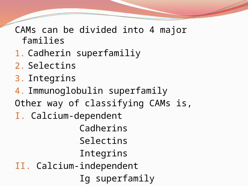
CAMs can be divided into 4 major families1. Cadherin superfamiliy2. Selectins3. Integrins4. Immunoglobulin superfamilyOther way of classifying CAMs is,I. Calcium-dependent Cadherins Selectins IntegrinsII. Calcium-independent Ig superfamily

CADHERINSMediate homophilic bindingStructure contains a short transmembrane
domain & a relatively long exrecellular domain wit 4 cadherin repeats (EC1 to EC4) each with a calcium binding sequence.
Interact with CateninsTypes :N-cadherinP-cadherinE-cadherin

SELECTINSCalcium dependent carbohydrate binding
proteinsMediate initial attachment of leukocytes to the
endothelium on the blood vessel wall during rolling step of Phagocytosis
Structure includes a NH2-terminal C-type Calcium binding lectin domain, a membrane spanning domain & a short cytoplasmic domain
Mediate heterophilic bindingTypes:L-selectinP-selectinE-selectin

INTEGRINSAre principally involved in cell-matrix adhesionAlso act as regulatory receptors that can initiate
intracellular signal pathwaysComposed of hetero dimers consisting of 2 non-
covalently associated subunits α and βBoth subunits are membrane glycoproteins with a
large extracellular domain, a single transmembrane domain & a short cytoplasmic domain
Types :β 1 integrins – VLA proteinsβ 2 integrins – Leu CAMsβ 3 integrins – cytoadhesionsβ 4 integrins

IMMUNOGLOBULIN SUPERFAMILYMediate many different functions including
acting as receptors for growth factors & mediating cell-cell & cell-matrix interactions
Structure is characterised by repeated domains similar to those found in immunoglobulins.
Mediate both heterophilic & homophilic binding
Types :ICAMs – Intracellular CAMsVCAMs – Vascular CAMsPECAMs – Platelet Endothelial CAMsNCAMs – Neural CAMs
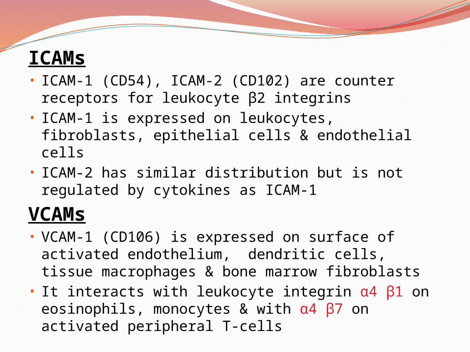
ICAMs• ICAM-1 (CD54), ICAM-2 (CD102) are counter
receptors for leukocyte β2 integrins• ICAM-1 is expressed on leukocytes, fibroblasts,
epithelial cells & endothelial cells• ICAM-2 has similar distribution but is not
regulated by cytokines as ICAM-1
VCAMs• VCAM-1 (CD106) is expressed on surface of
activated endothelium, dendritic cells, tissue macrophages & bone marrow fibroblasts
• It interacts with leukocyte integrin α4 β1 on eosinophils, monocytes & with α4 β7 on activated peripheral T-cells

PECAMs• PECAM-1 (CD31 or endoCAM) is found on
endothelial cells, on platelets, some monocytes & neutrophils
• Is involved in homophilic adhesion
NCAMs• Is expressed on most of the nerve cells• Play an important role in fine tuning of adhesive
interactions during development & regeneration“Although cadherins & Ig family members are
frequently expressed on the same cells, the adhesions mediated by cadherins are much stronger & are responsible for holding cells together, segregating cell collections into discrete tissues & maintaining tissue integrity”

APPLIED PHYSIOLOGY PEMPHIGUS
Role of Gap Junctions in Embryogenesis
E-cadherin & Prostate cancer
ICAM-1 & Melanoma
VCAM-1 & Tumour Metastases

REFERENCESReview of Medical Physiology by William
F.Ganong, 21st ed; 2003 by The McGraw-Hill Companies, Inc.
Alberts Molecular Biology of Cell 5th editionCell Junctions- Molecular Biology of Cell –
NCBI BookshelfCell Junctions- Biology Encyclopedia,
www.biologyreference.com/celljunctions.html#b
en.wikipedia.org/wiki/Cell _adhesion_molecule
Cellular Adhesion and Adhesion Molecules, Review Article by Zerrin Seller ; Turk Journal of Biology 25(2001)1-15

THANK YOU
Top Related