


Languages
Pages
Legal


Indication and contra-indications for cardiac catheterization
PUMCH
Shen zhujun

Cardiac catheterization
For diagnosis• Left heart cath.(inclu. CAG) : from artery, use
a catheter to measure left cardiac chamber and large vessel pressure or CAG or blood gas measurement.
• Right heart cath. : from vein, use a catheter to measure the pressure, angiogram or blood gas sample, EP test or cardiac biopsy.

Coronary angiogram

For stable angina or ischemia without symptom
• AP CCS III or IV on medication (B)• No matter the degree of AP, non-invasive test
show high risk* (A)• Aborted cardiac death, sustained VT (≥30sec) or
non-sustained (<30sec) polymorphic VT (B)

High-risk CAD (annual death >3%) on non-invasive test
• Severe LV dysfunction rest (LVEF< 35%)• High-risk on treadmill test (≤-11)• Severe LV dysfunction on exertion (LVEF< 35%)• Large area ischemic defect on stress test (esp.
anterior wall, Dobutamine or the others)• Multiple ischemic segment on sress test

For non-cardiac surgery assess the risk with know or suspect
CAD• High risk with non-invasive test (C)• Angina refractory to medication (C)• Unstable angina, esp. moderate or high risk
surgery (C)• Undetermined with non-invasive test with high
risk clinical risk factors before high risk surgery (C)
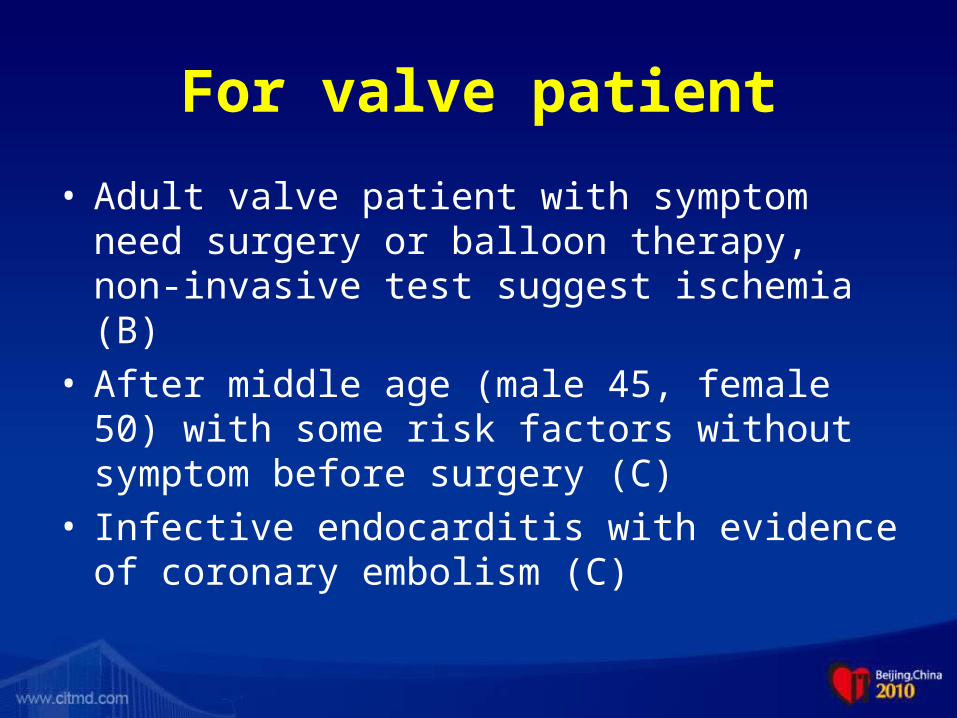
For valve patient
• Adult valve patient with symptom need surgery or balloon therapy, non-invasive test suggest ischemia (B)
• After middle age (male 45, female 50) with some risk factors without symptom before surgery (C)
• Infective endocarditis with evidence of coronary embolism (C)

For heart failure patient
• CHF with systolic dysfunction with angina or segmental wall motion abnomality or any evidence of reversible ischemia (B)
• Before heart transplantation (C)• CHF duo to MI with ventricular aneurysm or any
mechanical complication (C)

For STEMI --- indication
I • Suitable for primary or rescue PCI– within 12 hrs
or over 12 hrs with persistent ischemia, cathlab facility needed (A)
• Cardiogenic shock need revascularization– 36 hrs from onset or 18 hrs after shock (A)
• Complicated with VSD or severe MR need repair (B)
• Complicated with persistent hemodynamic unstable or electrical unstable condition (C)

For STEMI --- contra-indication
III • Multiple co-morbidities, revascularization may
not help the patient (C)

For UA/STEMI --- indication
• Recurrent ischemia after medication (B)• Moderate or High risk : TIMI score≥3.
Age≥65yrs; ≥3 CAD risk factors; Known CAD; Aspirin in past 7 days; Recent (within 24hrs) severe angina; ST deviation≥0.5mm; cardiac markers elevation (A).
• Low risk but non-invasive test show high risk :EF<0.35 、 large area ischemia (esp. anterior wall) or multiple segment ischemia (B)
• UA post-PCI or CABG (C)• Prinzmetal angina (C)

For UA/STEMI --- contra-indication
III • Multiple co-morbiditis, cannot benefit from
revascularization (C)• Chest pain with low risk UA (C)• Not a candidate for revascularization (C)

For recent MI --- indication
• Any ischemic evidence on low level stress test (B)

Relative contra-indication• Unknow fever or fever not controlled• Severe or acute liver or renal dysfunction or
failure• Severe anemia• Severe electrolyte disturbance• End stage carcinoma• Aortic valve IE• Severe bleeding disease or active bleeding
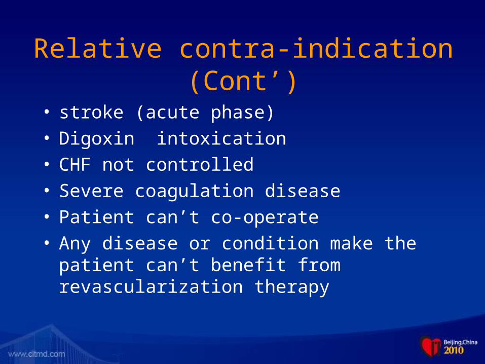
Relative contra-indication (Cont’)
• stroke (acute phase)• Digoxin intoxication• CHF not controlled• Severe coagulation disease• Patient can’t co-operate• Any disease or condition make the patient can’t
benefit from revascularization therapy

Factors increase the risk of CAG
• age> 70 yr• Complicated congenital heart disease• Severe obesity• Cachexia• Uncontrolled hyperglycemia• Hypoxia• Severe COPD• CRF• Hyperthyroidism

Factors increase the risk of CAG (cont’)
• Triple vessel disease• Left main disease• Heart failure, grade IV• Severe mitral valve 、 aortic valve disease or after
mechanical valve replacement• LVEF< 35 %• High risk on treadmill test (with hypotension or severe
ischemia)• Pulmonary hypertension• PCWP> 25 mm Hg

Factors increase the risk of CAG (cont’)
• Coagulation or bleeding disturbance• Uncontrolled hypertension• Severe peripheral artery disease• Recent stroke• Severe aortic insufficency

LV-gram indication
• Routine LV-gram should be done before or after CAG to evaluate the left ventricular function, mitral valve and aortic valve function
• LV was not done regularly in daily practice because the advantage of Echocardiography

LV-gram Contra-indication
• LV thrombus
• Severe LV dysfunction, LVEDP≥20mmHg
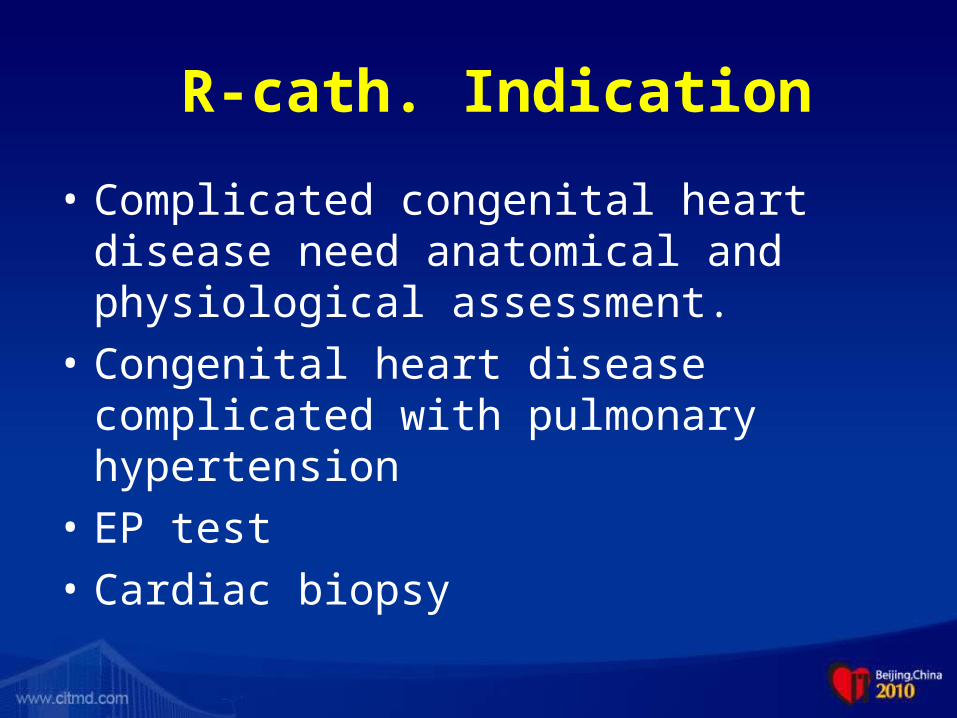
R-cath. Indication
• Complicated congenital heart disease need anatomical and physiological assessment.
• Congenital heart disease complicated with pulmonary hypertension
• EP test
• Cardiac biopsy

R-cath. Contra-indication
• Simple cases can be accurately diagnosed by echocardiography or other non-invasive procedures

Top Related