


Languages
Pages
Legal


Investigating Cells Homework Booklet 20/07/2011
Hyndland Secondary School 1 Biology Department
Hyndland Secondary School
Biology Department
Investigating Cells
Homework and Question Booklet1
Investigating Cells (a) Investigating Living Cells.............................................................. 2
Investigating Cells (b) – Investigating Diffusion ................................................................ 5
Diffusion ................................................................................................................... 5 Osmosis ................................................................................................................... 5
Investigating Cells (d) Investigating Enzymes .................................................................. 10
Enzyme Mechanisms & Investigation .................................................................... 10 Optimum Conditions .............................................................................................. 12 Problem Solving ..................................................................................................... 15
1 This booklet is available online from the Hyndland Secondary School website. Any grids for graphs are also
available on the site should you wish them for practice. There is also a handy hints booklet which will help you
should you get stuck, where appropriate this booklet also includes links to websites giving a more general help with
the section. Answers booklet will also be made available when the section has been completed in class. http://www.hyndland-sec.glasgow.sch.uk/PlainText/PlainText.aspx?SectionId=a0e6692f-ea72-48f6-b58f-cc274d47160b
Investigating Cells Homework Booklet 20/07/2011
Hyndland Secondary School 2 Biology Department
Investigating Cells (a) Investigating Living Cells Marks
1.
2.
3.
4.
5.
What are the basic units of life?
Why do we need to use stains when looking at plant and animal cells under
the microscope?
Draw and label a diagram of an animal cell.
Draw and label a diagram of a plant cell.
State the function of the following parts of a cell:
a. Cell Membrane
b. Cell Wall
c. Cytoplasm
d. Nucleus
e. Vacuole
f. Chloroplast
(1)
(1)
(1)
(1)
(3)
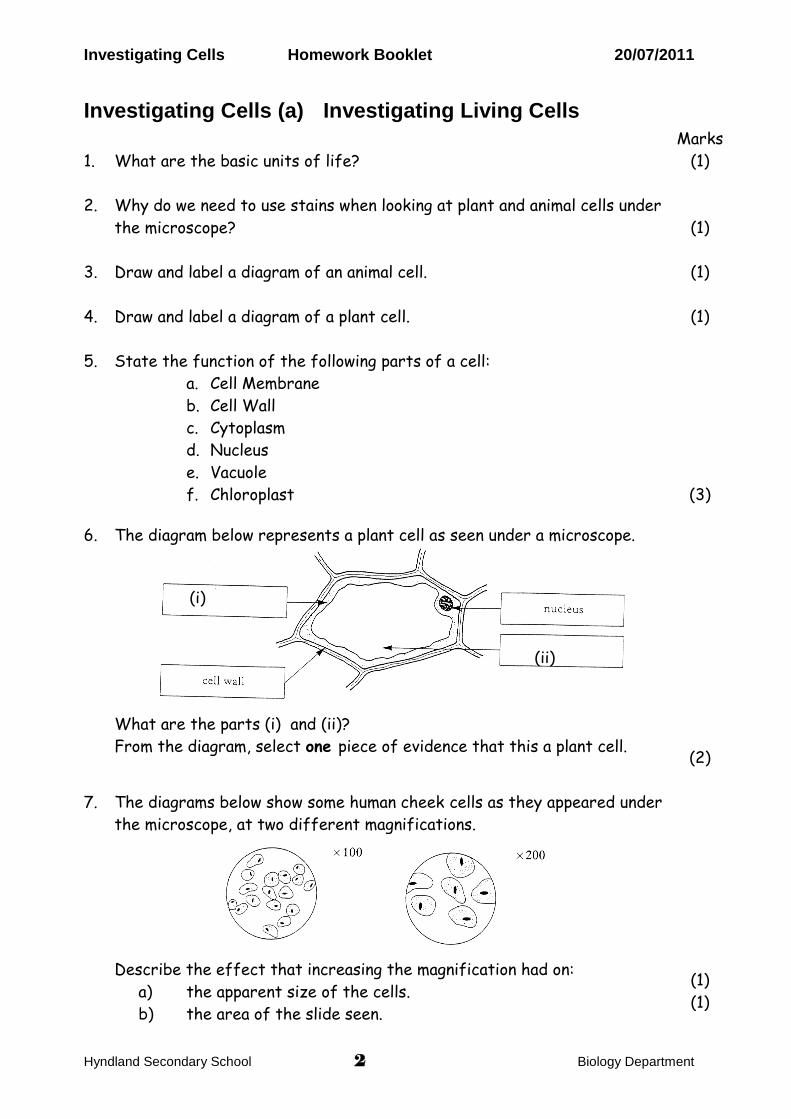
6. The diagram below represents a plant cell as seen under a microscope.
What are the parts (i) and (ii)?
From the diagram, select one piece of evidence that this a plant cell.
(2)
7. The diagrams below show some human cheek cells as they appeared under
the microscope, at two different magnifications.
Describe the effect that increasing the magnification had on:
a) the apparent size of the cells.
b) the area of the slide seen.
(1)
(1)
(i)
(ii)
Investigating Cells Homework Booklet 20/07/2011
Hyndland Secondary School 3 Biology Department
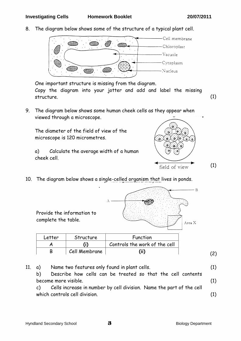
2. The diagram below shows some of the structures of a typical plant cell
8. The diagram below shows some of the structure of a typical plant cell. 2. The diagram below shows some of the structures of a typical plant cell
One important structure is missing from the diagram.
Copy the diagram into your jotter and add and label the missing
structure.
(1)
9.
The diagram below shows some human cheek cells as they appear when
viewed through a microscope.
The diameter of the field of view of the
microscope is 120 micrometres.
a) Calculate the average width of a human
cheek cell.
(1)
10.
The diagram below shows a single-celled organism that lives in ponds.
.
Provide the information to
complete the table.
Letter Structure Function
A (i) Controls the work of the cell
B Cell Membrane (ii)
(2)
11. a) Name two features only found in plant cells.
b) Describe how cells can be treated so that the cell contents
become more visible.
c) Cells increase in number by cell division. Name the part of the cell
which controls cell division.
(1)
(1)
(1)
2. The diagram below shows some of the structures of a typical plant cell
Investigating Cells Homework Booklet 20/07/2011
Hyndland Secondary School 4 Biology Department
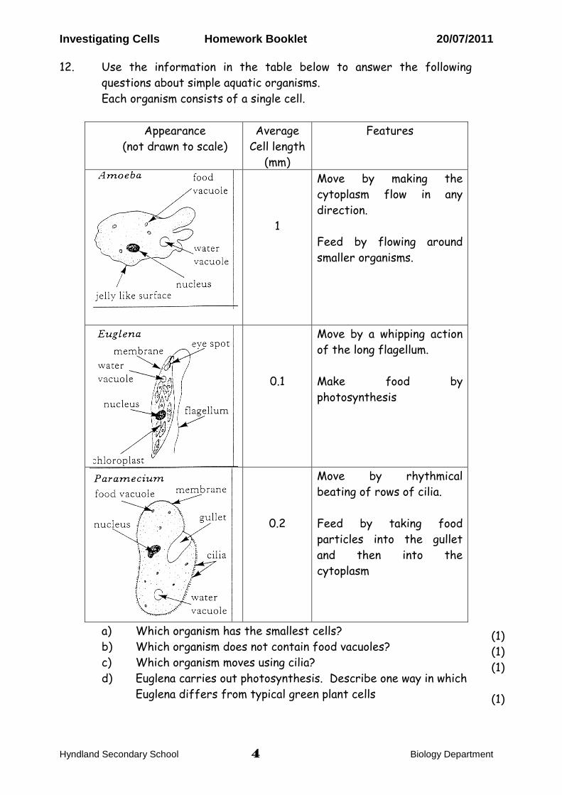
12. Use the information in the table below to answer the following
questions about simple aquatic organisms.
Each organism consists of a single cell.
Appearance
(not drawn to scale)
Average
Cell length
(mm)
Features
1
Move by making the
cytoplasm flow in any
direction.
Feed by flowing around
smaller organisms.
0.1
Move by a whipping action
of the long flagellum.
Make food by
photosynthesis
0.2
Move by rhythmical
beating of rows of cilia.
Feed by taking food
particles into the gullet
and then into the
cytoplasm
a) Which organism has the smallest cells?
b) Which organism does not contain food vacuoles?
c) Which organism moves using cilia?
d) Euglena carries out photosynthesis. Describe one way in which
Euglena differs from typical green plant cells
(1)
(1)
(1)
(1)
Investigating Cells Homework Booklet 20/07/2011
Hyndland Secondary School 5 Biology Department
Investigating Cells (b) – Investigating Diffusion
Diffusion
15.
16.
17.
18.
19.
State what is meant by diffusion.
Name 3 substances that can move by diffusion.
Which part of the cell controls the entry and exit of substances by
diffusion.
Why is diffusion important to living organisms?
The list below names three substances which diffuse into or out of an
animal cell.
Oxygen
Carbon Dioxide
Dissolved Food
State which of these would diffuse in and which would diffuse out of the
animal cell.
(1)
(1)
(1)
(1)
(1)
Osmosis
20.
21.
22.
23.
24.
25.
What is the difference between osmosis and diffusion?
State what is meant by osmosis.
Describe what happens to a plant cell placed in distilled water.
Describe what happens to a plant cell placed in a strong sugar
solution.
Describe what happens to an animal cell placed in distilled water.
Describe what happens to an animal cell placed in a strong sugar
solution.
(1)
(1)
(1)
(1)
(1)
(1)
Investigating Cells Homework Booklet 20/07/2011
Hyndland Secondary School 6 Biology Department
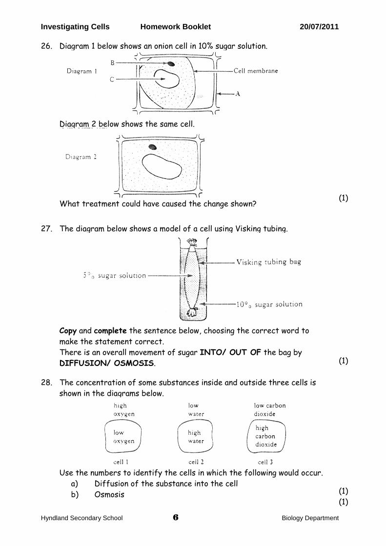
26.
Diagram 1 below shows an onion cell in 10% sugar solution.
Diagram 2 below shows the same cell.
What treatment could have caused the change shown?
(1)
27.
The diagram below shows a model of a cell using Visking tubing.
Copy and complete the sentence below, choosing the correct word to
make the statement correct.
There is an overall movement of sugar INTO/ OUT OF the bag by
DIFFUSION/ OSMOSIS.
(1)
28. The concentration of some substances inside and outside three cells is
shown in the diagrams below.
Use the numbers to identify the cells in which the following would occur.
a) Diffusion of the substance into the cell
b) Osmosis
(1)
(1)
Investigating Cells Homework Booklet 20/07/2011
Hyndland Secondary School 7 Biology Department
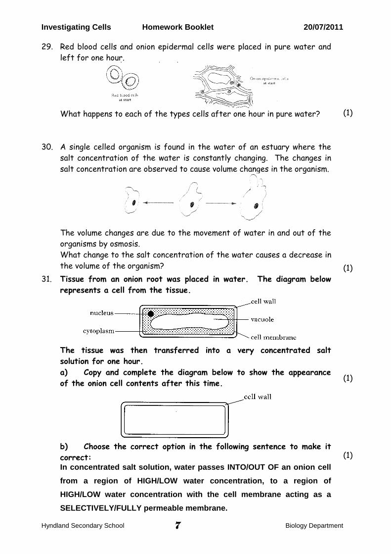
29. Red blood cells and onion epidermal cells were placed in pure water and
left for one hour.
What happens to each of the types cells after one hour in pure water?
(1)
30. A single celled organism is found in the water of an estuary where the
salt concentration of the water is constantly changing. The changes in
salt concentration are observed to cause volume changes in the organism.
The volume changes are due to the movement of water in and out of the
organisms by osmosis.
What change to the salt concentration of the water causes a decrease in
the volume of the organism?
(1)
31. Tissue from an onion root was placed in water. The diagram below
represents a cell from the tissue.
The tissue was then transferred into a very concentrated salt
solution for one hour.
a) Copy and complete the diagram below to show the appearance
of the onion cell contents after this time.
b) Choose the correct option in the following sentence to make it
correct: In concentrated salt solution, water passes INTO/OUT OF an onion cell
from a region of HIGH/LOW water concentration, to a region of
HIGH/LOW water concentration with the cell membrane acting as a
SELECTIVELY/FULLY permeable membrane.
(1)
(1)
Investigating Cells Homework Booklet 20/07/2011
Hyndland Secondary School 8 Biology Department
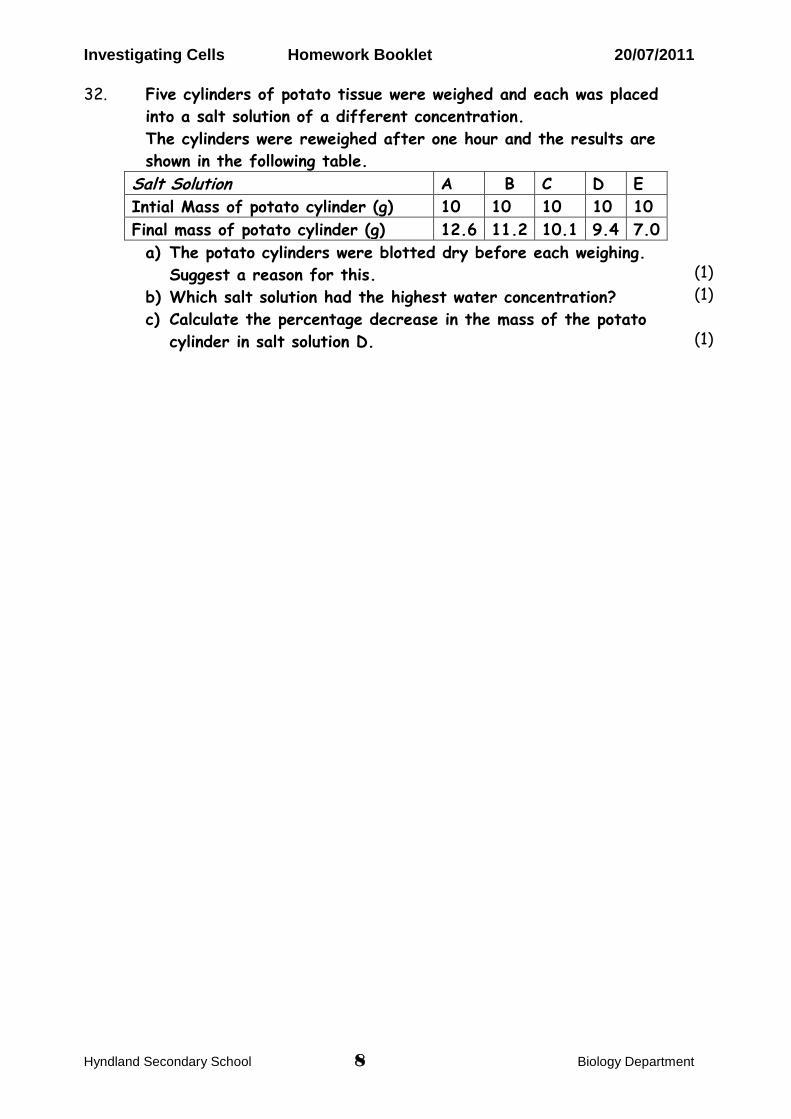
32. Five cylinders of potato tissue were weighed and each was placed
into a salt solution of a different concentration.
The cylinders were reweighed after one hour and the results are
shown in the following table.
Salt Solution A B C D E
Intial Mass of potato cylinder (g) 10 10 10 10 10
Final mass of potato cylinder (g) 12.6 11.2 10.1 9.4 7.0
a) The potato cylinders were blotted dry before each weighing.
Suggest a reason for this.
b) Which salt solution had the highest water concentration?
c) Calculate the percentage decrease in the mass of the potato
cylinder in salt solution D.
(1)
(1)
(1)
Investigating Cells Homework Booklet 20/07/2011
Hyndland Secondary School 9 Biology Department
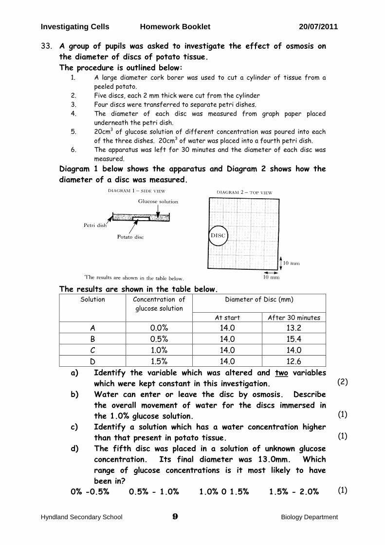
33.
A group of pupils was asked to investigate the effect of osmosis on
the diameter of discs of potato tissue.
The procedure is outlined below: 1. A large diameter cork borer was used to cut a cylinder of tissue from a
peeled potato.
2. Five discs, each 2 mm thick were cut from the cylinder
3. Four discs were transferred to separate petri dishes.
4. The diameter of each disc was measured from graph paper placed
underneath the petri dish.
5. 20cm3 of glucose solution of different concentration was poured into each
of the three dishes. 20cm3 of water was placed into a fourth petri dish.
6. The apparatus was left for 30 minutes and the diameter of each disc was
measured.
Diagram 1 below shows the apparatus and Diagram 2 shows how the
diameter of a disc was measured.
The results are shown in the table below.
Solution Concentration of
glucose solution
Diameter of Disc (mm)
At start After 30 minutes
A 0.0% 14.0 13.2
B 0.5% 14.0 15.4
C 1.0% 14.0 14.0
D 1.5% 14.0 12.6
a) Identify the variable which was altered and two variables
which were kept constant in this investigation.
b) Water can enter or leave the disc by osmosis. Describe
the overall movement of water for the discs immersed in
the 1.0% glucose solution.
c) Identify a solution which has a water concentration higher
than that present in potato tissue.
d) The fifth disc was placed in a solution of unknown glucose
concentration. Its final diameter was 13.0mm. Which
range of glucose concentrations is it most likely to have
been in?
0% -0.5% 0.5% - 1.0% 1.0% 0 1.5% 1.5% - 2.0%
(2)
(1)
(1)
(1)
Investigating Cells Homework Booklet 20/07/2011
Hyndland Secondary School 10 Biology Department
Investigating Cells (d) Investigating Enzymes
Enzyme Mechanisms & Investigation
34.
35.
36.
37.
38.
What is a catalyst?
What is an enzyme?
Why are enzymes important to living organisms?
Enzymes are said to be substrate specific, what does this mean?
What are enzymes made of?
(1)
(1)
(1)
(1)
(1)
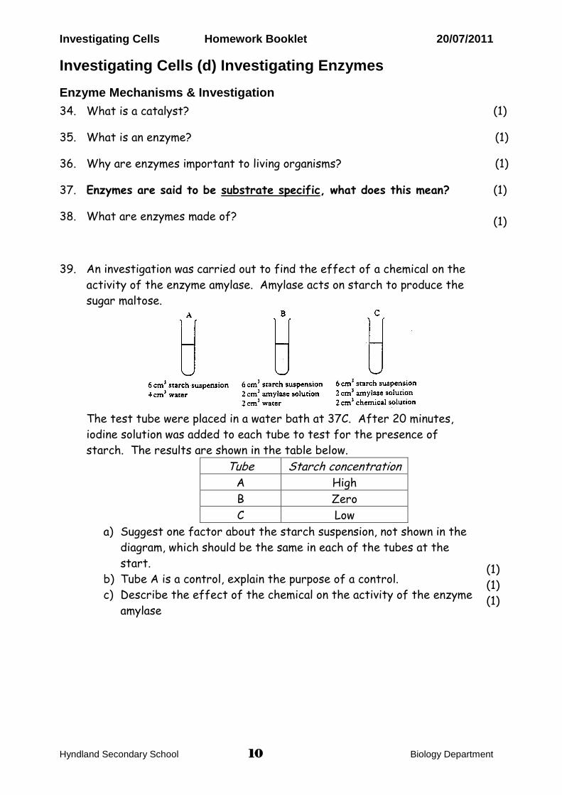
39.
An investigation was carried out to find the effect of a chemical on the
activity of the enzyme amylase. Amylase acts on starch to produce the
sugar maltose.
G
The test tube were placed in a water bath at 37C. After 20 minutes,
iodine solution was added to each tube to test for the presence of
starch. The results are shown in the table below.
Tube Starch concentration A High
B Zero
C Low
a) Suggest one factor about the starch suspension, not shown in the
diagram, which should be the same in each of the tubes at the
start.
b) Tube A is a control, explain the purpose of a control.
c) Describe the effect of the chemical on the activity of the enzyme
amylase
(1)
(1)
(1)
Investigating Cells Homework Booklet 20/07/2011
Hyndland Secondary School 11 Biology Department
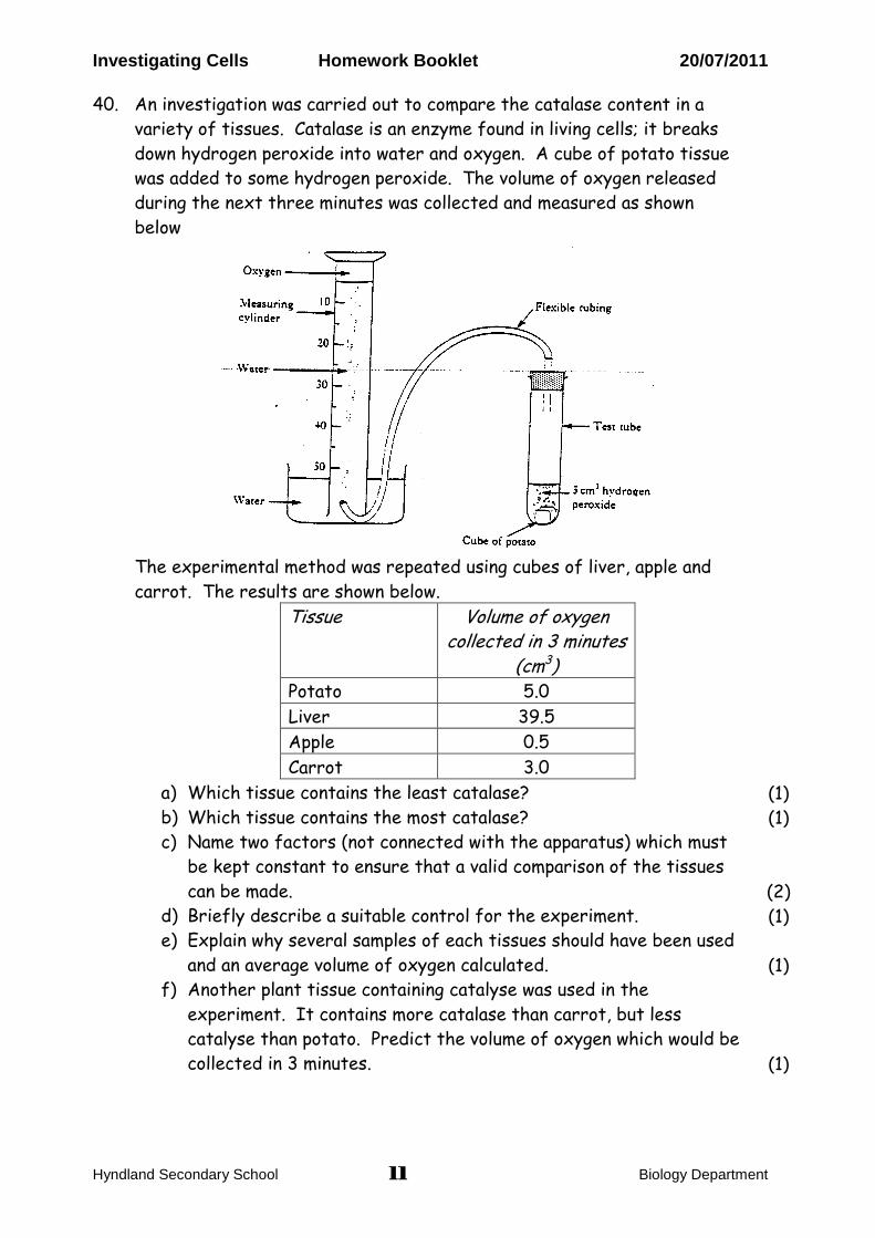
40. An investigation was carried out to compare the catalase content in a
variety of tissues. Catalase is an enzyme found in living cells; it breaks
down hydrogen peroxide into water and oxygen. A cube of potato tissue
was added to some hydrogen peroxide. The volume of oxygen released
during the next three minutes was collected and measured as shown
below
G
The experimental method was repeated using cubes of liver, apple and
carrot. The results are shown below.
Tissue Volume of oxygen collected in 3 minutes
(cm3) Potato 5.0
Liver 39.5
Apple 0.5
Carrot 3.0
a) Which tissue contains the least catalase?
b) Which tissue contains the most catalase?
c) Name two factors (not connected with the apparatus) which must
be kept constant to ensure that a valid comparison of the tissues
can be made.
d) Briefly describe a suitable control for the experiment.
e) Explain why several samples of each tissues should have been used
and an average volume of oxygen calculated.
f) Another plant tissue containing catalyse was used in the
experiment. It contains more catalase than carrot, but less
catalyse than potato. Predict the volume of oxygen which would be
collected in 3 minutes.
(1)
(1)
(2)
(1)
(1)
(1)
Investigating Cells Homework Booklet 20/07/2011
Hyndland Secondary School 12 Biology Department
Optimum Conditions
41.
42.
43.
Describe the effect of increasing the temperature on enzyme activity.
What is meant by the term optimum in relation to enzymes?
What are enzymes made of?
(1)
(1)
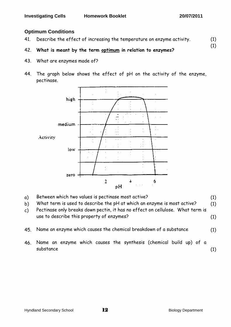
44.
a)
b)
c)
45.
46.
The graph below shows the effect of pH on the activity of the enzyme,
pectinase.
C
Between which two values is pectinase most active?
What term is used to describe the pH at which an enzyme is most active?
Pectinase only breaks down pectin, it has no effect on cellulose. What term is
use to describe this property of enzymes?
Name an enzyme which causes the chemical breakdown of a substance
Name an enzyme which causes the synthesis (chemical build up) of a
substance
(1)
(1)
(1)
(1)
(1)
Investigating Cells Homework Booklet 20/07/2011
Hyndland Secondary School 13 Biology Department
47.
a)
b)
c)
d)
e)
f)
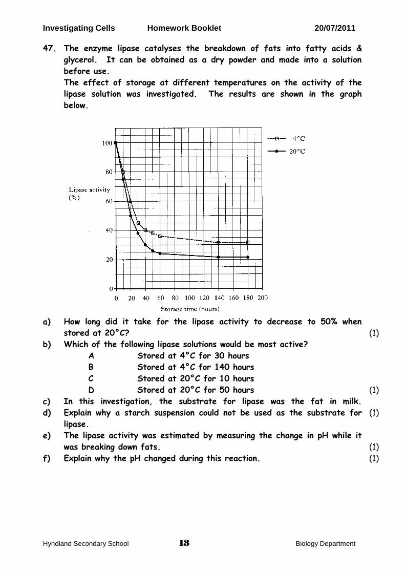
The enzyme lipase catalyses the breakdown of fats into fatty acids &
glycerol. It can be obtained as a dry powder and made into a solution
before use.
The effect of storage at different temperatures on the activity of the
lipase solution was investigated. The results are shown in the graph
below.
C
How long did it take for the lipase activity to decrease to 50% when
stored at 20°C?
Which of the following lipase solutions would be most active?
A Stored at 4°C for 30 hours
B Stored at 4°C for 140 hours
C Stored at 20°C for 10 hours
D Stored at 20°C for 50 hours
In this investigation, the substrate for lipase was the fat in milk.
Explain why a starch suspension could not be used as the substrate for
lipase.
The lipase activity was estimated by measuring the change in pH while it
was breaking down fats.
Explain why the pH changed during this reaction.
(1)
(1)
(1)
(1)
(1)
Investigating Cells Homework Booklet 20/07/2011
Hyndland Secondary School 14 Biology Department
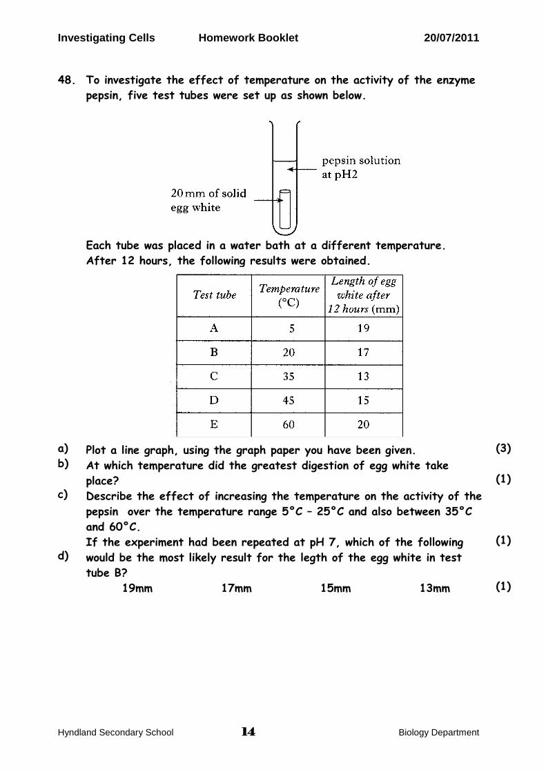
48.
a)
b)
c)
d)
To investigate the effect of temperature on the activity of the enzyme
pepsin, five test tubes were set up as shown below.
Plot a line graph of the data using the graph paper below (3)
G
Each tube was placed in a water bath at a different temperature.
After 12 hours, the following results were obtained.
Plot a line graph of the data using the graph paper below (3)
G
Plot a line graph, using the graph paper you have been given.
At which temperature did the greatest digestion of egg white take
place?
Describe the effect of increasing the temperature on the activity of the
pepsin over the temperature range 5°C – 25°C and also between 35°C
and 60°C.
If the experiment had been repeated at pH 7, which of the following
would be the most likely result for the legth of the egg white in test
tube B?
19mm 17mm 15mm 13mm
(3)
(1)
(1)
(1)
Investigating Cells Homework Booklet 20/07/2011
Hyndland Secondary School 15 Biology Department
Problem Solving
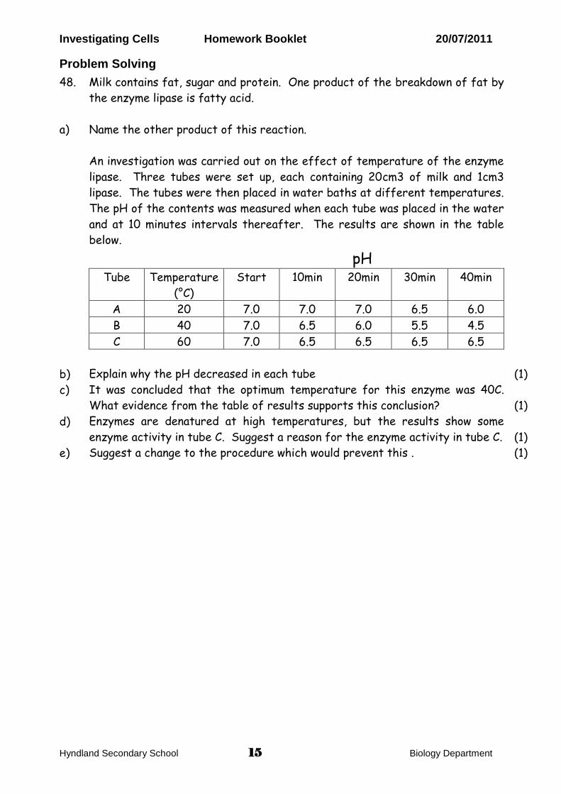
48.
a)
b)
c)
d)
e)
Milk contains fat, sugar and protein. One product of the breakdown of fat by
the enzyme lipase is fatty acid.
Name the other product of this reaction.
An investigation was carried out on the effect of temperature of the enzyme
lipase. Three tubes were set up, each containing 20cm3 of milk and 1cm3
lipase. The tubes were then placed in water baths at different temperatures.
The pH of the contents was measured when each tube was placed in the water
and at 10 minutes intervals thereafter. The results are shown in the table
below.
pH Tube Temperature
(°C)
Start 10min 20min 30min 40min
A 20 7.0 7.0 7.0 6.5 6.0
B 40 7.0 6.5 6.0 5.5 4.5
C 60 7.0 6.5 6.5 6.5 6.5
Explain why the pH decreased in each tube
It was concluded that the optimum temperature for this enzyme was 40C.
What evidence from the table of results supports this conclusion?
Enzymes are denatured at high temperatures, but the results show some
enzyme activity in tube C. Suggest a reason for the enzyme activity in tube C.
Suggest a change to the procedure which would prevent this .
(1)
(1)
(1)
(1)
Top Related