


Languages
Pages
Legal

Postgraduate Medical Journal (April 1974) 50, 199-201.
Gastro-oesophageal reflux and hiatus hernia-endoscopy
W. ROESCHM.D.
Department of Medicine, University of Erlangen-Niiremberg, West Germany
SummaryThe endoscopic diagnosis of hiatal hernia (slidingtype) relies on direct and indirect criteria during theoesophagoscopic or gastroscopic approach. A wideseparation between the anatomic and mucosaloesophago-gastric junction, the presence of a so-calledSchatzki-ring, a wandering junction with slidinggastric mucosal folds during inspiration and two ring-like structures are important. Whereas radiologyseems to be superior to endoscopy in the diagnosis ofasymptomatic hiatus hernia, oesophagoscopy mayreveal sequelae of reflux like oesophagitis, erosions,ulcers and strictures. Peptic oesophagitis is found inabout 10% to cause upper gastrointestinal haemor-rhage.
THE incidence of hiatal hernia varies between 1 and70°O in various studies depending on the enthusiasmof the examiner and the technique of provocation.Although certain endoscopic criteria of hiatus herniahave been established, radiology seems the diagnosticprocedure of choice to demonstrate the presence andsize of a hiatus hernia. From the endoscopic point ofview it is often difficult to differentiate betweenachalasia, cardiac sphincter insufficiency and a smallhiatus hernia; the identification also varies with thetechnique applied where the oesophagoscopic in-spection is usually more reliable (Roesch andOttenjann, 1969).The type of the instrument used may also be of
importance (Trujillo, Slaughter and Boyce, 1968).The squamocolumnar junction varies its locationfrequently forming an indented line which often lieswithin the thorax, 2 or 3 cm proximal to the ana-tomical union. A reflux oesophagitis may mask thiszig-zag line; Palmer's (1967) criteria for the oeso-phagoscopic diagnosis of sliding type hiatal hernia-the finding of the oesophagogastric mucosal junctionabove the diaphragm at 40 cm or less from theincisors, is therefore unreliable. The same is true forthe presence of gastro-oesophageal reflux, a pheno-menon which is only dependent upon the competenceof the gastro-oesophagic sphincter (Dagradi, 1969).Seifert and Kawai (1973) suggest the following directand indirect criteria for the oesophagoscopic exami-nation with prograde optics:
(A) Direct signs(1) Two wide-open ring-like structures (diaphragm-
atic hiatus and functional sphincter) with a dilatedbell-shaped segment in between;
(2) oesophagogastric junction within the dilatedsegment;
(3) location of the squamo-columnar union abovethe diaphragm;
(4) wandering junction during inspiration withsliding gastric mucosal folds.
(B) Indirect signs(1) Shortening of the distance between incisors
and the oesophagogastric junction (less than 38-40cm);
(2) free reflux of gastric contents into the oeso-phagus;
(3) presence of oesophagitis.Ortega (1972) added a new criterion: if the separa-
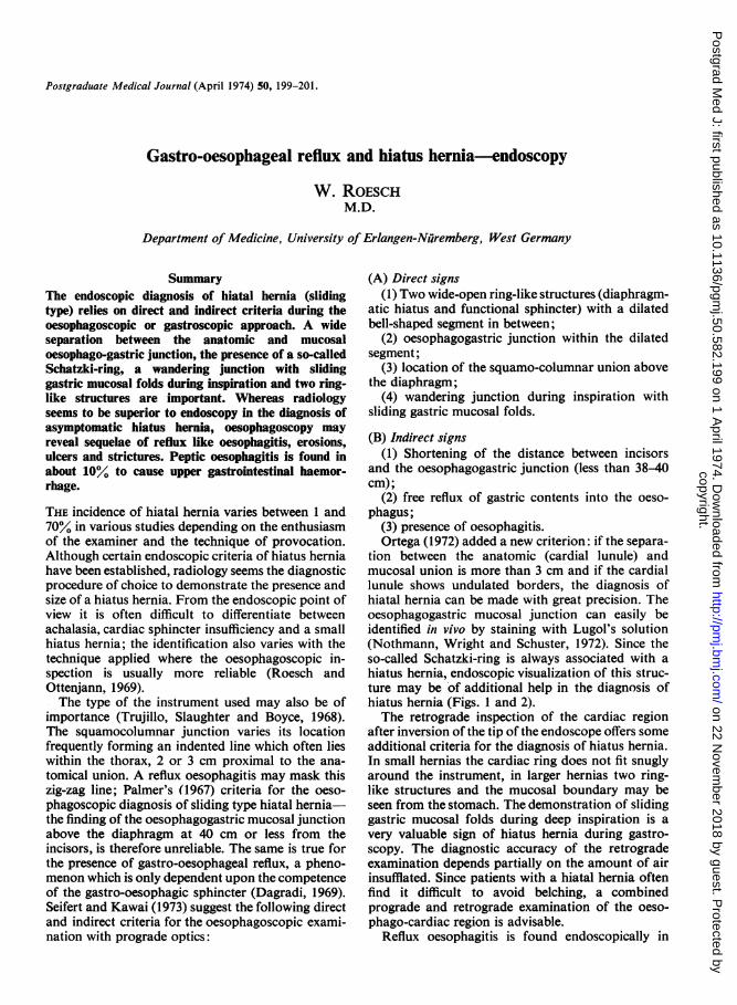
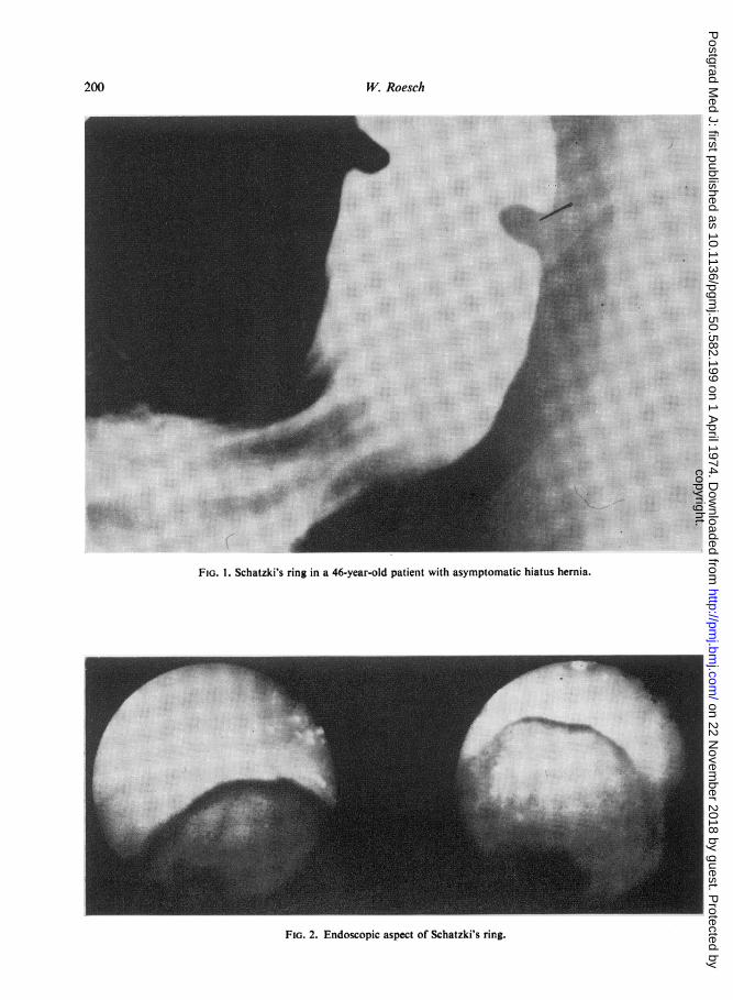
tion between the anatomic (cardial lunule) andmucosal union is more than 3 cm and if the cardiallunule shows undulated borders, the diagnosis ofhiatal hernia can be made with great precision. Theoesophagogastric mucosal junction can easily beidentified in vivo by staining with Lugol's solution(Nothmann, Wright and Schuster, 1972). Since theso-called Schatzki-ring is always associated with ahiatus hernia, endoscopic visualization of this struc-ture may be of additional help in the diagnosis ofhiatus hernia (Figs. 1 and 2).The retrograde inspection of the cardiac region
after inversion of the tip of the endoscope offers someadditional criteria for the diagnosis of hiatus hernia.In small hernias the cardiac ring does not fit snuglyaround the instrument, in larger hernias two ring-like structures and the mucosal boundary may beseen from the stomach. The demonstration of slidinggastric mucosal folds during deep inspiration is avery valuable sign of hiatus hernia during gastro-scopy. The diagnostic accuracy of the retrogradeexamination depends partially on the amount of airinsufflated. Since patients with a hiatal hernia oftenfind it difficult to avoid belching, a combinedprograde and retrograde examination of the oeso-phago-cardiac region is advisable.
Reflux oesophagitis is found endoscopically in
copyright. on 22 N
ovember 2018 by guest. P
rotected byhttp://pm
j.bmj.com
/P
ostgrad Med J: first published as 10.1136/pgm
j.50.582.199 on 1 April 1974. D
ownloaded from
W. Roesch
FIG. 1. Schatzki's ring in a 46-year-old patient with asymptomatic hiatus hernia.
FIG. 2. Endoscopic aspect of Schatzki's ring.
200
copyright. on 22 N
ovember 2018 by guest. P
rotected byhttp://pm
j.bmj.com
/P
ostgrad Med J: first published as 10.1136/pgm
j.50.582.199 on 1 April 1974. D
ownloaded from
Gastro-oesophageal reflux and hiatus hernia 201
,~"- ~·' ;:.. ". i,.' ... *~:. :.;: :'-'.~' ,...:~~~~~~~~~~~~~~~~~~~~...............................,:,.......*,~.....
4~P7,,~·,::, i'"":.'.':'- - .
~:~: .:..:::~~..
Cg.*..... :..:..
...::.:
::: ......
:r.":.:t'~*::.,:,i·!* . . : r: . . :,* ' :Y ::::,, :i*:::~g.L:'::::::::::i:::. .::i:!:::~
.:: :.....~
"" .... ... .g...:"..?. I:BI/::::. ,, : . ~ ~ ~ ~ ~~ ~~~~····;:::¢~i~:::~::i:j....======================
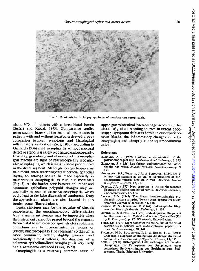
FIG. 3. Moniliasis in the biopsy specimen of membranous oesophagitis.
about 50% of patients with a large hiatal hernia(Seifert and Kawai, 1973). Comparative studiesusing suction biopsy of the terminal oesophagus inpatients with and without heartburn showed a poorcorrelation between symptoms and histologicalinflammatory infiltration (Zeus, 1970). According toGaillard (1956) mild oesophagitis without mucosaldefect or stenosis is rarely recognized endoscopically.Friability, granularity and ulceration of the oesopha-geal mucosa are signs of macroscopically recogniz-able oesophagitis, which is usually more pronouncedin the distal segment. Although forceps biopsy maybe difficult, often rendering only superficial epitheliallayers, an attempt should be made especially inmembranous oesophagitis to rule out moniliasis(Fig. 3). At the border zone between columnar andsquamous epithelium polypoid changes may oc-casionally be seen in extensive oesophagitis, whichcould lead to the false diagnosis of carcinoma. Deeptherapy-resistant ulcers are also located in thisborder zone (Barrett-ulcer).
Peptic strictures may be the sequelae of chronicoesophagitis; the oesophagoscopic differentiationfrom a malignant stenosis may be impossible whenthe instrument cannot be passed beyond the stenosis.When distal to a mid-oesophageal stricture columnarepithelium can be demonstrated by biopsy or(rarely) macroscopically (the columnar epithelium ismore prominent, redder, slightly granular andoccasionally almost villous), the diagnosis of acolumnar epithelium-lined oesophagus is very likelyand a carcinoma excluded (Trier, 1970).
Oesophagitis is a relatively common cause of
upper gastrointestinal haemorrhage accounting forabout 10% of all bleeding sources in urgent endo-scopy; asymptomatic hiatus hernia in our experiencenever bleeds, the inflammatory changes in refluxoesophagitis end abruptly at the squamocolumnarunion.
ReferencesDAGRADI, A.E. (1969) Endoscopic examination of the
gastrooesophageal area. Gastrointestinal Endoscopy, 3, 175.GAILLARD, J. (1956) Les formes endoscopiques de l'oeso-
phagite par reflux. Journal franfaise Oto-rhino-laryng, 5,530.
NOTHMANN, B.J., WRIGHT, J.R. & SCHUSTER, M.M. (1972)In vivo vital staining as an aid to identification of eso-phagogastric mucosal junction in man. American Journalof Digestive Diseases, 17, 919.
ORTEGA, J.A. (1972) New criterion in the esophagoscopicdiagnosis of sliding type hiatal hernia. American Journal ofGastroenterology, 57, 410.
PALMER, E.D. (1967) The hiatus hernia-esophagitis eso-phageal structure complex. Twenty years prospective study.American Journal of Medicine, 44, 566.
ROESCH, W. & OTTENJANN, R. (1969) Endoskopische Diag-nose der Hiatusgleithernie. Endoscopy, 1, 156.
SEIFERT, E. & KAWAI, K. (1973) Endoskopische Diagnostikder Hiatushernie. In: Refluxkrankheit der Speiserohre (Ed.by R. Ottenjann), p. 47. Witzstrock, Baden-Baden.
TRIER, J.H. (1970) Morphology of the epithelium of the distaloesophagus in patients with mid-esophageal peptic stric-tures. Gastroenterology, 58, 444.
TRUJILLO, N.P., SLAUGHTER, R.L. & BOYCE, H.W. (1968)Endoscopic diagnosis of sliding type diaphragmatic hiatalhernia. American Journal of Digestive Diseases, 13, 855.
ZEUS, J. (1970) Histologische Untersuchungen am distalenOesophagus zur Pathogenese der Oesophagitis unterbesonderer Beriicksichtigung der Beziehung zum Sod-brennen. Thesis, Erlangen University.
copyright. on 22 N
ovember 2018 by guest. P
rotected byhttp://pm
j.bmj.com
/P
ostgrad Med J: first published as 10.1136/pgm
j.50.582.199 on 1 April 1974. D
ownloaded from
Top Related