


![Choledocholithiasis, Ascending Cholangitis, and Gallstone ... · bilirubinate to form biliary sludge, which can aggregate eventually into a gallbladder stone [10]. Black pigment stones](https://static.fdocuments.net/doc/165x107/5e04b56d64882534e3400732/choledocholithiasis-ascending-cholangitis-and-gallstone-bilirubinate-to-form.jpg)
Languages
Pages
Legal

GALLSTONE:
DIAGNOSIS AND TREATMENT
Bijan Shahbazkhani
Imam Khomeini hospital
CLINICAL MANIFESTATIONS OF
GALLSTONES
Asymptomatic Gallstones
Symptomatic Gallstons
Biliary Colic
Acute Cholecystitis
Choledocholithiasis
Acute Cholangitis
Complications
DIAGNOSTIC EVALUATION
Laboratory Evaluation
Imaging
Abdominal radiographs
Trans-abdominal Ultrasound
Nuclear Medicine (HIDA scan)
Magnetic Resonance Imaging (MRC/MRCP)
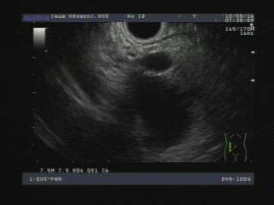
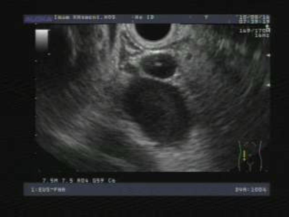
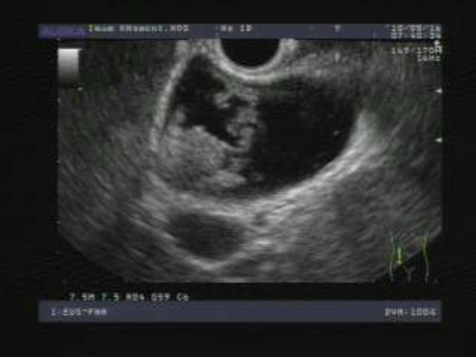
Endoscopic Ultrasound (EUS)
Endoscopic Retrograde Cholangiopancreatography (ERCP)
CT scan
IMPORTANT LABS
Infection
WBC
Blood Cultures
Hepatocyte inflammation and injury
AST/ALT
Biliary Obstruction and Cholangiocyte injury
Total Bilirubin
Alkaline Phosphatase
Gallstone Pancreatitis
Amylase (salivary, bowel)
Lipase (more specific to pancreas)
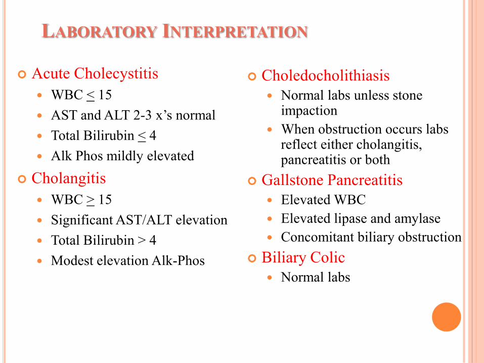
LABORATORY INTERPRETATION
Acute Cholecystitis
WBC < 15
AST and ALT 2-3 x’s normal
Total Bilirubin < 4
Alk Phos mildly elevated
Cholangitis
WBC > 15
Significant AST/ALT elevation
Total Bilirubin > 4
Modest elevation Alk-Phos
Choledocholithiasis
Normal labs unless stone impaction
When obstruction occurs labs reflect either cholangitis, pancreatitis or both
Gallstone Pancreatitis
Elevated WBC
Elevated lipase and amylase
Concomitant biliary obstruction
Biliary Colic
Normal labs
ABDOMINAL RADIOGRAPHS
Abdominal series:
supine, upright, decubitus and upright CXR
Low sensitivity and specificity for stones
Aid in the differential diagnosis
Calcification in pancreatitis
Ileus
Perforation
Intestinal pneumotosis
Pneumonia
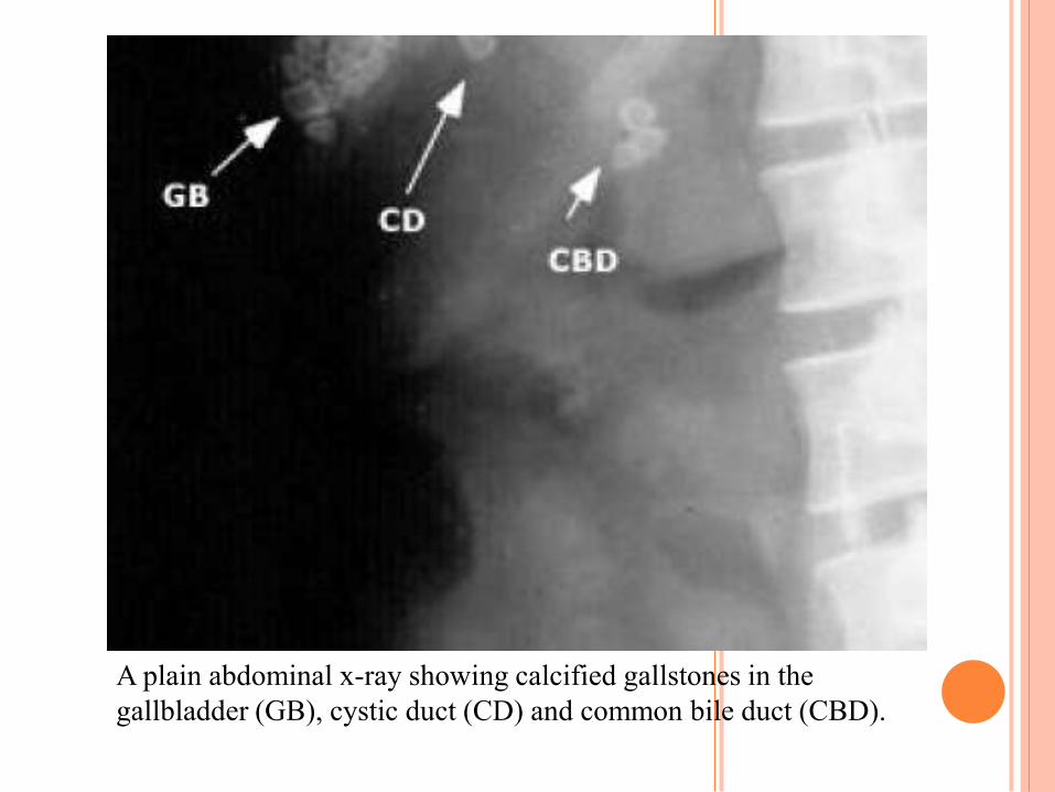
A plain abdominal x-ray showing calcified gallstones in the
gallbladder (GB), cystic duct (CD) and common bile duct (CBD).
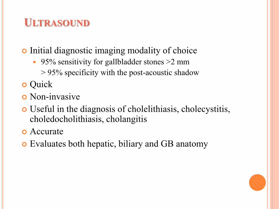
ULTRASOUND
Initial diagnostic imaging modality of choice
95% sensitivity for gallbladder stones >2 mm
> 95% specificity with the post-acoustic shadow
Quick
Non-invasive
Useful in the diagnosis of cholelithiasis, cholecystitis, choledocholithiasis, cholangitis
Accurate
Evaluates both hepatic, biliary and GB anatomy
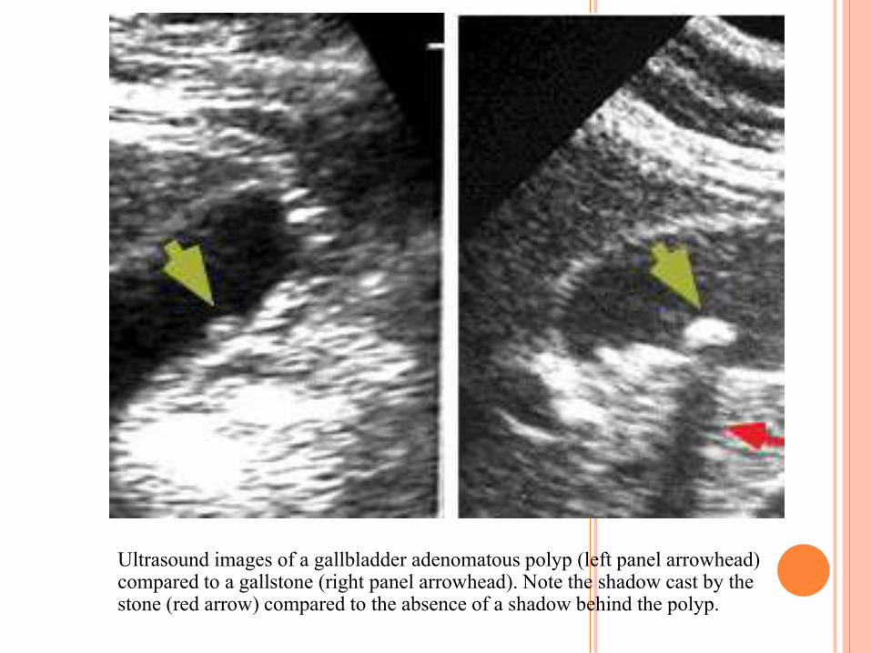
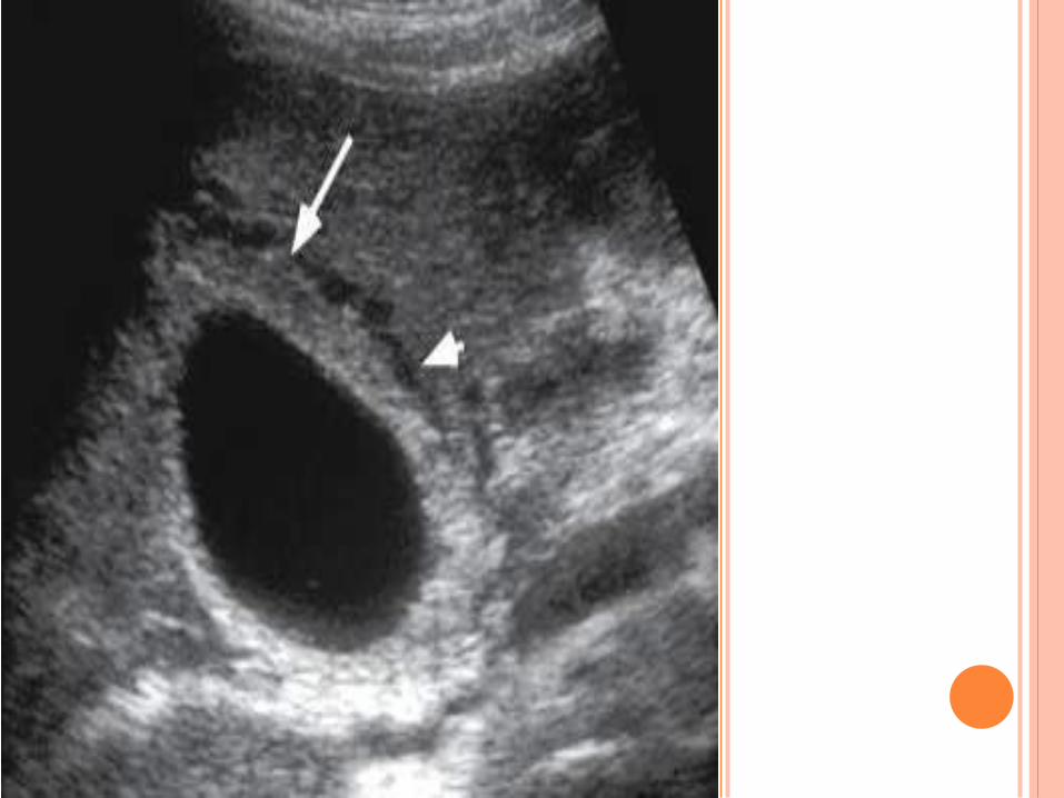
Ultrasound images of a gallbladder adenomatous polyp (left panel arrowhead) compared to a gallstone (right panel arrowhead). Note the shadow cast by the stone (red arrow) compared to the absence of a shadow behind the polyp.
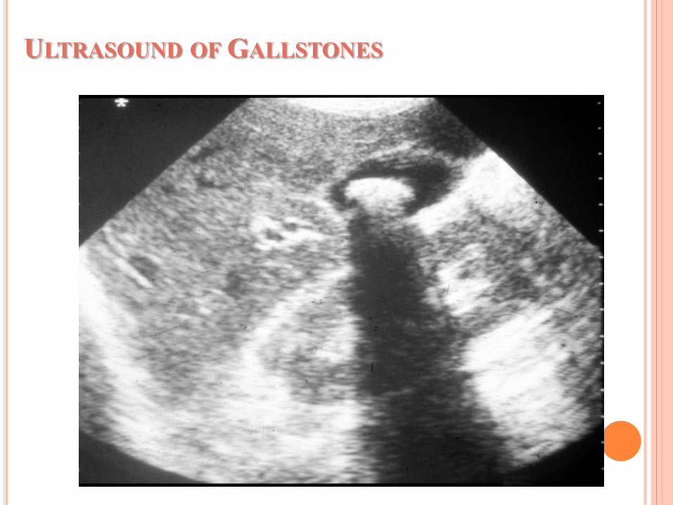
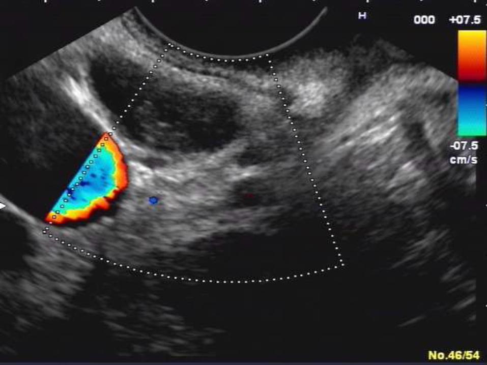
ULTRASOUND OF GALLSTONES
ULTRASOUND
Acute Cholecystitis
Pericholecystic fluid and/or stranding
Thickened gall bladder wall
Intramural gas
Cholelithiasis and/or GB sludge
Sonographic Murphy’s sign - PPV >90%
Choledocholithiasis
Extrahepatic stone localization
Sensitivity 50% - Common Bile Duct stones
Sensitivity 75% - dilated CBD
> 6 mm intact gallbladder
>10 mm post-cholecystectomy
CHOLESCINTIGRAPHY (HIDA)
Useful if the Ultrasound is non-diagnostic
A positive HIDA-scan for acute cholecystitis:
normal uptake of HIDA by the liver,
rapid excretion into the biliary system,
visualization of the extrahepatic bile ducts,
appearance of HIDA in the intestine,
failure to visualize the gallbladder
Sensitivity - 95%
Specificity - 90%
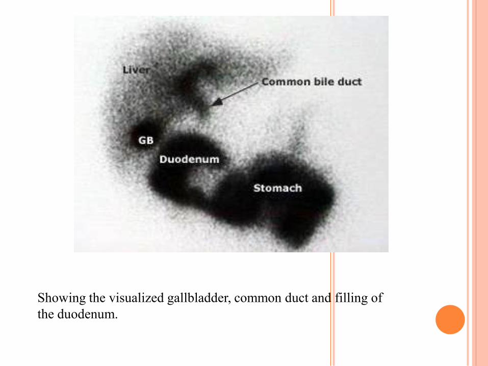
Showing the visualized gallbladder, common duct and filling of
the duodenum.
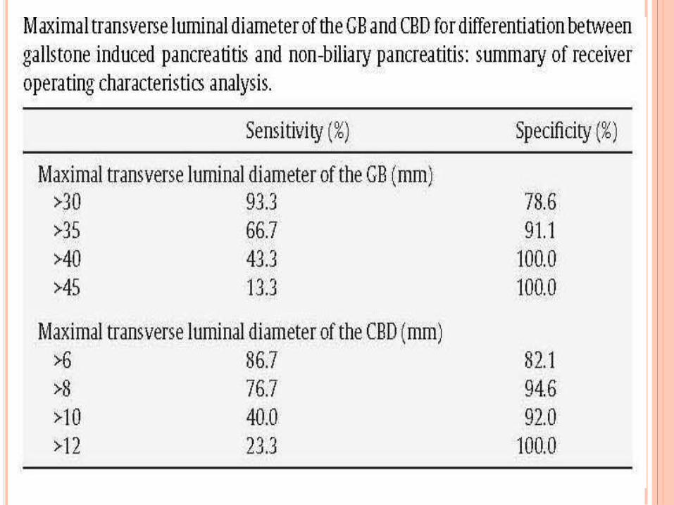
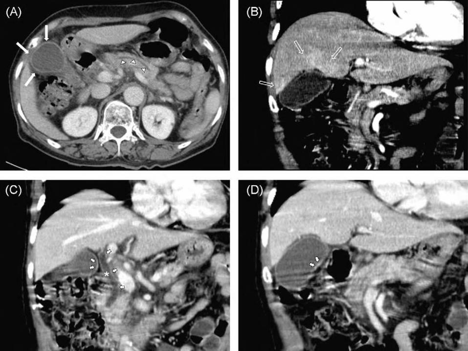
DIAGNOSTIC VALUE OF CT FEATURES OF
THE GALLBLADDER IN THE PREDICTION
OF GALLSTONE PANCREATITIS
Materials and methods: Eighty-six patients who
underwent a diagnostic computed tomography (CT) scan
for acute pancreatitis were included. The readers assessed
the presence of pericholecystic increased attenuation of the
liver parenchyma, enhancement of gallbladder (GB) and
common bile duct (CBD) wall, pericholecystic fat strands,
GB wall thickening, stone in the GB or CBD, and focal or
diffuse manifestations of pancreatitis on abdominal CT
scans. In addition, the maximal transverse luminal
diameters of the GB and CBD were measured.
EUROPEAN JOURNAL OF RADIOLOGY, 2010
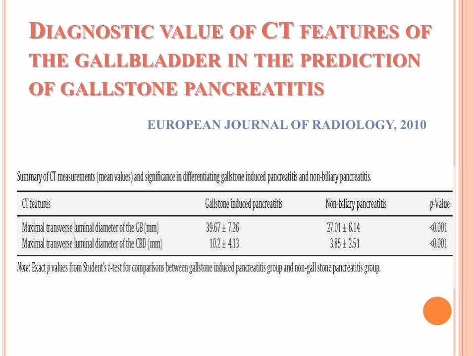
DIAGNOSTIC VALUE OF CT FEATURES OF
THE GALLBLADDER IN THE PREDICTION
OF GALLSTONE PANCREATITIS
EUROPEAN JOURNAL OF RADIOLOGY, 2010
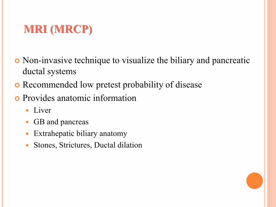
MRI (MRCP)
Non-invasive technique to visualize the biliary and pancreatic
ductal systems
Recommended low pretest probability of disease
Provides anatomic information
Liver
GB and pancreas
Extrahepatic biliary anatomy
Stones, Strictures, Ductal dilation
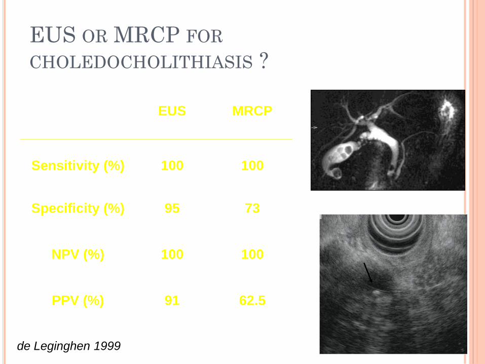
EUS OR MRCP FOR
CHOLEDOCHOLITHIASIS ?
EUS MRCP
Sensitivity (%) 100 100
Specificity (%) 95 73
NPV (%) 100 100
PPV (%) 91 62.5
de Leginghen 1999
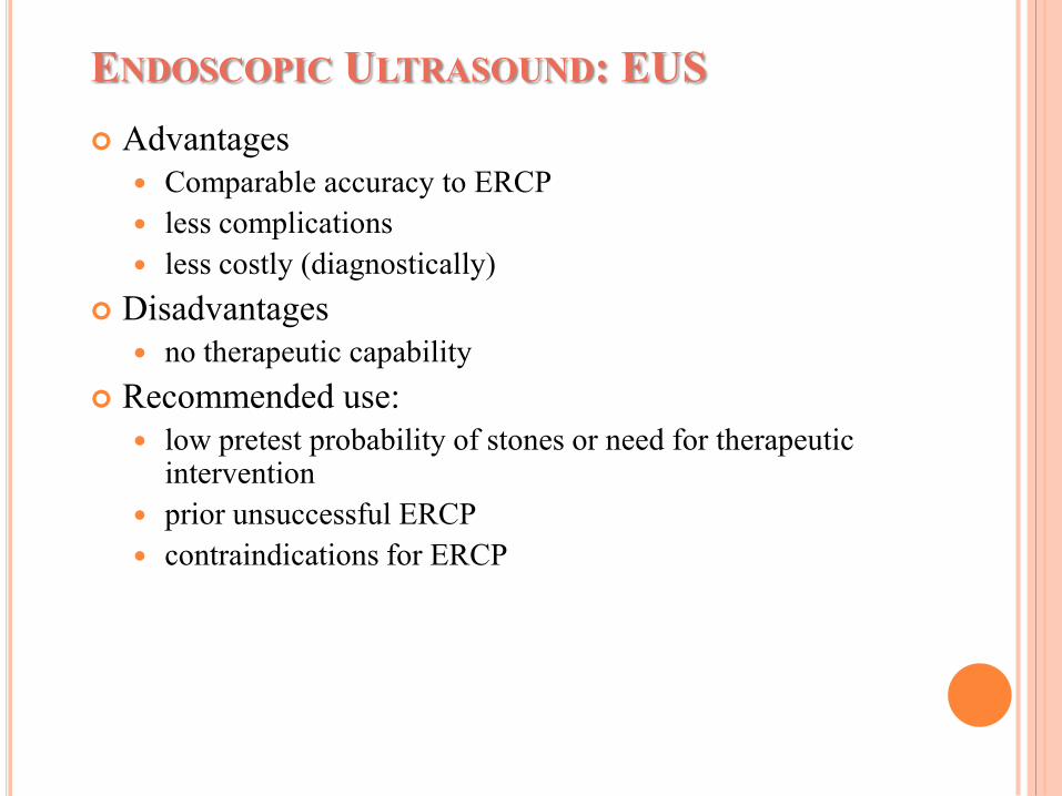
ENDOSCOPIC ULTRASOUND: EUS
Advantages
Comparable accuracy to ERCP
less complications
less costly (diagnostically)
Disadvantages
no therapeutic capability
Recommended use:
low pretest probability of stones or need for therapeutic intervention
prior unsuccessful ERCP
contraindications for ERCP
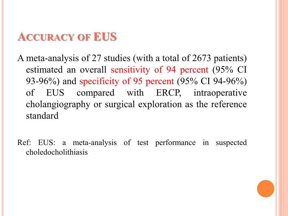
ACCURACY OF EUS
A meta-analysis of 27 studies (with a total of 2673 patients)
estimated an overall sensitivity of 94 percent (95% CI
93-96%) and specificity of 95 percent (95% CI 94-96%)
of EUS compared with ERCP, intraoperative
cholangiography or surgical exploration as the reference
standard
Ref: EUS: a meta-analysis of test performance in suspected
choledocholithiasis
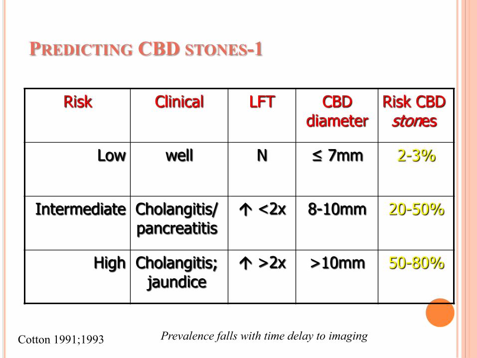
PREDICTING CBD STONES-1
Risk Clinical LFT CBD diameter
Risk CBD stones
Low well N ≤ 7mm 2-3%
Intermediate Cholangitis/ pancreatitis
<2x 8-10mm 20-50%
High Cholangitis; jaundice
>2x >10mm 50-80%
Cotton 1991;1993 Prevalence falls with time delay to imaging
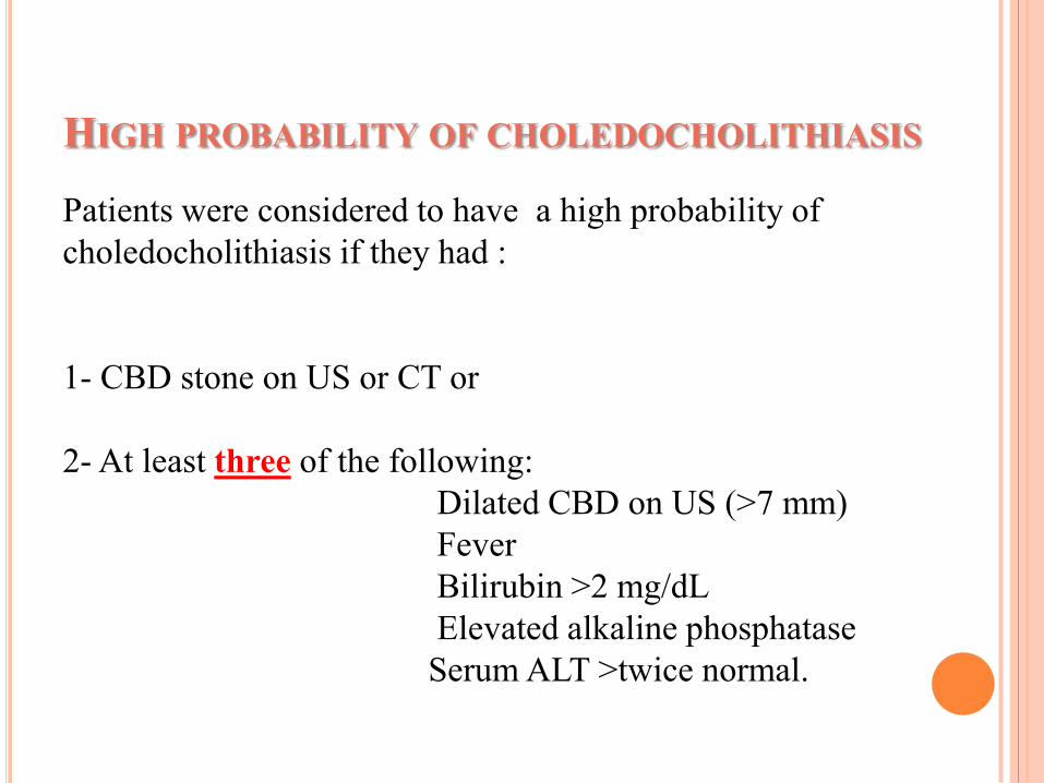
HIGH PROBABILITY OF CHOLEDOCHOLITHIASIS
Patients were considered to have a high probability of
choledocholithiasis if they had :
1- CBD stone on US or CT or
2- At least three of the following:
Dilated CBD on US (>7 mm)
Fever
Bilirubin >2 mg/dL
Elevated alkaline phosphatase
Serum ALT >twice normal.
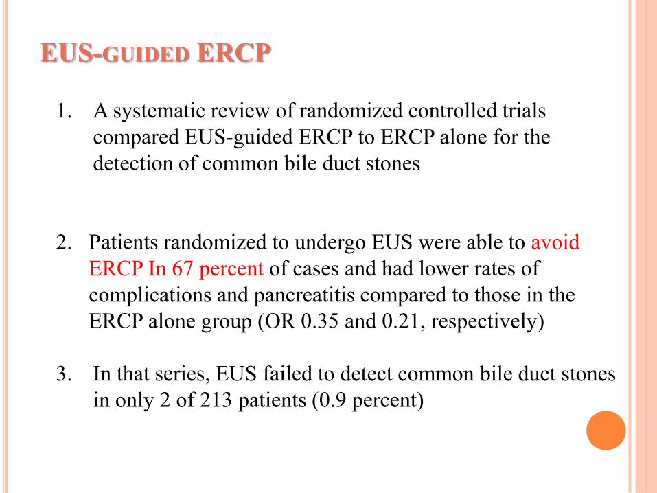
EUS-GUIDED ERCP
1. A systematic review of randomized controlled trials
compared EUS-guided ERCP to ERCP alone for the
detection of common bile duct stones
2. Patients randomized to undergo EUS were able to avoid
ERCP In 67 percent of cases and had lower rates of
complications and pancreatitis compared to those in the
ERCP alone group (OR 0.35 and 0.21, respectively)
3. In that series, EUS failed to detect common bile duct stones
in only 2 of 213 patients (0.9 percent)
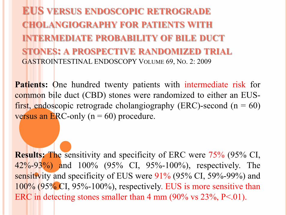
EUS VERSUS ENDOSCOPIC RETROGRADE
CHOLANGIOGRAPHY FOR PATIENTS WITH
INTERMEDIATE PROBABILITY OF BILE DUCT
STONES: A PROSPECTIVE RANDOMIZED TRIALGASTROINTESTINAL ENDOSCOPY VOLUME 69, NO. 2: 2009
Patients: One hundred twenty patients with intermediate risk for
common bile duct (CBD) stones were randomized to either an EUS-
first, endoscopic retrograde cholangiography (ERC)-second (n = 60)
versus an ERC-only (n = 60) procedure.
Results: The sensitivity and specificity of ERC were 75% (95% CI,
42%-93%) and 100% (95% CI, 95%-100%), respectively. The
sensitivity and specificity of EUS were 91% (95% CI, 59%-99%) and
100% (95% CI, 95%-100%), respectively. EUS is more sensitive than
ERC in detecting stones smaller than 4 mm (90% vs 23%, P<.01).
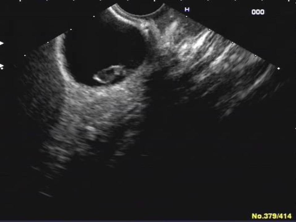
CASE REPORT
1-A 58 years old male
2-Recurrent severe RUQ pain& fever
3-AST=24, ALT=65,Alk.Ph=731
4-MRI=mild dilation of CBD +hydatic cyst
in liver
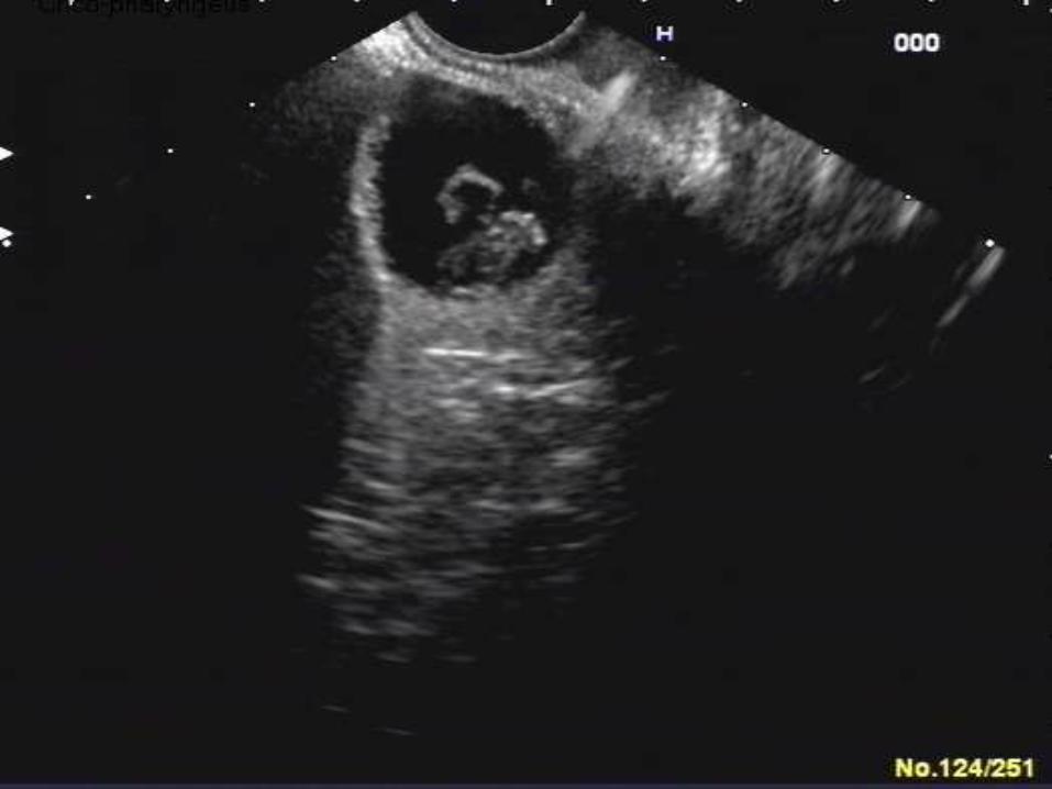
CASE REPORT
1. A 50 year old female
2. Recent mild pancreatitis
3. Elevated alkaline phosphatase
4. Sonography = Gallstone
ENDOSCOPIC RETROGRADE
CHOLANGIOPANCREATOGRAPHY: ERCP
Gold standard
Choledocholithiasis, unresolving cholangitis or gallstone pancreatitis
Both diagnostic and therapeutic potential
Sensitivity - 95%
Specificity - 95%
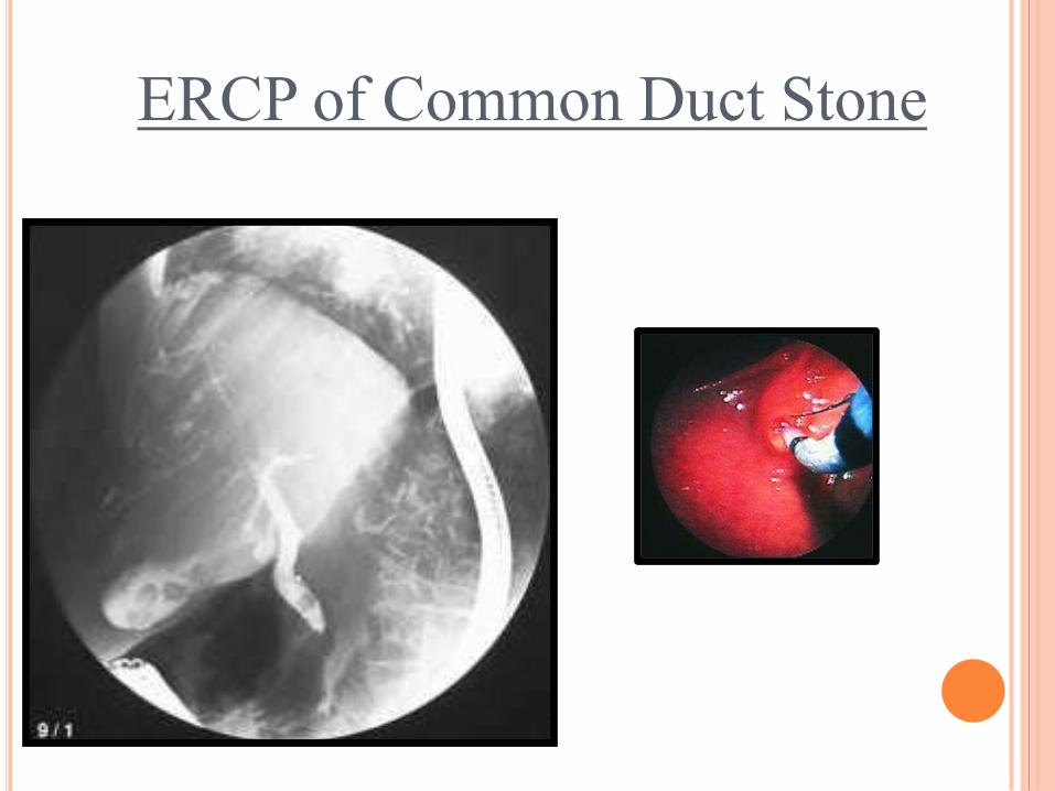
ERCP of Common Duct Stone
While gallstones are associated with cancers of the
gallbladder, the actual nature of their relationship needs to be
clarified. This would aid the recommendations on the need for
prophylactic cholecystectomy.
The evidence at the current time indicates that gallstones are a
cofactor in the causation of gallbladder cancer. Absolute proof of
their role as a cause for gallbladder cancer is lacking. The
recommendation for prophylactic cholecystectomy in countries
reporting a high incidence of gallbladder cancer and associated
gallstones needs to be tailored to the epidemiological profile of
the place.
Cholelithiasis in Gallbladder Cancer: Coincidence, Cofactor, or Cause!
EJSO 36 (2010); 514-519
In the case of gallstones, despite the lack of evidence
to support a recommendation, large stones (>3 cm)
or a gallbladder packed with stones (high stone/GB
volume ratio) could serve as potential indications for
prophylactic cholecystectomy.
Cholelithiasis in Gallbladder Cancer: Coincidence, Cofactor, or Cause!
EJSO 36 (2010); 514-519
SURGICAL TREATMENT OF GALLSTONES
The distinction between symptomatic and
asymptomatic gallstones can be difficult, as
symptoms can be mild and varied. The different
symptoms attributable to gallstones include
upper abdominal pain, biliary colic, and
dyspepsia. About 92% of patients with biliary
colic, 72% of patients with upper abdominal
pain, and 56% of patients with dyspepsia have
relief of symptoms after cholecystectomy.
GASTROENTEROL CLIN N AM 39 (2010); 229–244
TREATMENT
Asymptomatic GS
Prophylactic cholecystectomy
Symptomatic GS
Operative and Endoscopic management
Uncomplicated biliary colic
Acute cholecystitis, Cholangitis
Choledocholithiasis
Oral Bile acid therapy
Methyl-tert-butyl-ether (MTBE)
Extracorporal Shock Wave Lithotripsy (ESWL)
Endoscopic Sphincterotomy
TREATMENT ASYMPTOMATIC GS
Given the costs associated with cholecystectomy
The relatively benign natural history
Prophylactic cholecystectomy is NOT routinely recommended for
asymptomatic patients
Observation

PROPHYLACTIC CHOLECYSTECTOMY
Prophylactic cholecystectomy may be considered in those patients with high rates of complications:
Calcified (porcelain) gallbladder (high cancer risk)
Ileal resections,
Liver transplantation, Sickle cell disease
Children
Morbid obesity&gastrectomy
Choledocholithiasis
Immunocompromised
Chronic hemolysis+ splenectomy
Large stone > 2.5cm
GB polyp
TREATMENT SYMPTOMATIC GS
Elective cholecystectomy is the preferred treatment of patients with symptomatic GS
Biliary colic, Cholecystitis, Choledocholithiasis, Cholangitis, GS pancreatitis
Overall mortality for cholecystectomy 0.5%
Significantly lower for elective operations
Higher in emergencies
2-3x higher for CBD exploration
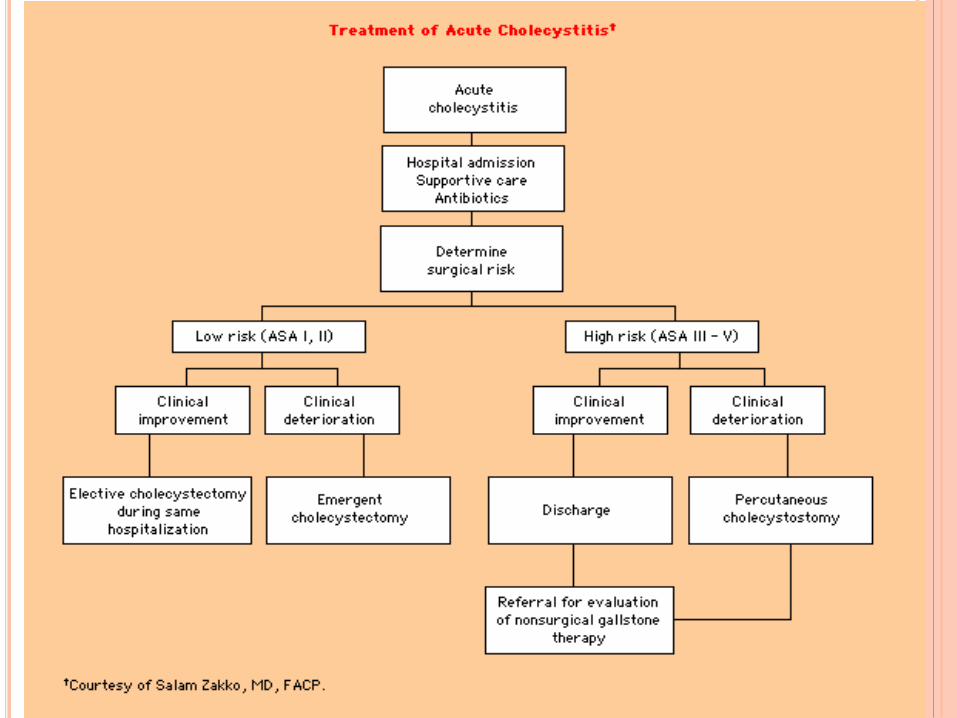
TREATMENT SYMPTOMATIC GS
Timing of cholecystectomy is variable
Always preferable to resolve acute infection and stabilize patient
prior to operative intervention
NPO, IVF
Appropriate antibiotics
Decompression and drainage
Endoscopic (Sphincterotomy and/or biliary stenting)
Percutaneous (Biliary or GB drain placement)
Operatively
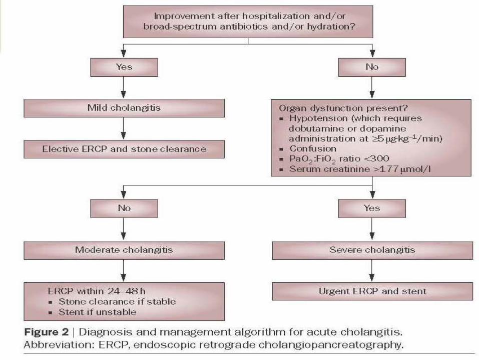
NATURE REVIEWS | GASTROENTEROLO
GY & EPATOLOGY,
SEP 2009
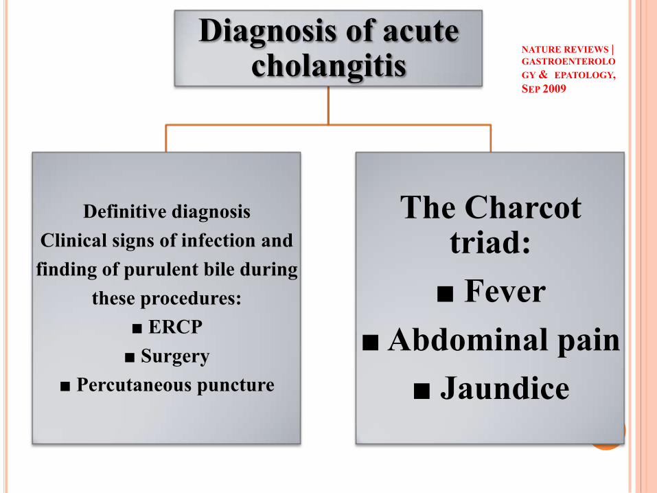
Diagnosis of acute cholangitis
Definitive diagnosis
Clinical signs of infection and
finding of purulent bile during
these procedures:
■ ERCP
■ Surgery
■ Percutaneous puncture
The Charcot triad:
■ Fever
■ Abdominal pain
■ Jaundice
Diagnosis of acute cholangitis has traditionally been made by the
Charcot triad criteria; that is, clinical findings of fever, biliary tract
pain and jaundice.
■ Approximately 80% of patients with acute cholangitis respond to broad-
spectrum antibiotics alone while the remainder require early biliary
drainage in addition to antibiotic therapy.
■ Endoscopic retrograde cholangiopancreatography (ERCP) and stent
placement are considerably safer than surgical biliary decompression.
■ Elective cholecystectomy should be performed after resolution of acute
cholangitis in patients with an intact gallbladder.
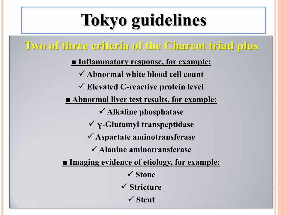
Two of three criteria of the Charcot triad plus
■ Inflammatory response, for example:
Abnormal white blood cell count
Elevated C-reactive protein level
■ Abnormal liver test results, for example:
Alkaline phosphatase
γ-Glutamyl transpeptidase
Aspartate aminotransferase
Alanine aminotransferase
■ Imaging evidence of etiology, for example:
Stone
Stricture
Stent
Tokyo guidelines
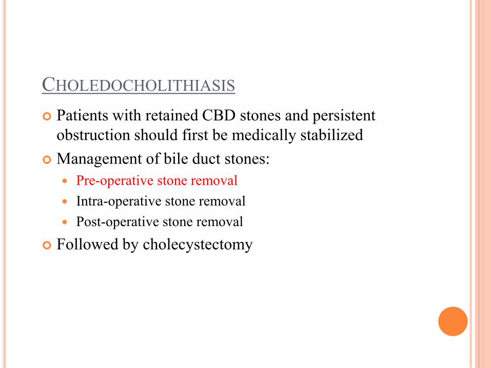
CHOLEDOCHOLITHIASIS
Patients with retained CBD stones and persistent
obstruction should first be medically stabilized
Management of bile duct stones:
Pre-operative stone removal
Intra-operative stone removal
Post-operative stone removal
Followed by cholecystectomy
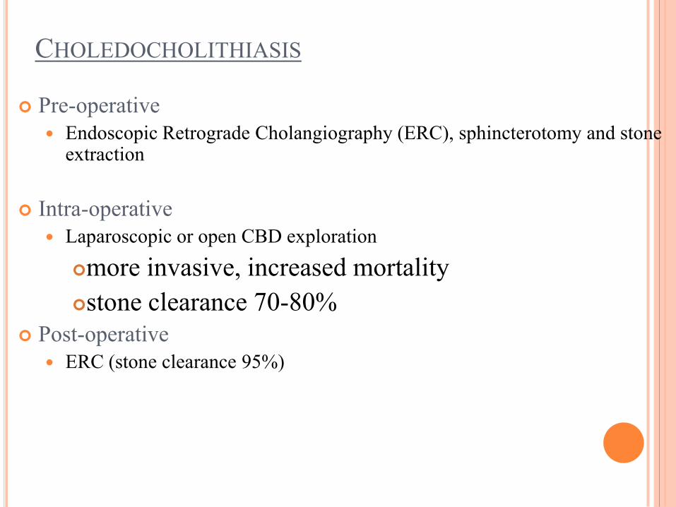
CHOLEDOCHOLITHIASIS
Pre-operative
Endoscopic Retrograde Cholangiography (ERC), sphincterotomy and stone extraction
Intra-operative
Laparoscopic or open CBD exploration
more invasive, increased mortality
stone clearance 70-80%
Post-operative
ERC (stone clearance 95%)
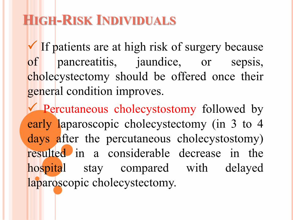
HIGH-RISK INDIVIDUALS
If patients are at high risk of surgery because
of pancreatitis, jaundice, or sepsis,
cholecystectomy should be offered once their
general condition improves.
Percutaneous cholecystostomy followed by
early laparoscopic cholecystectomy (in 3 to 4
days after the percutaneous cholecystostomy)
resulted in a considerable decrease in the
hospital stay compared with delayed
laparoscopic cholecystectomy.
PANCREATITIS
Gallstones are the most common cause.
The overall mortality is between 3% and 10%.
The role of early endoscopic sphincterotomy in the
management of gallstone pancreatitis is controversial.
the total number of complications is fewer after early
endoscopic sphincterotomy for predicted severe
pancreatitis.
There is of no benefit of early endoscopic
sphincterotomy for patients with acute gallstone
pancreatitis without cholangitis.
PANCREATITIS AGA PUBLISHED GUIDELINES
ERCP is recommended to be urgently performed “when acute
cholangitis has complicated acute biliary pancreatitis (about 10%
of patients)” and when “clinical or radiographic features
suggest a persistent common bile duct stone.”
Early ERCP, as defined as execution within 48 to 72 hours of the
onset of illness, should be considered “when biliary pancreatitis is
severe or is predicted to be severe (based on APACHE II, Ranson’s
criteria, or modified Glasgow criteria).”
cholecystectomy is indicated “as soon as possible,” but no later
than 4 weeks after discharge.
RECOMMENDATIONS
If the clinical presentation is consistent with acute cholangitis ,
urgent ERCP should be performed.(within 24h).
Early ERCP (within 24–48 h) is performed in patients who
have evidence of choledocholithiasis.
If there is persistent or episodic pain, or the laboratories do
not resolve as expected, then EUS or ERCP should be
considered because choledocholithiasis cannot be ruled out.
Endoscopic ultrasound may be performed first if the patient
has risk factors for an adverse outcome after ERCP such as
advanced age, comorbidities, or anticoagulation, or is
believed to be at low to moderate risk for choledocholithiasis.
Some patients may not have radiologic evidence for
choledocholithiasis, but this possibility still should be
considered if laboratory evidence of biliary stasis is found or
pain recurs.
However, in a few patients, choledocholithiasis will not be
detected despite multiple imaging studies. Therefore, serial
serum liver-associated enzyme and pancreatic enzyme tests are
performed, initially at a 12- to 24-hour interval.
FINALLY
we also consider performing early ERCP if the patient’s
clinical course becomes unstable or deviates from the expected
clinical course.
WHICH POLICY: OBSERVATION OR
CHOLECYSTECTOMY
Early laparoscopic cholecystectomy is safe and
can be completed successfully in most patients with
mild acute pancreatitis, delaying laparoscopic
cholecystectomy seems unnecessary and can expose
the patient to further gallstone-related complications.
Cholecystectomy appears safe as soon as the
general condition of the patient improves and the
pancreatic necrosis becomes sterile if infected (or
remains sterile if not infected)
CIRRHOTIC PATIENTS
The frequency of symptoms or complications does not
appear to be any different from other groups of patients
with gallstones. When patients develop complications,
however, they can be more severe.
Cholecystectomy is recommended for symptomatic
gallstones (grade B). There are no differences in the
timing of surgery for various indications between
compensated cirrhotic patients and other patients with
symptomatic gallstones.
For compensated cirrhotic patients with symptomatic
gallstones, laparoscopic cholecystectomy appears better
than open cholecystectomy.
Patients are more likely to be offered
cholecystectomy during pregnancy if the
biliary disease or biliary pancreatitis is
severe. Many patients who underwent
cholecystectomy would have been treated
conservatively first before they would have
been offered cholecystectomy.
The second trimester appears to be the best
time for performing cholecystectomy in
pregnant women.
PREGNANCY
ORAL BILE ACID THERAPY
Chenodeoxycholate (CDCA)
15mg/kg/d
Ursodeoxycholate (UDCA)
10mg/kg/d
Effective in dissolution of GS
Non-calcified, cholesterol GS
Functioning GB, patent cystic duct
Single or small stones
Reduce biliary cholesterol secretion
Complete dissolution in 30-40%
Recurrence rate high with cessation of drug
METHYL-TERT-BUTYL-ETHER (MTBE)
50-fold more potent the oral bile acid Rx
Non-calcified, cholesterol stones
Functioning GB
Percutaneous puncture of GB
Catheter placement
Serial infusions of MTBE (2 sessions)
Recurrence of stones common
Reserved for non-operative candidates in specialized centers
EXTRA-CORPOREAL SHOCK-WAVE LITHOTRIPSY
(ESWL)
Cholesterol stones
Single stone <30mm
Patent Cystic duct
ESWL administered with adjuvant bile acid therapy
47-68% stone-free 6 months
Recurrence rate 10% per year
Advantages
Non-invasive, low morbidity
Outpatient setting, lack of general anesthesia

COMPLICATIONS OF
LAPAROSCOPIC
CHOLECYSTECTOMY
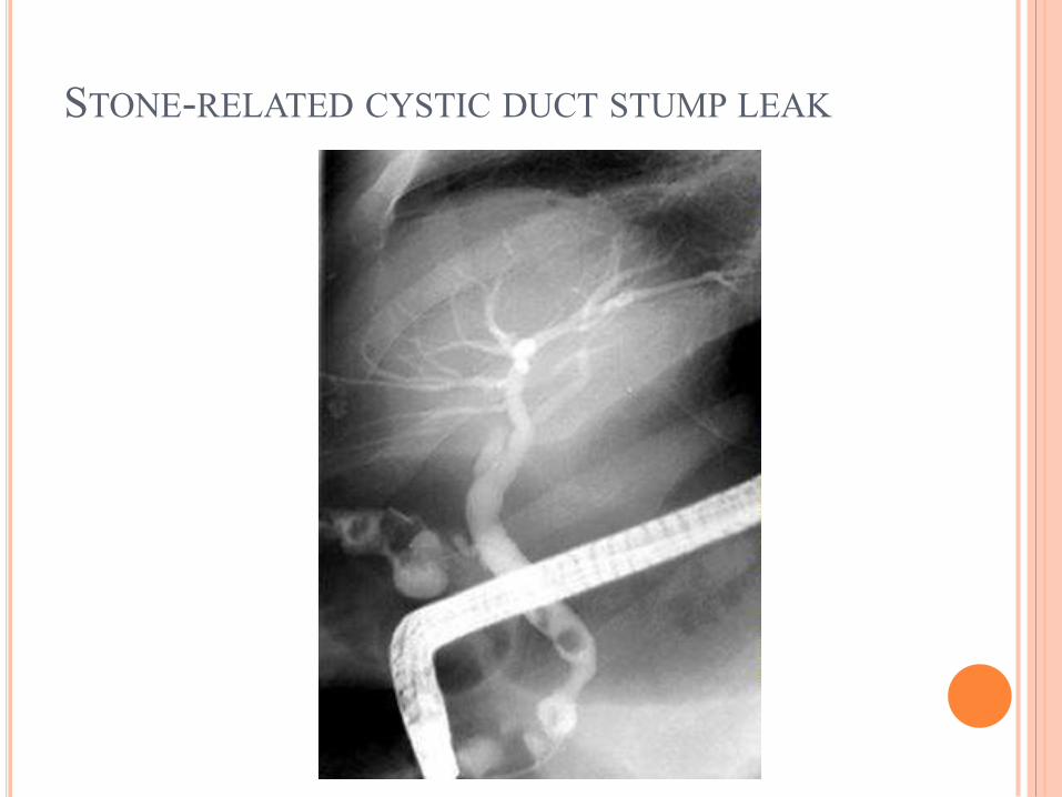
STONE-RELATED CYSTIC DUCT STUMP LEAK
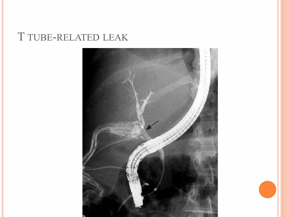
T TUBE-RELATED LEAK
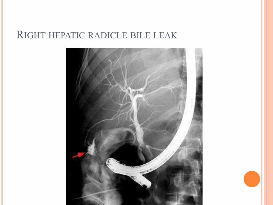
RIGHT HEPATIC RADICLE BILE LEAK
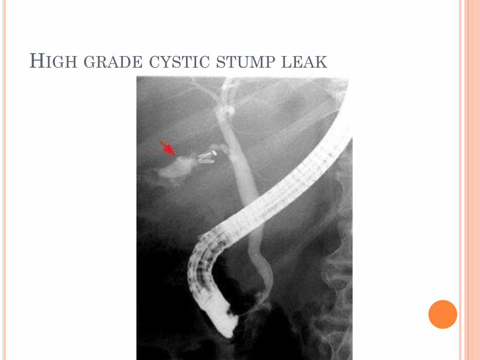
HIGH GRADE CYSTIC STUMP LEAK
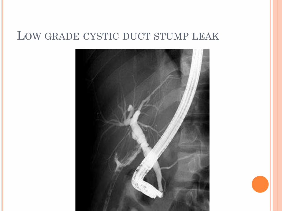
LOW GRADE CYSTIC DUCT STUMP LEAK

BILE LEAK FROM DUCT OF LUSCHKA
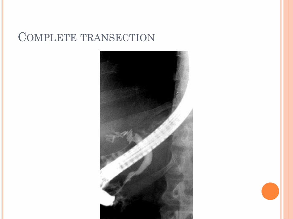
COMPLETE TRANSECTION
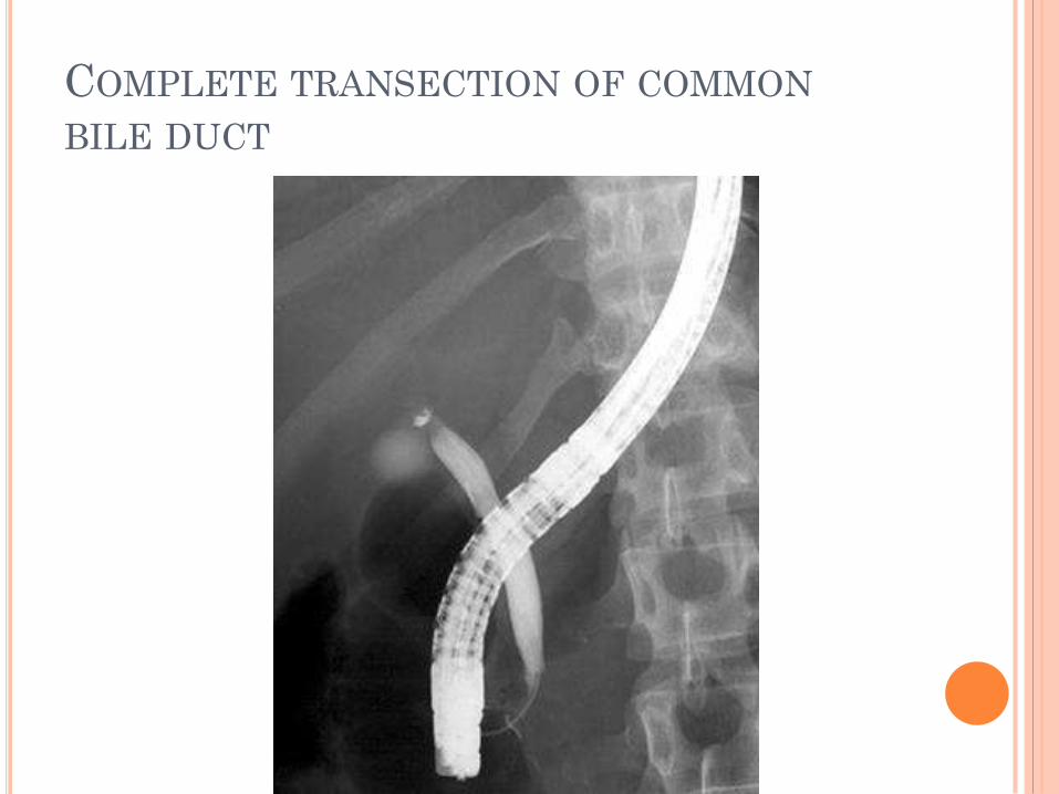
COMPLETE TRANSECTION OF COMMON
BILE DUCT
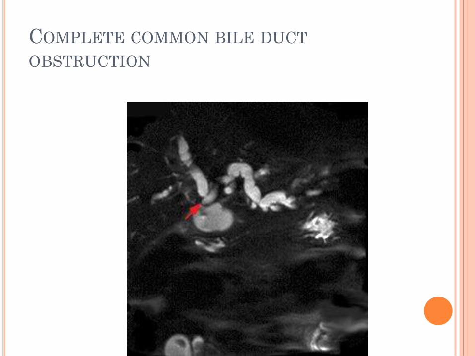
COMPLETE COMMON BILE DUCT
OBSTRUCTION
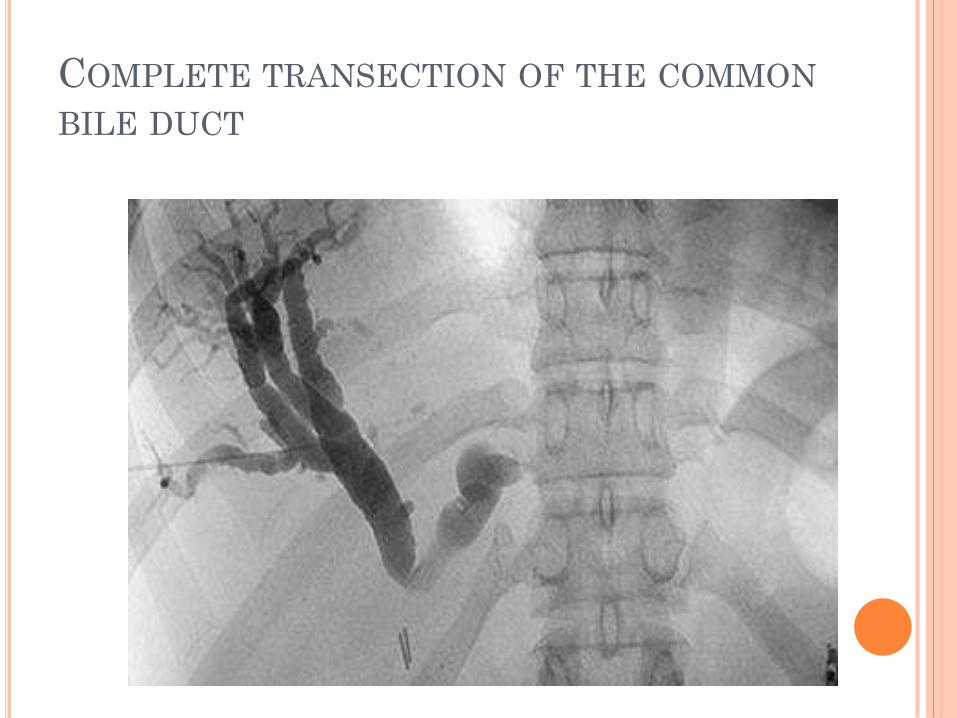
COMPLETE TRANSECTION OF THE COMMON
BILE DUCT

HEPATICOJEJUNOSTOMY
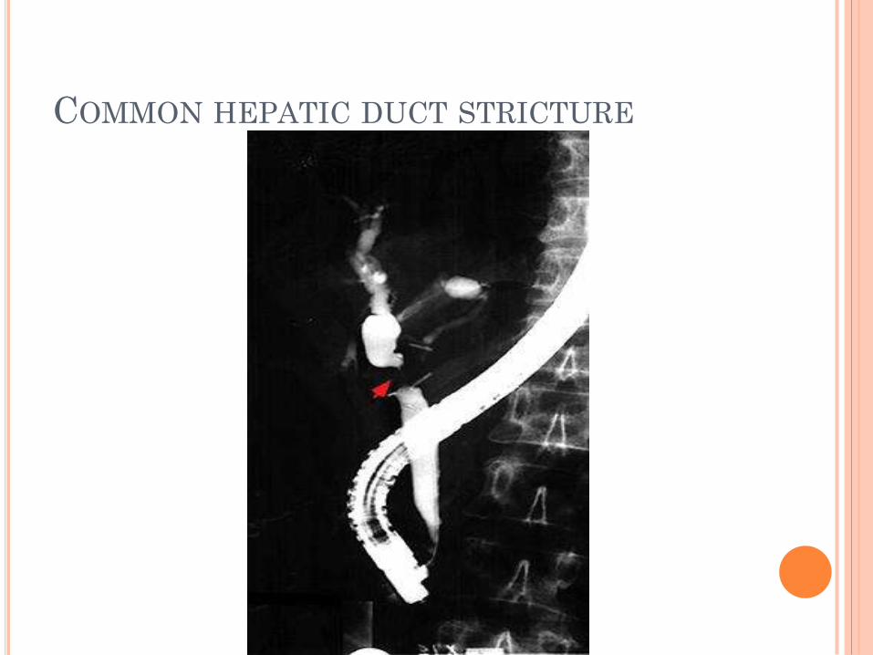
COMMON HEPATIC DUCT STRICTURE
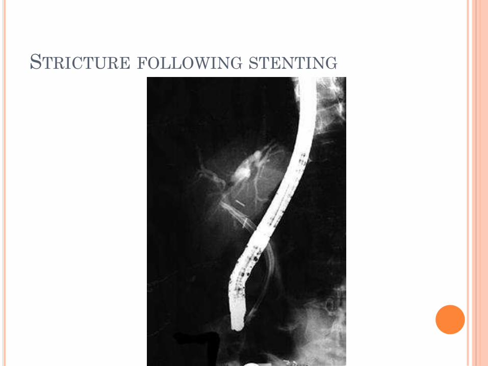
STRICTURE FOLLOWING STENTING
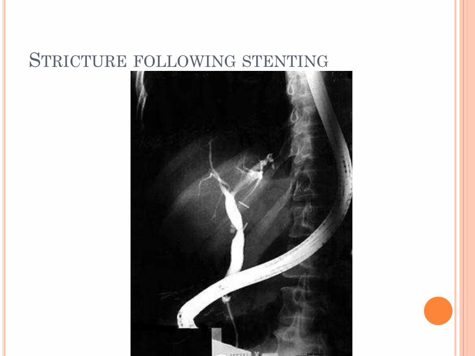
STRICTURE FOLLOWING STENTING
Top Related