


Languages
Pages
Legal

Ministry of Defence
Synopsis of Causation
Fractures Long Bones, Upper Limb (includes hand)
Author: Mr John A Dent, Ninewells Hospital and Medical School, Dundee Validator: Mr Sheo Tibrewal, Queen Elizabeth Hospital, London
September 2008

Disclaimer This synopsis has been completed by medical practitioners. It is based on a literature search at the standard of a textbook of medicine and generalist review articles. It is not intended to be a meta-analysis of the literature on the condition specified.
Every effort has been taken to ensure that the information contained in the synopsis is accurate and consistent with current knowledge and practice and to do this the synopsis has been subject to an external validation process by consultants in a relevant specialty nominated by the Royal Society of Medicine.
The Ministry of Defence accepts full responsibility for the contents of this synopsis, and for any claims for loss, damage or injury arising from the use of this synopsis by the Ministry of Defence.
2

1. Definition
1.1. A bone breaks as a result of a variety of injuring forces. The fracture produced in a long bone may be of its shaft or diaphysis; towards one of its ends where the bone widens (metaphysis); or of the end of the bone, where it forms a joint with the next bone (Figure 1). Fractures near a joint may involve the joint surface causing intra-articular fractures.
1.2. It must always be remembered that the soft tissues surrounding the bone will also be damaged to a varying extent by the injuring force. This may range from simple stripping of the periosteum at the fracture site to extensive laceration and crushing of the adjacent muscles, nerves, blood vessels and skin.
1.3. The most important aspect of a fracture, in terms of its management and prognosis, is whether it is an “open” fracture or a “closed” fracture, i.e. whether or not there is an open wound over the site of the fracture. There are various degrees of open fracture depending on the extent of contamination and soft tissue damage.
1.4. In young adults who have not reached skeletal maturity, an additional group of fractures is described in relation to the physis or growth plate. This area of growing cells is a weak area which may sometimes displace, taking a portion of the metaphysis or the whole of the epiphysis with it. The variety of fractures in this area have been classified (Figure 2).1
Figure 1: Regions of a long bone
Figure 2: Salter-Harris classification of epiphyseal injuries
3

1.5. In the diaphysis, a variety of fracture patterns can be seen on x-ray examination which indicates the mechanism of injury that has taken place (see Section 3.1.)
4
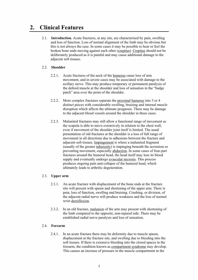
2. Clinical Features
2.1. Introduction. Acute fractures, at any site, are characterised by pain, swelling and loss of function. Loss of normal alignment of the limb may be obvious but this is not always the case. In some cases it may be possible to hear or feel the broken bone ends moving against each other (crepitus). Crepitus should not be deliberately produced as it is painful and may cause additional damage to the adjacent soft tissues.
2.2. Shoulder
2.2.1. Acute fractures of the neck of the humerus cause loss of arm movement, and in severe cases may be associated with damage to the axillary nerve. This may produce temporary or permanent paralysis of the deltoid muscle at the shoulder and loss of sensation in the “badge patch” area over the point of the shoulder.
2.2.2. More complex fractures separate the proximal humerus into 3 or 4 distinct pieces with considerable swelling, bruising and internal muscle disruption which affects the ultimate prognosis. There may be damage to the adjacent blood vessels around the shoulder in these cases.
2.2.3. Malunited fractures may still allow a functional range of movement as the scapula is able to move extensively in relation to the chest wall, even if movement of the shoulder joint itself is limited. The usual presentation of old fractures at the shoulder is a loss of full range of movement in all directions due to adhesions between the fracture and adjacent soft tissues. Impingement is where a malunited fragment (usually of the greater tuberosity) is impinging beneath the acromion so preventing movement, especially abduction. In some cases of four-part fractures around the humeral head, the head itself may lose its blood supply and eventually undergo avascular necrosis. This process produces ongoing pain and collapse of the humeral head, which ultimately leads to arthritic degeneration.
2.3. Upper arm
2.3.1. An acute fracture with displacement of the bone ends at the fracture site will present with spasm and shortening of the upper arm. There is pain, loss of function, swelling and bruising. Crushing, or division, of the adjacent radial nerve will produce weakness and the loss of normal wrist dorsiflexion.
2.3.2. In an old fracture, malunion of the arm may present with shortening of the limb compared to the opposite, non-injured side. There may be established radial nerve paralysis and loss of sensation.
2.4. Forearm
2.4.1. In an acute fracture there may be deformity due to muscle spasm, displacement at the fracture site, and swelling due to bleeding into the soft tissues. If there is extensive bleeding into the closed spaces in the forearm, the condition known as compartment syndrome may develop. This causes an increase of pressure in the muscle compartment in the
5

arm leading to muscle ischaemia and ultimately to compression of the nerve and artery in that compartment. Patients complain of pain on passive extension of the fingers and the loss of sensation and circulation in the digits.
2.4.2. Normal healing times are displayed at Figure 3. In fractures that do not heal as expected we may see delayed union, non-union, or malunion. An upper limb fracture may have failed to unite within the expected period of time, usually 6 weeks (delayed union), or have not united at all (non-union). In the first case, the fracture is still mobile and tender but some attempt at union can be seen on an x-ray. In non-union, the normal healing process has failed, and painless abnormal movement and crepitus persist at the fracture site. If a fracture has united fully but in an abnormal position (malunion) there may be obvious malalignment of the bone or loss of full range of movement in the adjacent joints. In the forearm, it is usually a loss of full supination and pronation (the ability to turn the hand over and back) which is apparent.
Spiral Transverse PERKINS CLASSIFICATION
OF FRACTURE HEALING union consolidation union consolidation
Upper Limb 3 6 6 12
Lower Limb 6 12 12 24
Figure 3: Perkins classification of fracture healing time (in weeks) for the fracture to unite and become fully healed
2.5. Wrist (Figure 4)
2.5.1. Distal radius. A fracture of the distal radius is commonly referred to as a broken wrist. In the common Colles’ fracture, particularly associated with postmenopausal females, there is usually a history of a fall on the outstretched hand resulting in posterior displacement of the distal part of the radius. This produces a characteristic dinner fork appearance to the wrist. Occasionally, numbness in the hand due to compression of the median nerve at the wrist may be found.
2.5.2. The opposite type of injury, with the distal radius angulated anteriorly, is known as a Smith’s fracture.
2.5.3. In old fractures, the presentation is usually of malunion. In Colles’ fractures, there is posterior malalignment and radial deviation of the wrist. A delayed, spontaneous rupture of the long extensor tendon of the thumb (extensor pollicis longus) causes inability to extend the tip of the thumb.2 More rarely, flexor tendon rupture and median nerve compression are seen.3
2.5.4. Carpal bones. Sports injuries are especially associated with fractures of the scaphoid,4 and direct blows or more severe trauma may cause fractures of the other carpal bones. In scaphoid fractures, there is
6

characteristic tenderness to palpation over the bone in the anatomical snuff box at the base of the thumb. Thumb movement is painful, especially if combined with axial pressure.
2.6. Hand (Figure 4)
2.6.1. In an acute metacarpal fracture, there may be pain and tenderness together with extensive soft tissue swelling and bruising on the back of the hand. There may be obvious malalignment, usually forward angulation of the distal part of the metacarpal, and loss of full extension of the associated finger at the metacarpophalangeal joint. Crepitus may be apparent. The common fracture of the neck of the fifth metacarpal is often associated with a skin wound on the knuckle.
2.6.2. In an old metacarpal fracture there will be loss of normal function, often with a persisting loss of full extension of the finger. Uncorrected forward angulation causes a persistent lump on the back of the hand and an associated lump in the palm, which is the displaced head of the metacarpal. This may cause pain on gripping solid objects.
2.7. Digits (Figure 4)
2.7.1. In an acute fracture, tenderness and deformity are apparent, depending on the severity of the injury. Malalignment of the digit, especially rotational malalignment, must also be looked for.
2.7.2. In an old fracture, rotational malunion or angulation of a phalanx may produce overriding of one finger on another. Loss of movement at the proximal interphalangeal joint is a cause of loss of function.
Figure 4: Bones of the hand and wrist
ERRATUM: Figure 4 includes a label that lists seven of the carpal bones. The eighth carpal bone, the pisiform, has been omitted.
7
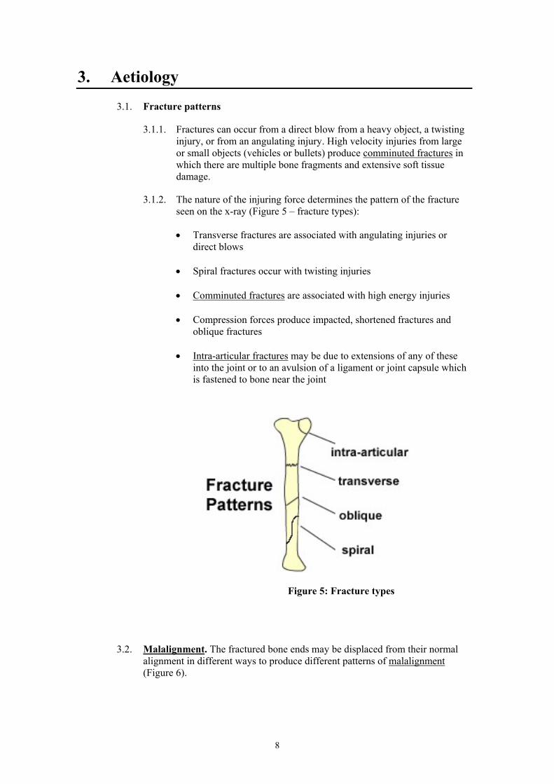
3. Aetiology
3.1. Fracture patterns
3.1.1. Fractures can occur from a direct blow from a heavy object, a twisting injury, or from an angulating injury. High velocity injuries from large or small objects (vehicles or bullets) produce comminuted fractures in which there are multiple bone fragments and extensive soft tissue damage.
3.1.2. The nature of the injuring force determines the pattern of the fracture seen on the x-ray (Figure 5 – fracture types):
Transverse fractures are associated with angulating injuries or direct blows
•
•
•
Spiral fractures occur with twisting injuries
Comminuted fractures are associated with high energy injuries
Compression forces produce impacted, shortened fractures and oblique fractures
•
•
Intra-articular fractures may be due to extensions of any of these into the joint or to an avulsion of a ligament or joint capsule which is fastened to bone near the joint
Figure 5: Fracture types
3.2. Malalignment. The fractured bone ends may be displaced from their normal alignment in different ways to produce different patterns of malalignment (Figure 6).
8

3.2.1. Angulated. The 2 bone ends are at an angle to each other. This may be in a forward/backward plane (anterior or posterior angulation) or in a sideways plane (medial or lateral angulation).
3.2.2. Displaced. The 2 bone ends are shifted on each other to various extents, usually described as a percentage of the width of the bone at the site of the fracture. The displacement may be in a forward/backwards direction (anterior or posterior displacement) or in a sideways direction (medial or lateral displacement).
3.2.3. Rotated. The 2 bone ends are rotated on each other. This may be difficult to recognise on the x-ray but is usually apparent when looking at the whole of the injured limb.
3.2.4. Shortened. The 2 bone ends are impacted into each other, as in the case of a comminuted fracture, where normal support for the length of the bone has been lost. The ends of the bone may slip on each other as in an oblique fracture, or else they may be completely displaced and override each other.
Figure 6: Malalignment
3.3. Shoulder. Fractures at the proximal end of the humerus usually occur following a fall. The neck of the humerus in this area is commonly broken in elderly females. More complex fractures which break the proximal humerus into 3 or 4 larger fragments are due to more severe injury and may be seen in younger patients. These fractures are usually associated with high energy injuries.
3.4. Upper arm. A twisting injury, such as might occur from arm wrestling, can cause a spiral fracture of the humerus. Direct blows and angulating forces produce transverse fractures.
9

3.5. Forearm. This fracture is often the result of a fall involving compression and twisting forces.5
3.6. Wrist (Figure 4)
3.6.1. Distal radius. A simple fall in an elderly person is a common cause of a Colles’ fracture of the end of the radius in the forearm.
3.6.2. A similar injury in a child causes a fracture around the growth plate, an epiphyseal injury (Figure 2).
3.6.3. Carpal bones. Fracture of the scaphoid is a common injury from a fall on the outstretched hand, usually associated with sports.4 This fracture may not be apparent on initial x-rays so, if suspected, repeat x-rays should always be taken two weeks later. Plaster of Paris immobilisation is usually adequate for treatment but, in some cases, internal fixation becomes necessary.
3.7. Hand (Figure 4)
3.7.1. Metacarpal fractures, especially of the neck of the fifth metacarpal, often occur from punching something. There is angulation at the fracture site and often a telltale wound on the knuckle.
3.7.2. Injuries to the first (thumb side) metacarpal often occur at its base and may involve the intra-articular surface (Bennett’s fracture).
3.8. Digits (Figure 4). Fractures of the proximal or middle phalanges are caused by rotation or crushing injuries. There may be extensive associated damage to the adjacent soft tissues.
3.9. Other causes. Rarely, fractures in the upper limb may be due to pre-existent bone disease, infection in the bone or secondary deposits (metastases) from malignant tumours elsewhere in the body.
10

4. Prognosis
4.1. Management
4.1.1. The treatment of fractures usually requires reduction, maintenance of reduction (immobilisation), and rehabilitation. Initial reduction of the fracture aims to correct any significant malalignment and usually requires anaesthesia. After this, the fracture must be maintained in its corrected position by either non-operative or operative methods. Non-operative treatment of an upper limb fracture may involve a sling or a splint, usually of plaster of Paris.
4.1.2. Operative treatment may use an internal fixation device such as a plate for a forearm fracture, an intermedullary rod for a humeral fracture, or pins or screws for fractures of small bones or where there are small fragments to secure. The risks associated with this treatment are of delayed non-union and, especially, infection. This has been reported as being in the region of 12% in the treatment of open fractures.6 Occasionally a fracture is stabilised by an external fixator which is secured to the fracture fragments by percutaneous pins or screws.
4.1.3. The prognosis of the fracture is influenced by the type of treatment used as well as by the initial type of injury and pattern of fracture.
4.1.4. The choice of treatment depends on the type of fracture, the degree of damage to the soft tissues, and the age and expectations of the patient.
4.2. Shoulder
4.2.1. Persisting pain and loss of function are the main problems after a fracture of the neck of the humerus. Non-union and myositis ossificans may also occur. Initial support in a sling followed by early active mobilisation allows for a quick return of shoulder function in the majority of cases. Physiotherapy is most important. Persisting symptoms from adhesions or a “frozen” stiff shoulder can be both painful and limiting. A 4-week rehabilitation programme combining exercises, electrical therapy and massage will produce a significant increase in shoulder strength and decrease in pain.7 Manipulation under anaesthesia also has a role in treatment.8
4.2.2. Patients with displaced fractures of the greater tuberosity of the humerus may develop impingement which requires surgical correction. This is a specialist procedure.
4.2.3. Cases treated by internal fixation may later have symptoms of bone pain or cold intolerance related to this and require removal of the metalwork. Occasionally, avascular necrosis of the humeral head fragment can be anticipated and a hemiarthroplasty performed.9
4.3. Upper arm
4.3.1. Internal fixation of a fractured shaft of the humerus is associated with a risk of infection, weakness, cold intolerance, or a second fracture of the
11
bone beyond the area splinted by the internal fixation. Further surgery, 18 months to 2 years later, may be required to remove the metalwork.
4.3.2. Cases associated with symptoms of radial nerve damage due to bruising, stretching or compression will usually improve within the first few months but those with partial or complete nerve division will require surgical exploration.
4.4. Forearm. Childhood fractures of the radius and ulna can be treated by manipulation and an above elbow plaster splint. The risks are of compartment syndrome and joint stiffness. In adults, internal fixation of fractures is required to achieve an anatomical reduction and so avoid malunion and the resulting loss of full movement, especially pronation and supination.10 The risks of internal fixation are infection, delayed union and non-union, as well as the risks associated with any operative procedure and anaesthesia.
4.5. Wrist
4.5.1. Distal radius. Colles’ fracture in an elderly person is usually treated by immobilisation in plaster. The complications of this are stiffness and malunion, resulting in loss of full wrist flexion and weak grip. Up to a third of patients will not be able to carry out all basic activities of daily living at 5 weeks after coming out of plaster.11,12 Prolonged pain, swelling and stiffness in the hand and fingers (Sudek’s atrophy) occasionally is seen as a neurological complication of injury and immobilisation. Extensive physiotherapy and sympathetic nerve block with guanethidine are the treatments required. Grip strength may take up to a year to return.13 Smith’s fracture is often treated by open reduction and internal fixation, and so may have all the risks associated with a surgical procedure.
4.5.2. Carpal bones. Fracture of the scaphoid may be slow to heal and may develop delayed union or non-union.14 Internal fixation and bone grafting is required in this case. Patients usually continue to have some loss of full range of wrist movements.
4.5.3. Failure to recognise and immobilise a scaphoid fracture runs the risk of allowing non-union to take place. This results in the early development of arthritic degeneration in the wrist. The joint around the scaphoid bone and the radio-carpal joint in general become involved, giving a stiff painful wrist. Surgical treatment in these cases requires intercarpal arthrodesis and, in some cases, total wrist fusion.
4.6. Hand
4.6.1. Support in a removable canvas splint is usually sufficient for fractures of the metacarpal necks. There may be some residual prominence or recession of the normal knuckle at the fracture site on the back of the hand. This malalignment can usually be accepted unless there is loss of full movement in the adjacent joint (metacarpophalangeal joint) or any rotation of the digits.
4.6.2. Internal fixation using a small plate or wires may be used in the treatment of metacarpal fractures, especially for unstable or intra-articular fractures, but the risk of complications may be high.15
12

4.7. Digits
4.7.1. Splinting may be sufficient if the fracture is stable. Stiffness is the main complication. Physiotherapy and occupational therapy are important. Patients may require up to 3 months rest from heavy work and may have residual weakness and the loss of full finger movements and dexterity. Sometimes in open crush injuries, there may be ongoing effects of nerve damage resulting in numbness and cold intolerance in the affected finger. Overriding fingers may require further surgery to correct the deformity. This may involve re-fracture, fixation and bone grafting.
4.7.2. Adhesions between the fracture site and the adjacent extensor tendons may cause loss of the ability to extend the finger fully. Shortening at the fracture site also influences finger tendon function.16
4.7.3. Fractures of the terminal phalanx, often associated with crush injuries to the finger tip, can usually be treated as soft tissue injuries.
4.8. Intra-articular fractures
4.8.1. Fractures which involve the joint surface of a bone may produce a permanent irregularity of the joint surface causing malalignment of the joint and loss of full range of movement. Damage to the articular surface may progress to cause premature degenerative changes and post-traumatic osteoarthritis, characterised by pain, swelling, stiffness and loss of normal function (secondary osteoarthritis).
4.8.2. Secondary osteoarthritis. Intra-articular fractures are at risk of developing secondary osteoarthritis due to disruption of the normal articular surface. Other fractures resulting in angulations or rotation of the bone usually do not have this risk
13

5. Summary 5.1. In common with fractures elsewhere in the body, fractures of the bones of the
upper limb occur in recognised patterns, according to the type of injury suffered. The type of fracture indicates the appropriate treatment and the likely risks of complications.
5.2. The treatment of fractures includes the management of the associated soft tissue injury as well as the bone. Appropriate rehabilitation of the joints and muscles are required to achieve a good recovery.
5.3. Fractures in children which involve the growing end of a bone may be associated with premature growth arrest leading to deformity.
5.4. Fractures involving the articular surfaces of joints may lead to the development of early osteoarthritis (secondary osteoarthritis).
14

6. Related Synopses
Fractures of the Lower Limb (includes foot)
Acute and Chronic Soft Tissue Injuries: the Shoulder and Elbow
15

7. Glossary
abduction Movement of a limb away from the midline.
adduction Movement of a limb towards the midline.
angulation The description of a fracture when the distal fragment is at an angle to the proximal fragment.
arthrodesis The surgical fusion of a joint.
avascular necrosis A disease resulting from the temporary or permanent loss of the blood supply to the bones. Without blood, the bone tissue dies and causes the bone to collapse.
carpal bones The name for the group of eight small angular bones, which are situated between the forearm bones and metacarpals, making up the wrist. Also known as the carpus.
closed fracture A fracture with no associated damage to the overlying skin
comminuted fracture A fracture with many fragments.
compartment syndrome Increased pressure in a compartment closed by firm fascia and bone. This may be due to swelling caused by blood or tissue fluid and results in squashing of muscles, nerves and blood vessels that are situated within the compartment.
crepitus The sound made by broken bone ends rubbing together.
delayed union Union which is proceeding more slowly than expected.
diaphysis The middle, shaft region of long bones.
dorsiflexion The movement of extension of a joint, usually referring to the wrist.
epiphysis The end of a long bone, which sits on the physis or growth plate and usually forms the articulating surface of the bone.
extension/flexion A joint’s ability to move backwards and forwards due to the muscles acting across it.
external fixator A rigid device attached to the broken bone by percutaneous wires or screws, used to secure or manipulate a fracture.
16

fracture
A break in the normal continuity of a bone which may be complete or incomplete (see fracture patterns).
humerus The bone of the upper arm
hemiarthroplasty Surgical procedure where a single degenerate joint surface is replaced by artificial material.
immobilisation Holding a fracture or joint in its anatomically correct reduced position by external splint (e.g. plaster of Paris or external fixation) or by an internal fixation device (e.g. a plate or intermedullary rod).
impingement Usually refers to the shoulder where the proximal humerus may nip the intervening soft tissues beneath the acromion. This may occur in rotator cuff degeneration and malunion of the greater tuberosity.
interphalangeal joint The joint between the phalanges in a digit. Proximal and distal joints are seen in the toes and fingers, but only one interphalangeal joint is present in the thumb and big toe.
intra-articular fracture A fracture which extends into the adjacent joint.
intermedullary Lying within the central cavity of a long bone.
ischaemia A low oxygen state usually due to obstruction of the arterial blood supply or inadequate blood flow.
malalignment The description of a fracture which is out of line with the normal appearance of the bone. This includes medial/lateral and anterior/posterior displacement, as well as angular and rotational malalignment at the fracture site.
malunion
A fracture which has united in an abnormal position due to either initial displacement of the fracture or loss of position after acute reduction.
metacarpals The long thin bones which make up the hand (not the fingers).
metacarpophalangeal joint The joint between the hand bones (metacarpals) and the digits (phalanges).
metaphysis The widened part of the shaft of a long bone as it nears the physis.
myositis ossificans Ectopic bone formation in soft tissue as a result of trauma.
17

non-union Failure of a fracture to unite (heal) after the expected
time for its location and type.
open fracture A fracture with an overlying skin wound.
percutaneous Passing through the skin.
periosteum The membrane of tissue, which normally covers a bone providing it with a nerve and blood supply.
phalanx (pl. phalanges) The bones which make up the digits; 3 in the fingers and 2 in the thumb.
proximal Referring to an area of the body, or part of a long bone or limb, that is nearer to the central axis of the body.
radius The bone on the thumb-side of the forearm.
reduction Realigning a fracture or dislocation in its normal position.
rehabilitation The return to normal comfort and function; usually with the help of exercise or other therapy.
scaphoid The scaphoid bone of the wrist is found on the thumb side of the hand, within the anatomical snuffbox. It is approximately the size and shape of a cashew, and is slow to heal because of the poor circulation to the bone.
Sudek’s atrophy Prolonged pain, swelling and stiffness in an extremity (predominantly wrist and hand) after trauma or surgery.
supination/pronation Supination describes the action of turning the forearm to position the hand upwards. Pronation is the reverse.
tuberosity A bulging growth on a bone especially for attachment of a muscle or ligament.
ulna The bone on the little finger-side of the forearm.
18
8. References
1.
2.
3.
4.
5.
6.
7.
8.
9.
10.
11.
12.
13.
14.
15.
Slater RB, Harris WR. Injuries involving the epiphyseal plate. J Bone Joint Surg 1963;45A(3):587-622.
Helal B, Chen SC, Iwegbu G. Rupture of the extensor pollicis longus tendon in undisplaced Colles’ type of fracture. Hand 1982 Feb;14(1):41-7.
Wong FY, Pho RW. Median nerve compression, with tendon ruptures, after Colles’ fracture. J Hand Surg [Br] 1984 Jun;9(2):139-41.
Dent JA, Mitchell CA, Sharma MM. Herbert screw: results of a single-centre trial. Injury 1992;23(4):228-30.
Rockwood CA, Wilkins KE, King RE, editors. Fractures in children. Philadelphia, PA: Lippincott; 1984.
Gustilo RB, Simpson L, Nixon R, Ruiz A, Indeck W. Analysis of 511 open fractures. Clin Orthop Relat Res 1969 Sep-Oct;66:148-54.
Jurgel J, Rannama L, Gapeyeva H, Ereline J, Kolts I, Paasuke M. Shoulder function in patients with frozen shoulder before and after 4-week rehabilitation. Medicina (Kaunas) 2005;41(1):30-8.
Hamdan TA, Al-Essa KA. Manipulation under anaesthesia for the treatment of frozen shoulder. Int Orthop 2003;27(2):107-9.
Neer CS 2nd. Displaced proximal humeral fractures. II. Treatment of three-part and four-part displacement. J Bone Joint Surg Am 1970 Sep;52(6):1090-103.
Anderson LD. Fractures of the shafts of the radius and ulna. In: Rockwood CA, Green DJ, editors. Fractures in adults. 2nd ed. Philadelphia, PA: Lippincott; 1984. p. 511-58.
Dekkers M, Søballe K. Activities and impairments in the early stage of rehabilitation after Colles’ fracture. Disabil Rehabil 2004 Jun 3;26(11):662-8.
Vaneker M, van Egeraat SP, Haerkens MH, Wilder-Smith OH. Letter to the editor in response to the article of Dekkers and Søballe (Activities and impairments in the early stage of rehabilitation after Colles’ fracture; Vol. 26, No. 11, June 3, 2004). Disabil Rehabil 2005 May 6;27(9):535-6.
Lagerstrom C, Nordgren B, Rahme H. Recovery of isometric grip strength after Colles’ fracture: a prospective two-year study. Scand J Rehabil Med 1999 Mar;31(1):55-62.
Herbert TJ, Fischer WE. Management of the fractured scaphoid using a new bone screw. J Bone Joint Surg Br 1984 Jan;66(1):114-23.
Ouellette EA, Freeland AE. Use of the minicondylar plate in metacarpal and phalangeal fractures. Clin Orthop Relat Res 1996 Jun;(327):38-46.
19

20
16. Vahey JW, Wegner DA, Hastings H 3rd. Effect of proximal phalangeal fracture deformity on extensor tendon function. J Hand Surg [Am] 1998 Jul;23(4):673-81.