


Languages
Pages
Legal


404/ CURRENT CONCEPTS REVIEWSOUBORNÝ REFERÁT
ACTA CHIRURGIAE ORTHOPAEDICAEET TRAUMATOLOGIAE ČECHOSL., 79, 2012, p. 404–410
Fixation of Osteoporotic Fractures in the UpperLimb with a Locking Compression Plate
Stabilizace osteoporotických zlomenin horní končetiny pomocí úhlově stabilníuzamykatelné dlahy (LPC) – přehled současných přístupů
NEUHAUS, V., KING, J. D., JUPITER, J. B.
Hand and Upper Extremity Service, Massachusetts General Hospital, Orthopaedic Surgery, Boston, Massachusetts, USA
SUMMARY
Locking Compression Plate (LCP) has the advantageous feature that screws can be locked in the plate leaving an angu-lar stable construct. There is no need to have contact between the plate and the bone to achieve stability resulting fromfriction of the plate-bone-construct. Therefore the plate does not need to be contoured exactly to the bone and the healingbone’s periosteal blood supply is not affected. The LCP is used as a bridging plate to gain relative stability in multi-frag-mentary, diaphyseal or metaphyseal fractures. Depending on the fracture, the combination hole can also allow the LCP toachieve absolute stability similar to conventional fixation techniques.
Osteoporotic fractures have significant impact on morbidity and mortality. Proximal humeral and distal radius fracturesare typical examples. These osteoporotic and often comminuted fractures are ideal settings/indications for LCP utilizationin the upper extremity. However, the data quality is due to mostly small study populations not so powerful. Unquestionablythere has been a clear and fashionable trend to choose operative treatment for these fractures, because the angular sta-bility allows stable fixation and early functional mobilization.
Key words: concepts, current, fractures, Locking Compression Plate, osteoporotic, review.
INTRODUCTION
Reconstruction of complex fractures and nonunionsin the upper limb in the elderly patient with underlyingosteoporosis and prior to the development of the Loc-king Compression Plate (LCP) was challenging (20, 23).The development of LCP in the last ten years has dra-matically changed the treatment not only of distal ra diusand proximal humeral fractures, but also of complexfractures and nonunions (Fig. 1a–f). This article will pre-sent an overview about the principles of LCP and thecurrent concepts utilizing LCP in treating osteoporoticupper extremity fractures.
LCP
Locking Compression Plate (LCP) has the advanta-geous feature that screws can be locked easily in the pla-te achieving an angular stable construct. There is no needto have contact between the plate and the bone to getstability resulting from friction of the plate-bone-con-struct (42). As a consequence the plate does not need to
be contoured as exactly as possible to the bone. Ano theradvantageous aspect of this technique is that the healingbone’s periosteal blood supply is not affected. The LCPis broadly used as a bridging plate to gain relative sta-bility in multi-fragmentary diaphyseal or metaphysealfractures (12). An open approach is possible, but a lessinvasive, as in MIPO (minimal invasive plate osteosynt-hesis), is preferred and affords less harm to the soft-tis-sue coverage. The reduction is mostly achieved by indi-rect means. Yet there are important features to beconsidered according to Gauthier and Sommer (12).First the plate must be of appropriate length. The pro-portion of plate length and fracture length has to be con-sidered. They concluded that the plate length should be2 to 3 times the fracture length in comminuted fracturesand 8 to 10 times in simple fractures. Second the platemust be positioned well, so that all the screws can beadequately positioned in the bone to maximize ancho-rage of the bicortical screws. Third the appropriate num-ber of screws is needed, usually three on each main frag-
s_404_410_neuhaus_test_acta_sloupce 8.10.12 12:15 Stránka 404

405/ ACTA CHIRURGIAE ORTHOPAEDICAEET TRAUMATOLOGIAE ČECHOSL., 79, 2012 CURRENT CONCEPTS REVIEW
SOUBORNÝ REFERÁT
ment. One last issue is the complete loss of the surgicalfeeling of the quality of the bone during screw inser tionand tightening.
Depending on the fracture, the combination hole canallow the LCP to achieve absolute stability similar toconventional open reduction and internal fixation tech-niques using Low Contact Dynamic Compression Pla-tes (LC-DCP). The main indications therefore are arti-cular fractures or simple diaphyseal (AO type A)fractures (42). However, a precise contouring is manda-tory in this situation. In osteoporosis the loss of reduc-tion is often encountered with conventional plate-screw-
bone-constructs due to the thin bone cortex and conse-cutively short working length of the screws in the cor-tex (12). This leads to malalignment and in the case ofinstability to nonunion. A combination of conventionaland locking screws is possible and is showed in dia-gram 1. Anatomical reduction is achieved with the pre-cise contoured plate and conventional screws. Supple-mental locking head screws improve the pullout forcesof the osteosynthesis and the resistance against bendingand torsion.
Another example of the combination of conventionaland locking screws is the use of absolute and relative
Fig. 1a–f. This 38-year-old patient had a complete amputa tionof his arm and had a replantation four years ago. He presen-ted to our office with this complex nonunion of the left distalhumerus. Radiographs are showing the peri-operative sequ-ence of events and treatment (open reduction and internal fixa-tion nonunion with anterior and posterior capsular release).After two years he can do pushups and the x-rays show goodhealing of the nonunion.
Diagram 1. In osteoporosis loss of reductionis often encountered with conventional pla-te-screw-bone-constructs due to the thinbone cortex and consecutively short workinglength. A combination of conventional andlocking screws is possible. (Reprinted fromInjury, Int. J. Care Injured, Vol 34, MichaelWagner, General principles for the clinicaluse of the LCP, Page No. S-B35, Copyright(2003), with permission from Elsevier).
good bone quality
osteoporotic bone
a b c de f
s_404_410_neuhaus_test_acta_sloupce 8.10.12 12:15 Stránka 405
406/ ACTA CHIRURGIAE ORTHOPAEDICAEET TRAUMATOLOGIAE ČECHOSL., 79, 2012 CURRENT CONCEPTS REVIEW
SOUBORNÝ REFERÁT
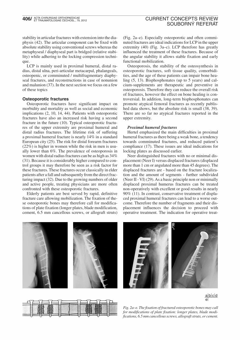
(Fig. 2a–e). Especially osteoporotic and often commi-nuted fractures are ideal indications for LCP in the upperextremity (40) (Fig. 3a–c). LCP therefore has greatlyinfluenced the treatment of these fractures. Because ofthe angular stability it allows stable fixation and earlyfunctional mobilization.
Osteoporosis, the stability of the osteosynthesis inosteoporotic fractures, soft tissue quality, comorbidi -ties, and the age of these patients can impair bone hea-ling (5, 13). Bisphosphonates (up to 5 years) and cal -cium-supplements are therapeutic and preventive inosteoporosis. Therefore they can reduce the overall riskof fractures, however the effect on bone healing is con-troversial. In addition, long-term bisphosphonates canpromote atypical femoral fractures as recently publis-hed data shows, but the absolute risk is small (38, 39).There are so far no atypical fractures reported in theupper extremity.
Proximal humeral fracturesHertel emphasized the main difficulties in proximal
humeral fractures as there being a weak bone, a tendencytowards comminuted fractures, and reduced patient’scompliance (17). These issues are ideal indications forlocking plates as discussed earlier.
Neer distinguished fractures with no or minimal dis-placement (Neer I) versus displaced fractures (displacedmore than 1 cm or angulated more than 45 degrees). Thedisplaced fractures are - based on the fracture localiza-tion and the amount of segments - further subdivided(Neer II - VI) (29). As a basic principle non or minimallydisplaced proximal humerus fractures can be treatednon-operatively with excellent or good results in nearly90% (11). In contrast, conservative treatment of displa-ced proximal humeral fractures can lead to a worse out-come. Therefore the number of fragments and their dis-placement influences the decision to proceed withoperative treatment. The indication for operative treat-
stability in articular fractures with extension into the dia-physis (42). The articular component can be fixed withabsolute stability using conventional screws whereas themetaphyseal / diaphyseal part is bridged (relative stabi-lity) while adhering to the locking compression techni-que.
LCP is mainly used in proximal humeral, distal ra -dius, distal ulna, peri-articular metacarpal, phalangeals,osteopenic, or comminuted / multifragmentary diaphy-seal fractures, and reconstructions in case of nonunionand malunion (37). In the next section we focus on a fewof these topics
Osteoporotic fracturesOsteoporotic fractures have significant impact on
morbidity and mortality as well as social and economicimplications (2, 10, 14, 44). Patients with osteoporoticfractures have also an increased risk having a secondfracture in the future (10). Typical osteoporotic fractu-res of the upper extremity are proximal humeral anddistal radius fractures. The lifetime risk of sufferinga proximal humeral fracture is nearly 14% in a standardEuropean city (25). The risk for distal forearm fractures(22%) is higher in women while the risk in men is usu-ally lower than 6%. The prevalence of osteoporosis inwomen with distal radius fractures can be as high as 34%(31). Because it is considerably higher compared to con-trol groups it may therefore be seen as a risk factor forthese fractures. These fractures occur classically in elderpatients after a fall and subsequently from the direct frac-turing impact (32). Due to the growing numbers of olderand active people, treating physicians are more oftenconfronted with these osteoporotic fractures.
Elderly patients are best served by rapid, definitivefracture care allowing mobilization. The fixation of the-se osteoporotic bones may therefore call for modifica -tions of plate fixation (longer plates, blade modifica tion,cement, 6.5 mm cancellous screws, or allograft struts)
Fig. 2a–e. The fixation of fractured osteoporotic bones may callfor modifications of plate fixation: longer plates, blade modi-fications, 6.5 mm cancellous screws, allograft struts, or cement.
a b c de
s_404_410_neuhaus_test_acta_sloupce 8.10.12 12:15 Stránka 406

407/ ACTA CHIRURGIAE ORTHOPAEDICAEET TRAUMATOLOGIAE ČECHOSL., 79, 2012 CURRENT CONCEPTS REVIEW
SOUBORNÝ REFERÁT
ment not only depends on these criteria, but also on manyother factors not limited to but including the existenceof osteoporosis, the predictors of humeral head perfu -sion (Hertel criteria), and the preference of the surgeonor institution. However, the quality of published studiesis due to mostly small study populations sometimes notideally powered (15).
Open reduction and internal fixation is the most popu-lar surgical method, whereas percutaneous pinning is notwidely used (34). Internal fixation can be achieved viaconventional or locking techniques. The delto-pectoralapproach is mostly used. Worse outcome can result fromloss of reduction, malunion or even nonunion due to loo-sening of conventional screws and plates. In contrast,angular-stable implants have biologic and biomechani-cal advantages as well as fewer complications compa-red to conventional fixation techniques (33). In manybiomechanical studies the angular-stable plates showedsignificantly greater stiffness, higher load before failu-re and significantly less displacement at higher loadsthan conventional plates (4, 35). An excellent or goodfunctional outcome can therefore be achieved in 92% ofpatients with two or three part fractures treated with LCPafter an average time to union of 18 weeks (40). Pa tientsattained nearly 90% range of motion compared with theuninjured shoulder in a recently published long-term fol-low-up study (18).
However, the reoperation rate was 29%. This high re -operation rate is consistent with other studies (16, 18,43). Screw penetration, varus fracture collapse, subac-romial impingement, and loosening of implant werementioned as indications for revision surgery (33, 34,40). Screw penetration in 16% was the major complica-tion in a retrospective analysis. We agree with the aut-hors that the appropriate number and length of screwsare of great importance (8). More displaced fractures,initial varus fracture configuration and reduced headvascularity were the main risk factor for failure (16). Inaddition to the high reoperation rate, up to 18% candemonstrate a fair result due do pain or restricted abduc-tion. The most predictive factors for these insufficientresults were revision surgery and increasing age (18).Additionally, studies from Norway and Sweden showedno functional benefit after one year of operative treat-ment even in displaced proximal humeral fractures (9,45).
As a result, the complication and reoperation ratesneed to be estimated in every patient. The indication toproceed with surgery is crucial and as mentioned by Yanget al. that larger study cohorts are needed to better cry-stallize the ideal indications (43).
Hemiarthroplasties are usually indicated in non-res-torable situations (complex head split fractures, osteo-porosis, avascular head) and as a salvage option. Onestudy compared the 2-years results of hemiarthroplastyversus non-operative treatment of displaced 4-part pro-ximal humeral fractures in elderly patients. The qualityof life was significantly higher in the hemiarthroplastygroup due to less pain, but the range of motion was simi-lar (30).
Distal radius fractures (Fig. 4a–f)Distal radius fractures have a great impact on functi-
onal independence in elderly patients. Casting of distalradius fractures affected functional outcome in olderpatients more than in younger patients. The functionaloutcome as well as the satisfaction - but not the x-ray -were yet similar for open reduction and internal fixati-on vs. casting in unstable distal radius fracture in elder-ly people after a follow-up period of one year (1).
To proceed with the ideal treatment, it is fundamen-tal to understand the fracture pattern as well as the pa -tient. Different classification-systems evolved over timeand greatly help us to improve the comprehension ofthese fractures and their stability. The fracture stabilityand reduction, the presence of associated injuries andpatient factors mainly guide the treatment (19). Howe-ver the treatment is debatable, the trials lack adequatepower and the indication to operate is beyond the focusof this review (21, 27). Unquestionably there has beena clear trend towards operative treatment of distal ra diusfractures.
Before the development of LCP the dorsal approachwas preferred for dorsally displaced distal radius frac-tures. The conventional plate was used as a buttress pla-te. Additionally a dorsal incision of the joint capsule allo-wed visualizing the adequacy of the open reduction of
Fig. 3a–c. Osteoporotic andcomminuted fractures areideal indications for LCP inthe upper extremity.
a bc
s_404_410_neuhaus_test_acta_sloupce 8.10.12 12:15 Stránka 407

408/ ACTA CHIRURGIAE ORTHOPAEDICAEET TRAUMATOLOGIAE ČECHOSL., 79, 2012 CURRENT CONCEPTS REVIEW
SOUBORNÝ REFERÁT
intra-articular fractures. However, the positioning of theplate can cause tendinitis and tendon ruptures and aretherefore often removed typically after six months (26).Currently, the dorsal approach is rarely used and themain indications are displaced dorso-ulnar fragments,need for repair of carpal ligaments, and articular surfa-ce reconstructions. For the volar approach, the incisionis made between the flexor carpi radialis and the radialartery, then the pronator elevated and the fracture iden-tified, reduced, and fixed.
A biomechanical study showed superiority of volarlocked plating versus dorsal non-locked plating in distalradius fracture (3). These results are more distinct inosteoporotic fractures (24). A recent study comparingvolar versus dorsal approach and internal fixation withlocking plates in 305 patients revealed an earlier and bet-ter functional outcome in the volar group (27). Howe-ver, the complication rate in the volar treated group wasconsiderably higher (15% vs. 5%). Mostly transient softtissue / wound problems were encountered. Loss ofreduction and fracture healing problems were seen inless than 2%. A multicenter prospective case series in125 patients treated with open reduction and internal
fixation with a volar 2.4 mm locking plate showed good-to-excellent outcomes with low complication rates (22).Motion, grip strength, satisfaction, Gartland and Werleyscore, and DASH-score were measured as outcome va -riables. Loss of reduction, malunion, and nonunion wereencountered in less than 3%.
The choice of the plate most likely does not affect theoutcome. A study comparing a titanium 2.4 mm pre-con-toured plate with that of a stainless-steel oblique 3.5 mmT-shaped plate revealed similar results (41).
The main goal in operative treatment is definite andstable fixation of the fracture. The most important pro-vider of stability in volar plate fixation remains yet thesubchondral positioning of the screws (6). More screwsseem also more stable in osteoporotic bones. Loss ofvolar tilt as a consequence of malpositioned screws cancause reduced range of motion, incongruity of the distalradioulnar joint as well as flexor tendon ruptures due tothe more prominent plate (7, 36). A volar plate shouldtherefore not overlap the Watershed line. A second pla-te right-angled to a volar plate as well as bone substitu-te (allograft and cement) may improve further the sta-bility.
Fig. 4a–f. A comminuteddistal radius fracture ina 52-year-old woman withosteoporosis. Pre- and post -operative anteroposteriorand lateral radiographs af -ter volar locking plate fixa-tion as well as after one year.
a b cd e f
s_404_410_neuhaus_test_acta_sloupce 8.10.12 12:15 Stránka 408
409/ ACTA CHIRURGIAE ORTHOPAEDICAEET TRAUMATOLOGIAE ČECHOSL., 79, 2012 CURRENT CONCEPTS REVIEW
SOUBORNÝ REFERÁT
As a summary, Mudgal and Jupiter stated that lockingplate fixation of distal radius fractures can have satisfy-ing outcome even in the face of osteoporosis (28). In theactive elderly population there is an acceptable compli-cation rate to justify open reduction and internal fixati-on for appropriate fractures. Osteoporotic bone does notpreclude internal fixation. And they usually have good(functional) outcomes.
References
1. ARORA, R., LUTZ, M., DEML, C., KRAPPINGER, D.,HAUG, L., GABL, M.: A prospective randomized trial comparingnonopera tive treatment with volar locking plate fixation for dis-placed and unstable distal radial fractures in patients sixty-fiveyears of age and older. J. Bone Jt Surg., 93-A: 2146–2153, 2011.
2. CALVO, E., MORCILLO, D., FORURIA, A. M., REDONDO-SANTAMARIA, E., OSORIO-PICORNE, F., CAEIRO, J. R.:Nondisplaced proximal humeral fractures: high incidence amongoutpatient-treated osteoporotic fractures and severe impact onupper extremity function and patient subjective health perception.J. Shoulder Elbow Surg., 20: 795–801, 2011.
3. CAPO, J. T., KINCHELOW, T., BROOKS, K., TAN, V., MANI-GRASSO, M., FRANCISCO, K.: Biomechanical stability of fourfixation constructs for distal radius fractures. Hand (N Y), 4:272–278, 2009.
4. CHUDIK, S. C., WEINHOLD, P., DAHNERS, L. E.: Fixed-ang-le plate fixation in simulated fractures of the proximal humerus:a biomechanical study of a new device. J. Shoulder Elbow Surg.,12: 578–588, 2003.
5. DIMITRIOU, R., JONES, E., McGONAGLE, D., GIANNOUDIS,P. V.: Bone regeneration: current concepts and future directions.BMC Med., 9: 66, 2011.
6. DROBETZ, H., BRYANT, A. L., POKORNY, T., SPITALER, R.,LEIXNERING, M., JUPITER, J. B.: Volar fixed-angle plating ofdistal radius extension fractures: influence of plate position onsecondary loss of reduction--a biomechanic study in a cadavericmodel. J. Hand Surg. Am., 31: 615–622, 2006.
7. DROBETZ, H., KUTSCHA-LISSBERG, E.: Osteosynthesis ofdistal radial fractures with a volar locking screw plate system. Int.Orthop., 27: 1–6, 2003.
8. EGOL, K. A., ONG, C. C., WALSH, M., JAZRAWI, L. M., TEJ-WANI, N. C., ZUCKERMAN, J. D.: Early complications in pro-ximal humerus fractures (OTA Types 11) treated with locked pla-tes. J. Orthop. Trauma, 22: 159–164, 2008.
9. FJALESTAD, T., HOLE, M. O., HOVDEN, I. A., BLUCHER, J.,STROMSOE, K.: Surgical treatment with an angular stable platefor complex displaced proximal humeral fractures in elderly pati-ents: A randomized controlled trial. J. Orthop. Trauma, 26: 98–106,2012.
10. FOOTE, J. E., ROZENTAL, T. D.: Osteoporosis and upper extre-mity fragility fractures. J. Hand Surg. Am., 37: 165–167, 2012.
11. GAEBLER, C., McQUEEN, M. M., COURT-BROWN, C. M.:Minimally displaced proximal humeral fractures: epidemiologyand outcome in 507 cases. Acta Orthop. Scand., 74: 580–585,2003.
12. GAUTIER, E., SOMMER, C.: Guidelines for the clinical appli-cation of the LCP. Injury, 34 Suppl 2: B63–76, 2003.
13. GIANNOUDIS, P., TZIOUPIS, C., ALMALKI, T., BUCKLEY,R.: Fracture healing in osteoporotic fractures: is it really different?A basic science perspective. Injury, 38 Suppl 1: S90–99, 2007.
14. GREENDALE, G. A., BARRETT-CONNOR, E., INGLES, S.,HAILE, R.: Late physical and functional effects of osteoporoticfracture in women: the Rancho Bernardo Study. J. Am. Geriatr.Soc., 43: 955–961, 1995.
15. HANDOLL, H. H., GIBSON, J. N., MADHOK, R.: Interventionsfor treating proximal humeral fractures in adults. Cochrane Data-base Syst. Rev., CD000434, 2003.
16. HARDEMAN, F., BOLLARS, P., DONNELLY, M., BELLE-MANS, J., NIJS, S.: Predictive factors for functional outcome andfailure in angular stable osteosynthesis of the proximal humerus.Injury, 43: 153–158, 2012.
17. HERTEL, R.: Fractures of the proximal humerus in osteoporoticbone. Osteoporos Int., 16 Suppl 2: S65–572, 2005.
18. HIRSCHMANN, M. T., FALLEGGER, B., AMSLER, F.,REGAZZONI, P., GROSS, T.: Clinical longer-term results afterinternal fixation of proximal humerus fractures with a lockingcompression plate (PHILOS). J. Orthop. Trauma, 25: 286–293,2011.
19. ILYAS, A. M., JUPITER, J. B.: Distal radius fractures-classifica-tion of treatment and indications for surgery. Orthop. Clin. NorthAm., 38: 167–173, 2007.
20. JUPITER, J. B.: The management of nonunion and malunion ofthe distal humerus a 30-year experience. J.Orthop. Trauma, 22:742–750, 2008.
21. JUPITER, J. B., MARENT-HUBER, M.: Operative managementof distal radial fractures with 2.4 millimeter locking plates. A mul-ticenter prospective case series. J. Bone Jt Surg., 91-A: 55–65,2009.
22. JUPITER, J. B., MARENT-HUBER, M.: Operative managementof distal radial fractures with 2.4 millimeter locking plates: a mul-ticenter prospective case series. Surgical technique. J. Bone JtSurg., 92-A: Suppl 1 Pt 1: 96–106, 2010.
23. JUPITER, J. B., WYSS, H.: Stable fixation of osteoporotic frac-tures and nonunions in the upper limb – life before the „lockingplate“. Acta Chir. orthop. Traum. čech., 77: 361–364, 2010.
24. KANDEMIR, U., MATITYAHU, A., DESAI, R., PUTTLITZ, C.:Does a volar locking plate provide equivalent stability as a dorsalnonlocking plate in a dorsally comminuted distal radius fracture?:a biomechanical study. J. Orthop. Trauma, 22: 605–610, 2008.
25. KANIS, J. A., JOHNELL, O., ODEN, A., SEMBO, I., REDLUND-JOHNELL, I., DAWSON, A., DE LAET, C., JONSSON, B.: Long-term risk of osteoporotic fracture in Malmo. Osteoporos Int., 11:669–674, 2000.
26. KELLER, M., STEIGER, R.: The pi plate: an implant for unstab-le extension fractures of the distal radius in patients with osteo-porotic bone. Tech. Hand Up Extrem Surg., 8: 212–218, 2004.
27. MATSCHKE, S., WENTZENSEN, A., RING, D., MARENT-HUBER, M., AUDIGE, L., JUPITER, J. B.: Comparison of ang-le stable plate fixation approaches for distal radius fractures. Inju-ry, 42: 385–392, 2011.
28. MUDGAL, C. S., JUPITER, J. B.: Plate fixation of osteoporoticfractures of the distal radius. J. Orthop. Trauma, 22: S106–115,2008.
29. NEER, C. S., 2nd: Displaced proximal humeral fractures. I. Classi-fication and evaluation. J. Bone Jt Surg., 52-A: 1077–1089, 1970.
30. OLERUD, P., AHRENGART, L., PONZER, S., SAVING, J.,TIDERMARK, J.: Hemiarthroplasty versus nonoperative treat-ment of displaced 4-part proximal humeral fractures in elderly pa -tients: a randomized controlled trial. J. Shoulder Elbow Surg., 20:1025–1033, 2011.
31. OYEN, J., BRUDVIK, C., GJESDAL, C. G., TELL, G. S., LIE,S. A., HOVE, L. M.: Osteoporosis as a risk factor for distal radi-al fractures: a case-control study. J. Bone Jt Surg., 93-A: 348–356,2011.
32. PALVANEN, M., KANNUS, P., PARKKARI, J., PITKAJAR-VI, T., PASANEN, M., VUORI, I., JARVINEN, M.: The injurymechanisms of osteoporotic upper extremity fractures amongolder adults: a controlled study of 287 consecutive patients andtheir 108 controls. Osteoporos Int., 11: 822–831, 2000.
33. PARNES, N., JUPITER, J. B.: Fixed-angle locking plating of dis-placed proximal humerus fractures. Instr. Course Lect., 59:539–552, 2010.
34. RING, D.: Current concepts in plate and screw fixation of osteo-porotic proximal humerus fractures. Injury, 38 Suppl 3: S59–68,2007.
35. RODERER, G., GEBHARD, F., KRISCHAK, G., WILKE, H. J.,CLAES, L.: Biomechanical in vitro assessment of fixed angle pla-ting using a new concept of locking for the treatment of osteo -porotic proximal humerus fractures. Int. Orthop., 35: 535–541,2011.
s_404_410_neuhaus_test_acta_sloupce 8.10.12 12:15 Stránka 409

410/ ACTA CHIRURGIAE ORTHOPAEDICAEET TRAUMATOLOGIAE ČECHOSL., 79, 2012 CURRENT CONCEPTS REVIEW
SOUBORNÝ REFERÁT
36. ROZENTAL, T. D., BLAZAR, P. E.: Functional outcome and com-plications after volar plating for dorsally displaced, unstable frac-tures of the distal radius. J. Hand Surg. Am., 31: 359–365, 2006.
37. RUCHELSMAN, D. E., MUDGAL, C. S., JUPITER, J. B.: Therole of locking technology in the hand. Hand Clin., 26: 307–319;2010.
38. SCHILCHER, J., MICHAELSSON, K., ASPENBERG, P.: Bis -phosphonate use and atypical fractures of the femoral shaft.N. Engl. J. Med., 364: 1728–1737, 2011.
39. SHANE, E., BURR, D., EBELING, P. R., ABRAHAMSEN, B.,ADLER, R. A., BROWN, T. D., CHEUNG, A. M., COSMAN, F.,CURTIS, J. R., DELL, R., DEMPSTER, D., EINHORN, T. A.,GENANT, H. K., GEUSENS, P., KLAUSHOFER, K., KOVAL,K., LANE, J. M., MCKIERNAN, F., MCKINNEY, R., NG, A.,NIEVES, J., O'KEEFE, R., PAPAPOULOS, S., SEN, H. T., VANDER MEULEN, M. C., WEINSTEIN, R. S., WHYTE, M.: Aty-pical subtrochanteric and diaphyseal femoral fractures: report ofa task force of the American Society for Bone and Mineral Rese-arch. J. Bone Miner. Res., 25: 2267–2294, 2010.
40. SIWACH, R., SINGH, R., ROHILLA, R. K., KADIAN, V. S.,SANGWAN, S. S., DHANDA, M.: Internal fixation of proximal
humeral fractures with locking proximal humeral plate (LPHP) inelderly patients with osteoporosis. J. Orthop. Traumatol., 9:149–153, 2008.
41. SOUER, J. S., RING, D., MATSCHKE, S., AUDIGE, L.,MAREN-HUBERT, M., JUPITER, J.: Comparison of functionaloutcome after volar plate fixation with 2.4 mm titanium versus3.5 mm stainless-steel plate for extra-articular fracture of distalradius. J. Hand Surg. Am., 35: 398–405, 2010.
42. WAGNER, M.: General principles for the clinical use of the LCP.Injury, 34 Suppl 2: B31–42, 2003.
43. YANG, H., LI, Z., ZHOU, F., WANG, D., ZHONG, B.: A pro-spective clinical study of proximal humerus fractures treated witha locking proximal humerus plate. J. Orthop. Trauma, 25: 11–17,2011.
44. ZIADE, N., JOUGLA, E., COSTE, J.: Population-level impact ofosteoporotic fractures on mortality and trends over time: a nationwide analysis of vital statistics for France, 1968–2004. Am. J Epidemiol., 172: 942–951, 2010.
45. ZYTO, K., AHRENGART, L., SPERBER, A., TORNKVIST, H.:Treatment of displaced proximal humeral fractures in elderly pa -tients. J. Bone Jt Surg., 79-B: 412–417, 1997.
Corresponding author:Prof. Jesse B. Jupiter, M.D., AO Professor of Orthopedic Surgery Hand and UpperExtremity Service, Yawkey Center, Suite 2100, Massachusetts General Hospital, 55 Fruit Street, Boston, MA 02114, USAE-mail: [email protected]
s_404_410_neuhaus_test_acta_sloupce 8.10.12 12:15 Stránka 410
Top Related