


Languages
Pages
Legal

Fetal Cell Count™ kit Diagnosis of Fetomaternal Hemorrhage (FMH)
by flow cytometry
[REF]1 IQP-363 s 25 tests I package insert
[IVD] |||| In Vitro Diagnostic medical device
INTERNATIONAL PACKAGE INSERT ENGLISH – DEUTSCH – FRANÇAIS – ITALIANO - SVENSKA
English …………………………………………………………… …. 3 Français …………………………………………………………… …. 8 Deutsch …………………………………………………………… …. 13 Italiano …………………………………………………………… …. 18 Svenska …………………………………………………………… …. 23 This product is registered as “in vitro diagnostic use” in the countries that belong to the European Community. In all other countries it should be labeled “for research use only”.
©2017 - IQ Products bv. All rights reserved. No parts of these works may be reproduced in any form without permission in writing.

PI IQP-363 version 1
Page 2 of 27
PI IQP-363 version 1
Page 3 of 27
Fetal Cell Count™ kit Diagnosis of Fetomaternal Hemorrhage (FMH) by flow cytometry [REF]1 IQP-363 s 25 tests I package insert [IVD] |||| In Vitro Diagnostic medical device
Intended use The Fetal Cell Count™ kit is intended for the discrimination and quantitative detection of human fetal red blood cells in maternal blood. The Fetal Cell Count™ kit is based on a sensitive and accurate flow cytometric method, which offers a dual fluorescent detection of two intracellular antigens, Hemoglobin F (HbF) and Carbonic Anhydrase (CA). Both HbF and CA are detected in red blood cells obtained from EDTA anti-coagulated or Heparin-treated human peripheral whole blood. The complete dual-color staining and analysis of up to 5 samples can be concluded within 2 hour from blood collection. Principle of the test The Fetal Cell Count™ methodology is based on a combination of two antibodies. One is directed against fetal hemoglobin (HbF), which is present in fetal RBCs and in a small percentage of adult RBCs (called F cells). The second antibody is directed against Carbonic Anhydrase (CA), an enzyme only present in adult RBCs and very late stage fetal cells. The dual-color flow cytometric method allows simultaneous detection of these two intracellular antigens, while the use of formaldehyde as fixative and sodium dodecyl sulfate (SDS) for permeabilization of fixed RBCs results in low background staining, negligible HbF leakage, and minimal cell clumping. With every patient sample an adult (male) blood sample spiked with 5% cord blood as a positive control and without the cord blood spike as a negative control should be included with the test. Kit content
Each kit contains sufficient reagents to perform 25 tests. Laboratory material required but not included Laboratory centrifuge; 5 mL sterile, test tubes; sterile, conically bottomed micro centrifuge tubes; phosphate buffered saline (PBS), pH 7.4; demineralized water; blood collection tubes with anticoagulant; adjustable micropipettes and tips; vortex; hemo cytometer or automated cell counter; stop watch/timer. Storage Upon receipt, store reagents at 2-8 °C. Avoid direct sunlight. Reagents stored according to stated storage instructions are stable until the expiration date indicated on the label. For repeatedly testing store the reagents immediately after usage at 2-8 °C.
! D K Warning and precautions Reagents containing sodium azide may react with lead or copper plumbing to form explosive metal azides. On disposal, flush with large amounts of water to prevent azide build-up. All reagents should be handled in accordance with good laboratory practices using appropriate precautions. In addition, handle all patient samples with appropriate precautions. Do not pipette by mouth and wear gloves during the procedure. Reagent B contains formaldehyde, a highly toxic allergenic and potentially carcinogenic reagent, which should be handled in accordance with good laboratory practices using appropriate precautions. Avoid skin or eye contact. The test must be performed by well-trained and authorized laboratory technicians. Please contact the manufacturer if the original test kit is damaged. Specimen collection and preparation Reagent preparation
� Prior to testing the 10x concentrated washing solution (10x reagent D) should be diluted. Per sample about 16 mL of 1x reagent D is needed. 18 mL of 0.2 µm filtered demineralized water to 2 mL of 10x reagent D washing solution. The total volume is 20 mL of 1x D washing solution (maximum volume). For example, when testing a patient sample, a negative and a positive control a total of 60 mL of 1x reagent D is used.
� All reagents should be at room temperature before use. Especially reagent C should be at room temperature (any precipitates should be dissolved before use).
Collection and processing of a patient sample
� Collect (at least) 1.0 mL venous blood into an EDTA or Heparin-treated tube, using aseptic venapuncture. Blood samples should be stored at either 2-8 °C or at room temperature (20 – 25 °C) until processing. After 12 hours the sample should be stored at 2-8 °C and should be tested within 72 hours.
� A patient sample that was stored (12 – 72 hours), should be washed three times using 1x reagent D (3 x 2 mL at 300 g for 3 minutes, low brake) before starting the tests. When possible use the soft start and stop of the centrifuge.
Processing of cord blood and adult blood to be used for spiking experiments
� Cord and adult blood to be used for spiking experiments may also be stored up to 72 hours.
� Cord and adult blood should always be washed three times using 1x reagent D (3 x 2 mL at 300 g for 3 minutes, low brake) before spiking and the start f the staining procedure. When possible use the soft start and stop of the centrifuge.
Control samples Always run a positive and negative control sample with every patient sample. A mix of cord blood and adult (male) blood is advised as positive control sample. When no cord blood is available FETALtrol (FH101) can be used. Adult (male) blood without spike is advised as negative control sample.
Positive control and to use for set up of cytometer
� Mix approximately 5% cord blood in normal adult blood (v/v). Only washed cord blood and adult blood should be mixed.
� When the mixture is not only to be used for set up and control, but also for an accurate quantification of the spiked cells the erythrocytes in both cord and adult blood samples should be counted on an hematology analyser. From these numbers the spike can be calculated accurately.
Reagent A Fixative Solution (A) - Containing < 0.1% sodium azide
2.5 mL
Reagent B Fixative Solution (B) - buffered Formaldehyde
DANGER
2.5 mL
Reagent C Permeabilization Solution (C) – containing sodium dodecyl sulfate (SDS)
2.5 mL
Reagent D (10x)
Washing Solution (10xD), 10x concentrated - PBS containing heparin
1x50 mL
Reagent E Monoclonal antibody to human Carbonic Anhydrase conjugated with FITC, containing < 0.1% sodium azide
1.3 mL
Reagent F Monoclonal antibody to human fetal hemoglobin conjugated with R-PE, containing < 0.1% sodium azide
1.3 mL
PI IQP-363 version 1
Page 4 of 27
Negative control (no fetal cells)
� As a negative control it is advised to use blood from an adult man. Treat this material as patient sample in procedure.
Test procedure Fetal Cell Count™ kit
Fixation and Permeabilization control (spiked) sample and patient sample
1. Label for each patient sample and the positive and
negative external controls a separate 5 mL conical bottom centrifuge tube.
2. Add 100 µL Reagent A to each tube. 3. Add 10 µL EDTA-anticoagulated whole blood, mix and
vortex. When FetalTrol is used as a control sample 5µl should be used.
4. Add 100 µL Reagent B and vortex. 5. Incubate the mixed cell suspension at room
temperature for exactly 30 minutes. Mix the suspension gently every 10 minutes.
6. Add 2 mL 1x reagent D and mix the cells by inverting the tubes a few times.
7. Centrifuge the cell suspension at 300 g for 3 minutes. 8. Discard the supernatant. 9. Add 100 µL 1x reagent D. 10. Resuspend the cell pellet and vortex gently. 11. Add 100 µL reagent C and vortex (the incubation time
of exactly 3 minutes is started with the first tube). Reagent C should be at room temperature (any precipitates should be dissolved before use).
12. After exactly 3 minutes: add 2 mL 1x reagent D and mix the cells by inverting the tubes a few times.
13. Centrifuge the cell suspension at 300 g for 3 minutes. 14. Discard the supernatant. 15. Add 2 mL 1x reagent D and resuspend cell pellet by
inverting the tubes a few times. 16. Centrifuge the cell suspension at 300 g for 3 minutes. 17. Discard the supernatant. 18. Resuspend the cell pellet in 1 mL 1x reagent D and
resuspend the cells by gentle vortexing.
Immunofluorescent staining control samples
19. Label four conical bottom tubes which can be used with the flow cytometer with S1, S2, S3, and S4.
20. Add the different components to the tubes following table 1. and mix.
21. Incubate at room temperature for 15 minutes in the dark (avoid direct light).
Tube 5% spiked sample
Reagent E
Reagent F
S1 50 µl --- ---
S2 50 µl 50 µl ---
S3 50 µl --- 50 µl
S4 50 µl 50 µl 50 µl
Table 1. Components to add together for adjustment of the settings of the flow cytometer.
22. Add 2 mL 1x reagent D and centrifuge the cell
suspension at 300 g for 3 minutes. 23. Discard the supernatant. 24. Resuspend the cell pellet in 500 µL 1x reagent D. 25. The cells are now ready for data acquisition by flow
cytometry. The cells should be assessed within 30 minutes.
Immunofluorescent staining patient sample 26. Add together in a new conical bottomed tube and mix
well: a) 50 µL Reagent E - anti-human CA FITC b) 50 µL Reagent F - anti-human HbF-R PE c) 50 µL Erythrocyte suspension (the obtained cell
suspension from step 18) 27. Incubate at room temperature for 15 minutes in the
dark (avoid direct light). 28. Add 2 mL 1x reagent D and centrifuge the cell
suspension at 300 g for 3 minutes. 29. Discard the supernatant. 30. Resuspend the cell pellet in 500 µL 1x reagent D. 31. The cells are now ready for data acquisition by flow
cytometry. The cells should be assessed within 30 minutes.
Data Acquisition
� List mode files of at least 100,000 events should be
collected for log FSC, log SSC, and log fluorescence signals for both fluorochrome conjugated antibodies with the region gated at the erythrocytes.
� Less than 100,000 events will influence the accuracy of the assay.
� To prevent coincidence of a fetal and a maternal cells passing the laser it is advised to run the samples at a low to medium speed.
Instrument Requirements � Make sure that the flow cytometer is calibrated correctly
according to manufacturer’s instruction. � It is advised to perform instrument calibration and
maintenance on regular basis. � The flow cytometer should be operated by a technician
skilled in the art. Evaluation of the results should be done by someone skilled in the interpretation of flow cytometric data.
Instrument settings
This procedure describes setting up the flow cytometer prior to acquisition and analysis of Fetal Cell Count™ kit data. During analysis it is easier to interpret the data when the number of events in each dot plot is limited to 10,000 events.
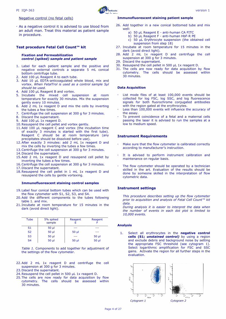
Analysis 1. Select all erythrocytes in the negative control
cells (S1; unstained control) by using a region and exclude debris and background noise by setting the appropriate FSC threshold (see cytogram 1). Select logarithmic amplification for FSC and SSC gains. Activate the region for all further steps in the evaluation.
Cytogram 1 Cytogram 2
PI IQP-363 version 1
Page 5 of 27
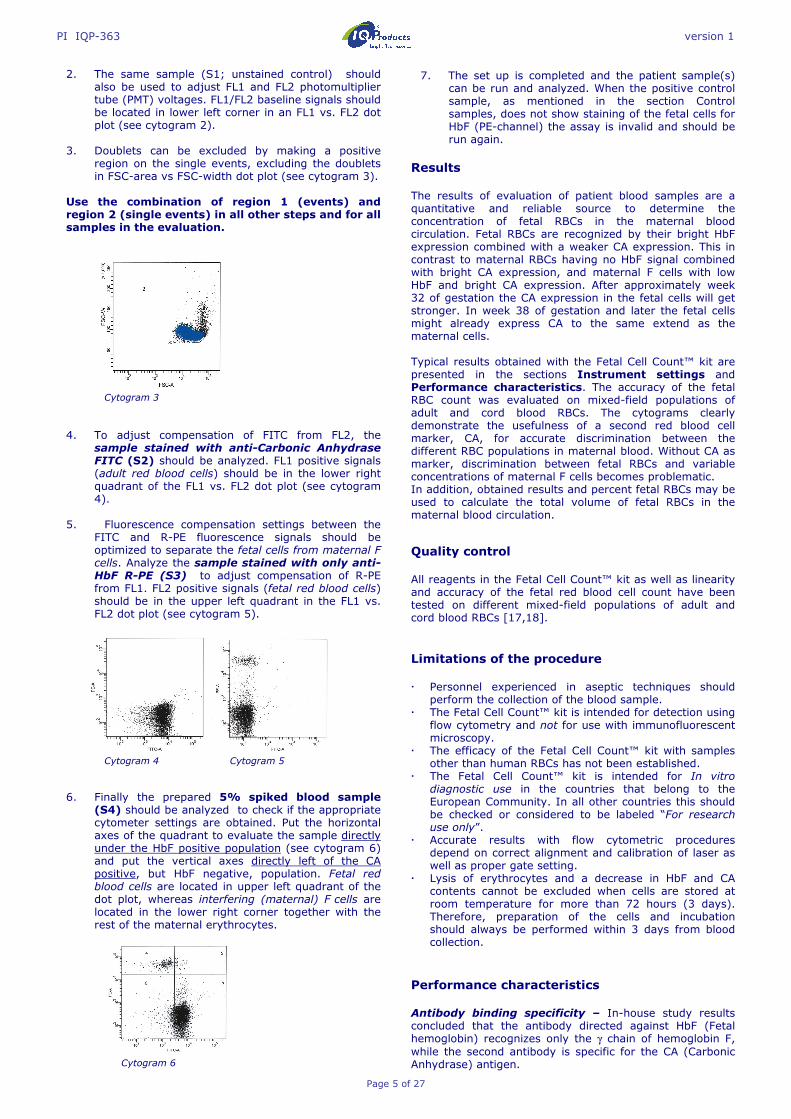
2. The same sample (S1; unstained control) should also be used to adjust FL1 and FL2 photomultiplier tube (PMT) voltages. FL1/FL2 baseline signals should be located in lower left corner in an FL1 vs. FL2 dot plot (see cytogram 2).
3. Doublets can be excluded by making a positive
region on the single events, excluding the doublets in FSC-area vs FSC-width dot plot (see cytogram 3).
Use the combination of region 1 (events) and region 2 (single events) in all other steps and for all samples in the evaluation.
Cytogram 3
4. To adjust compensation of FITC from FL2, the
sample stained with anti-Carbonic Anhydrase FITC (S2) should be analyzed. FL1 positive signals (adult red blood cells) should be in the lower right quadrant of the FL1 vs. FL2 dot plot (see cytogram 4).
5. Fluorescence compensation settings between the
FITC and R-PE fluorescence signals should be optimized to separate the fetal cells from maternal F cells. Analyze the sample stained with only anti-HbF R-PE (S3) to adjust compensation of R-PE from FL1. FL2 positive signals (fetal red blood cells) should be in the upper left quadrant in the FL1 vs. FL2 dot plot (see cytogram 5).
Cytogram 4 Cytogram 5
6. Finally the prepared 5% spiked blood sample
(S4) should be analyzed to check if the appropriate cytometer settings are obtained. Put the horizontal axes of the quadrant to evaluate the sample directly under the HbF positive population (see cytogram 6) and put the vertical axes directly left of the CA positive, but HbF negative, population. Fetal red blood cells are located in upper left quadrant of the dot plot, whereas interfering (maternal) F cells are located in the lower right corner together with the rest of the maternal erythrocytes.
Cytogram 6
7. The set up is completed and the patient sample(s)
can be run and analyzed. When the positive control sample, as mentioned in the section Control samples, does not show staining of the fetal cells for HbF (PE-channel) the assay is invalid and should be run again.
Results The results of evaluation of patient blood samples are a quantitative and reliable source to determine the concentration of fetal RBCs in the maternal blood circulation. Fetal RBCs are recognized by their bright HbF expression combined with a weaker CA expression. This in contrast to maternal RBCs having no HbF signal combined with bright CA expression, and maternal F cells with low HbF and bright CA expression. After approximately week 32 of gestation the CA expression in the fetal cells will get stronger. In week 38 of gestation and later the fetal cells might already express CA to the same extend as the maternal cells. Typical results obtained with the Fetal Cell Count™ kit are presented in the sections Instrument settings and Performance characteristics. The accuracy of the fetal RBC count was evaluated on mixed-field populations of adult and cord blood RBCs. The cytograms clearly demonstrate the usefulness of a second red blood cell marker, CA, for accurate discrimination between the different RBC populations in maternal blood. Without CA as marker, discrimination between fetal RBCs and variable concentrations of maternal F cells becomes problematic. In addition, obtained results and percent fetal RBCs may be used to calculate the total volume of fetal RBCs in the maternal blood circulation.
Quality control All reagents in the Fetal Cell Count™ kit as well as linearity and accuracy of the fetal red blood cell count have been tested on different mixed-field populations of adult and cord blood RBCs [17,18]. Limitations of the procedure � Personnel experienced in aseptic techniques should
perform the collection of the blood sample. � The Fetal Cell Count™ kit is intended for detection using
flow cytometry and not for use with immunofluorescent microscopy.
� The efficacy of the Fetal Cell Count™ kit with samples other than human RBCs has not been established.
� The Fetal Cell Count™ kit is intended for In vitro diagnostic use in the countries that belong to the European Community. In all other countries this should be checked or considered to be labeled “For research use only”.
� Accurate results with flow cytometric procedures depend on correct alignment and calibration of laser as well as proper gate setting.
� Lysis of erythrocytes and a decrease in HbF and CA contents cannot be excluded when cells are stored at room temperature for more than 72 hours (3 days). Therefore, preparation of the cells and incubation should always be performed within 3 days from blood collection.
Performance characteristics Antibody binding specificity – In-house study results concluded that the antibody directed against HbF (Fetal hemoglobin) recognizes only the γ chain of hemoglobin F, while the second antibody is specific for the CA (Carbonic Anhydrase) antigen.

PI IQP-363 version 1
Page 6 of 27
Correlation to the improved version of the Fetal Cell Count™ kit (IQP-363) - This version is the improved version of the Fetal Cell Count™ kit that was based on the direct staining of the two used markers (IQP-379). Studies demonstrate identical performance of the versions. The correlation coefficient (r2) between the two versions is > 0.999. Linearity – Measurement of artificial mixtures for the (theoretical) concentration range 0.02 – 5.0 % (v/v) show a high correlation (r = 0.999 ), when 100,000 cells are measured. This correlation increases when larger number of cells are evaluated. Specificity - Tested samples from control blood donors did not show staining in the upper left (UL) area. These data demonstrate that in there is no interference in the UL area leading to inaccurate counting of fetal cells. Detection limit – The detection limit of the assay is based on the measurement of artificial mixtures and determined to be 0.014% when 100.000 cells are evaluated. Accuracy is improved when the number of events is increased. Clinical evaluation – In total a series of 737 samples have been tested during two different clinical studies. Only part of the studies is represented here. The publications containing all data can be obtained via [email protected] � During the clinical evaluation the Fetal Cell Count™ kit
(IQP-379) has been compared to an earlier version of the Fetal Cell Count™ kit (IQP-370) that was based on the indirect staining of the markers. The correlation between the two versions has shown to be r2 > 0.995
� A clinical evaluation was performed to study the Fetal Cell Count™ kit (IQP-370) performance by comparison with the generally used Kleihauer-Betke test. In this study 130 patient samples were screened.
� In 13,1% (17/130) of the cases feto-maternal transfusion was detected using both methods.
� On a total of 130 patients, 28 (28/130 – 21.59%) were shown to contain fetal cells by using the Kleihauer-Betke test. Of these, only 17 patients (17/28; 61%) contained true fetal cells using the Fetal Cell Count™ kit (range 0.17 to 11.2%) .
The other 11 positive tested patients (11/28; 39%) had a non-typical Kleihauer-Betke test pattern with very faint staining of a number of cells.
� Out of the 11 Kleihauer-Betke positive and Fetal Cell Count™ kit negative patients 7 had a non-typical Kleihauer-Betke test pattern with very faint staining of the cells. These samples showed a typical pattern for thalassemia. These corresponding patients were diagnosed as being thalassemic.
Bibliography 1. DIN EN ISO 15223-1 Medical devices – Symbols to be
used with medical device labels, labeling and information to be supplied-Part 1: General requirements.
2. Sebring, E.S., Polesky, H.F. 1990. Fetomaternal hemorrhage: incidence, risk factors, time of occurence, and clinical effects. Transfusion 30: 344-357.
3. Garratty, G., and Arndt, P.A. 1999. Applications of flow
cytofluorometry to red blood cell immunology. Cytometry (Communications in Clinical Cytometry) 38: 259-267.
4. Nance, S.J., Nelson, J.M., Arndt, P.A., et al. 1989. Quantitation of Feto-maternal hemorrhage by flow cytometry, a simple and accurate method. Am.J.Clin.Pathol. 91: 288-292.
5. Hadley, A.G. 1998. A comparison of in vitro tests for predicting the severity of haemolytic disease of the fetus and newborn. Vox Sang. 74: 375-383.
6. Lee, D., Contreras, M., Robson, S.C., Rodeck, C.H., Whittle, M.J. 1999. Recommendations for the use of anti-D immunoglobulin for Rh prophylaxis. Transf.Med. 9: 93-97.
7. Kleihauer, P., Braun, H., and Betke, K. 1957. Demonstration of fetal hemoglobin in erythrocytes of a blood smear. Klin.Wochenschr. 35: 637-638.
8. Davis, B.H., Olsen, S., Bigelow, N.C., Chen, J.C. 1998. Detection of fetal red cells in fetomaternal hemorrhage using a fetal hemoglobin monoclonal antibody by flow cytometry. Immunohematology 38: 749-756.
9. Johnson, P.R., Tait, R.C., Austin, E.B., et al. 1995. Flow cytometry in diagnosis and management of large fetomaternal haemorrhage. J.Clin.Pathol.48: 1005-1008.
10. Nelson, M., Popp, H., Horky, K., Forsyth, C., Gibson, J. 1994. Development of a flow cytometric test for the detection of D-positive fetal cells after foetalmaternal hemorrhage and a survey of the prevalence in D-negative women. Immunohematology 10: 55-59.
11. Corsetti, J.P., Cox, C., Leary, J.F., Cox, M.T., Blumberg, N., Doherty, R.A. 1987. Comparison of quantitative acid-elution technique and flow cytometry for detecting fetomaternal hemorrhage. Ann.Clin.Lab.Sci. 17: 197-206.
12. Navenot, J.M., Merghoub, T., Ducrocq, R., Krishnamoorthy, R., Blanchard, D. 1998. A new method for quantitative determination of fetal hemoglobin-containing red blood cells by flow cytometry: application to sickle cell disease. Cytometry 32: 186-190.
13. Nelson, M., Zarkos, K., Popp, H., Gilson, J. 1998. A flow-cytometric equivalent of the Kleihauer test. Vox Sang. 75: 234-241.
14. Navenot, J.M., Blandin, A.M., Willem, C., Bernard, D., Muller, J.Y., Blanchard, D. 1995. In situ detection of foetal hemoblobin by flow cytometry: evaluation of a simple procedure for quantitating foetal erythrocytes in maternal peripheral blood. In: International Society of Blood Transfusion – 5th Regional Congress; Venice 2-5 July, abstract POS 309, p239.
15. Blanchard, D., Bernard, D., Loirat, M.J., Frioux, Y., Guimbretière, J., Guimbretière, L. 1992. Caractérisation d’anticorps monoclonaux murins dirigés contre les érythrocytes foetaux. Rev.Fr.Transfus.Hémobiol. 35: 239-254.
16. Brady, HJ.M., Edwards, M., Linch, D.C., 1990 Expression of the human carbonic anhydrase I gene is activated late in fetal erythriod development and regulated by stage-specific trans-acting factors. British Journal of Haematology, 76, 135-142.
17. Bernaud, J., Rigal, D., Porra, V., Follea, G., Blanchard, D. Fetal Cell Count™ - a commercial kit for quantification of fetal cells in maternal blood by flow cytometry. 5th Euroconference on Clinical Cell Analysis. Athens, Greece, 20-25 September 2005.
18. Porra, V., Bernaud, J., Gueret, P., Bricca, P., Rigal, D., Follea, G., and Blanchard, D. 2007 Identification and quantification of fetal red blood cells in maternal blood by a dual-color flow cytometric method: evaluation of the Fetal Cell Count kit. Transfusion, 47:7 , 1281 – 1289.
19. Leers MP, Pelikan HM, Salemans TH, Giordano PC, Scharnhorst V. Discriminating fetomaternal hemorrhage from maternal HbF-containing erythrocytes by dual- parameter flow cytometry. Eur J Obstet Gynecol Reprod Biol. 2006 Jul 24.
Fetal Cell Count™ + - Total
Kleihauer-Betke + 17 11 28 - 0 102 102
Total 17 113 130
PI IQP-363 version 1
Page 7 of 27
Warranty Products sold hereunder are warranted only to conform to the quantity and contents stated on the label at the time of delivery to the customer. There are no warranties, expressed or implied, which extend beyond the description on the label of the product. IQ Products BV is not liable for property damage, personal injury, or economic loss caused by the product. Explanation of used symbols I
Consult instructions for use
[REF]
Catalogue number
s
Sufficient for
[IVD]
In Vitro Diagnostic medical device
!
Caution, consult accompanying document
K
Keep away from (sun)light
D
Biological risks
t
Temperature limitation (°C)
[RUO]
For Research Use Only
[LOT]
Batch code
e
Use by yyyy-mm-dd
M
Manufacturer
[EC_|REP]
Authorized Representative in the European Community
Contact information M IQ Products BV www.iqproducts.nl Rozenburglaan 13a 9727 DL Groningen The Netherlands T +31 (0)50 5757000 F +31 (0)50 5757002 [email protected] This product is registered as “in vitro diagnostic use” in the countries that belong to the European Community. In all other countries it should be labeled “for research use only”.
©2017 - IQ Products bv. All rights reserved. No parts of these works may be reproduced in any form without permission in writing.
PI IQP-363 version 1
Page 8 of 27
Fetal Cell Count™ Kit Détection et quantification des hématies fœtales par cytométrie en flux [REF]1 IQP-363 s25 tests
I Instructions d’utilisation [IVD] |||| Dispositif médical de diagnostic in vitro
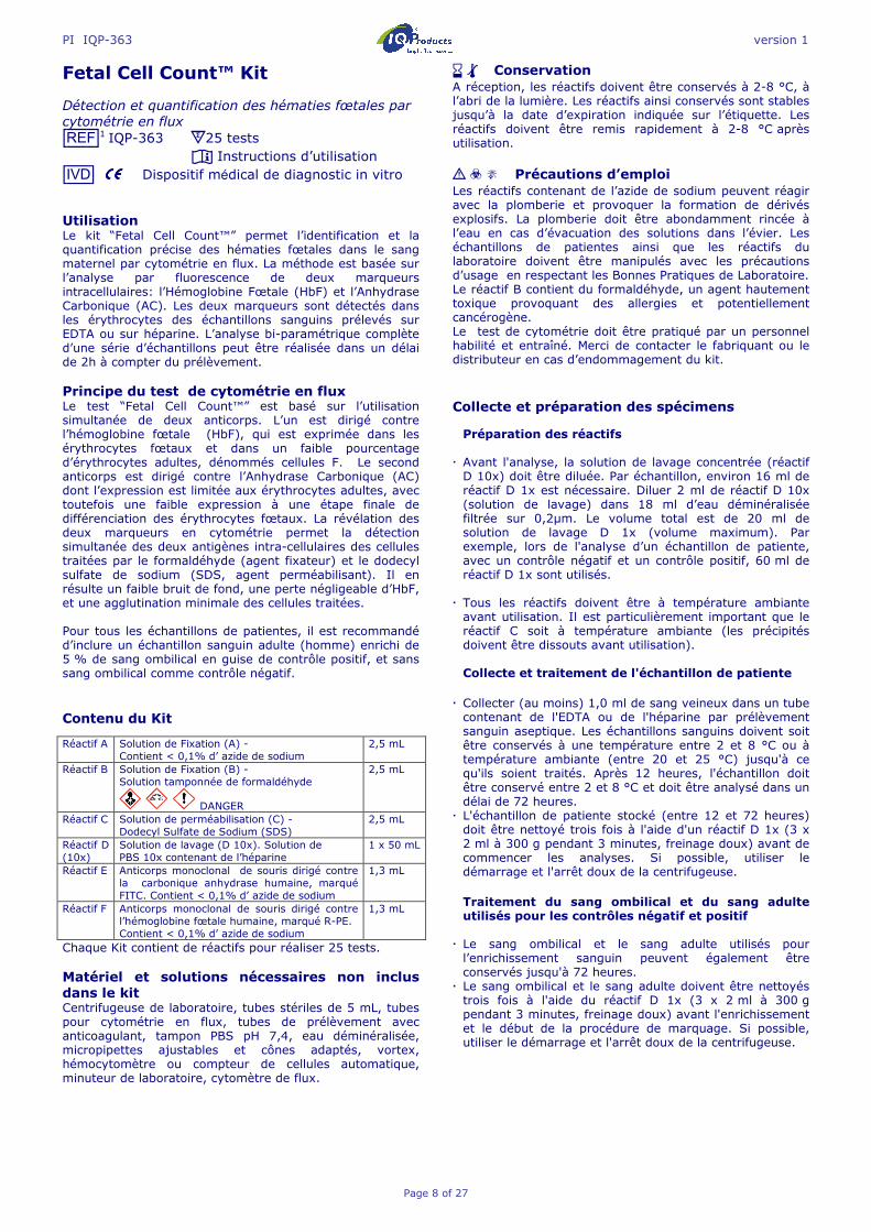
Utilisation Le kit “Fetal Cell Count™” permet l’identification et la quantification précise des hématies fœtales dans le sang maternel par cytométrie en flux. La méthode est basée sur l’analyse par fluorescence de deux marqueurs intracellulaires: l’Hémoglobine Fœtale (HbF) et l’Anhydrase Carbonique (AC). Les deux marqueurs sont détectés dans les érythrocytes des échantillons sanguins prélevés sur EDTA ou sur héparine. L’analyse bi-paramétrique complète d’une série d’échantillons peut être réalisée dans un délai de 2h à compter du prélèvement. Principe du test de cytométrie en flux Le test “Fetal Cell Count™” est basé sur l’utilisation simultanée de deux anticorps. L’un est dirigé contre l’hémoglobine fœtale (HbF), qui est exprimée dans les érythrocytes fœtaux et dans un faible pourcentage d’érythrocytes adultes, dénommés cellules F. Le second anticorps est dirigé contre l’Anhydrase Carbonique (AC) dont l’expression est limitée aux érythrocytes adultes, avec toutefois une faible expression à une étape finale de différenciation des érythrocytes fœtaux. La révélation des deux marqueurs en cytométrie permet la détection simultanée des deux antigènes intra-cellulaires des cellules traitées par le formaldéhyde (agent fixateur) et le dodecyl sulfate de sodium (SDS, agent perméabilisant). Il en résulte un faible bruit de fond, une perte négligeable d’HbF, et une agglutination minimale des cellules traitées. Pour tous les échantillons de patientes, il est recommandé d’inclure un échantillon sanguin adulte (homme) enrichi de 5 % de sang ombilical en guise de contrôle positif, et sans sang ombilical comme contrôle négatif. Contenu du Kit
Chaque Kit contient de réactifs pour réaliser 25 tests. Matériel et solutions nécessaires non inclus dans le kit Centrifugeuse de laboratoire, tubes stériles de 5 mL, tubes pour cytométrie en flux, tubes de prélèvement avec anticoagulant, tampon PBS pH 7,4, eau déminéralisée, micropipettes ajustables et cônes adaptés, vortex, hémocytomètre ou compteur de cellules automatique, minuteur de laboratoire, cytomètre de flux.
e t Conservation A réception, les réactifs doivent être conservés à 2-8 °C, à l’abri de la lumière. Les réactifs ainsi conservés sont stables jusqu’à la date d’expiration indiquée sur l’étiquette. Les réactifs doivent être remis rapidement à 2-8 °C après utilisation. ! D K Précautions d’emploi Les réactifs contenant de l’azide de sodium peuvent réagir avec la plomberie et provoquer la formation de dérivés explosifs. La plomberie doit être abondamment rincée à l’eau en cas d’évacuation des solutions dans l’évier. Les échantillons de patientes ainsi que les réactifs du laboratoire doivent être manipulés avec les précautions d’usage en respectant les Bonnes Pratiques de Laboratoire. Le réactif B contient du formaldéhyde, un agent hautement toxique provoquant des allergies et potentiellement cancérogène. Le test de cytométrie doit être pratiqué par un personnel habilité et entraîné. Merci de contacter le fabriquant ou le distributeur en cas d’endommagement du kit. Collecte et préparation des spécimens Préparation des réactifs
� Avant l'analyse, la solution de lavage concentrée (réactif D 10x) doit être diluée. Par échantillon, environ 16 ml de réactif D 1x est nécessaire. Diluer 2 ml de réactif D 10x (solution de lavage) dans 18 ml d’eau déminéralisée filtrée sur 0,2µm. Le volume total est de 20 ml de solution de lavage D 1x (volume maximum). Par exemple, lors de l'analyse d’un échantillon de patiente, avec un contrôle négatif et un contrôle positif, 60 ml de réactif D 1x sont utilisés.
� Tous les réactifs doivent être à température ambiante avant utilisation. Il est particulièrement important que le réactif C soit à température ambiante (les précipités doivent être dissouts avant utilisation).
Collecte et traitement de l'échantillon de patiente
� Collecter (au moins) 1,0 ml de sang veineux dans un tube contenant de l'EDTA ou de l'héparine par prélèvement sanguin aseptique. Les échantillons sanguins doivent soit être conservés à une température entre 2 et 8 °C ou à température ambiante (entre 20 et 25 °C) jusqu'à ce qu'ils soient traités. Après 12 heures, l'échantillon doit être conservé entre 2 et 8 °C et doit être analysé dans un délai de 72 heures.
� L'échantillon de patiente stocké (entre 12 et 72 heures) doit être nettoyé trois fois à l'aide d'un réactif D 1x (3 x 2 ml à 300 g pendant 3 minutes, freinage doux) avant de commencer les analyses. Si possible, utiliser le démarrage et l'arrêt doux de la centrifugeuse.
Traitement du sang ombilical et du sang adulte utilisés pour les contrôles négatif et positif
� Le sang ombilical et le sang adulte utilisés pour l’enrichissement sanguin peuvent également être conservés jusqu'à 72 heures.
� Le sang ombilical et le sang adulte doivent être nettoyés trois fois à l'aide du réactif D 1x (3 x 2 ml à 300 g pendant 3 minutes, freinage doux) avant l'enrichissement et le début de la procédure de marquage. Si possible, utiliser le démarrage et l'arrêt doux de la centrifugeuse.
Réactif A Solution de Fixation (A) - Contient < 0,1% d’ azide de sodium
2,5 mL
Réactif B Solution de Fixation (B) - Solution tamponnée de formaldéhyde
DANGER
2,5 mL
Réactif C Solution de perméabilisation (C) - Dodecyl Sulfate de Sodium (SDS)
2,5 mL
Réactif D (10x)
Solution de lavage (D 10x). Solution de PBS 10x contenant de l’héparine
1 x 50 mL
Réactif E Anticorps monoclonal de souris dirigé contre la carbonique anhydrase humaine, marqué FITC. Contient < 0,1% d’ azide de sodium
1,3 mL
Réactif F Anticorps monoclonal de souris dirigé contre l’hémoglobine fœtale humaine, marqué R-PE. Contient < 0,1% d’ azide de sodium
1,3 mL
PI IQP-363 version 1
Page 9 of 27
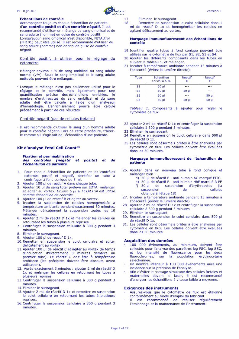
Échantillons de contrôle Accompagner toujours chaque échantillon de patiente d'un contrôle positif et d'un contrôle négatif. Il est recommandé d'utiliser un mélange de sang ombilical et de sang adulte (homme) en guise de contrôle positif. Lorsqu'aucun sang ombilical n'est disponible, FETALtrol (FH101) peut être utilisé. Il est recommandé d'utiliser du sang adulte (homme) non enrichi en guise de contrôle négatif.
Contrôle positif, à utiliser pour le réglage du cytomètre
� Mélanger environ 5 % de sang ombilical au sang adulte normal (v/v). Seuls le sang ombilical et le sang adulte nettoyés peuvent être mélangés.
� Lorsque le mélange n’est pas seulement utilisé pour le réglage et le contrôle, mais également pour une quantification précise des échantillons enrichies, le nombre d'érythrocytes du sang ombilical et du sang adulte doit être calculé à l'aide d'un analyseur d'hématologie. L'enrichissement pourra être calculé précisément à partir de ces résultats. Contrôle négatif (pas de cellules fœtales)
� Il est recommandé d'utiliser le sang d'un homme adulte pour le contrôle négatif. Lors de cette procédure, traitez-le comme s'il s'agissait de l'échantillon d'une patiente.
Kit d'analyse Fetal Cell Count™
Fixation et perméabilisation des contrôles (négatif et positif) et de l’échantillon de patiente
1. Pour chaque échantillon de patiente et les contrôles
externes positif et négatif, identifier un tube à centrifuger à fond conique de 5 ml.
2. Ajouter 100 µl de réactif A dans chaque tube. 3. Ajouter 10 µl de sang total prélevé sur EDTA, mélanger
et agiter au vortex. Utiliser 5 µl si FETALTrol est utilisé comme échantillon de contrôle.
4. Ajouter 100 µl de réactif B et agiter au vortex. 5. Incuber la suspension de cellules homogénéisée à
température ambiante pendant exactement 30 minutes. Mélanger délicatement la suspension toutes les 10 minutes.
6. Ajouter 2 ml de réactif D 1x et mélanger les cellules en retournant les tubes à plusieurs reprises.
7. Centrifuger la suspension cellulaire à 300 g pendant 3 minutes.
8. Éliminer le surnageant. 9. Ajouter 100 µl de réactif D 1x. 10. Remetter en suspension le culot cellulaire et agiter
délicatement au vortex. 11. Ajouter 100 µl de réactif C et agiter au vortex (le temps
d'incubation d'exactement 3 minutes démarre au premier tube). Le réactif C doit être à température ambiante (les précipités doivent être dissouts avant utilisation).
12. Après exactement 3 minutes : ajouter 2 ml de réactif D 1x et mélanger les cellules en retournant les tubes à plusieurs reprises.
13. Centrifuger la suspension cellulaire à 300 g pendant 3 minutes.
14. Éliminer le surnageant. 15. Ajouter 2 mL de réactif D 1x et remetter en suspension
le culot cellulaire en retournant les tubes à plusieurs reprises.
16. Centrifuger la suspension cellulaire à 300 g pendant 3 minutes.
17. Éliminer le surnageant. 18. Remettre en suspension le culot cellulaire dans 1
ml de réactif D 1x et homogénéiser les cellules en agitant délicatement au vortex.
Marquage immunofluorescent des échantillons de contrôle
19. Identifier quatre tubes à fond conique pouvant être utilisés sur le cytomètre de flux par S1, S2, S3 et S4.
20. Ajouter les différents composants dans les tubes en suivant le tableau 1. et mélanger.
21. Incuber à température ambiante pendant 15 minutes à l'obscurité (évitez la lumière directe).
Tube Échantillon
enrichi à 5 % Réactif
E Réactif
F
S1 50 µl --- ---
S2 50 µl 50 µl ---
S3 50 µl --- 50 µl
S4 50 µl 50 µl 50 µl
Tableau 1. Composants à ajouter pour régler le cytomètre de flux.
22. Ajouter 2 ml de réactif D 1x et centrifuger la suspension
cellulaire à 300 g pendant 3 minutes. 23. Éliminer le surnageant. 24. Remettre en suspension le culot cellulaire dans 500 µl
de réactif D 1x. 25. Les cellules sont désormais prêtes à être analysées par
cytométrie en flux. Les cellules doivent être évaluées dans les 30 minutes. Marquage immunofluorescent de l'échantillon de patiente
26. Ajouter dans un nouveau tube à fond conique et mélanger bien :
d) 50 µl de réactif E - anti-humain AC marqué FITC e) 50 µl de réactif F - anti-humain HbF marqué R PE f) 50 µl de suspension d'érythrocytes (la
suspension de cellules obtenue à l'étape 18)
27. Incuber à température ambiante pendant 15 minutes à l'obscurité (évitez la lumière directe).
28. Ajouter 2 ml de réactif D 1x et centrifuger la suspension cellulaire à 300 g pendant 3 minutes.
29. Éliminer le surnageant. 30. Remettre en suspension le culot cellulaire dans 500 µl
de réactif D 1x. 31. Les cellules sont désormais prêtes à être analysées par
cytométrie en flux. Les cellules doivent être évaluées dans les 30 minutes.
Acquisition des données � 100 000 évènements, au minimum, doivent être
collectés pour l’analyse des paraètres log FSC, log SSC, et log intensité de fluorescence pour les deux fluorochromes, sur la population érythrocytaire sélectionnée.
� Un nombre inférieur à 100 000 évènements aura une incidence sur la précision de l'analyse.
� Afin d'éviter le passage simultané des cellules fœtales et maternelles devant le laser, il est recommandé d'analyser les échantillons à vitesse faible à moyenne.
Exigences des instruments � Assurez-vous que le cytomètre de flux est étalonné
conformément au mode d'emploi du fabricant. � Il est recommandé de réaliser régulièrement
l'étalonnage et la maintenance de l'instrument.
PI IQP-363 version 1
Page 10 of 27
� Le cytomètre de flux doit être manipulé par un technicien compétent en la matière. L'évaluation des résultats doit être réalisée par une personne compétente dans l'interprétation des données de cytométrie en flux.
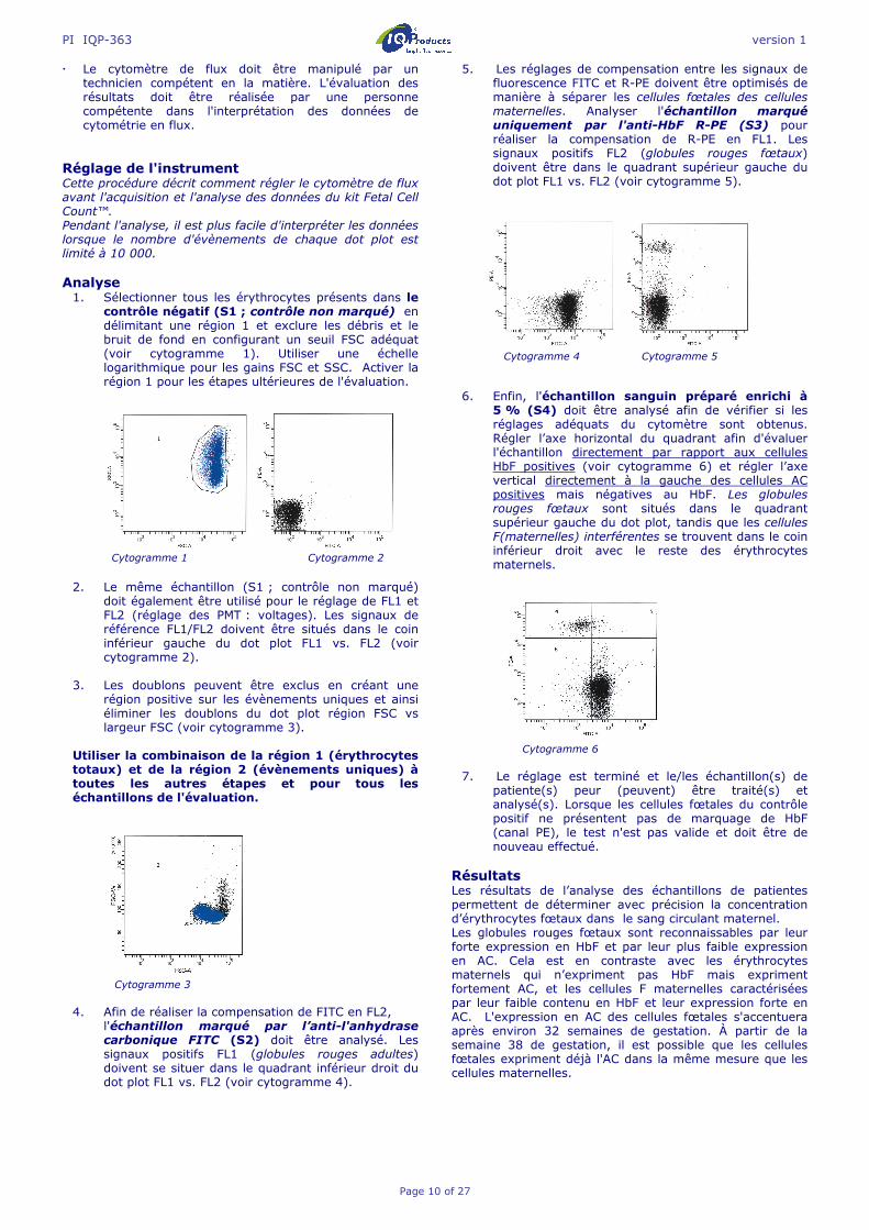
Réglage de l'instrument Cette procédure décrit comment régler le cytomètre de flux avant l'acquisition et l'analyse des données du kit Fetal Cell Count™. Pendant l'analyse, il est plus facile d'interpréter les données lorsque le nombre d'évènements de chaque dot plot est limité à 10 000. Analyse 1. Sélectionner tous les érythrocytes présents dans le
contrôle négatif (S1 ; contrôle non marqué) en délimitant une région 1 et exclure les débris et le bruit de fond en configurant un seuil FSC adéquat (voir cytogramme 1). Utiliser une échelle logarithmique pour les gains FSC et SSC. Activer la région 1 pour les étapes ultérieures de l'évaluation.
Cytogramme 1 Cytogramme 2
2. Le même échantillon (S1 ; contrôle non marqué)
doit également être utilisé pour le réglage de FL1 et FL2 (réglage des PMT : voltages). Les signaux de référence FL1/FL2 doivent être situés dans le coin inférieur gauche du dot plot FL1 vs. FL2 (voir cytogramme 2).
3. Les doublons peuvent être exclus en créant une
région positive sur les évènements uniques et ainsi éliminer les doublons du dot plot région FSC vs largeur FSC (voir cytogramme 3).
Utiliser la combinaison de la région 1 (érythrocytes totaux) et de la région 2 (évènements uniques) à toutes les autres étapes et pour tous les échantillons de l'évaluation.
Cytogramme 3
4. Afin de réaliser la compensation de FITC en FL2,
l'échantillon marqué par l’anti-l'anhydrase carbonique FITC (S2) doit être analysé. Les signaux positifs FL1 (globules rouges adultes) doivent se situer dans le quadrant inférieur droit du dot plot FL1 vs. FL2 (voir cytogramme 4).
5. Les réglages de compensation entre les signaux de fluorescence FITC et R-PE doivent être optimisés de manière à séparer les cellules fœtales des cellules maternelles. Analyser l'échantillon marqué uniquement par l'anti-HbF R-PE (S3) pour réaliser la compensation de R-PE en FL1. Les signaux positifs FL2 (globules rouges fœtaux) doivent être dans le quadrant supérieur gauche du dot plot FL1 vs. FL2 (voir cytogramme 5).
Cytogramme 4 Cytogramme 5
6. Enfin, l'échantillon sanguin préparé enrichi à
5 % (S4) doit être analysé afin de vérifier si les réglages adéquats du cytomètre sont obtenus. Régler l’axe horizontal du quadrant afin d'évaluer l'échantillon directement par rapport aux cellules HbF positives (voir cytogramme 6) et régler l’axe vertical directement à la gauche des cellules AC positives mais négatives au HbF. Les globules rouges fœtaux sont situés dans le quadrant supérieur gauche du dot plot, tandis que les cellules F(maternelles) interférentes se trouvent dans le coin inférieur droit avec le reste des érythrocytes maternels.
Cytogramme 6
7. Le réglage est terminé et le/les échantillon(s) de
patiente(s) peur (peuvent) être traité(s) et analysé(s). Lorsque les cellules fœtales du contrôle positif ne présentent pas de marquage de HbF (canal PE), le test n'est pas valide et doit être de nouveau effectué.
Résultats Les résultats de l’analyse des échantillons de patientes permettent de déterminer avec précision la concentration d’érythrocytes fœtaux dans le sang circulant maternel. Les globules rouges fœtaux sont reconnaissables par leur forte expression en HbF et par leur plus faible expression en AC. Cela est en contraste avec les érythrocytes maternels qui n’expriment pas HbF mais expriment fortement AC, et les cellules F maternelles caractérisées par leur faible contenu en HbF et leur expression forte en AC. L'expression en AC des cellules fœtales s'accentuera après environ 32 semaines de gestation. À partir de la semaine 38 de gestation, il est possible que les cellules fœtales expriment déjà l'AC dans la même mesure que les cellules maternelles.

PI IQP-363 version 1
Page 11 of 27
Des résultats représentatifs obtenus avec le kit sont présentés dans les sections « Réglages de l’appareil » et « Performances ». La précision de comptage des érythrocytes a été évaluée en utilisant des mélanges artificiels de sang de cordons et d’adultes normaux. Les profils démontrent clairement l’importance du second marqueur, AC, pour une identification précise des populations érythrocytaires dans le sang maternel. En l’absence du marqueur, la séparation des cellules fœtales et des cellules F maternelles est délicate. Les résultats obtenus peuvent être utilisés pour déterminer le volume de sang fœtal présent dans le sang maternel. Contrôle Qualité Tous les réactifs du kit Fetal Cell Count™ ont été évalués avec des mélanges artificiels de sang de cordons et d’adultes normaux. Linéarité et précision de comptage des cellules fœtales ont été analysées. [17,18] Limites de la méthode � La méthode nécessite l’utilisation de sang prélevé dans
des conditions aseptiques, par du personnel compétent.
� Le kit Fetal Cell Count™ est prévu pour la quantification des érythrocytes fœtaux par cytométrie; il n’est pas adapté pour des études en microscopie.
� La performance du kit Fetal Cell Count™ n’a pas été évalué avec des échantillons autres que ceux d’origine humaine.
� Le kit Fetal Cell Count™ doit être utilisé pour le Diagnostic In Vitro dans les pays de la Communauté Européenne. Dans les autres pays, il doit être utilisé dans un cadre de recherche uniquement.
� La précision de la méthode de cytométrie dépend de l’utilisation d’un appareil de cytométrie dûment réglé, entretenu et contrôlé selon les recommandations du fabricant.
� La lyse des globules rouges et la diminution de l’expression des marqueurs HbF et AC ne peuvent pas être exclues quand les cellules sont conservées à température ambiante pendant plus de 72 heures (3 jours). Ainsi, la préparation des cellules et leur incubation avec les anticorps doivent être effectuées dans les 3 jours qui suivent le prélèvement.
Performance de la méthode Spécificité des anticorps - l’anticorps monoclonal est spécifique de la chaîne γ de l’hémoglobine fœtale F (HbF) (ref. 12). L’anticorps monoclonal reconnaît spécifiquement l’anhydrase carbonique.
Corrélation avec la version améliorée du kit Fetal Cell Count ™ (IQP-363) - Cette version est la version améliorée du kit Fetal Cell Count™ qui était basée sur la coloration directe des deux marqueurs utilisés (IQP-379). L’évaluation du test à démontré des performances équivalentes pour les deux méthodes. Le coefficient de corrélation (r2) pour les deux méthodes est > 0,99. Linéarité - L’analyse de mélanges artificiels d’érythrocytes contenant de 0.00 à 5.00% de cellules de cordons (v/v) a montré une très forte corrélation (r=0,999) avec les valeurs théoriques attendues, pour des comptages de 100 000 cellules. Spécificité du test – Les échantillons de donneurs de sang utilisés comme témoins n’ont présenté aucune cellule dans le quadrant UL (zone des cellules HbF+, CA-). Ces résultats démontrent que les échantillons d’une population témoin ne contiennent pas d’éléments susceptibles d’interférer avec l’analyse spécifique des érythrocytes fœtaux.
Limite de détection – La limite de détection de la méthode est basée sur l’analyse de mélanges artificiels. Elle a été déterminée à 0,014% par comptage de 100 000 cellules. Elle est améliorée par un comptage d’un nombre plus important de cellules. Evaluation clinique du test- Un total de 737 échantillons a été analysé au cours des évaluations cliniques. Une partie des résultats est présenté ci-dessous ; les publications rassemblant l’ensemble des résultats peuvent être obtenues via [email protected] . � Au cours de l’évaluation clinique du kit Fetal Cell
Count™ (IQP-379), ses performances ont été évalués en comparaison avec celle du kit utilisant la méthode indirecte (IQP-370). La corrélation entre les deux méthodes est r2 > 0,995.
� L’évaluation clinique a également permis l’analyse du kit Fetal Cell Count™ (IQP-370) en comparaison avec le test de Kleihauer-Betke (KB). Dans cette étude, 130 échantillons de patients ont été analysés.
� 13,1% (17/130) de cas d’hémorragie fœto-maternelle
ont été identifiés par les deux méthodes. � Sur un total de 130 échantillons de patientes, 28
(28/130 - 21,59%) ont été identifiés comme des échantillons positifs en utilisant le test KB.
� Sur ces 28 échantillons positifs selon le test KB, seuls 17 patientes (17/28 - 61,00%) possédaient des cellules fœtales identifiées par cytométrie en flux en utilisant le Fetal Cell Count™ kit (valeurs de 0,17 to 11,2%). Les 11 autres patientes de la série (11/28 – 39,00 %) ont présenté un profil atypique en test KB, avec une faible coloration des cellules identifiées comme positives.
� Sur les 11 patientes positives avec le test KB et négatives avec le kit ™ de comptage de cellules fœtales, 7 avaient un profil atypique en test KB, avec une très faible coloration des cellules. Ces échantillons ont montré un profil typique de la thalassémie. Ces patientes correspondantes ont été diagnostiquées comme étant thalassémique.
Bibliographie 1. NEN EN ISO 15223-1 Medical devices - Symbols to be
used with medical device labels, labeling and information to be supplied – Part 1: General requirements.
2. Sebring, E.S., Polesky, H.F. 1990. Fetomaternal hemorrhage: incidence, risk factors, time of occurence, and clinical effects. Transfusion 30: 344-357.
3. Garratty, G., and Arndt, P.A. 1999. Applications of flow cytofluorometry to red blood cell immunology. Cytometry (Communications in Clinical Cytometry) 38: 259-267.
4. Nance, S.J., Nelson, J.M., Arndt, P.A., et al. 1989. Quantitation of Feto-maternal hemorrhage by flow cytometry, a simple and accurate method. Am.J.Clin.Pathol. 91: 288-292.
5. Hadley, A.G. 1998. A comparison of in vitro tests for predicting the severity of haemolytic disease of the fetus and newborn. Vox Sang. 74: 375-383.
6. Lee, D., Contreras, M., Robson, S.C., Rodeck, C.H., Whittle, M.J. 1999. Recommendations for the use of anti-D immunoglobulin for Rh prophylaxis. Transf.Med. 9: 93-97.
7. Kleihauer, P., Braun, H., and Betke, K. 1957. Demonstration of fetal hemoglobin in erythrocytes of a blood smear. Klin.Wochenschr. 35: 637-638.
Fetal Cell Count™ + - Total
Kleihauer-Betke + 17 11 28 - 0 102 102
Total 17 113 130
PI IQP-363 version 1
Page 12 of 27
8. Davis, B.H., Olsen, S., Bigelow, N.C., Chen, J.C. 1998. Detection of fetal red cells in fetomaternal hemorrhage using a fetal hemoglobin monoclonal antibody by flow cytometry. Immunohematology 38: 749-756.
9. Johnson, P.R., Tait, R.C., Austin, E.B., et al. 1995. Flow cytometry in diagnosis and management of large fetomaternal haemorrhage. J.Clin.Pathol.48: 1005-1008.
10. Nelson, M., Popp, H., Horky, K., Forsyth, C., Gibson, J. 1994. Development of a flow cytometric test for the detection of D-positive fetal cells after foetalmaternal hemorrhage and a survey of the prevalence in D-negative women. Immunohematology 10: 55-59.
11. Corsetti, J.P., Cox, C., Leary, J.F., Cox, M.T., Blumberg, N., Doherty, R.A. 1987. Comparison of quantitative acid-elution technique and flow cytometry for detecting fetomaternal hemorrhage. Ann.Clin.Lab.Sci. 17: 197-206.
12. Navenot, J.M., Merghoub, T., Ducrocq, R., Krishnamoorthy, R., Blanchard, D. 1998. A new method for quantitative determination of fetal hemoglobin-containing red blood cells by flow cytometry: application to sickle cell disease. Cytometry 32: 186-190.
13. Nelson, M., Zarkos, K., Popp, H., Gilson, J. 1998. A flow-cytometric equivalent of the Kleihauer test. Vox Sang. 75: 234-241.
14. Navenot, J.M., Blandin, A.M., Willem, C., Bernard, D., Muller, J.Y., Blanchard, D. 1995. In situ detection of foetal hemoblobin by flow cytometry: evaluation of a simple procedure for quantitating foetal erythrocytes in maternal peripheral blood. In: International Society of Blood Transfusion – 5th Regional Congress; Venice 2-5 July, abstract POS 309, p239.
15. Blanchard, D., Bernard, D., Loirat, M.J., Frioux, Y., Guimbretière, J., Guimbretière, L. 1992. Caractérisation d’anticorps monoclonaux murins dirigés contre les érythrocytes foetaux. Rev.Fr.Transfus.Hémobiol. 35: 239-254.
16. Brady, HJ.M., Edwards, M., Linch, D.C., 1990 Expression of the human carbonic anhydrase I gene is activated late in fetal erythroid development and regulated by stage-specific trans-acting factors. British Journal of Haematology 1990, 76, 135-142.
17. Bernaud, J., Rigal, D., Porra, V., Follea, G., Blanchard, D. Fetal Cell Count™ - a commercial kit for quantification of fetal cells in maternal blood by flow cytometry. 5th Euroconference on Clinical Cell Analysis. Athens, Greece, 20-25 September 2005.
18. Porra, V., Bernaud, J., Gueret, P., Bricca, P., Rigal, D., Follea, G., and Blanchard, D. 2007 Identification and quantification of fetal red blood cells in maternal blood by a dual-color flow cytometric method: evaluation of the Fetal Cell Count kit. Transfusion, 47:7, 1281 – 1289.
19. Leers MP, Pelikan HM, Salemans TH, Giordano PC, Scharnhorst V. Discriminating fetomaternal hemorrhage from maternal HbF-containing erythrocytes by dual- parameter flow cytometry. Eur J Obstet Gynecol Reprod Biol. 2006 Jul 24. Garantie Quantité et contenu des produits composant le kit sont garantis conformes à l’étiquetage, au moment de la livraison. Aucune garantie, implicite ou explicite, n’est donnée au delà de l’étiquetage. Le fabricant, IQ Products bv, ne pourrait être tenu pour responsable de tout dommage de propriété, accident du personnel, ou perte économique causée par le produit. Mise aux déchets Respecter les exigences réglementaires en vigueur dans le pays d’utilisation. Pour la France: GBEA du 26 Novembre 1999 relatif à la bonne exécution des analyses de biologie médicale.
Tableau des Symboles I Consulter les instructions d’utilisation [REF] Référence du catalogue s Suffisant pour [IVD] Dispositif médical de diagnostic in vitro ! Attention voir notice d’instructions K Ne pas exposer aux rayons (solaires) D Risques biologiques t Limites de température (°C) [RUO] Pour la recherche uniquement [LOT] Code du lot e Utiliser jusque M Fabricant [EC_|REP] Mandataire dans la Communauté
européenne |||| Conformité Européenne Ce produit est enregistré pour “diagnostic in vitro” dans les pays de la Communauté Européenne. Dans les autres pays, il sera utilisé comme produit de recherche et libellé « for research use only ». M IQ Products bv
Rozenburglaan 13a 9727 DL Groningen, The Netherlands ℡ +31 (0)50 57 57 000 � +31 (0)50 57 57 002 � Technical [email protected] � Orders [email protected] � www.iqproducts.nl
©2017 - IQ Products bv. Tout droit réservé. Les éléments contenus dans cette notice en peuvent pas être reproduits sans accord écrit .
PI IQP-363 version 1
Page 13 of 27
Fetal Cell CountTM Kit Durchflußzytometrische Diagnose fetomaternaler Hämorrhagie [REF]1 IQP-363 s 25 Tests I Packungsbeilage [IVD] |||| In-Vitro-Diagnostikum
Verwendungszweck Der Fetal Cell Count™ kit wird zum quantitativen Nachweis humaner fetaler Erythrozyten im maternalem Blut verwendet. Der Fetal Cell Count™ kit basiert auf einer sensitiven und genauen durchflusszytometrischen Methode, die einen Zwei-Farben-Nachweis zweier intrazellulärer Antigenen, Hämoglobin F (HbF) und Carboanhydrase (CA), ermöglicht. Der Nachweis von HbF und CA erfolgt aus EDTA- oder heparinisiertem humanen Vollblut. Einschließlich der Färbung und der Analyse von bis zu 5 Proben kann der Test innerhalb von 2 Stunden durchgeführt werden. Testprinzip Die Fetal Cell Count™ Methode basiert auf einer Kombination zweier Antikörper. Der erste Antikörper ist gegen fetales Hämoglobin (HbF) gerichtet, nachweisbar in fetalen Erythrozyten und einem geringen Prozentsatz adulter Erythrozyten (sog. F-Zellen). Der zweite Antikörper ist gegen die Carbonanhydrase gerichtet, ein Enzym, das nur in adulten Erythrozyten vorkommt und in fetalen Erythrozyten zum Ende der Schwangerschaft. Die durchflußzytometrische Zwei-Farben-Methode erlaubt den simultanen Nachweis dieser zwei intrazellulären Antigene. Die Verwendung von Formaldehyd als Fixativ und Natriumdodezylsulfat (SDS) zur Permeabilisierung der fixierten Erythrozyten verursacht nur geringes Hintergrundrauschen und minimale Zellverklumpung, dadurch kann der Verlust von HbF vernachlässigt werden. Für jeden Patienten muss eine Blutprobe eines (männlichen) Erwachsenen, angereichert mit 5 % Nabelschnurblut, als positive Kontrollprobe und eine solche Blutprobe ohne Nabelschnurblut als negative Kontrollprobe mit dem Test einbezogen werden. Kit-Inhalt
Jeder Kit enthält ausreichend Reagentien zur Durchführung von 25 Tests.
Zusätzlich benötigtes Labormaterial Laborzentrifuge; 5 mL-Teströhrchen, steril; konische Mikrozentrifugenröhrchen, steril; phosphatgepufferte Saline (PBS), pH 7.4; mit EDTA oder Heparin beschichtetes Röhrchen, verstellbare Mikropipetten und Einwegspitzen, Vortex-Mixer, Hämozytometer oder automatisierter Zellzähler, Kurzzeitmesser. e t Lagerung Nach Erhalt Reagenzien bei 2-8 °C aufbewahren. Direktes Sonnenlicht vermeiden. Reagenzien, die unter den vorgegebenen Bedingungen gelagert werden, sind bis zum Verfallsdatum (siehe Etikett) haltbar.
Bei mehrmaliger Verwendung Reagenzien sofort nach Gebrauch wieder bei 2-8 °C aufbewahren. ! D K Warnung und Vorsichtsmaßnahmen Reagenzien, die Natriumazid enthalten, können mit Blei oder Kupfer explosive Metallazide bilden. Bei der Beseitigung mit reichlich Wasser nachspülen, um die Azidbildung zu verhindern. Alle Reagenzien sind entsprechend herrschender Laborpraxis anzuwenden. Zusätzlich sind Patientenproben mit geeigneten Vorsichtsmaßnahmen zu behandeln. Nicht mit dem Mund pipettieren, Handschuhe tragen. Reagenz B enthält Formaldehyd, ein hochtoxisches, allergenes und potentiell krebserregendes Reagenz, das entsprechend herrschender Laborpraxis mit geeigneten Vorsichtsmaßnahmen zu verwenden ist. Haut- oder Augenkontakt vermeiden. Der Test darf nur von ausgebildetem und autorisiertem Laborpersonal durchgeführt werden. Bitte informieren Sie den Hersteller, wenn der originale Kit beschädigt ist. Gewinnung und Aufbereitung der Proben Aufbereitung der Reagenzien
� Vor dem Testen sollte die 10-fach konzentrierte Waschlösung (10x Reagens D) verdünnt werden. Pro Probe werden dafür etwa 16 ml von 1x Reagens D benötigt. 18 ml von 0,2 µm gefiltertem, demineralisierten Wasser zu 2 ml der Waschlösung mit 10-fach konzentriertem Reagens D. Das Gesamtvolumen beträgt 20 ml der Waschlösung mit 1-facher D-Konzentration (Höchstvolumen). Zum Testen einer Patientenprobe werden zum Beispiel eine negative und eine positive Kontrollprobe mit insgesamt 60 ml 1-fach konzentriertem Reagens D verwendet.
� Alle Reagenzien sollten vor Gebrauch auf Raumtemperatur gebracht werden. Vor allem Reagens C sollte Raumtemperatur besitzen (zur Lösung möglicher Präzipitate).
Gewinnung und Verarbeitung einer Patientenprobe
� Mithilfe einer aseptischen Venenpunktion (mindestens) 1,0 ml Venenblut entnehmen und in einem mit EDTA oder Heparin beschichteten Röhrchen auffangen. Bis zur Verarbeitung sollten die Blutproben entweder bei 2-8 °C oder bei Raumtemperatur (20-25 °C) gelagert werden.Nach 12 Stunden sollte die Probe bei 2-8 °C gelagert und innerhalb von 72 Stunden getestet werden.
� Eine Patientenprobe, die zwischengelagert wurde (12-72 Stunden), sollte vor Beginn der Tests drei Mal mit 1-fachem Reagens D (3 x 2 ml bei 300 g, 3 min, langsames Bremsen) gewaschen werden. Wenn möglich, ist die Soft-Funktion für das Anlaufen und Bremsen der Zentrifuge zu verwenden.
Verarbeitung von Nabelschnurblut und Blut von Erwachsenen zur Verwendung in Anreicherungsexperimenten
� Nabelschnurblut und Blut von Erwachsenen für die Verwendung in Anreicherungsexperimenten kann ebenfalls bis zu 72 Stunden gelagert werden.
� Nabelschnurblut und Blut von Erwachsenen sollte vor der Anreicherung und dem Beginn der Färbung stets drei Mal mit 1-fachem Reagens D (3 x 2 ml bei 300 g, 3 min, langsames Bremsen) gewaschen werden. Wenn möglich, ist die Soft-Funktion für das Anlaufen und Bremsen der Zentrifuge zu verwenden.
Reagenz A Fixativlösung (A) - Enthält < 0.1% Natriumazid
2,5 ml
Reagenz B Fixativlösung (B) - gepuffertes Formaldehyd
GEFAHR
2,5 ml
Reagenz C Permeabilitätslösung (C) – Enthält Natrium Dodecylsulfat (SDS)
2,5 ml
Reagenz D (10x) Waschpuffer (10xD), 10fach konzentriert – PBS enthält Heparin
1x50 ml
Reagenz E Monoklonale Antikörper gegen humane Carboanhydrase konjugiert mit FITC, enthält< 0,1% Natriumazid
1,3 ml
Reagenz F Monoklonale Antikörper gegen humanes Hämoglobin F konjugiert mit R-PE, enthält < 0,1% Natriumazid
1,3 ml
PI IQP-363 version 1
Page 14 of 27
Kontrollproben
Für jede Patientenprobe immer eine positive und eine negative Kontrollprobe durchführen. Eine Mischung aus Nabelschnurblut und Blut eines (männlichen) Erwachsenen muss als positive Kontrollprobe verwendet werden. Wenn kein Nabelschnurblut verfügbar ist, kann FETALtrol (FH101) verwendet werden. Blut eines (männlichen) Erwachsenen ohne Anreicherung sollte als negative Kontrollprobe verwendet werden. Positive Kontrollprobe, auch zur Verwendung für die Einrichtung des Zytometers
� Etwa 5 % Nabelschnurblut mit normalem Erwachsenenblut (v/v) mischen. Nur gewaschenes Nabelschnurblut und Blut von Erwachsenen sollte miteinander vermischt werden.
� Wenn die Mischung nicht nur zum Einrichten und zur Kontrolle, sondern auch für eine genaue Quantifizierung der angereicherten Zellen verwendet werden soll, sollten die Erythrozyten sowohl in der Nabelschnurblutprobe als auch in der Blutprobe des Erwachsenen mit einem Hämatologie-Analysegerät gezählt werden. Auf Grundlage dieser Daten kann die Anreicherung genau berechnet werden. Negative Kontrollprobe (ohne fetale Zellen)
� Als negative Kontrollprobe wird Blut eines männlichen Erwachsenen empfohlen. Dieses Material ist im Verfahren als Patientenprobe zu behandeln.
Testverfahren Fetal Cell Count™ Kit
Fixierung und Permeabilisierung (angereicherte) Kontrollprobe und Patientenprobe
1. Für jede Patientenprobe und die positiven und negativen, externen Kontrollproben jeweils ein konisches 5-ml-Zentrifugenröhrchen beschriften.
2. Jedem Röhrchen 100 µl Reagens A hinzufügen. 3. 10 µl EDTA-antikoaguliertes Vollblut hinzufügen und
auf dem Vortex-Mischer mischen. Bei Verwendung von FetalTrol als Kontrollprobe, nur 5 µl verwenden.
4. 100 µl Reagens B hinzufügen und auf dem Vortex-Mischer mischen.
5. Die vermischte Zellsuspension bei Raumtemperatur exakt 30 Minuten lang inkubieren. Die Suspension aller 10 Minuten vorsichtig mischen.
6. 2 ml 1x Reagens D hinzufügen und die Zellen durch mehrmaliges Hin- und Herwenden der Röhrchen mischen.
7. Die Zellsuspension 3 Minuten bei 300 g zentrifugieren. 8. Überstand verwerfen. 9. 100 µl 1x Reagens D hinzufügen. 10. Zellpellet resuspendieren und auf dem Vortexmischer
vorsichtig mischen. 11. 100 µl Reagens C hinzufügen und auf dem Vortex-
Mischer mischen (die Inkubationszeit von exakt 3 Minuten beginnt mit dem ersten Röhrchen). Reagens C sollte Raumtemperatur besitzen (zur Lösung möglicher Präzipitate).
12. Nach exakt 3 Minuten: 2 ml 1x Reagens D hinzufügen und die Zellen durch mehrmaliges Hin- und Herwenden der Röhrchen mischen.
13. Die Zellsuspension 3 Minuten bei 300 g zentrifugieren. 14. Überstand verwerfen. 15. 2 ml 1x Reagens D hinzufügen und das Zellpellet
durch mehrmaliges Hin- und Herwenden der Röhrchen resuspendieren.
16. Die Zellsuspension 3 Minuten bei 300 g zentrifugieren. 17. Überstand verwerfen. 18. Das Zellpellet in 1 ml 1x Reagens D resuspendieren
und die Zellen durch vorsichtiges Mischen auf dem Vortex-Mischer resuspendieren.
Immunfluoreszenzfärbung der Kontrollproben
19. Vier konische Röhrchen zur Verwendung mit dem Durchflusszytometer mit S1, S2, S3 und S4 beschriften.
20. Die verschiedenen Komponenten entsprechend Tabelle 1 zu den Röhrchen hinzufügen und mischen.
21. Für 15 Minuten bei Raumtemperatur inkubieren (direktes Licht vermeiden).
Röhrchen mit 5 % angereicherte
Probe
Reagens E
Reagens F
S1 50 µl --- ---
S2 50 µl 50 µl ---
S3 50 µl --- 50 µl
S4 50 µl 50 µl 50 µl
Tabelle 1. Komponenten, die für die Einstellung des Durchflusszytometers zu vermischen sind.
22. 2 ml 1x Reagens D hinzufügen und die Zellsuspension 3
Minuten bei 300 g zentrifugieren. 23. Überstand verwerfen. 24. Das Zellpellet in 500 µl 1x Reagens D resuspendieren. 25. Die Zellen sind jetzt für die Datenerfassung am
Durchflusszytometer bereit. Die Messung der Zellen sollte innerhalb von 30 Minuten erfolgen. Immunfluoreszenzfärbung der Patientenprobe
26. In ein neues, konisches Röhrchen pipettieren und gut mischen:
g) 50 µl Reagens E - Anti-human CA FITC h) 50 µl Reagens F - Anti-human HbF-R PE i) 50 µl Erythrozytensuspension (die Zellsuspension
aus Schritt 18) 27. Für 15 Minuten bei Raumtemperatur im Dunkeln
inkubieren (direktes Licht vermeiden). 28. 2 ml 1x Reagens D hinzufügen und die Zellsuspension
bei 300 g 3 Minuten lang zentrifugieren. 29. Überstand verwerfen. 30. Das Zellpellet in 500 µl 1x Reagens D resuspendieren. 31. Die Zellen sind jetzt für die Datenerfassung am
Durchflusszytometer bereit. Die Messung der Zellen sollte innerhalb von 30 Minuten erfolgen.
Datenerfassung
� Für beide fluorochrom-konjugierten Antikörper sollten
mindestens 100.000 Ereignisse für log FSC, log SSC und log Fluoreszenzsignale mit einem Gate um die Region der Erythrozyten im Listenmodus erfasst werden.
� Bei Erfassung von weniger als 100.000 Ereignissen kann es zu Ungenauigkeiten im Test kommen.
� Um das gleichzeitige Passieren einer fetalen und einer mütterlichen Zelle vor dem Laser zu verhindern, wird empfohlen, die Proben bei geringer bis moderater Geschwindigkeit zu prüfen.
Geräteanforderungen � Durchflusszytometer auf korrekte Kalibrierung
entsprechend der Herstelleranweisungen überprüfen. � Es wird empfohlen, das Gerät regelmäßig zu kalibrieren
und zu warten. � Das Durchflusszytometer sollte von einem dafür
ausgebildeten Techniker bedient werden. Die Auswertung der Ergebnisse sollte durch eine Fachkraft erfolgen, die dazu ausgebildet ist, die Daten des Durchflusszytometers zu interpretieren.
PI IQP-363 version 1
Page 15 of 27
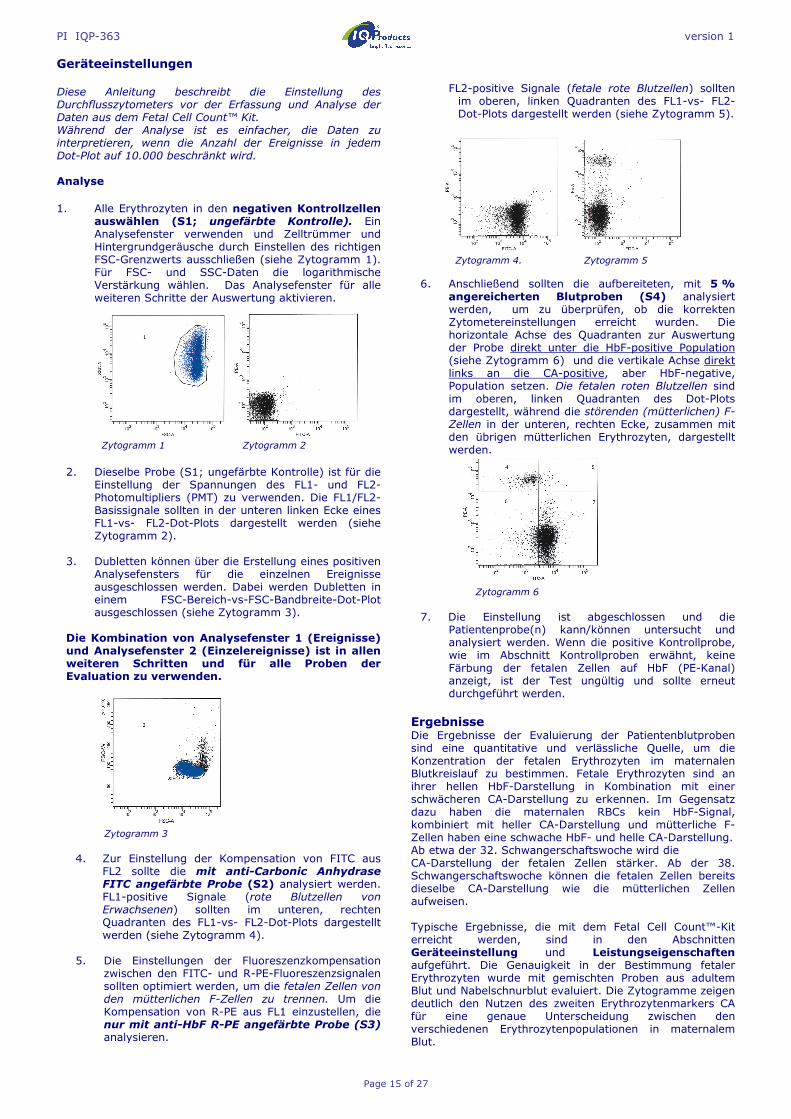
Geräteeinstellungen Diese Anleitung beschreibt die Einstellung des Durchflusszytometers vor der Erfassung und Analyse der Daten aus dem Fetal Cell Count™ Kit. Während der Analyse ist es einfacher, die Daten zu interpretieren, wenn die Anzahl der Ereignisse in jedem Dot-Plot auf 10.000 beschränkt wird.
Analyse 1. Alle Erythrozyten in den negativen Kontrollzellen
auswählen (S1; ungefärbte Kontrolle). Ein Analysefenster verwenden und Zelltrümmer und Hintergrundgeräusche durch Einstellen des richtigen FSC-Grenzwerts ausschließen (siehe Zytogramm 1). Für FSC- und SSC-Daten die logarithmische Verstärkung wählen. Das Analysefenster für alle weiteren Schritte der Auswertung aktivieren.
Zytogramm 1 Zytogramm 2
2. Dieselbe Probe (S1; ungefärbte Kontrolle) ist für die
Einstellung der Spannungen des FL1- und FL2-Photomultipliers (PMT) zu verwenden. Die FL1/FL2-Basissignale sollten in der unteren linken Ecke eines FL1-vs- FL2-Dot-Plots dargestellt werden (siehe Zytogramm 2).
3. Dubletten können über die Erstellung eines positiven
Analysefensters für die einzelnen Ereignisse ausgeschlossen werden. Dabei werden Dubletten in einem FSC-Bereich-vs-FSC-Bandbreite-Dot-Plot ausgeschlossen (siehe Zytogramm 3).
Die Kombination von Analysefenster 1 (Ereignisse) und Analysefenster 2 (Einzelereignisse) ist in allen weiteren Schritten und für alle Proben der Evaluation zu verwenden.
Zytogramm 3
4. Zur Einstellung der Kompensation von FITC aus
FL2 sollte die mit anti-Carbonic Anhydrase FITC angefärbte Probe (S2) analysiert werden. FL1-positive Signale (rote Blutzellen von Erwachsenen) sollten im unteren, rechten Quadranten des FL1-vs- FL2-Dot-Plots dargestellt werden (siehe Zytogramm 4).
5. Die Einstellungen der Fluoreszenzkompensation
zwischen den FITC- und R-PE-Fluoreszenzsignalen sollten optimiert werden, um die fetalen Zellen von den mütterlichen F-Zellen zu trennen. Um die Kompensation von R-PE aus FL1 einzustellen, die nur mit anti-HbF R-PE angefärbte Probe (S3) analysieren.
FL2-positive Signale (fetale rote Blutzellen) sollten im oberen, linken Quadranten des FL1-vs- FL2-Dot-Plots dargestellt werden (siehe Zytogramm 5).
Zytogramm 4. Zytogramm 5
6. Anschließend sollten die aufbereiteten, mit 5 % angereicherten Blutproben (S4) analysiert werden, um zu überprüfen, ob die korrekten Zytometereinstellungen erreicht wurden. Die horizontale Achse des Quadranten zur Auswertung der Probe direkt unter die HbF-positive Population (siehe Zytogramm 6) und die vertikale Achse direkt links an die CA-positive, aber HbF-negative, Population setzen. Die fetalen roten Blutzellen sind im oberen, linken Quadranten des Dot-Plots dargestellt, während die störenden (mütterlichen) F-Zellen in der unteren, rechten Ecke, zusammen mit den übrigen mütterlichen Erythrozyten, dargestellt werden.
Zytogramm 6
7. Die Einstellung ist abgeschlossen und die
Patientenprobe(n) kann/können untersucht und analysiert werden. Wenn die positive Kontrollprobe, wie im Abschnitt Kontrollproben erwähnt, keine Färbung der fetalen Zellen auf HbF (PE-Kanal) anzeigt, ist der Test ungültig und sollte erneut durchgeführt werden.
Ergebnisse Die Ergebnisse der Evaluierung der Patientenblutproben sind eine quantitative und verlässliche Quelle, um die Konzentration der fetalen Erythrozyten im maternalen Blutkreislauf zu bestimmen. Fetale Erythrozyten sind an ihrer hellen HbF-Darstellung in Kombination mit einer schwächeren CA-Darstellung zu erkennen. Im Gegensatz dazu haben die maternalen RBCs kein HbF-Signal, kombiniert mit heller CA-Darstellung und mütterliche F-Zellen haben eine schwache HbF- und helle CA-Darstellung. Ab etwa der 32. Schwangerschaftswoche wird die CA-Darstellung der fetalen Zellen stärker. Ab der 38. Schwangerschaftswoche können die fetalen Zellen bereits dieselbe CA-Darstellung wie die mütterlichen Zellen aufweisen. Typische Ergebnisse, die mit dem Fetal Cell Count™-Kit erreicht werden, sind in den Abschnitten Geräteeinstellung und Leistungseigenschaften aufgeführt. Die Genauigkeit in der Bestimmung fetaler Erythrozyten wurde mit gemischten Proben aus adultem Blut und Nabelschnurblut evaluiert. Die Zytogramme zeigen deutlich den Nutzen des zweiten Erythrozytenmarkers CA für eine genaue Unterscheidung zwischen den verschiedenen Erythrozytenpopulationen in maternalem Blut.
PI IQP-363 version 1
Page 16 of 27
Ohne den Marker CA wird die Differenzierung zwischen fetalen Erythrozyten und variierenden Konzentrationen maternaler F-Zellen problematisch. Zusätzlich können die Ergebnisse (Anteil der fetalen Erythrozyten in Prozent) für die Berechnung des transfundierten Volumens der fetalen Zellen im mütterlichen Blut herangezogen werden.
Qualitätskontrolle Sowohl sämtliche Reagenzien im Fetal Cell Count™ Kit als auch die Linearität und Genauigkeit des Nachweises fetaler Erythrozyten wurden mit unterschiedlichen gemischten Proben von fetalen und adulten Erythrozyten getestet. [17,18] Produkteinschränkungen � Blutabnahmen sollten nur durch erfahrenes Fachpersonal vorgenommen werden.
� Der Fetal Cell Count™ Kit ist für die Messung am Durchflußzytometer vorgesehen und nicht für den Gebrauch am Immunfluoreszenzmikroskop.
� Die Wirksamkeit des Fetal Cell Count™ Kits bei Erythrozyten anderer Spezies wurde nicht ermittelt.
� Dieses Produkt ist in allen Ländern der Europäischen Union zur In-vitro-Diagnostik zugelassen. Für alle anderen Länder ist dieses Produkt nur für Forschungszwecke bestimmt.
� Akkurate Ergebnisse bei durchflußzytometrischen Verfahren sind abhängig von der korrekten Ausrichtung und Kalibrierung des Lasers sowie der sachgerechten Einstellung des Analysefensters.
� Erythrocytenlyse und eine Verminderung der HbF- und CA-Konzentrationen kann nicht ausgeschlossen werden, sollten die Zellen länger als 72 Stunden (3 Tage) bei Raumtemperatur aufbewahrt werden. Die Verarbeitung der Zellen sollte deswegen innerhalb dreier Tage nach Blutabnahme erfolgen.
Leistungsmerkmale Antikörperbindungsspezifität - Hausinterne Studien ergaben, dass der Antikörper gegen HbF (Fetales Hämoglobin) die γ-Kette des Hämoglobins F erkennt und der polyklonale Antikörper spezifisch für die Carbonanhydrase ist.
Korrelation der verbesserten Version des Fetal Cell Count™ Kits (IQP-363) Diese Version ist die verbesserte Version des Fetal Cell Count™ Kits, die auf der direkten Färbung der beiden benutzten Marker basierte (IQP-379). Studien demonstrieren identische Leistungen der Versionen. Der Korrelationskoeffizient (r2) zwischen den beiden Versionen ist > 0,99. Linearität - Die Messung künstlicher Mischungen mit 0,00-1,00% fetalen Zellen zeigte eine hohe Korrelation (r > 0.999) bei 100.000 gemessenen Zellen für die theoretischen Konzentrationsbereiche 0,02-5,0% (v/v) an fetalen Zellen in adultem Blut. Diese Korrelation steigt, wenn größere Mengen Zellen evaluiert werden. Spezifität - Getestete Proben von Kontrollblutspenden zeigten keine Färbung im oberen linken Quadranten (UL). Diese Daten demonstrieren, dass es zu keiner Beeinträchtigung im UL-Bereich kommt, die zu einer ungenauen Zählung der Fetalzellen führt. Nachweisgrenze - Die Nachweisgrenze des Kits basiert auf der Messung künstlicher Mixturen und ist festgelegt auf 0,014%, wenn 100.000 Zellen evaluiert werden. Die Genauigkeit der Messung erhöht sich mit der Zahl der gemessenen Ereignisse.
Klinische Evaluierung – Insgesamt wurden Serien von 737 Proben in zwei verschiedenen klinischen Studien getestet. Nur ein Teil der Studien wird hier dargestellt. Die Publikationen mit allen Daten können über [email protected] bezogen werden. � Während der klinischen Evaluaierung wurde dieser
verbesserte Fetal Cell Count™ Kit (IQP-379) mit einer früheren Version des Fetal Cell Count™ Kits (IQP-370), der auf einer indirekten Färbung der Marker basierte, verglichen. Es zeigte sich, dass die Korrelation zwischen den beiden Versionen bei r2 > 0,995 lag.
� Eine klinische Evaluation wurde durchgeführt, um die Leistung des Fetal Cell Count™ Kits (IQP-370) im Vergleich zu dem allgemein gebräuchlichen Kleihauer-Betke-Test zu untersuchen. In dieser Studie wurden 130 Patientenproben gescreent.
� In 13,1% (17/130) der Fälle wurden feto-maternale Blutübertragungen mit beiden Methoden nachgewiesen. � Von insgesamt 130 Patienten zeigten 28 (28/130 –
21,59%) fetale Zellen im Kleihauer-Betke-test; von diesen enthielten nur 17 (17/28 – 61,00%) Patienten echte fetale Zellen beim Gebrauch des Fetal Cell Count™-kits (Bereich 0,17 bis 11,2%). Die anderen 11 positiv getesteten Patienten (11/28- 39%) hatten ein untypisches Kleihauer-Betke Testmuster mit sehr schwacher Färbung einiger Zellen.
� Von den 11 Patienten die positiv getestet wurden mit dem Kleihauer-Betke Test und negativ mit dem Fetal Cell Count™-kit hatten 7 Patienten ein untypisches Kleihauer-Betke Testmuster mit sehr schwacher Färbung einiger Zellen. Diese Patienten zeigten aber das typische Muster für Thalassämie. Die entsprechenden Patienten wurden als thalassämisch diagnostiziert.
Literaturverzeichnis 1. NEN EN ISO 15223-1 Medical devices - Symbols to be
used with medical device labels, labeling and information to be supplied – Part 1: General requirements.
2. Sebring, E.S., Polesky, H.F. 1990. Fetomaternal hemorrhage: incidence, risk factors, time of occurence, and clinical effects. Transfusion 30: 344-357.
3. Garratty, G., and Arndt, P.A. 1999. Applications of flow cytofluorometry to red blood cell immunology. Cytometry (Communications in Clinical Cytometry) 38: 259-267.
4. Nance, S.J., Nelson, J.M., Arndt, P.A., et al. 1989. Quantitation of Feto-maternal hemorrhage by flow cytometry, a simple and accurate method. Am.J.Clin.Pathol. 91: 288-292.
5. Hadley, A.G. 1998. A comparison of in vitro tests for predicting the severity of haemolytic disease of the fetus and newborn. Vox Sang. 74: 375-383.
6. Lee, D., Contreras, M., Robson, S.C., Rodeck, C.H., Whittle, M.J. 1999. Recommendations for the use of anti-D immunoglobulin for Rh prophylaxis. Transf. Med. 9: 93-97.
7. Kleihauer, P., Braun, H., and Betke, K. 1957. Demonstration of fetal hemoglobin in erythrocytes of a blood smear. Klin. Wochenschr. 35: 637-638.
8. Davis, B.H., Olsen, S., Bigelow, N.C., Chen, J.C. 1998. Detection of fetal red cells in fetomaternal hemorrhage using a fetal hemoglobin monoclonal antibody by flow cytometry. Immunohematology 38: 749-756.
Fetal Cell Count™
+ - Total Kleihauer-Betke
+ 17 11 28 - 0 102 102
Total 17 113 130
PI IQP-363 version 1
Page 17 of 27
9. Johnson, P.R., Tait, R.C., Austin, E.B., et al. 1995. Flow
cytometry in diagnosis and management of large fetomaternal haemorrhage. J.Clin.Pathol.48: 1005-1008.
10. Nelson, M., Popp, H., Horky, K., Forsyth, C., Gibson, J. 1994. Development of a flow cytometric test for the detection of D-positive fetal cells after foetal maternal hemorrhage and a survey of the prevalence in D-negative women. Immunohematology 10: 55-59.
11. Corsetti, J.P., Cox, C., Leary, J.F., Cox, M.T., Blumberg, N., Doherty, R.A. 1987. Comparison of quantitative acid-elution technique and flow cytometry for detecting fetomaternal hemorrhage. Ann.Clin.Lab.Sci. 17: 197-206.
12. Navenot, J.M., Merghoub, T., Ducrocq, R., Krishnamoorthy, R., Blanchard, D. 1998. A new method for quantitative determination of fetal hemoglobin-containing red blood cells by flow cytometry: application to sickle cell disease. Cytometry 32: 186-190.
13. Nelson, M., Zarkos, K., Popp, H., Gilson, J. 1998. A flow-cytometric equivalent of the Kleihauer test. Vox Sang. 75: 234-241.
14. Navenot, J.M., Blandin, A.M., Willem, C., Bernard, D., Muller, J.Y., Blanchard, D. 1995. In situ detection of foetal hemoblobin by flow cytometry: evaluation of a simple procedure for quantitating foetal erythrocytes in maternal peripheral blood. In: International Society of Blood Transfusion – 5th Regional Congress; Venice 2-5 July, abstract POS 309, p239.
15. Blanchard, D., Bernard, D., Loirat, M.J., Frioux, Y., Guimbretière, J., Guimbretière, L. 1992. Caractérisation d’anticorps monoclonaux murins dirigés contre les érythrocytes foetaux. Rev.Fr.Transfus.Hémobiol. 35: 239-254.
16. Brady, HJ.M., Edwards, M., Linch, D.C., 1990 Expression of the human carbonic anhydrase I gene is activated late in fetal erythriod development and regulated by stage-specific trans-acting factors. British Journal of Haematology, 76, 135-142.
17. Bernaud, J., Rigal, D., Porra, V., Follea, G., Blanchard, D. Fetal Cell Count™ - a commercial kit for quantification of fetal cells in maternal blood by flow cytometry. 5th Euroconference on Clinical Cell Analysis. Athen, Griechenland, 20. –25. September 2005.
18. Porra, V., Bernaud, J., Gueret, P., Bricca, P., Rigal, D., Follea, G., and Blanchard, D. 2007 Identification and quantification of fetal red blood cells in maternal blood by a dual-color flow cytometric method: evaluation of the Fetal Cell Count kit. Transfusion, 47:7 , 1281 – 1289.
19. Leers MP, Pelikan HM, Salemans TH, Giordano PC, Scharnhorst V. Discriminating fetomaternal hemorrhage from maternal HbF-containing erythrocytes by dual- parameter flow cytometry. Eur J Obstet Gynecol Reprod Biol., 24. Juli 2006. Garantie Die Gewährleistung für die hierunter verkauften Produkte bezieht sich nur auf die auf dem Etikett angegebene Menge und den Inhalt zum Zeitpunkt der Auslieferung an den Kunden. Es gibt keine Garantie, weder ausdrücklich noch stillschweigend, die über die Beschreibung des Produkts auf dem Etikett hinausgeht. IQ Products bv haftet nicht für durch das Produkt hervorgerufene Sachschäden, Personenschäden oder wirtschaftlichen Verlust.
Erklärung der verwendeten Symbole I Gebrauchsanweisung beachten [REF] Bestellnummer s Ausreichend für [IVD] In Vitro Diagnostikum ! Achtung, Begleitdokumente beachten K Vor (Sonnen)licht schützen D Biologische Gefahr t Zulässiger Temperaturbereich (°C) [RUO] Nur für Forschungszwecke [LOT] Chargenbezeichnung e Verwendbar bis M Hersteller [EC_|REP] Bevollmächtigter in der Europäischen
Gemeinschaft |||| Conformité Européenne (Europäische
Konformität) Dieses Produkt ist in allen Ländern der Europäischen Union zur In-Vitro-Diagnostik zugelassen. Für alle anderen Ländern ist dieses Produkt nur für Forschungszwecke bestimmt.
M IQ Products bv
Rozenburglaan 13a 9727 DL Groningen, The Netherlands ℡ +31 (0)50 57 57 000 � +31 (0)50 57 57 002 � Technical [email protected] � Orders [email protected] � www.iqproducts.nl
©2017- IQ Products bv. Sämtliche Rechte vorbehalten. Keinerlei Bestandteile dieser Arbeiten dürfen ohne schriftliche Genehmigung in irgendeiner Form reproduziert werden.
PI IQP-363 version 1
Page 18 of 27
Kit Fetal Cell Count™ Diagnosi di trasfusione feto-materna tramite citometria a flusso [REF]1 IQP-363 s 25 test I foglietto illustrativo [IVD] |||| Dispositivo medico-diagnostico in vitro Uso previsto Il kit Fetal Cell Count™ è stato concepito per la discriminazione e il rilevamento quantitativo di emazie fetali umane nel sangue materno. Il kit Fetal Cell Count™ è basato su un accurato e sensibile metodo citometrico a flusso che offre un duplice rilevamento fluorescente di due antigeni intracellulari, l’emoglobina F (HbF) e l’anidrasi carbonica (CA). Entrambi, HbF e CA, sono rilevati nelle emazie del sangue ottenute da sangue intero periferico umano anticoagulato con EDTA o eparinizzato. La completa colorazione a due colori e l’analisi di un massimo di 5 campioni può essere conclusa entro 2 ore dal prelievo sanguigno. Principio del test Il metodo del Fetal Cell Count™ si basa su una combinazione di due anticorpi. Uno è diretto contro l’emoglobina fetale (HbF) presente nei Globuli Rossi fetali ed in percentuale minima nei Globuli Rossi adulti (cellule F). Il secondo anticorpo è diretto contro l’anidrasi carbonica (CA), un enzima presente solo nei Globuli Rossi adulti e in cellule fetali in stadio molto avanzato. Il metodo di citometria a flusso a due colori consente il rilevamento contemporaneo di questi due antigeni intracellulari, mentre l’uso della formaldeide come fissativo e del dodecilsolfato di sodio (SDS) per la permeabilizzazione di Globuli Rossi fissati risulta in una bassa colorazione dello sfondo, una perdita di HbF trascurabile e un’agglutinazione minima delle cellule. Con ogni campione del paziente insieme al test occorre includere un campione di sangue adulto (maschile) con l'aggiunta del 5% di sangue del funicolo ombelicale come controllo positivo e, come controllo negativo, un campione di sangue adulto (maschile) normale senza l'aggiunta di sangue cordonale. Contenuto del kit
Ciascu kit contiene i reagenti sufficienti per eseguire 25 esami. Materiale di laboratorio necessario ma non incluso Centrifuga da laboratorio, provetta da laboratorio sterile da 5 ml, provette da microcentrifuga sterili a fondo conico; tampone fosfato salino (PBS), ph 7,4; acqua demineralizzata; provette con anticoagulante per il prelievo di sangue; micropipette regolabili e punte; vortex; emocitometro o contacellule automatico; cronometro/timer.
e t Conservazione Al momento della consegna, conservare i reagenti a 2-8 °C. Evitare l’esposizione diretta alla luce solare. I reagenti conservati conformemente alle istruzioni rimangono stabili fino alla data di scadenza indicata sull’etichetta. Per test ripetuti, conservare i reagenti immediatamente dopo l’uso a 2-8 °C. ! D K Avvertimenti e precauzioni I reagenti contengono sodio azide che può reagire con le tubature di piombo o rame formando azidi metalliche esplosive. Al momento dello smaltimento, sciacquare con acqua abbondante per prevenire la formazione di azidi. Tutti i reagenti saranno maneggiati nel rispetto delle corrette pratiche di laboratorio, adottando le adeguate precauzioni. Maneggiare inoltre tutti i campioni di paziente con l’adeguata cura. Non aspirare alla pipetta con la bocca ed indossare guanti durante la procedura. Il Reagente B contiene formaldeide, un allergenico altamente tossico e reagente potenzialmente cancerogeno da trattare secondo le corrette procedure di laboratorio, adottando adeguate precauzioni. Evitare il contatto con pelle o occhi. Il test sarà eseguito da un operatore di laboratorio autorizzato e competente. Rivolgersi al produttore se il kit originale è danneggiato. Raccolta e preparazione del campione Preparazione dei reagenti
� Prima di procedere con il test, diluire la soluzione di lavaggio concentrata 10 volte (reagente D 10x). Per ciascun campione sono necessari circa 16 ml di reagente D 1x. Aggiungere 18 ml di acqua demineralizzata filtrata a 0,2 µm a 2 ml di soluzione di lavaggio con reagente D 10x. Il volume totale sarà 20 ml di soluzione di lavaggio con reagente D 1x (volume massimo). Ad esempio, per il test di un campione del paziente, di un controllo negativo e di uno positivo viene utilizzato un totale di 60 ml di reagente D 1x.
� Tutti i reagenti devono essere a temperatura ambiente prima dell'uso. Soprattutto il reagente C deve essere a temperatura ambiente (è necessario che tutti i precipitati siano dissolti prima dell'uso).
Raccolta e processazione di un campione del paziente
� Raccogliere (almeno) 1 ml di sangue venoso in una provetta contenente EDTA o eparina tramite prelievo venoso asettico. I campioni di sangue devono essere conservati a 2-8 °C o a temperatura ambiente (20-25 °C) fino alla processazione. Dopo 12 ore il campione dovrà essere conservato a 2-8 °C e testato entro 72 ore.
� Il campione del paziente conservato (12-72 ore) deve essere lavato tre volte con reagente D 1x (3 x 2 ml a 300 g per 3 minuti, frenata lenta) prima di iniziare i test. Se possibile, usare l'avvio e l'arresto lento della centrifuga.
Processazione di sangue del cordone ombelicale e di sangue adulto da usare per esperimenti di spiking
� Anche il sangue del cordone ombelicale e il sangue adulto da usare per esperimenti di spiking possono essere conservati fino a 72 ore.
� Il sangue del cordone ombelicale e il sangue adulto devono sempre essere lavati tre volte con reagente D 1x (3 x 2 ml a 300 g per 3 minuti, frenata lenta) prima dello spiking e dell'inizio della procedura di colorazione. Se possibile, usare l'avvio e l'arresto lento della centrifuga.
Reagente A Soluzione fissante (A) - Contenente < 0,1% sodio azide
2,5 ml
Reagente B Soluzione fissante (B) - Formaldeide tamponata
PERICOLO
2,5 ml
Reagente C Soluzione permeabilizzante (C) - Contenente dodecilsolfato di sodio (SDS)
2,5 ml
Reagente D (10x)
Soluzione di lavaggio (10xD) concentrata 10 volte – PBS contenente eparina
1x50 ml
Reagente E Anticorpo monoclonale all’anidrasi carbonica umana coniugato con FITC, contenente < 0,1% sodio azide
1,3 ml
Reagente F Anticorpo monoclonale all’emoglobina fetale umana coniugato con R-PE, contenente < 0,1% sodio azide.
1,3 ml
PI IQP-363 version 1
Page 19 of 27
Campioni di controllo Analizzare sempre un campione di controllo positivo e negativo con ciascun campione del paziente. Si consiglia come campione di controllo positivo una miscela di sangue del funicolo ombelicale e sangue adulto (maschile). Qualora non fosse disponibile sangue del funicolo ombelicale è possibile utilizzare FETALtrol (FH101). Si consiglia come campione di controllo negativo sangue adulto (maschile) senza aggiunta.
Utilizzo del controllo positivo per l'impostazione del citometro
� Miscelare circa 5% di sangue del cordone ombelicale in sangue adulto normale (v/v). Miscelare solo sangue del cordone ombelicale e sangue adulto lavati.
� Quando la miscela non viene usata solo per l'impostazione e il controllo ma anche per una quantificazione accurata delle cellule ottenute tramite spiking, gli eritrociti nei campioni di sangue del cordone ombelicale e di sangue adulto devono essere contati su un analizzatore ematologico. Da questi numeri è possibile calcolare lo spike accuratamente. Controllo negativo (senza cellule fetali)
� Per un controllo negativo è consigliabile usare il sangue di un uomo adulto. Nella procedura trattare questo materiale come campione del paziente.
Procedura del test con il kit Fetal Cell Count™
Fissazione e permeabilizzazione Campione di controllo (spiked) e campione del paziente
1. Etichettare una provetta per centrifuga da 5 ml a
fondo conico separata per ciascun campione di paziente e per i controlli esterni positivo e negativo.
2. Aggiungere 100 µl di Reagente A in ogni provetta. 3. Aggiungere 10 µl di sangue intero con anticoagulante
EDTA, mescolare e agitare su vortex. Se come campione di controllo viene usato FetalTrol, aggiungerne 5 µl.
4. Aggiungere 100 µl di Reagente B e agitare su vortex. 5. Incubare la sospensione di cellule mescolate a
temperatura ambiente esattamente per 30 minuti. Mescolare la sospensione delicatamente ogni 10 minuti.
6. Aggiungere 2 ml di reagente D 1x e miscelare più volte per inversione le provette con le cellule.
7. Centrifugare la sospensione di cellule a 300 g per 3 minuti.
8. Rimuovere il surnatante. 9. Aggiungere 100 µl di reagente D 1x. 10. Sospendere nuovamente il pellet di cellule e agitare su
vortex delicatamente. 11. Aggiungere 100 µl di reagente C e agitare su vortex; il
tempo esatto di incubazione di 3 minuti ha inizio con la prima provetta. Il reagente C deve essere a temperatura ambiente (è necessario che tutti i precipitati siano dissolti prima dell'uso).
12. Dopo esattamente 3 minuti, aggiungere 2 ml di reagente D 1x e miscelare più volte per inversione le provette con le cellule.
13. Centrifugare la sospensione di cellule a 300 g per 3 minuti.
14. Rimuovere il surnatante. 15. Aggiungere 2 ml di reagente D 1x e sospendere
nuovamente il pellet di cellule agitando le provette più volte per inversione.
16. Centrifugare la sospensione di cellule a 300 g per 3 minuti.
17. Rimuovere il surnatante.
18. Sospendere nuovamente il pellet di cellule in 1 ml di
reagente D 1x e risospendere le cellule agitandole su vortex delicatamente. Campioni di controllo per la colorazione immunofluorescente
19. Etichettare quattro provette a fondo conico da usare con il citometro a flusso con S1, S2, S3 e S4.
20. Aggiungere i diversi componenti alle provette in base alla tabella 1 e miscelare.
21. Incubare a temperature ambiente per 15 minuti al buio (evitare la luce diretta).
Provetta Campione con l'aggiunta del
5%
Reagente E
Reagente F
S1 50 µl --- ---
S2 50 µl 50 µl ---
S3 50 µl --- 50 µl
S4 50 µl 50 µl 50 µl
Tabella 1. Componenti da mettere insieme per la regolazione delle impostazioni del citometro a flusso.
22. Aggiungere 2 ml di reagente D 1x e centrifugare la sospensione di cellule a 300 g per 3 minuti.
23. Rimuovere il surnatante. 24. Sospendere nuovamente il pellet di cellule in 500 µl di
reagente D 1x. 25. Le cellule sono ora pronte per l'acquisizione dei dati
tramite citometria a flusso. Le cellule devono essere esaminate entro 30 minuti. Campione del paziente per la colorazione immunofluorescente
26. Mettere insieme in una nuova provetta a fondo conico e mescolare bene:
j) 50 µl di Reagente E – CA FITC antiumano k) 50 µl di Reagente F – HbF R-PE antiumano l) 50 µl di sospensione di eritrociti (la sospensione
di cellule ottenuta al punto 18)
27. Incubare a temperature ambiente per 15 minuti al buio (evitare la luce diretta).
28. Aggiungere 2 ml di reagente D 1x e centrifugare la sospensione di cellule a 300 g per 3 minuti.
29. Rimuovere il surnatante. 30. Sospendere nuovamente il pellet di cellule in 500 µl di
reagente D 1x. 31. Le cellule sono ora pronte per l'acquisizione dei dati
tramite citometria a flusso. Le cellule devono essere esaminate entro 30 minuti.
Acquisizione dei dati � Devono essere raccolti dati in modalità elenco di almeno
100.000 eventi per i parametri FSC, SSC e segnali di fluorescenza per entrambi gli anticorpi coniugati con fluorocromi, con la regione chiusa agli eritrociti.
� Meno di 100.000 eventi influiranno sulla precisione dell'analisi.
� Per impedire la coincidenza del passaggio di cellule fetali e cellule materne attraverso il laser, è consigliabile analizzare i campioni a una velocità medio-bassa.
Requisiti della strumentazione � Accertarsi che il citometro a flusso sia calibrato
correttamente secondo le istruzioni del produttore. � Si consiglia di eseguire regolarmente la calibrazione e
la manutenzione dello strumento. � Il citometro a flusso deve essere utilizzato da un
tecnico esperto. La valutazione dei risultati deve essere eseguita da una persona esperta nell'interpretazione dei dati di citometria a flusso.
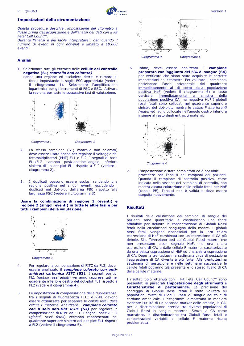
PI IQP-363 version 1
Page 20 of 27
Impostazioni della strumentazione Questa procedura descrive l'impostazione del citometro a flusso prima dell'acquisizione e dell'analisi dei dati con il kit Fetal Cell Count™. Durante l'analisi è più facile interpretare i dati quando il numero di eventi in ogni dot-plot è limitato a 10.000 eventi.
Analisi 1. Selezionare tutti gli eritrociti nelle cellule del controllo
negativo (S1; controllo non colorato) usando una regione ed escludere detriti e rumore di
fondo impostando la soglia FSC appropriata (vedere il citogramma 1). Selezionare l'amplificazione logaritmica per gli incrementi di FSC e SSC. Attivare la regione per tutte le successive fasi di valutazione.
Citogramma 1 Citogramma 2
2. Lo stesso campione (S1; controllo non colorato)
deve essere usato anche per regolare il voltaggio dei fotomoltiplicatori (PMT) FL1 e FL2. I segnali di base FL1/FL2 saranno posizionatinell'angolo inferiore sinistro di un dot-plot FL1 rispetto a FL2 (vedere il citogramma 2).
3. I duplicati possono essere esclusi rendendo una
regione positiva nei singoli eventi, escludendo i duplicati nel dot-plot dell'area FSC rispetto alla larghezza FSC (vedere il citogramma 3).
Usare la combinazione di regione 1 (eventi) e regione 2 (singoli eventi) in tutte le altre fasi e per tutti i campioni della valutazione.
Citogramma 3
4. Per regolare la compensazione di FITC da FL2, deve
essere analizzato il campione colorato con anti-anidrasi carbonica FITC (S2). I segnali positivi FL1 (globuli rossi adulti) verranno rappresentati nel quadrante inferiore destro del dot-plot FL1 rispetto a FL2 (vedere il citogramma 4).
5. Le impostazioni di compensazione della fluorescenza
tra i segnali di fluorescenza FITC e R-PE devono essere ottimizzate per separare le cellule fetali dalle cellule F materne. Analizzare il campione colorato con il solo anti-HbF R-PE (S3) per regolare la compensazione di R-PE da FL1. I segnali positivi FL2 (globuli rossi fetali) verranno rappresentati nel quadrante superiore sinistro del dot-plot FL1 rispetto a FL2 (vedere il citogramma 5).
Citogramma 4 Citogramma 5
6. Infine, deve essere analizzato il campione
preparato conl'aggiunta del 5% di sangue (S4) per verificare che siano state acquisite le corrette impostazioni del citometro. Per valutare il campione, posizionare l'asse orizzontale del quadrante immediatamente al di sotto della popolazione positiva HbF (vedere il citogramma 6) e l'asse verticale immediatamente a sinistra della popolazione positiva CA ma negativa HbF.I globuli rossi fetali sono collocati nel quadrante superiore sinistro del dot-plot, mentre le cellule F interferenti (materne) sono collocate nell'angolo destro inferiore insieme al resto degli eritrociti materni.
Citogramma 6
7. L'impostazione è stata completata ed è possibile
procedere con l'analisi dei campioni dei pazienti. Quando il campione di controllo positivo, come indicato nella sezione dei campioni di controllo, non mostra alcuna colorazione delle cellule fetali per HbF (canale PE), l'analisi non è valida e deve essere eseguita nuovamente.
Risultati I risultati della valutazione dei campioni di sangue dei pazienti sono quantitativi e costituiscono una fonte affidabile per definire la concentrazione di Globuli Rossi fetali nella circolazione sanguigna della madre. I globuli rossi fetali vengono riconosciuti per la loro chiara espressione di HbF combinata con un'espressione di CA più debole. Si differenziano così dai Globuli Rossi materni che non presentano alcun segnale HbF, ma una chiara espressione di CA, e dalle cellule F materne, caratterizzate da una bassa espressione di HbF ed una chiara espressione di CA. Dopo la trentaduesima settimana circa di gestazione l'espressione di CA diventerà più forte. Alla trentottesima settimana di gestazione e nelle settimane successive le cellule fetali potranno già presentare lo stesso livello di CA delle cellule materne. I risultati tipici ottenuti con il kit Fetal Cell Count™ sono presentati ai paragrafi Impostazione degli strumenti e Caratteristiche di performance. La precisione del conteggio di Globuli Rossi fetali è stata valutata su popolazioni miste di Globuli Rossi di sangue adulto e di cordone ombelicale. I citogrammi dimostrano in maniera evidente l’utilità di un secondo marker delle emazie, la CA, per la discriminazione precisa tra diverse popolazioni di Globuli Rossi in sangue materno. Senza la CA come marcatore, la discriminazione tra Globuli Rossi fetali e concentrazioni variabili di cellule F materne risulta problematica.
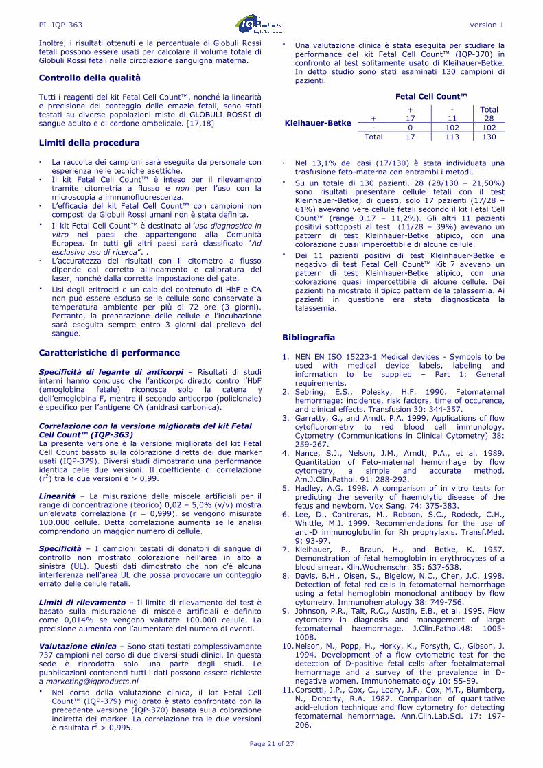
PI IQP-363 version 1
Page 21 of 27
Inoltre, i risultati ottenuti e la percentuale di Globuli Rossi fetali possono essere usati per calcolare il volume totale di Globuli Rossi fetali nella circolazione sanguigna materna.
Controllo della qualità Tutti i reagenti del kit Fetal Cell Count™, nonché la linearità e precisione del conteggio delle emazie fetali, sono stati testati su diverse popolazioni miste di GLOBULI ROSSI di sangue adulto e di cordone ombelicale. [17,18] Limiti della procedura � La raccolta dei campioni sarà eseguita da personale con
esperienza nelle tecniche asettiche. � Il kit Fetal Cell Count™ è inteso per il rilevamento
tramite citometria a flusso e non per l’uso con la microscopia a immunofluorescenza.
� L’efficacia del kit Fetal Cell Count™ con campioni non composti da Globuli Rossi umani non è stata definita.
� Il kit Fetal Cell Count™ è destinato all’uso diagnostico in vitro nei paesi che appartengono alla Comunità Europea. In tutti gli altri paesi sarà classificato “Ad esclusivo uso di ricerca”. .
� L’accuratezza dei risultati con il citometro a flusso dipende dal corretto allineamento e calibratura del laser, nonché dalla corretta impostazione del gate.
� Lisi degli eritrociti e un calo del contenuto di HbF e CA non può essere escluso se le cellule sono conservate a temperatura ambiente per più di 72 ore (3 giorni). Pertanto, la preparazione delle cellule e l’incubazione sarà eseguita sempre entro 3 giorni dal prelievo del sangue.
Caratteristiche di performance Specificità di legante di anticorpi – Risultati di studi interni hanno concluso che l’anticorpo diretto contro l’HbF (emoglobina fetale) riconosce solo la catena γ dell’emoglobina F, mentre il secondo anticorpo (policlonale) è specifico per l’antigene CA (anidrasi carbonica). Correlazione con la versione migliorata del kit Fetal Cell Count™ (IQP-363) La presente versione è la versione migliorata del kit Fetal Cell Count basato sulla colorazione diretta dei due marker usati (IQP-379). Diversi studi dimostrano una performance identica delle due versioni. Il coefficiente di correlazione (r2) tra le due versioni è > 0,99. Linearità – La misurazione delle miscele artificiali per il range di concentrazione (teorico) 0,02 – 5,0% (v/v) mostra un’elevata correlazione (r = 0,999), se vengono misurate 100.000 cellule. Detta correlazione aumenta se le analisi comprendono un maggior numero di cellule. Specificità – I campioni testati di donatori di sangue di controllo non mostrato colorazione nell’area in alto a sinistra (UL). Questi dati dimostrato che non c’è alcuna interferenza nell’area UL che possa provocare un conteggio errato delle cellule fetali. Limiti di rilevamento – Il limite di rilevamento del test è basato sulla misurazione di miscele artificiali e definito come 0,014% se vengono valutate 100.000 cellule. La precisione aumenta con l’aumentare del numero di eventi. Valutazione clinica – Sono stati testati complessivamente 737 campioni nel corso di due diversi studi clinici. In questa sede è riprodotta solo una parte degli studi. Le pubblicazioni contenenti tutti i dati possono essere richieste a [email protected]
� Nel corso della valutazione clinica, il kit Fetal Cell Count™ (IQP-379) migliorato è stato confrontato con la precedente versione (IQP-370) basata sulla colorazione indiretta dei marker. La correlazione tra le due versioni è risultata r2 > 0,995.
� Una valutazione clinica è stata eseguita per studiare la performance del kit Fetal Cell Count™ (IQP-370) in confronto al test solitamente usato di Kleihauer-Betke. In detto studio sono stati esaminati 130 campioni di pazienti.
� Nel 13,1% dei casi (17/130) è stata individuata una
trasfusione feto-materna con entrambi i metodi.
� Su un totale di 130 pazienti, 28 (28/130 – 21,50%) sono risultati presentare cellule fetali con il test Kleinhauer-Betke; di questi, solo 17 pazienti (17/28 – 61%) avevano vere cellule fetali secondo il kit Fetal Cell Count™ (range 0,17 – 11,2%). Gli altri 11 pazienti positivi sottoposti al test (11/28 – 39%) avevano un pattern di test Kleinhauer-Betke atipico, con una colorazione quasi impercettibile di alcune cellule.
� Dei 11 pazienti positivi di test Kleinhauer-Betke e negativo di test Fetal Cell Count™ Kit 7 avevano un pattern di test Kleinhauer-Betke atipico, con una colorazione quasi impercettibile di alcune cellule. Dei pazienti ha mostrato il tipico pattern della talassemia. Ai pazienti in questione era stata diagnosticata la talassemia.
Bibliografia 1. NEN EN ISO 15223-1 Medical devices - Symbols to be
used with medical device labels, labeling and information to be supplied – Part 1: General requirements.
2. Sebring, E.S., Polesky, H.F. 1990. Fetomaternal hemorrhage: incidence, risk factors, time of occurence, and clinical effects. Transfusion 30: 344-357.
3. Garratty, G., and Arndt, P.A. 1999. Applications of flow cytofluorometry to red blood cell immunology. Cytometry (Communications in Clinical Cytometry) 38: 259-267.
4. Nance, S.J., Nelson, J.M., Arndt, P.A., et al. 1989. Quantitation of Feto-maternal hemorrhage by flow cytometry, a simple and accurate method. Am.J.Clin.Pathol. 91: 288-292.
5. Hadley, A.G. 1998. A comparison of in vitro tests for predicting the severity of haemolytic disease of the fetus and newborn. Vox Sang. 74: 375-383.
6. Lee, D., Contreras, M., Robson, S.C., Rodeck, C.H., Whittle, M.J. 1999. Recommendations for the use of anti-D immunoglobulin for Rh prophylaxis. Transf.Med. 9: 93-97.
7. Kleihauer, P., Braun, H., and Betke, K. 1957. Demonstration of fetal hemoglobin in erythrocytes of a blood smear. Klin.Wochenschr. 35: 637-638.
8. Davis, B.H., Olsen, S., Bigelow, N.C., Chen, J.C. 1998. Detection of fetal red cells in fetomaternal hemorrhage using a fetal hemoglobin monoclonal antibody by flow cytometry. Immunohematology 38: 749-756.
9. Johnson, P.R., Tait, R.C., Austin, E.B., et al. 1995. Flow cytometry in diagnosis and management of large fetomaternal haemorrhage. J.Clin.Pathol.48: 1005-1008.
10. Nelson, M., Popp, H., Horky, K., Forsyth, C., Gibson, J. 1994. Development of a flow cytometric test for the detection of D-positive fetal cells after foetalmaternal hemorrhage and a survey of the prevalence in D-negative women. Immunohematology 10: 55-59.
11. Corsetti, J.P., Cox, C., Leary, J.F., Cox, M.T., Blumberg, N., Doherty, R.A. 1987. Comparison of quantitative acid-elution technique and flow cytometry for detecting fetomaternal hemorrhage. Ann.Clin.Lab.Sci. 17: 197-206.
Fetal Cell Count™
+ - Total
Kleihauer-Betke + 17 11 28 - 0 102 102
Total 17 113 130
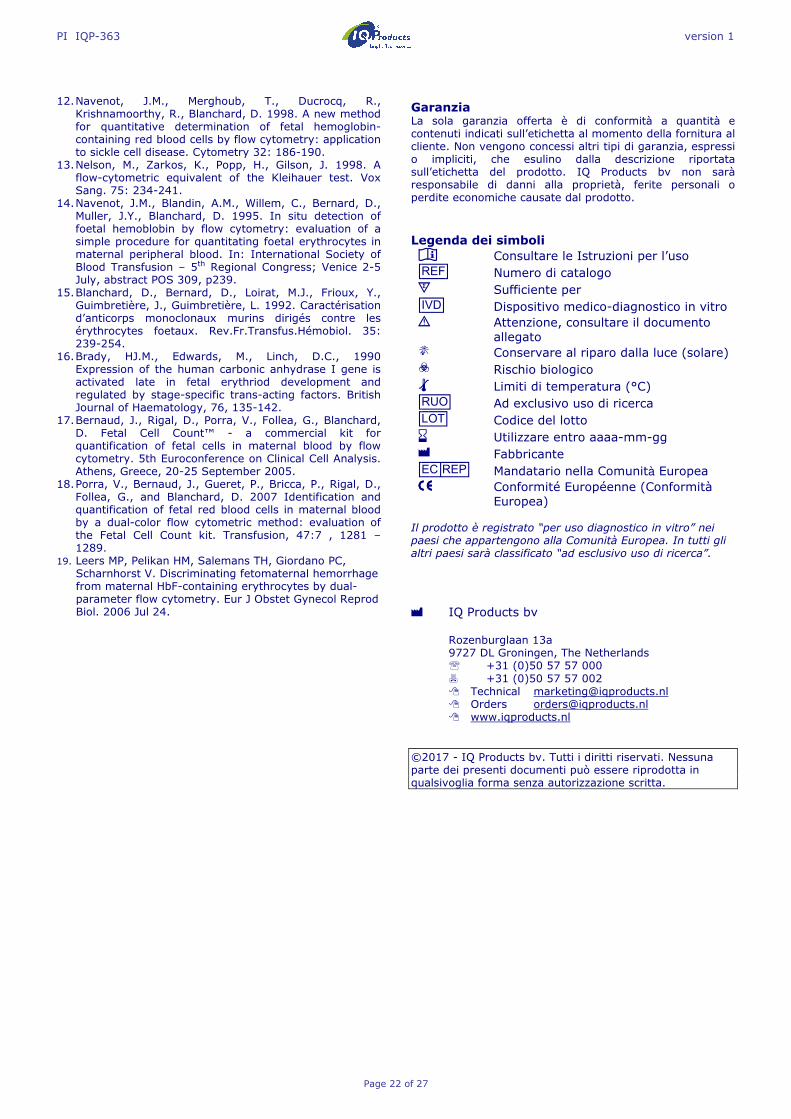
PI IQP-363 version 1
Page 22 of 27
12. Navenot, J.M., Merghoub, T., Ducrocq, R.,
Krishnamoorthy, R., Blanchard, D. 1998. A new method for quantitative determination of fetal hemoglobin-containing red blood cells by flow cytometry: application to sickle cell disease. Cytometry 32: 186-190.
13. Nelson, M., Zarkos, K., Popp, H., Gilson, J. 1998. A flow-cytometric equivalent of the Kleihauer test. Vox Sang. 75: 234-241.
14. Navenot, J.M., Blandin, A.M., Willem, C., Bernard, D., Muller, J.Y., Blanchard, D. 1995. In situ detection of foetal hemoblobin by flow cytometry: evaluation of a simple procedure for quantitating foetal erythrocytes in maternal peripheral blood. In: International Society of Blood Transfusion – 5th Regional Congress; Venice 2-5 July, abstract POS 309, p239.
15. Blanchard, D., Bernard, D., Loirat, M.J., Frioux, Y., Guimbretière, J., Guimbretière, L. 1992. Caractérisation d’anticorps monoclonaux murins dirigés contre les érythrocytes foetaux. Rev.Fr.Transfus.Hémobiol. 35: 239-254.
16. Brady, HJ.M., Edwards, M., Linch, D.C., 1990 Expression of the human carbonic anhydrase I gene is activated late in fetal erythriod development and regulated by stage-specific trans-acting factors. British Journal of Haematology, 76, 135-142.
17. Bernaud, J., Rigal, D., Porra, V., Follea, G., Blanchard, D. Fetal Cell Count™ - a commercial kit for quantification of fetal cells in maternal blood by flow cytometry. 5th Euroconference on Clinical Cell Analysis. Athens, Greece, 20-25 September 2005.
18. Porra, V., Bernaud, J., Gueret, P., Bricca, P., Rigal, D., Follea, G., and Blanchard, D. 2007 Identification and quantification of fetal red blood cells in maternal blood by a dual-color flow cytometric method: evaluation of the Fetal Cell Count kit. Transfusion, 47:7 , 1281 – 1289.
19. Leers MP, Pelikan HM, Salemans TH, Giordano PC, Scharnhorst V. Discriminating fetomaternal hemorrhage from maternal HbF-containing erythrocytes by dual- parameter flow cytometry. Eur J Obstet Gynecol Reprod Biol. 2006 Jul 24.
Garanzia La sola garanzia offerta è di conformità a quantità e contenuti indicati sull’etichetta al momento della fornitura al cliente. Non vengono concessi altri tipi di garanzia, espressi o impliciti, che esulino dalla descrizione riportata sull’etichetta del prodotto. IQ Products bv non sarà responsabile di danni alla proprietà, ferite personali o perdite economiche causate dal prodotto. Legenda dei simboli I Consultare le Istruzioni per l’uso [REF] Numero di catalogo s Sufficiente per [IVD] Dispositivo medico-diagnostico in vitro ! Attenzione, consultare il documento
allegato K Conservare al riparo dalla luce (solare) D Rischio biologico t Limiti di temperatura (°C) [RUO] Ad exclusivo uso di ricerca [LOT] Codice del lotto e Utilizzare entro aaaa-mm-gg M Fabbricante [EC_|REP] Mandatario nella Comunità Europea |||| Conformité Européenne (Conformità
Europea) Il prodotto è registrato “per uso diagnostico in vitro” nei paesi che appartengono alla Comunità Europea. In tutti gli altri paesi sarà classificato “ad esclusivo uso di ricerca”. M IQ Products bv
Rozenburglaan 13a 9727 DL Groningen, The Netherlands ℡ +31 (0)50 57 57 000 � +31 (0)50 57 57 002 � Technical [email protected] � Orders [email protected] � www.iqproducts.nl
©2017 - IQ Products bv. Tutti i diritti riservati. Nessuna parte dei presenti documenti può essere riprodotta in qualsivoglia forma senza autorizzazione scritta.
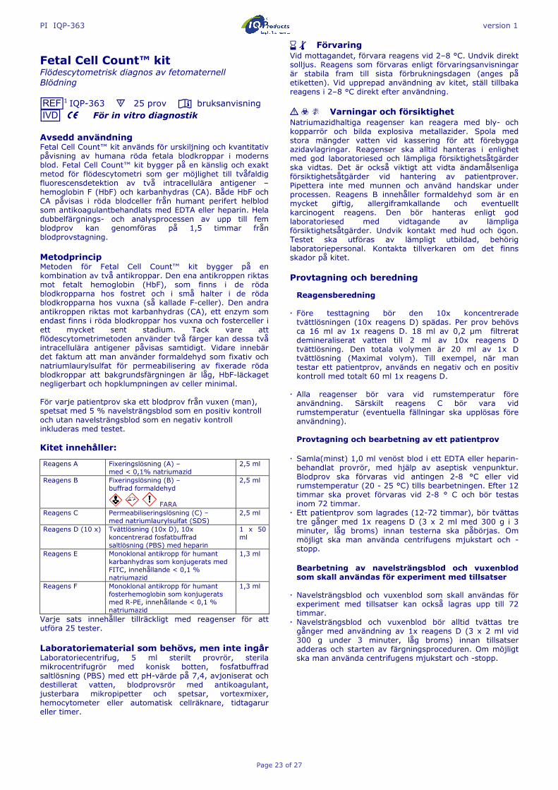
PI IQP-363 version 1
Page 23 of 27
Fetal Cell Count™ kit Flödescytometrisk diagnos av fetomaternell Blödning [REF]1 IQP-363 s 25 prov I bruksanvisning [IVD] |||| För in vitro diagnostik
Avsedd användning Fetal Cell Count™ kit används för urskiljning och kvantitativ påvisning av humana röda fetala blodkroppar i moderns blod. Fetal Cell Count™ kit bygger på en känslig och exakt metod för flödescytometri som ger möjlighet till tvåfaldig fluorescensdetektion av två intracellulära antigener – hemoglobin F (HbF) och karbanhydras (CA). Både HbF och CA påvisas i röda blodceller från humant perifert helblod som antikoagulantbehandlats med EDTA eller heparin. Hela dubbelfärgnings- och analysprocessen av upp till fem blodprov kan genomföras på 1,5 timmar från blodprovstagning. Metodprincip Metoden för Fetal Cell Count™ kit bygger på en kombination av två antikroppar. Den ena antikroppen riktas mot fetalt hemoglobin (HbF), som finns i de röda blodkropparna hos fostret och i små halter i de röda blodkropparna hos vuxna (så kallade F-celler). Den andra antikroppen riktas mot karbanhydras (CA), ett enzym som endast finns i röda blodkroppar hos vuxna och fosterceller i ett mycket sent stadium. Tack vare att flödescytometrimetoden använder två färger kan dessa två intracellulära antigener påvisas samtidigt. Vidare innebär det faktum att man använder formaldehyd som fixativ och natriumlaurylsulfat för permeabilisering av fixerade röda blodkroppar att bakgrundsfärgningen är låg, HbF-läckaget negligerbart och hopklumpningen av celler minimal. För varje patientprov ska ett blodprov från vuxen (man), spetsat med 5 % navelsträngsblod som en positiv kontroll och utan navelsträngsblod som en negativ kontroll inkluderas med testet. Kitet innehåller:
Varje sats innehåller tillräckligt med reagenser för att utföra 25 tester. Laboratoriematerial som behövs, men inte ingår Laboratoriecentrifug, 5 ml sterilt provrör, sterila mikrocentrifugrör med konisk botten, fosfatbuffrad saltlösning (PBS) med ett pH-värde på 7,4, avjoniserat och destillerat vatten, blodprovsrör med antikoagulant, justerbara mikropipetter och spetsar, vortexmixer, hemocytometer eller automatisk cellräknare, tidtagarur eller timer.
e t Förvaring Vid mottagandet, förvara reagens vid 2–8 °C. Undvik direkt solljus. Reagens som förvaras enligt förvaringsanvisningar är stabila fram till sista förbrukningsdagen (anges på etiketten). Vid upprepad användning av kitet, ställ tillbaka reagens i 2–8 °C direkt efter användning. ! D K Varningar och försiktighet Natriumazidhaltiga reagenser kan reagera med bly- och kopparrör och bilda explosiva metallazider. Spola med stora mängder vatten vid kassering för att förebygga azidavlagringar. Reagenser ska alltid hanteras i enlighet med god laboratoriesed och lämpliga försiktighetsåtgärder ska vidtas. Det är också viktigt att vidta ändamålsenliga försiktighetsåtgärder vid hantering av patientprover. Pipettera inte med munnen och använd handskar under processen. Reagens B innehåller formaldehyd som är en mycket giftig, allergiframkallande och eventuellt karcinogent reagens. Den bör hanteras enligt god laboratoriesed med vidtagande av lämpliga försiktighetsåtgärder. Undvik kontakt med hud och ögon. Testet ska utföras av lämpligt utbildad, behörig laboratoriepersonal. Kontakta tillverkaren om det finns skador på kitet. Provtagning och beredning Reagensberedning
� Före testtagning bör den 10x koncentrerade tvättlösningen (10x reagens D) spädas. Per prov behövs ca 16 ml av 1x reagens D. 18 ml av 0,2 µm filtrerat demineraliserat vatten till 2 ml av 10x reagens D tvättlösning. Den totala volymen är 20 ml av 1x D tvättlösning (Maximal volym). Till exempel, när man testar ett patientprov, används en negativ och en positiv kontroll med totalt 60 ml 1x reagens D.
� Alla reagenser bör vara vid rumstemperatur före användning. Särskilt reagens C bör vara vid rumstemperatur (eventuella fällningar ska upplösas före användning).
Provtagning och bearbetning av ett patientprov
� Samla(minst) 1,0 ml venöst blod i ett EDTA eller heparin-behandlat provrör, med hjälp av aseptisk venpunktur. Blodprov ska förvaras vid antingen 2-8 °C eller vid rumstemperatur (20 - 25 °C) tills bearbetningen. Efter 12 timmar ska provet förvaras vid 2-8 ° C och bör testas inom 72 timmar.
� Ett patientprov som lagrades (12-72 timmar), bör tvättas tre gånger med 1x reagens D (3 x 2 ml med 300 g i 3 minuter, låg broms) innan testerna ska påbörjas. Om möjligt ska man använda centrifugens mjukstart och -stopp.
Bearbetning av navelsträngsblod och vuxenblod som skall användas för experiment med tillsatser
� Navelsträngsblod och vuxenblod som skall användas för experiment med tillsatser kan också lagras upp till 72 timmar.
� Navelsträngsblod och vuxenblod bör alltid tvättas tre gånger med användning av 1x reagens D (3 x 2 ml vid 300 g under 3 minuter, låg broms) innan tillsatser adderas och starten av färgningsproceduren. Om möjligt ska man använda centrifugens mjukstart och -stopp.
Reagens A Fixeringslösning (A) – med < 0,1% natriumazid
2,5 ml
Reagens B Fixeringslösning (B) – buffrad formaldehyd
FARA
2,5 ml
Reagens C Permeabiliseringslösning (C) – med natriumlaurylsulfat (SDS)
2,5 ml
Reagens D (10 x) Tvättlösning (10x D), 10x koncentrerad fosfatbuffrad saltlösning (PBS) med heparin
1 x 50 ml
Reagens E Monoklonal antikropp för humant karbanhydras som konjugerats med FITC, innehållande < 0,1 % natriumazid
1,3 ml
Reagens F Monoklonal antikropp för humant fosterhemoglobin som konjugerats med R-PE, innehållande < 0,1 % natriumazid
1,3 ml
PI IQP-363 version 1
Page 24 of 27
Kontrollprover Kör alltid ett positivt och negativt kontrollprov med varje patientprov. En blandning av navelsträngsblod och blod från en vuxen (man) rekommenderas som ett positivt kontrollprov. När det inte finns något navelsträngsblod tillgängligt kan FETALtrol (FH101) användas. Blod från vuxen (man) som inte är spetsat rekommenderas som negativt kontrollprov. Positiv kontroll och att använda för konfiguration av cytometer
� Blanda ca 5 % navelsträngsblod i normalt vuxenblod (volym/volym). Endast tvättad navelsträngsblod och vuxenblod ska blandas.
� När blandningen inte bara används för konfiguration och kontroll, utan även för en exakt kvantifiering av de spetsade cellerna, ska erytrocyterna i både navelsträngs- och vuxna blodprover räknas i en hematologi-analysator. Med hjälp av dessa siffror kan tillsatserna beräknas exakt. Negativ kontroll (inga fetala celler)
� Som en negativ kontroll rekommenderas det att använda blod från en vuxen man. Behandla detta material som patientprov i proceduren.
Testprocedur Fetal Cell Count™-kit
Fixering och permeabilisering kontroll (spetsat) prov och patientprov
1. Märk varje patientprov och de positiva och negativa
externa kontrollerna ett separat 5 ml-centrifugrör med konisk botten.
2. Tillsätt 100 µL reagens A i varje rör. 3. Tillsätt 10 µL EDTA-antikoagulerat helblod, blanda och
skaka. När FetalTrol används som ett kontrollprov bör 5 µl användas.
4. Tillsätt 100 µl reagens B och skaka på vortex. 5. Inkubera den blandade cellsuspensionen vid
rumstemperatur i exakt 30 minuter. Blanda suspensionen försiktigt var 10:e minut.
6. Tillsätt 2 ml 1x reagens D och blanda cellerna genom att vända rören några gånger.
7. Centrifugera cellsuspensionen vid 300 g i 3 minuter. 8. Kassera supernatanten. 9. Tillsätt 100 µL 1x reagens D. 10. Återsuspendera cellpelleten och skaka försiktigt på
vortex. 11. Tillsätt 100 µL reagens C och skaka på vortex
(inkubationstiden på exakt 3 minuter startas med det första röret). Reagens C bör vara vid rumstemperatur (eventuella fällningar ska upplösas före användning).
12. Efter exakt 3 minuter: Tillsätt 2 ml 1x reagens D och blanda cellerna genom att vända rören några gånger.
13. Centrifugera cellsuspensionen vid 300 g i 3 minuter. 14. Kassera supernatanten. 15. Tillsätt 2 ml 1x reagens D och återsuspendera
cellpelleten genom att vända rören några gånger. 16. Centrifugera cellsuspensionen vid 300 g i 3 minuter. 17. Kassera supernatanten. 18. Återsuspendera cellpelleten i 1 ml 1x reagens D och
återsuspendera cellerna genom försiktig blandning på vortex.
Immunofluorescerande färgning kontrollprov
19. Märk fyra koniska bottenrör som kan användas med flödescytometern med S1, S2, S3 och S4.
20. Tillsätt de olika komponenterna till rören genom att följa tabell 1 och blanda.
21. Inkubera vid rumstemperatur i 15 minuter i mörker (undvika direkt ljus).
Rör 5 % spetsat prov Reagens E
Reagens F
S1 50 µl --- ---
S2 50 µl 50 µl ---
S3 50 µl --- 50 µl
S4 50 µl 50 µl 50 µl
Tabell 1. Komponenter som ska blandas ihop för justering av inställningarna för flödescytometern.
22. Tillsätt 2 ml 1x reagens D och centrifugera
cellsuspensionen vid 300 g i 3 minuter. 23. Kassera supernatanten. 24. Återsuspendera cellpelleten i 500 µl 1x reagens D. 25. Cellerna är nu redo för datainsamling genom
flödescytometri. Cellerna bör utvärderas inom 30 minuter. Immunofluorescerande färgning av patientprov
26. Tillsätt ihop i ett nytt rör med konisk botten och blanda väl:
m) 50 µl reagens E - antihuman CA FITC n) 50 µl reagens E - antihuman HbF-R PE o) 50 µl erytrocytsuspension (den erhållna cell-
suspensionen från steg 18) 27. Inkubera vid rumstemperatur i 15 minuter i mörker
(undvik direkt ljus). 28. Tillsätt 2 ml 1x reagens D och centrifugera
cellsuspensionen vid 300 g i 3 minuter. 29. Kassera supernatanten. 30. Återsuspendera cellpelleten i 500 µl 1x reagens D. 31. Cellerna är nu redo för datainsamling genom
flödescytometri. Cellerna bör utvärderas inom 30 minuter.
Datainsamling � List mode-filer av minst 100 000 händelser ska samlas
in för log-FSC, log-SSC, och log-fluorescens signaler för både fluorokromkonjugerade antikroppar med området avgränsat vid erytrocyterna genom gating.
� Mindre än 100 000 händelser kommer att påverka analysens noggrannhet.
� För att förhindra sammanträffande då en fostercell och en moderscell passerar lasern rekommenderas det att köra proverna vid en låg till medelhög hastighet.
Instrumentens kravspecifikationer � Kontrollera att flödescytometern har kalibrerats på rätt
sätt enligt tillverkarens instruktioner. � Det rekommenderas att utföra instrumentkalibrering
och underhåll med jämna mellanrum. � Flödescytometern bör användas av en utbildad tekniker.
Utvärderingen av resultaten bör göras av någon som är utbildad i tolkningen av flödescytometriska data.
Instrumentinställningar Denna procedur beskriver konfigurationen av flödescytometern före insamlingen och analysen av data från Fetal Cell Count™ kit. Vid analys är det lättare att tolka data när antalet händelser i varje punktdiagram är begränsat till 10 000 händelser.
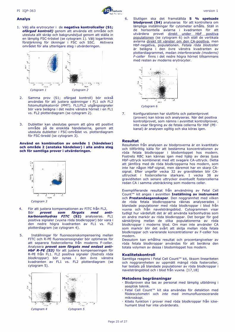
PI IQP-363 version 1
Page 25 of 27
Analys 1. Välj alla erytrocyter i de negativa kontrollceller (S1;
ofärgad kontroll) genom att använda ett område och utesluta allt skräp och bakgrundsljud genom att ställa in en lämplig FSC-tröskel (se cytogram 1). Välj logaritmisk förstärkning för ökningar i FSC och SSC. Aktivera området för alla ytterligare steg i utvärderingen.
Cytogram 1 Cytogram 2
2. Samma prov (S1; ofärgad kontroll) bör också
användas för att justera spänningar i FL1 och FL2 fotomultiplikatorrör (PMT). FL1/FL2 utgångssignaler bör vara belägna i det nedre vänstra hörnet i en FL1 vs. FL2 plotterdiagram (se cytogram 2).
3. Dubletter kan uteslutas genom att göra ett positivt
område på de enskilda händelserna, genom att utesluta dubletter i FSC-området vs. plotterdiagram för FSC-bredd (se cytogram 3).
Använd en kombination av område 1 (händelser) och område 2 (enstaka händelser) i alla andra steg och för samtliga prover i utvärderingen.
Cytogram 3
4. För att justera kompensationen av FITC från FL2,
Bör provet som färgats med anti-karbonanhydras FITC (S2) analyseras. FL1 positiva signaler (vuxna röda blodkroppar) bör vara i den nedre högra kvadranten av FL1 vs. FL2 plotterdiagram (se cytogram 4).
5. Inställningar för fluorescenskompensering mellan
FITC och R-PE fluorescenssignaler bör optimeras för att separera fostercellerna från moderns F-celler. Analysera provet som färgats med endast anti-HbF R-PE (S3) för att justera kompenseringen för R-PE från FL1. FL2 positiva signaler (fostrets röda blodkroppar) bör synas i den övre vänstra kvadranten av FL1 vs. FL2 plotterdiagram (se cytogram 5).
Cytogram 4 Cytogram 5
6. Slutligen ska det framställda 5 % spetsade blodprovet (S4) analyseras för att kontrollera om lämpliga inställningar för cytometer erhålls. Ställ in de horisontella axlarna i kvadranten för att utvärdera provet direkt under HbF positiva populationen (se cytogram 6) och ställ de vertikala axlarna direkt till vänster om den CA-positiva, men HbF-negativa, populationen. Fetala röda blodceller är belägna i den övre vänstra kvadranten av plotterdiagrammet, medan interfererande (moderns) F-celler finns i det nedre högra hörnet tillsammans med resten av moderns erytrocyter.
Cytogram 6
7. Konfigurationen har slutförts och patientprovet
(proven) kan köras och analyseras. När det positiva kontrollprovet, som nämns i avsnittet kontrollprover, inte visar färgning av de fetala cellerna för HbF (PE-kanal) är analysen ogiltig och ska köras igen.
Resultat Resultaten från analysen av blodproverna är en kvantitativ och tillförlitlig källa för att bestämma koncentrationen av röda fetala blodkroppar i blodomloppet hos modern. Fostrets RBC kan kännas igen med hjälp av deras ljusa HbF-uttryck kombinerat med ett svagare CA-uttryck. Detta att jämföra med de röda blodkropparna hos modern, som inte har någon HbF-signal, men däremot har en skarp CA-signal. Efter ungefär vecka 32 av graviditeten blir CA-uttrycket i fostercellerna starkare. I vecka 38 av graviditeten och senare uttrycker eventuellt fostercellerna redan CA i samma utsträckning som moderns celler. Exemplifierande resultat från användning av Fetal Cell Count™ kit anges i avsnitten Inställning av instrument och Prestandaegenskaper. Den noggrannhet med vilken de röda fetala blodkropparna räknas analyserades i blandade populationer med röda blodkroppar i blod från vuxna och från navelsträngsblod. Cytogrammen visar tydligt hur värdefullt det är att använda karbonhydras som en andra markör av röda blodkroppar. Det borgar för god särskiljning mellan de olika populationerna av röda blodkroppar i moderns blod. Om man inte använder CA som markör blir det svårt att skilja mellan röda fetala blodkroppar och varierande koncentrationer av F-celler hos modern. Dessutom kan erhållna resultat och procentangivelser av röda fetala blodkroppar användas för att beräkna den totala volymen av dessa i blodomloppet hos modern.
Kvalitetskontroll Samtliga reagens i Fetal Cell Count™ kit, liksom lineariteten och noggrannheten av uppmätt mängd röda fosterceller, har testats på blandade populationer av röda blodkroppar i navelsträngsblod och i blod från vuxna. [17,18] Metodens begränsningar � Blodproven ska tas av personal med lämplig utbildning i aseptisk teknik.
� Fetal Cell Count™ kit ska användas för detektion med flödescytometri och inte med immunofluorescerande mikroskopi.
� Kitets funktion i prover med röda blodkroppar från icke-humant blod har inte utvärderats.
PI IQP-363 version 1
Page 26 of 27
� Fetal Cell Count™ kit är avsett för in vitro diagnostik i EU-länder och ska märkas "endast för forskning" i alla andra länder.
� För att den flödescytometriska analysen ska ge noggranna resultat krävs korrekt inställning och kalibrering av lasern samt riktig inställning av gaten.
� Lys av erytrocyter och det kan inte uteslutas att mängden HbF- och CA minskar om cellerna förvaras i rumstemperatur i mer än 72 immar (3 dagar). Det är därför viktigt att cellerna prepareras och inkuberas inom 3 dagar från blodprovstagning.
Prestandaegenskaper Antikropparnas specificitet - Interna resultat visar att den antikropp som är riktad mot HbF (fetalt hemoglobin)
endast identifierar γ-kedjan hos hemoglobin F, medan den andra antikroppen (polyklonal) är specifik för karbonhydrasantigenen (CA). Korrelation med version av Fetal Cell Count™ kit (IQP-363) - Föreliggande version är en förbättrad version av det Fetal Cell Count™ kit som var baserat på direkt infärgning av de två markörer som användes (IQP-379). Studier visar att de två versionerna visar identiska resultat. Korrelationskoefficienten (r2) mellan de två versionerna ligger på > 0,99. Linearitet – Mätning av artificiella prov med ett (teoretiskt) koncentrationsintervall på 0,02–5,0 % (v/v) visar på hög korrelation (r = 0,999) vid mätning av 100 000 celler. Korrelationen blir högre när fler celler analyseras. Specificitet - Vid analys av kontrollprover erhölls inte någon infärgning i övre vänstra området (UL). Detta visar att det inte finns någon interferens i UL-området som skulle leda till en felaktig räkning av fetala celler. Detektionsgräns – Provets detektionsgräns har beräknats utifrån mätningar av artificiella provblandningar och uppgick till 0,014 % vid analys av 100 000 celler. Noggrannheten förbättras när fler händelser analyseras. Klinisk utvärdering – Sammanlagt 737 prover har analyserats i två skilda kliniska studier. Endast delar av dessa studier är representerade s här. De fullständiga
publikationerna kan erhållas via [email protected].
� Under den kliniska utvärderingen jämfördes det förbättrade Fetal Cell Count™ kit (IQP-379) med en tidigare version av Fetal Cell Count™ kit (IQP-370) som byggde på indirekt infärgning av markörerna. Korrelationen mellan de två versionerna har bevisats vara r2 > 0,995
� En klinisk utvärdering genomfördes för att undersöka den kliniska relevansen med Fetal Cell Count™ kit (IQP-379) i jämförelse med det allmänt använda Kleihauer-Betke-testet. I den utredningen kontrollerades 130 patientprover.
� I 13,1 % (17/130) av fallen upptäcktes fetomaternell
transfusion med båda metoderna.
� Av sammanlagt 130 patientprover påvisades, med hjälp av Kleihauer-Betke-testet, 28 (28/130 – 21,59 %) innehålla fosterceller. Av dessa påvisades, med hjälp av Fetal Cell Count™ kit (intervallet 0.17–11,2 %), endast 17 av proverna (17/28 - 61%) innehålla äkta fosterceller. De andra 11 positiva testade patienter (11/28- 39%) hade ett icke-typiskt Kleihauer-Betke test mönster med mycket svag färgning av ett antal celler.
� Av de 11 Kleihauer-Betke positiva och Fetal Cell Count™ kit negativa patienterna, hade 7 stycken ett icke-typiskt Kleihauer-Betke mönster med mycket svag infärgning av celler. Dessa prov visade ett typiskt thalassemiamönster. Dessa patienter blev diagnostiserade som thalassemipatienter.
Bibliografi 1. DIN EN ISO 15223-1 Medical devices – Symbols to be
used with medical device labels, labeling and information to be supplied-Part 1: General requirements.
2. Sebring, E.S., Polesky, H.F. 1990. Fetomaternal hemorrhage: incidence, risk factors, time of occurence, and clinical effects. Transfusion 30: 344-357.
3. Garratty, G., and Arndt, P.A. 1999. Applications of flow cytofluorometry to red blood cell immunology. Cytometry (Communications in Clinical Cytometry) 38: 259-267.
4. Nance, S.J., Nelson, J.M., Arndt, P.A., et al. 1989. Quantitation of Feto-maternal hemorrhage by flow cytometry, a simple and accurate method. Am.J.Clin.Pathol. 91: 288-292.
5. Hadley, A.G. 1998. A comparison of in vitro tests for predicting the severity of haemolytic disease of the fetus and newborn. Vox Sang. 74: 375-383.
6. Lee, D., Contreras, M., Robson, S.C., Rodeck, C.H., Whittle, M.J. 1999. Recommendations for the use of anti-D immunoglobulin for Rh prophylaxis. Transf.Med. 9: 93-97.
7. Kleihauer, P., Braun, H., and Betke, K. 1957. Demonstration of fetal hemoglobin in erythrocytes of a blood smear. Klin.Wochenschr. 35: 637-638.
8. Davis, B.H., Olsen, S., Bigelow, N.C., Chen, J.C. 1998. Detection of fetal red cells in fetomaternal hemorrhage using a fetal hemoglobin monoclonal antibody by flow cytometry. Immunohematology 38: 749-756.
9. Johnson, P.R., Tait, R.C., Austin, E.B., et al. 1995. Flow cytometry in diagnosis and management of large fetomaternal haemorrhage. J.Clin.Pathol.48: 1005-1008.
10. Nelson, M., Popp, H., Horky, K., Forsyth, C., Gibson, J. 1994. Development of a flow cytometric test for the detection of D-positive fetal cells after foetalmaternal hemorrhage and a survey of the prevalence in D-negative women. Immunohematology 10: 55-59.
11. Corsetti, J.P., Cox, C., Leary, J.F., Cox, M.T., Blumberg, N., Doherty, R.A. 1987. Comparison of quantitative acid-elution technique and flow cytometry for detecting fetomaternal hemorrhage. Ann.Clin.Lab.Sci. 17: 197-206.
12. Navenot, J.M., Merghoub, T., Ducrocq, R., Krishnamoorthy, R., Blanchard, D. 1998. A new method for quantitative determination of fetal hemoglobin-containing red blood cells by flow cytometry: application to sickle cell disease. Cytometry 32: 186-190.
13. Nelson, M., Zarkos, K., Popp, H., Gilson, J. 1998. A flow-cytometric equivalent of the Kleihauer test. Vox Sang. 75: 234-241.
14. Navenot, J.M., Blandin, A.M., Willem, C., Bernard, D., Muller, J.Y., Blanchard, D. 1995. In situ detection of foetal hemoblobin by flow cytometry: evaluation of a simple procedure for quantitating foetal erythrocytes in maternal peripheral blood. In: International Society of Blood Transfusion – 5th Regional Congress; Venice 2-5 July, abstract POS 309, p239.
Fetal Cell Count™ kit + - Summa Kleihauer-Betke
+ 17 11 28 - 0 102 102
Summa 17 113 130
PI IQP-363 version 1
Page 27 of 27
15. Blanchard, D., Bernard, D., Loirat, M.J., Frioux, Y., Guimbretière, J., Guimbretière, L. 1992. Caractérisation d’anticorps monoclonaux murins dirigés contre les érythrocytes foetaux. Rev.Fr.Transfus.Hémobiol. 35: 239-254.
16. Brady, HJ.M., Edwards, M., Linch, D.C., 1990 Expression of the human carbonic anhydrase I gene is activated late in fetal erythriod development and regulated by stage-specific trans-acting factors. British Journal of Haematology, 76, 135-142.
17. Bernaud, J., Rigal, D., Porra, V., Follea, G., Blanchard, D. Fetal Cell Count™ - a commercial kit for quantification of fetal cells in maternal blood by flow cytometry. 5th Euroconference on Clinical Cell Analysis. Athens, Greece, 20-25 September 2005
18. Porra, V., Bernaud, J., Gueret, P., Bricca, P., Rigal, D., Follea, G., and Blanchard, D. 2007 Identification and quantification of fetal red blood cells in maternal blood by a dual-color flow cytometric method: evaluation of the Fetal Cell Count kit. Transfusion, 47:7 , 1281 – 1289.
19. Leers MP, Pelikan HM, Salemans TH, Giordano PC, Scharnhorst V. Discriminating fetomaternal hemorrhage from maternal HbF-containing erythrocytes by dual- parameter flow cytometry. Eur J Obstet Gynecol Reprod Biol. 2006 Jul 24.
Garanti Produkterna som säljs härunder garanteras endast överensstämma med den kvantitet och det innehåll som anges på etiketten vid tidpunkten för leverans till kunden. Inga garantier, varken uttryckliga eller underförstådda, ges utöver den beskrivning av produkten som finns på etiketten. IQ Products bv ansvarar inte för sakskada, personskada eller ekonomiska förluster som orsakats av produkten. Symbolförklaring I
Läs bruksanvisningen
[REF]
Listnummer
s
Räcker till
[IVD]
Medicinteknisk produkt avsedd för in in vitro-diagnostik
!
Viktigt
K
Skyddas mot ljus
D
Biologisk risk
t
Temperaturbegränsning (°C)
[RUO]
Endast för forskning
[LOT]
Satsnummer
e
Används före åååå-mm-dd
M
Tillverkare
[EC_|REP]
Auktoriserad representant inom Europeiska gemenskapen
M IQ Products bv
Rozenburglaan 13a 9727 DL Groningen, The Netherlands ℡ +31 (0)50 57 57 000 � +31 (0)50 57 57 002 � Technical [email protected] � Orders [email protected] � www.iqproducts.nl
Produkten är registrerad som "endast för in vitro diagnostik" i EU-länder och ska märkas "endast för forskning" i alla andra länder. ©2017 - IQ Products bv. Med ensamrätt. Detta verk får inte återges utan skriftligt tillstånd, varken helt eller delvis.
Top Related