


Languages
Pages
Legal


April 2016
EXIT procedure in a patient
with a severe lymphangioma
colli
SWISS SOCIETY OF NEONATOLOGY

Gubler D, Jöhr M, Winiker H, Hodel M, Berger TM,
Neonatal and Pediatric Intensive Care Unit (GD, BTM),
Pediatric Anesthesiology (JM), Pediatric Surgery (WH),
Children’s Hospital of Lucerne, Department of Fetal
Medicine, Neue Frauenklinik Lucerne (HM), Switzerland
© Swiss Society of Neonatology, Thomas M Berger, Webmaster
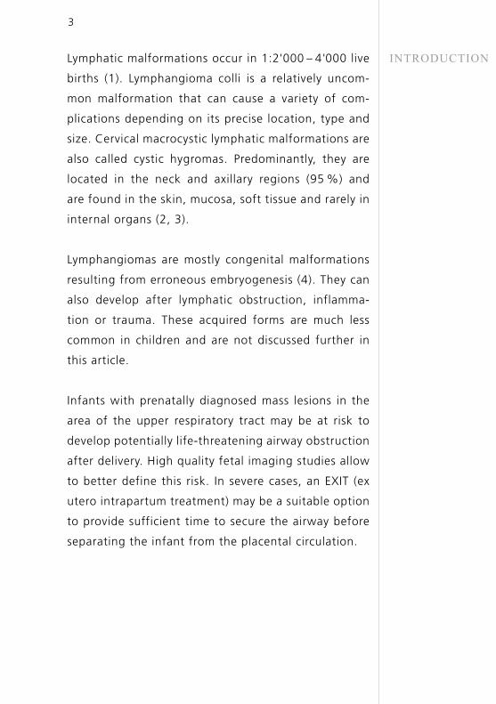
Lymphatic malformations occur in 1:2'000 – 4'000 live
births (1). Lymphangioma colli is a relatively uncom-
mon malformation that can cause a variety of com-
plications depending on its precise location, type and
size. Cervical macrocystic lymphatic malformations are
also called cystic hygromas. Predominantly, they are
located in the neck and axillary regions (95 %) and
are found in the skin, mucosa, soft tissue and rarely in
internal organs (2, 3).
Lymphangiomas are mostly congenital malformations
resulting from erroneous embryogenesis (4). They can
also develop after lymphatic obstruction, inflamma-
tion or trauma. These acquired forms are much less
common in children and are not discussed further in
this article.
Infants with prenatally diagnosed mass lesions in the
area of the upper respiratory tract may be at risk to
develop potentially life-threatening airway obstruction
after delivery. High quality fetal imaging studies allow
to better define this risk. In severe cases, an EXIT (ex
utero intrapartum treatment) may be a suitable option
to provide sufficient time to secure the airway before
separating the infant from the placental circulation.
INTRODUCTION
3

CASE REPORT
4
At 22 weeks of pregnancy, routine fetal ultrasound
examination revealed a cystic mass in the area of the
fetal neck of this female infant (Fig. 1). The findings
were felt to be consistent either with a lymphangi-
oma colli or a teratoma. Over the following weeks, the
mass continued to increase in size (Fig. 2) and preg-
nancy was complicated by polyhydramnios most likely
secondary to impaired fetal swallowing. Given the
impressive size of the lesion and its location, a mul-
tidisciplinary team from fetal medicine, neonatology,
pediatric otolaryngology, surgery and anesthesiology
decided to propose an EXIT procedure. The parents
agreed, and, after meticulous planning, the infant
was delivered by elective Caesarean section at 37 5/7
weeks of gestation.
Following uterotomy, the head and the left arm
were brought to the surface while the umbilical cord
remained attached to the placenta. The vocal cords
could be visualized by direct laryngoscopy without dif-
ficulty and the infant was intubated with a 3.5 endo-
tracheal tube (Fig. 3, 4). Once the airway was secured,
the female baby was fully delivered and the umbilical
cord was cut seven minutes after uterotomy.
The extent of the neck mass was further examined
with ultrasound and MRI (Fig. 5). Postnatal histo-
pathology confirmed polycystic lymphangioma colli
with out signs of malignancy. On day eight of life, the

5
larger cysts were aspirated, followed by instillation
of OK-432 (Picibanil®). Although the procedure was
repeated two weeks later, no significant reduction of
the mass was achieved. Therefore, extensive surgical
excision was performed at seven weeks of age. At
the same time, a tracheostomy was done. One month
later, a gastrostomy tube was placed. Soon thereafter,
the girl was discharged from hospital and followed on
an outpatient basis.

**
6
Fetal ultrasound examination at 22 weeks of
gestation: a cystic mass in the neck region
is demonstrated (asterisks).
Fig. 1
A
* * **
**
Fig. 2
**
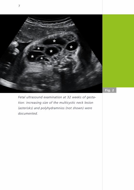
7
Fetal ultrasound examination at 32 weeks of gesta-
tion: increasing size of the multicystic neck lesion
(asterisks) and polyhydramnios (not shown) were
documented.

8
EXIT procedure: successful nasotracheal intubation
following partial delivery of the infant.
Fig. 3
A

Fig. 4A B
maternal
abdomen
abdominal
incision𐇠 𐇠
𐇠
9
EXIT procedure: following nasotracheal intubation,
the endotracheal tube was sutured in place (A) and
positive pressure ventilation was initiated with a
self-inflating bag (B).

*
A*
A
***
**
A B
10
Postnatal T2- weighted MRI demonstrating a large
multicystic neck lesion (asterisks): A) sagittal view, B)
coronal view.
Fig. 5
A

11
DISCUSSIONThe ex utero intrapartum treatment (EXIT) procedure
was originally developed to reverse temporary tra-
cheal occlusion in patients who had undergone fetal
surgery for severe congenital diaphragmatic hernia
(5). In recent years, it has increasingly been used to
deliver babies who have airway compression due to
cervical teratomas, cystic hygromas or blockage of
the airway such as congenital high airway obstruction
syndrome (CHAOS) (EXIT-to-Airway) (6). The procedure
has also been used to stabilize and perform resection
for babies with massive lung lesions (EXIT-to-Resec-
tion) and infants with severe forms of diaphragmatic
hernia (EXIT-to-ECMO).
EXIT procedures are performed under maternal gene-
ral anesthesia. A special uterine stapling device is
used to prevent bleeding from the uterine incision,
which could be excessive since the uterus is prevented
from contracting. The baby is then partially delivered
through the incision while remaining attached to the
placenta. Once the airway is secured (in case of an
EXIT-to-Airway procedure), oxytocin is administered
and delivery is completed.
Most lymphatic malformations are benign lesions. In
some cases, observation may be appropriate, as spon-
taneous regression has been reported to occur in up to
45% of cases. However, patients with lymphangioma
colli frequently show signs of respiratory compromise
and impaired swallowing; trauma (not seldom in active
12
toddlers), infection or intracystic hemorrhage are
other potential complications that will lead to active
interventions. Possible treatment options include scle-
rotherapy (4, 7) (Table), radiofrequency ablation, laser
therapy or surgical resection. More recently, the use
of propranolol (a well established therapy for infan-
tile hemangioma), sildenafil and sirolimus has been
reported (1). Unfortunately, lymphatic malformations
have a high recurrence rate. In the future, better
understanding of the genetics and biology of these
lesions will hopefully lead to improved treatment stra-
tegies.
See also: COTM April 2006 «Intralesional injection
therapy wit OK-432 (Picibanil®) in a full term infant
with multicystic lymphangioma colli».

Agent Assumed mechanism of action
Side effects / com-plications
Use in macrocy-stic LM
Use in microcy-stic /other LM
Picibanil® (OK-432), lyophilized mixture: Streptococcus pyogenes group A benzyl penicillin
Inflammatory response: increased cytokine pro-duction by leucocytes
Anaphylaxis yes no
Doxycycline, tetra-cycline antibiotic
Inhibition of matrix metalloproteinases and cell proliferation; suppression of VEGF-induced lymph-/angio-genesis; deposition of collagen and fibrin with cyst involution
Discoloration of teeth; electrolyte imbalances
yes no
Bleomycin, chemo-therapeutic agent
Inhibition of DNA synthesis; inflammatory reaction on endothelial cells
Interstitial pneu-monia; pulmonary fibrosis (if cumula-tive IV dose > 400 mg)
yes no
Pingyangmycin, chemotherapeutic agent
Destruction of lympha-tic endothelial cells; increased collagen deposition in cyst cavity
Hair loss; gastro-intestinal reaction; alteration of skin pigmentation; pulmonary fibrosis
yes yes
Sodium tetrade-cyl sulfate (STS), detergent
Emulsion of cell membrane lipoproteins; increase of membrane permeability; enhance-ment of cell death and fibrosis, if used with doxycycline or ethanol
Increased risk of infection
yes orbital
Ethanol, desiccant Dehydration of lympha-tic endothelial cells
Respiratory depression; cardiac arrhythmias; rhab-omyolysis; hypogly-caemia; seizures
(yes) no
Table. Sclerosing agents used in the treatment of
lymphatic malformations (LM) (1).
13

1. Defnet AM, Bagrodia N, Hernandez SL, Gwilliam N, Kandel JJ.
Pediatric lymphatic malformations: evolving understanding and
therapeutic options. Pediatr Surg Int 2016 Jan 27 (epub ahead
of print) (Abstract)
2. Levy AD, Cantisani V, Miettinen M. Abdominal lymphangiomas:
imaging features with pathologic correlation. Pictorial essay.
AJR 2004;182:1485-1491 (no abstract available)
3. Lugo-Oliveieri CH, Taylor GA. CT differentiation of large
abdominal lymphangioma from ascites. Pediatr Radiol
1993;23(2):129-130 (Abstract)
4. Rautio R, Keski-Kisula L, Laranne J, Laasonen E. Treatment of
lymphangiomas with OK-432 (Picibanil). Cardiovasc Intervent
Radiol 2003;26:31-36 (Abstract)
5. Harrison MR, Adzick NS, Flake AW, et al. Correction of
congenital diaphragmatic hernia in utero VIII: Response of
the hypoplastic lung to tracheal occlusion. J Pediatr Surg
1996;31:1339-1348 (Abstract)
6. Olutoye OO, Olutoye OA. EXIT procedure for fetal neck masses.
Curr Opin Pediatr 2012;24:386-393 (Abstract)
7. Balakrishnan K, Menezes MD, Chen BS, Magit AE, Perkins JA.
Primary surgery vs primary sclerotherapy for head and neck
lymphatic malformations. JAMA Otolaryngol Head Neck Surg
2014;140:41-45 (Abstract)
REFERENCES
14

15

SUPPORTED BY
CONTACT
Swiss Society of Neonatology
www.neonet.ch
con
cep
t &
des
ign
by
mes
ch.c
h
Top Related