


Languages
Pages
Legal


Purdue UniversityPurdue e-PubsWeldon School of Biomedical Engineering FacultyWorking Papers Weldon School of Biomedical Engineering
6-30-1992
Evidence That Free Fatty Acid-Iron ComplexesDirectly Initiate Lipid Peroxidation in Vitro and inVivo: A New Mechanism of Oxidative StressSteven C. Salaris
Charles F. BabbsPurdue University, [email protected]
Joann Pham
John J. Turek
Follow this and additional works at: http://docs.lib.purdue.edu/bmewp
Part of the Biomedical Engineering and Bioengineering Commons
This document has been made available through Purdue e-Pubs, a service of the Purdue University Libraries. Please contact [email protected] foradditional information.
Recommended CitationSalaris, Steven C.; Babbs, Charles F.; Pham, Joann; and Turek, John J., "Evidence That Free Fatty Acid-Iron Complexes Directly InitiateLipid Peroxidation in Vitro and in Vivo: A New Mechanism of Oxidative Stress" (1992). Weldon School of Biomedical EngineeringFaculty Working Papers. Paper 14.http://docs.lib.purdue.edu/bmewp/14
Free Fatty Acids, Fe, & Oxidative Stress 1 SC Salaris et al.
EVIDENCE THAT FREE FATTY ACID-IRON COMPLEXES
DIRECTLY INITIATE LIPID PEROXIDATION IN VITRO
AND IN VIVO: A NEW MECHANISM OF OXIDATIVE
STRESS
STEVEN C. SALARIS, CHARLES F. BABBS, JOANN PHAM, and JOHN J. TUREK
Biomedical Engineering Center and School of Veterinary Medicine, Purdue University,
West Lafayette, Indiana, USA
June 30, 1992
Abstract -- Through a series of biochemical and histochemical experiments we explored the novel
hypothesis that iron and free fatty acids, liberated after tissue injury, combine to form liposoluble
complexes that directly initiate lipid peroxidation. The addition of 100 M ferric iron to 30 mM
linoleate suspensions at pH 7.4 produced time dependent lipid peroxidation, measured as
conjugated diene formation. Complexes of 100 M ferric iron and 600 M pentanoate also
initiated formation of conjugated dienes in linoleate suspensions and formation of
malondialdehyde-like materials in rat liver slices. A histochemical stain for free fatty acids
revealed positive reactions within cell membranes in traumatized regions of rat liver tissue that
underwent compression injury followed by thirty minutes of blood perfusion, but not in non-
traumatized control regions. The diaminobenzidine-H2O2 histochemical reaction for iron, revealed
increased levels of redox cyclable iron in the membranes and the cytoplasm of traumatized
hepatocytes. We propose that traumatic injury initiates cascades leading to liberation of iron from
storage proteins and free fatty acids from membranes, which combine, distribute to the lipid
domains of cell membranes, and directly initiate lipid peroxidation.
Keywords--Conjugated dienes, Free fatty acid-iron complexes, Initiation, Lipid hydroperoxide,
Liposomes, Lipid peroxidation, Oxidative Stress, Perferryl Fe-oxo complexes, Perferryl iron, Oxo-
iron complexes, Trauma
Free Fatty Acids, Fe, & Oxidative Stress 2 SC Salaris et al.
INTRODUCTION
Oxidative stress leading to lipid peroxidation, as well as oxidative modification of proteins, low
density lipoproteins, and nucleic acids, has been linked to the pathophysiology of many disease
states, including rheumatoid arthritis, [1] ischemia/reperfusioninjury, [2-5] atherosclerosis, [6, 7]
paraquat poisoning, [8] and traumatic injury to the central nervous system. [9, 10] Although
oxidative modification of non-lipid species has received some attention, [11-15] lipids are
generally considered to be the most important targets of oxidative stress in biological systems.[10,
16-24] Lipid oxidation has been suggested to disrupt the architecture of cell membranes by the
introduction of polar (-OOH and -OH) groups into the lipophilic central regions of phospholipid
bilayers, [23] by subsequent degradation of lipid peroxides into smaller molecules, and by toxic
effects of lipid oxidation products, including 4-hydroxynonenal, which is both a direct cytotoxin
and a potent chemoattractant for leukocytes. [25-28] Activated leukocytes, in turn, are prodigious
sources of biological oxidants that may easily perpetuate the cycle of oxidative stress. [29, 30]
The most quoted chemical mechanism for initiation of lipid peroxidation has been the abstraction
of hydrogen atoms from polyunsaturated lipids by hydroxyl radicals (HO•) generated by the
superoxide driven Fenton reaction. [2, 4, 12] Superoxide (O2), the primary oxygen radical in
biological systems, may be produced by oxidoreductases, [31, 32] endothelial cells, [30, 33] and
activated leukocytes. [29, 34, 35] According to the prevailing view, [36] the redox cycling of
suitably chelated iron in the presence of superoxide and hydrogen peroxide may lead to the
generation of the hydroxyl radical,
followed by the initiation of lipid peroxidation,
LH + HO• L• + HOH ,
where LH is a polyunsaturated lipid, and subsequent chain propagation,
L• + O2 LOO•
LOO• + LH + LOOH + L• , etc.
The hydroxyl radical (HO•) is one of the most effective oxidants in nature, which is ordinarily
created under extreme physical conditions, such as those prevailing in fields of high intensity
gamma irradiation, [37, 38] high temperature flames, [39-41] or intergalactic dust clouds. [42] The
intellectual appeal of the superoxide driven Fenton reaction as a pathophysiologic mechanism is
that it provides a route by which such a powerful oxidant may be readily generated in aqueous
solutions under physiologic conditions of temperature, pH, and ionic strength. Further, there is

Free Fatty Acids, Fe, & Oxidative Stress 3 SC Salaris et al.
rather abundant evidence that interventions such as administration of superoxide dismutase (SOD)
or strong iron chelators (like deferoxamine), which would be expected to disrupt Fenton
chemistry, attenuate oxidative stress caused by ischemia/reperfusion in the rabbit heart, [43] rabbit
kidney, [22, 44] cat intestine, [45] and dog heart, [46-48] as well as oxidative stress associated
with CNS trauma, [49, 50] cardiac arrest and resuscitation, [51-53] and hemorrhagic shock. [54-
58]
Although the roles of superoxide, iron, and lipid peroxidation in the forgoing scheme are rather
well established, several lines of evidence cast doubt upon the importance of HO• as a direct
initiator of lipid peroxidation and related pathophysiologic alterations in vivo. A major physical
limitation for initiation of lipid peroxidation by HO• is the requirement that the highly reactive
HO• radical, initially generated in the aqueous phase, penetrate the hydrophobic central region of
the membrane. Hydroxyl radicals are generally considered to be such indiscriminate oxidants that
they react within one or two molecular diameters with aliphatic or aromatic carbon compounds
[59-61], including amines, amino acids, lipids, and nucleic acids that are likely to be present in the
immediate environment or on the membrane surface. Aust and coworkers [62] noted HO•
scavengers and catalase prevented lipid peroxidation in detergent solubilized lipids but not
microsomal lipids in micellar form. Although HO• can initiate lipid peroxidation in solubilized,
homogenous reaction systems, [62, 63] lipid peroxidation in liposomes or microsomes initiated by
adding iron-ATP, ferrous-ferric complexes, or ferric ascorbate is not usually inhibited by catalase
or scavengers of HO•. [63] Rush and Koppenol, in an uncomplicated chemical system, found
kinetic evidence against direct formation of HO• from chelated ferrous iron and hydrogen peroxide
at pH 6. [64] Steiner [65], using dimethyl sulfoxide as a molecular probe for HO•, found marginal
evidence for a burst of hydroxyl radicals during ischemia/reoxygenation of rat kidney slices, even
though lipid peroxidation, preventable by iron chelators and antioxidants, readily occurs in this
model. [66, 67] Similarly, sensitive cytochemical [58] and biochemical [59] probes for
intracellular bursts of hydrogen peroxide in postischemic liver have failed to reveal evidence of
heightened Fenton chemistry; although lipid peroxidation in postischemic liver is readily
demonstrated in the laboratory. [66-70]
Review of this prior work and some intriguing preliminary studies in our own laboratory led us to
propose a new hypothesis regarding the initiation of lipid peroxidation in biological membranes.
The alternative initiation mechanism involves the direct oxidation of polyunsaturated fatty acids
by free fatty acid-ferric iron complexes, without the necessary involvement of HO• or H2O2. In
brief, this alternative mechanism begins with the liberation of low molecular weight iron species,
the concurrent liberation of free fatty acids, the formation of free fatty acid-iron complexes and
their uptake into phospholipid bilayers, and direct, non-enzymatic initiation of lipid peroxidation
by such complexes (Figure 1). The liberation of "free" or low molecular weight forms of iron from
high molecular weight forms, notably ferritin, may occur either via the action of calcium activated
proteases following tissue trauma or ischemia [71, 72] or via the release of low molecular weight
iron from ferritin by superoxide [73] generated by xanthine oxidase, [2, 5] mitochondrial
metabolism, [74] and activated leukocytes. [29, 30, 34, 75]

Free Fatty Acids, Fe, & Oxidative Stress 4 SC Salaris et al.
Fig. 1. Proposed pathophysiological cascade of cellular events leading to lipid peroxidation
and cell death. Note positive feedback loops leading to accelerated release of iron and free
fatty acids. The scheme shows proposed routes for sustained lipid peroxidation after
trauma or ischemia.

Free Fatty Acids, Fe, & Oxidative Stress 5 SC Salaris et al.
Thus, lipid peroxidation and attendant tissue damage in vivo may not necessarily be dependent
upon superoxide and may [76, 77] or may not [78, 79] be inhibited by superoxide dismutase. The
concurrent liberation of free fatty acids, most probably by calcium activated phospholipases
following tissue ischemia, trauma, thermal injury, etc. [80, 81] may provide lipid soluble chelators
of iron, capable of carrying the iron into the hydrophobic interior regions of phospholipid bilayers
within cell membranes. [82] Here, in the immediate proximity of the oxidizable double bonds of
membrane lipids, we propose that the ferric iron-free fatty acid complexes may directly initiate
lipid peroxidation, without the involvement of HO• or H2O2.
We further propose that lipid oxidation is accompanied by reduction of the iron to the ferrous
form, followed by autoxidation or reoxidation to the ferric form and further redox cycling of the
iron, thus creating a self perpetuating mechanism that leads to continued lipid peroxidation,
calcium intoxication, and membrane destabilization that hastens membrane disruption and cell
death (Figure 1).
The present paper provides evidence from a diverse series of experiments suggesting that such
complexes form both in vitro and in vivo and are capable of initiating lipid peroxidation. In
particular, biochemical studies were performed to determine if free fatty acid-iron complexes
could initiate lipid peroxidation in vitro. Then companion histochemical studies were performed to
search for evidence for the proposed free fatty acid-iron complexes in vivo.
MATERIALS AND METHODS
Reagents and pharmaceuticals
Ferric-EDTA, linoleic acid, oleic acid, palmitic acid, pentanoic acid, quinacrine (6-chloro-9-[(4-
diethylamine)-1-methyl-butyl]amino-2-methoxyacridine), rubeanic acid (dithiooxamide),
spectrophotometric grade cyclohexane, 2-thiobarbituric acid and Tris (hydroxymethyl-
aminomethane) buffer were obtained from Sigma Chemical Company, St. Louis, Missouri, USA.
Copper sulfate, hydrogen peroxide, methanol, and trichloroacetic acid were obtained from Fisher
Scientific Company, Fair Lawn, New Jersey, USA. Ferric chloride, ferrous sulfate, glacial acetic
acid, potassium ferrocyanide and spectrophotometric grade chloroform were obtained from
Mallinckrodt, Inc., Paris, Kentucky, USA. 3,3'diaminobenzidine-4HCL was obtained from Aldrich
Chemical Company, Milwaukee, Wisconsin, USA. The Tris-Ringer buffer solution consisted of
4.0 mM KCl, 140 mM NaCl, 2.0 mM CaCl2 , 4.0 mM Tris buffer, and 200 mg/dL d-glucose, pH
7.4. In some experiments, lactate buffer was used, which consisted of 4.0 mM KCl, 100 mM NaCl,
2.0 mM CaCl2, 40.0 mM lactate, and 200 mg/dL d-glucose, pH 7.4.
Conjugated diene (CD) production in micellar suspensions of polyunsaturated fatty acids
Unlike other indicators of lipid oxidation such as malondialdehyde and ethane formation,
conjugated dienes provide a more quantitative yield of reaction products that are proximal to the
actual initiation of lipid peroxidation. [36, 83, 84] Also, using conjugated dienes as the end-point
permitted studies of the possible anoxic oxidation of lipids by ferric iron, thereby providing

Free Fatty Acids, Fe, & Oxidative Stress 6 SC Salaris et al.
evidence excluding oxygen based initiators. Such work is not possible with malondialdehyde-like
materials (MDA-LM), since the formation of MDALM from conjugated dienes requires
molecular oxygen. [36 , 83]
To investigate the hypothesis that iron can directly initiate lipid peroxidation in micellar
suspensions of polyunsaturated fatty acids (PUFAs), conjugated diene formation in vitro was
studied. To test tubes containing 1.0 ml of 30 mM free fatty acid salt (linoleate, oleate, or
palmitate) in suspension at pH 7.4, 0.1 ml of 1.0 mM FeSO4 , FeCl3 , ZnCl2 or 0.9% NaCl was
added while vortexing. The final concentration of iron or zinc was 100 M. This concentration of
low molecular weight chelate iron ("free iron") was chosen to be similar to that previously
measured in postischemic dog brain and heart. [85, 86] The test tubes were then incubated in room
air for 30 minutes at 37 °C in a shaking water bath. To study the hypothesis that ferric iron and not
ferrous iron, was the initiator of conjugated diene (CD) formation in PUFAs, linoleate samples
with either FeSO4 or FeCl3 additions were also performed in an argon saturated (anoxic) test tube,
in which autoxidation of ferrous iron to the ferric form would not occur. In these experiments,
anoxia was maintained by continuous bubbling of the fatty acid suspensions with argon gas.
To investigate the time course of iron initiated CD formation in linoleate, 3.5 ml of 30 mM
linoleate were mixed with 0.35 ml of 1000 M ferric chloride by vortexing. The mixture was then
placed in a shaking water bath in room air at 37 °C. A time 0 sample was taken from fresh
linoleate. (In preliminary experiments, similar results were obtained if the time 0 sample was taken
either just before or just after iron addition, indicating that no iron dependent lipid peroxidation
occurred during the conjugated diene assay itself). Subsequent 0.5 ml samples 5, 10, 15, 30, 45,
and 60 minutes after iron addition were taken from the reaction mixture and assayed for
conjugated dienes, as subsequently described. To test the hypothesis that lipid soluble free fatty
acid-iron complexes could initiate CD formation, 0.1 ml of 1 mM FeCl3 / 6 mM pentanoate was
added to 1.0 ml of 30 mM linoleate (pH 7.4) vortexed and incubated in room air for 30 minutes at
37 °C. Pentanoic acid only and pentanoic acid / ferric chloride only (no linoleate) controls were
also studied.
Measurement of conjugated dienes
Conjugated dienes were measured by a modification of the spectrophotometric method described
by Recknagel and Glende. [84] A 0.5 ml aliquot of sample was taken and added to 3.0 ml of
chloroform methanol 2:1. The mixture was then vortexed for 2 minutes and then centrifuged for 5
minutes at 1700g. A 0.5 ml aliquot of the bottom chloroform layer was removed, added to a clean
test tube and taken to dryness under a stream of argon gas. Then, 2.0 ml of spectrophotometric
grade cyclohexane were added, and the test tube was gently vortexed. Conjugated dienes were
then assayed by scanning the sample against a cyclohexane blank and reading the absorbance from
300 to 200 nm. The conjugated dienes exhibited an absorbance peak at 232 nm, as expected.
The absorbances at 220, 225, 232, 240 and 245 nm were obtained and conjugated diene
concentrations were calculated using a computer program for peak extraction implementing the
double derivative method of O'Haver [87, 88] and based on reported UV spectra [89] and an
extinction coefficient [89] of 27,000 M-1
cm-1
.

Free Fatty Acids, Fe, & Oxidative Stress 7 SC Salaris et al.
Lipid peroxidation in fresh rat hepatic tissue by free fatty acid / iron complexes in vitro.
To investigate the hypothesis that iron / free fatty acid complexes initiate lipid peroxidation in
organized tissue, we used an in vitro liver slice model developed in our laboratory and described
previously. [66, 67, 90] Tissues were taken from male Wistar rats which had been anesthetized
with sodium pentobarbital (60 mg/kg, i.p.). Rat liver was selected for study because it readily
undergoes lipid peroxidation in similar models of tissue trauma and oxidative stress, [67, 90] and
so provides a convenient basis for the study of free fatty acid and iron effects. Upon reaching the
stage of surgical anesthesia, each rat was given a 0.5 ml bolus of 1000U/ml heparin via the dorsal
penile vein. After a 2 min circulation time, the liver was surgically exposed and the portal vein
was cannulated. The inferior vena cava was then cut and the liver was perfused in situ with cold
lactate buffer to remove blood elements. The liver was then excised, and tissue samples were
processed according to protocols described subsequently. The tissue slices were placed into 50 ml
Erlenmeyer flasks containing air equilibrated lactate buffer solution.
Tissue manipulations
Previous research in our laboratory [90] had indicated that tissue trauma is a very effective inducer
of lipid peroxidation in the short term organ culture model. Trauma is several times more effective
at inducing lipid peroxidation in this model, measured as MDA-LM formation, as anoxia /
reoxygenation without trauma. Accordingly, we chose trauma as our model of oxidative stress in
rat hepatic tissue. Minimally traumatized control tissue samples were prepared as follows: a large
lobe of rat liver was excised, moistened with buffer, and gently cut into thin (approx. 1 mm) strips
with a #11 scalpel blade. Then, the tissue strips were gently cut again, perpendicular to the planes
of the first series of cuts. This technique produced small tissue blocks about 1 mm x 1 mm x 2-3
mm. These tissues were then washed three times in cold lactate buffer, divided into samples of
approximately 0.4 grams, then added to the buffer-containing 50 ml Erlenmeyer flasks. The
cutting was done prior to any tissue incubations. The tissues were incubated with shaking in
room-air equilibrated buffer at 37 °C for 30 min. In addition to the "minimally traumatized" tissues
described above, samples of "maximally traumatized" tissue, which were vigorously minced
without washing into similar sized pieces using iris scissors, were also studied for comparison with
minimally traumatized tissue samples.
The fresh hepatic tissue slices were incubated in a 37 °C shaking water bath for 30 minutes in
room air. In some experiments, the tissues were exposed to lipid-soluble iron in the form of 100
M Fe+++
complexed with 600 M pentanoate. In other experiments, tissues were exposed to 100
M ferric-EDTA (1:1), a water soluble form of iron. Pentanoate only and tissue only controls were
included. For comparison, samples of minimally and maximally traumatized liver tissue were
prepared, and incubated as above for 30 minutes. In a time course study with the pentanoate /
ferric iron complex, 6 separate flasks were incubated for 5, 10, 15, 30, 45 or 60 minutes. A time 0
sample was taken directly from fresh liver tissue. The extent of lipid peroxidation was determined
by measuring malondialdehyde-like materials (MDA-LM). Flask contents, including tissue and
buffer, were homogenized in a Teflon / glass homogenizer and assayed for MDALM by the
spectrophotometric method of Buege and Aust, [91] as modified by Salaris and Babbs. [66]

Free Fatty Acids, Fe, & Oxidative Stress 8 SC Salaris et al.
Traumatic injury to rat hepatic tissue in vivo and in situ
To study free fatty acid liberation and iron liberation in traumatized rat liver tissue a model of
tissue trauma in vivo was developed. Rats were anesthetized with sodium pentobarbital (60
mg/kg). The liver was then surgically exposed and a supporting steel plate was placed under the
liver. Trauma was induced by dropping a 5.5 g steel bolt (1.0 cm diameter hex-head-side down)
through a 1.5 cm inside diameter plastic guide tube five times on the liver from a height of 13 cm.
Trauma was evident as a circular, bruised region on the surface of the liver. The steel plate was
then removed and the incision loosely closed with towel clips. A saline moistened gauze was
placed over the incision to prevent tissue drying and the rat was placed under a warm lamp for
thirty minutes to allow tissue responses to traumatic injury to develop. Rectal temperature was
monitored and maintained at 37-38 °C. Then one of several histochemical methods was used to
detect the presence of either free fatty acids or iron in and around the traumatized region.
Histochemical detection of free fatty acid liberation during traumatic injury to hepatic tissue
To detect free fatty acids in situ, a modification of Holczinger's histochemical technique for free
fatty acids [92, 93] was employed. This method utilizes the formation of insoluble complexes of
free fatty acids and copper, which then reacts with rubeanic acid to form a visible reaction product
for light microscopy. Thirty minutes after trauma, the portal vein in the rat was cannulated with an
18 gauge cannula. The inferior vena cava was cut and the liver was perfused with 60 ml of Tris
buffer solution to remove blood constituents. The liver was then perfused with 50 ml of an
isotonic solution of 75 mM copper sulfate in 75 mM NaCl, followed by 80 ml of Tris buffer
solution to remove the excess copper sulfate. Then, the liver was perfused with 50 ml of 0.1% w/v
rubeanic acid in 70% v/v ethanol. Finally, the liver was then perfused with 40 ml of isotonic saline
to remove excess rubeanic acid, followed by 50 ml of Trumps fixative solution (1%
gluteraldehyde, 4% formaldehyde in neutral phosphate buffer).
The liver was then excised and trimmed into sections representing traumatized and non-
traumatized regions of the liver, which were submitted for routine tissue processing for light
microscopy. Seven micron thick sections were stained with hematoxylin and eosin. Two control
groups of rats were studied. In one group, just prior to traumatizing the tissue, the rats were treated
with 10 mg/kg of the phospholipase inhibitor, quinacrine, [94, 95] dissolved in saline, given via
the dorsal penile vein. In a second control group, an organic solvent cocktail consisting of 50 ml
ethanol, 50 ml methanol and 1 ml glacial acetic acid was perfused through the liver prior to the
copper / rubeanic staining sequence. The acidic alcohol perfusion was intended to produce
conditions in which the free fatty acids would be extracted from the cell membranes [93] and thus
not be detected.
Tissue processing for iron staining
In another series of weight drop experiments utilizing intact, blood perfused livers, the portal vein
of each rat was cannulated with an 18 gauge cannula after 30 minutes of post-traumatic blood
perfusion. The inferior vena cava was cut and the liver was perfused with 60 ml Tris buffer
solution to remove blood constituents, followed by 50 ml of Trumps fixative solution. A

Free Fatty Acids, Fe, & Oxidative Stress 9 SC Salaris et al.
traumatized block and a non-traumatized control block of the same liver were then removed,
stored in fixative and processed for light or electron microscopy. In other experiments, the liver
was traumatized (as described above) just prior to the Tris buffer flush without post-traumatic
blood perfusion. This provided a control in which the presence or absence of redox cyclable iron
could be observed immediately after trauma. Unstained, paraffin embedded sections were
dewaxed and rehydrated by standard methods. To detect iron histochemically, the slides were then
incubated for 60 minutes either in Perl's reagent or in a hydrogen peroxide and diaminobenzidine
solution as described subsequently. At the end of the incubation period, the tissues were rinsed,
counterstained with eosin (Perl's reagent treated) or methyl green (DAB reagent treated), and
permanently mounted.
Conventional histochemical stain for iron
Initial attempts to detect iron in cell membranes of traumatized hepatic cells employed a
modification of Perl's method for iron, [96] in which ferric iron in the tissue combines with added
ferrocyanide to form ferric ferrocyanide precipitate (Prussian blue). Seven micron thick sections of
dewaxed and rehydrated tissues were placed into a mixture of 3% HCl w/v and 7% w/v potassium
ferrocyanide solution which had been previously saturated with Prussian blue by the addition of a
small amount of ferric chloride solution and subsequent low speed centrifugation. After 1 h, the
tissues were washed and counterstained with eosin and examined under the light microscope.
Some light blue staining around the cell membranes was visible in the cells near the traumatized
region, but it was concluded that a more sensitive method of staining and detecting iron was
desirable.
In situ histochemical detection of redox cyclable iron using hydrogen peroxide and
diaminobenzidine (DAB)
To detect redox cyclable iron at neutral pH in situ, with greater sensitivity than conventional iron
stains, we modified the method of Kuo and Fridovich [97] for staining iron-containing proteins in
electrophoretic gels. This method exploits the catalytic activity of iron on the diaminobenzidine
polymerization reaction as a histochemical procedure, presumably based upon the following
coupled reactions:
Fe+++
+ DAB Fe++
+ DAB•,
Fe++
+ H2O2 Fe+++
+ HO• +HO ,
followed by chain propagation in the usual manner,
DAB• + DAB •DAB-DAB ,
• (DAB)n +DAB • (DAB)n+1, etc.
until chain termination occurs
• (DAB)n + • (DAB)m (DAB)n+m .

Free Fatty Acids, Fe, & Oxidative Stress 10 SC Salaris et al.
The result is a brown DAB polymer, which persists through further tissue processing and is readily
visible by light microscopy. To implement this approach, the dewaxed and rehydrated slides were
placed in 4 mM Tris Ringer buffer (pH 7.4) containing 40 mM H2O2 and 2.5 mM DAB.
Incubation time at room temperature was 60 minutes.
Quantitative morphometry
Cellular profiles of H&E counterstained slides were examined by light microscopy at 200X
magnification by an observer who was unaware of the treatment or control group designation of
the specimen. In the free fatty acid studies, the copper-rubeanic acid reaction product appeared as
amorphous, amber-brown deposits in and around cell membranes, which were scored positively if
reaction product was clearly visible. In the free redox cyclable iron studies, the DAB reaction
product appeared as amber-brown deposits in and around cell membranes, which were scored
positively if reaction product was clearly visible. Counts of profiles of positively stained
hepatocyte membranes were made within a 400 x 400 2 test area, delimited by a micrometer
eyepiece on the light microscope. The tissue on a 1 in. x 3 in. glass slide was scanned in vertical
strips at successive 2.5 mm horizontal increments from left to right.
Defining trauma
In order to independently define traumatized areas of the tissues microscopically, the following
five criteria were established. At least three of the five criteria had to be present for the area to be
defined as traumatized. These criteria were independent of any histochemical staining that may
have been observed and are as follows: (1) irregular tears in the normal cellular matrix not
attributable to sectioning artifact, (2) "naked nuclei" or those without surrounding cytoplasmic
components, (3) cytoplasmic fragments without nuclei, (4) lighter staining, coagulated cytoplasm
in cells near traumatized area, and (5) hemorrhage. These characteristics were not observed in non-
traumatized control tissues.
Stereology
To quantify the surface area, Sv, of stained cell membranes per unit volume of tissue in units of
mm2/mm
3 , we employed the stereological techniques of Weibel [98] and Elias [99], in which Sv
was computed from the observed number of intersections of positively stained membranes with
measured test lines superimposed on the light microscopic field by a micrometer eyepiece.
Microscopic sections of non-traumatized tissues were randomly labeled and studied by an observer
who was unaware of the treatment protocol. The tissue was scanned in vertical strips, each 2.5 mm
apart. The ten horizontal lines of the micrometer eyepiece grid served as the test line for the
stereology counts (total length = 10 x 400m = 4000m) in non-overlapping fields. Each time a
positively stained membrane crossed a horizontal test line, a count was recorded. The traumatized
tissues were similarly labeled and studied. For these tissues, the sections were scanned vertically
until a region of tissue trauma was encountered and then and extensive sampling of the area was
made to maximize the information obtained in the traumatized areas. The numbers of counts and
total test line length were then added and the Sv calculated as

Free Fatty Acids, Fe, & Oxidative Stress 11 SC Salaris et al.
L
I2Sv ,
where I is the sum of the positive membrane intersections observed for all the grids counted
and L is the sum of the test line lengths for all of the grids counted. [99]
Electron microscopy
To detect iron in cell membranes at the ultrastructural level, we employed the DAB/hydrogen
peroxide reaction used in light microscopic studies. Liver tissue was traumatized and blood
perfused in vivo and in situ and then fixed with Trump's solution as described previously.
Representative trimmed regions of traumatized and non-traumatized liver tissues were then cut
into small (< 0.25 mm thick) sections using a Smith and Farquhar TC-2 tissue sectioner (Sorvall).
These small sections were then placed in the DAB and H2O2 reaction mixture for 1 hour. The liver
slices were then rinsed in phosphate buffered 1% glutaraldehyde--4% formaldehyde and postfixed
with 1% osmium tetroxide in phosphate buffer for 1 hour. Samples were then dehydrated through
a graded 30% to 100% ethanol series, rinsed twice in propylene oxide and infiltrated with graded
propylene oxide-epoxy resin mixture (Poly/Bed 812, Polysciences, Warrington, Pennsylvania,
USA). Tissue blocks were all sectioned at 70 nm and photographed at the same intensity on the
JEOL, JEM-1000X electron microscope. Negatives were printed using an automatic processor and
exposed for identical times and intensities to minimize photographic variations in the apparent
density of the reaction product.
Data analysis
An analysis of variance (ANOVA) was run to test each of the null hypotheses that 1) iron alone
did not initiate formation of conjugated dienes from poly-unsaturated free fatty acids, 2) ferric and
ferrous were not different as initiators of conjugated diene formation, 3) conjugated diene
formation did not change as a function of time after iron addition, 4) complexes of ferric iron and
free fatty acids did not cause conjugated diene formation in PUFAs, 5) complexes of ferric iron
and free fatty acids did not induce lipid peroxidation in fresh rat hepatic tissue, 6) free fatty acids
did not increase in traumatized hepatic cells, and 7) the amounts of free, redox cyclable iron
detected in traumatized hepatic cells were not different from those in control cells. The ANOVA's
were preceded by Bartlett's chi-square tests for homogeneity of variance. [100] If the variances of
compared data sets were not similar, a logarithmic or square root transformation of the data was
performed [101] and the ANOVA repeated on the transformed data. Specific comparisons were
made using Scheffe's multiple range test. A p-value of 0.05 was considered significant.

Free Fatty Acids, Fe, & Oxidative Stress 12 SC Salaris et al.
RESULTS
Oxidation of purified fatty acids
Figure 2 illustrates the formation of conjugated dienes following the simple addition of iron to
linoleate suspensions at pH 7.4. Conjugated dienes (CDs) are formed by the spontaneous
rearrangement of the polyunsaturated lipid alkyl radical immediately after hydrogen atom
abstraction. [83, 102] Hence, they are the earliest measurable products of lipid oxidation. There
was a significant effect of iron on conjugated diene formation (F = 38.6, p < 0.01). Compared to
Fe++
and Fe+++
ions, salts of the non-transition metal ion Zn++
did not induce CD formation. In the
argon sparged (anoxic) samples, CD formation by ferrous iron was significantly less than in the air
exposed samples (F = 75.6, p < 0.01), while CD formation in response to ferric iron was
unaffected by anoxia. The quantitative yield of CDs in linoleate suspensions was nearly equal to
the molar amount of ferric iron added (100 M). No CD formation was detected upon iron
addition to either saturated (oleic) or monounsaturated (palmitic) fatty acids, by the methods
described above (data not shown). CD formation required Fe+++
iron.
Fig. 2. Formation of conjugated dienes following the addition of 100 M iron or zinc to 30
mM suspensions of purified linoleate at pH 7.4. Bars represent mean ± SE for n = 6
experiments. Legend: 1 = linoleate only; 2 = linoleate plus Zn++
; 3 = linoleate plus ferrous
iron; 4 = linoleate plus ferric iron; 5 = anoxic linoleate plus ferrous iron; 6 = anoxic
linoleate plus ferric iron. * = significant from linoleate only control at p < 0.05.

Free Fatty Acids, Fe, & Oxidative Stress 13 SC Salaris et al.
Figure 3 illustrates the time course of conjugated diene formation from linoleate at pH 7.4 over a
60 minute period after iron addition. The increase in CDs with respect to time was significant
(F = 10.1, p < 0.05). There was an initial phase of rapid CD formation during the first 5 minutes,
followed by a phase of much slower CD formation after that time. Quantitatively, the initial rise
matched the initial concentration of Fe+++
iron (100 µM). Thereafter, the continued slow rise in
CD concentration is consistent with slow autoxidation of Fe++
back to Fe+++
in the presence of
molecular oxygen.
Fig. 3. Time course of conjugated diene formation in 30 mM suspensions of purified
linoleate incubated with 100 M ferric iron at pH 7.4. Graph shows means ± SE for n = 4
experiments.
Free Fatty Acids, Fe, & Oxidative Stress 14 SC Salaris et al.
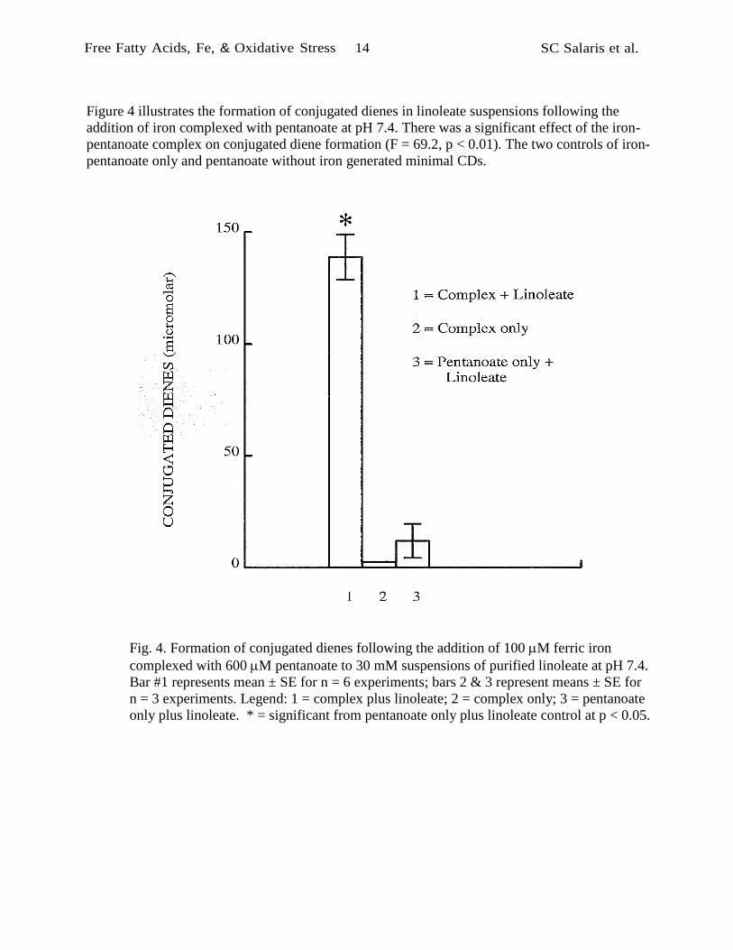
Figure 4 illustrates the formation of conjugated dienes in linoleate suspensions following the
addition of iron complexed with pentanoate at pH 7.4. There was a significant effect of the iron-
pentanoate complex on conjugated diene formation (F = 69.2, p < 0.01). The two controls of iron-
pentanoate only and pentanoate without iron generated minimal CDs.
Fig. 4. Formation of conjugated dienes following the addition of 100 M ferric iron
complexed with 600 M pentanoate to 30 mM suspensions of purified linoleate at pH 7.4.
Bar #1 represents mean ± SE for n = 6 experiments; bars 2 & 3 represent means ± SE for
n = 3 experiments. Legend: 1 = complex plus linoleate; 2 = complex only; 3 = pentanoate
only plus linoleate. * = significant from pentanoate only plus linoleate control at p < 0.05.

Free Fatty Acids, Fe, & Oxidative Stress 15 SC Salaris et al.
Oxidation of rat liver slices
Figure 5 demonstrates the formation of MDA-LM as indicators of lipid peroxidation in rat hepatic
tissues following the addition of either lipid soluble or water soluble iron salts. The addition of
pentanoate only to non-traumatized tissues had no effect on MDA-LM production when compared
to untreated, non-traumatized tissue (F = 1.5, p > 0.05). Water soluble iron in the form of ferric-
EDTA slightly and significantly increased MDA-LM production (F = 9.5, p < 0.05). However, the
addition of ferric iron complexed with pentanoate, caused a large increase in the amount of
measured MDALM compared to all the other treatments of non-traumatized tissue (F = 252.4, p
<< 0.01). The effect of the more lipid soluble ferric-pentanoate complex was similar to that
produced by maximal tissue trauma.
Fig. 5. Formation of MDA-LM in rat hepatic tissue slices following the addition of either
lipid soluble or water soluble iron salts. Bars represent mean ± SE for n = 6 experiments.
Legend: 1 = non-traumatized tissues (NT); 2 = non-traumatized tissues plus 600 M
pentanoate (P5); 3 = non-traumatized tissues plus 100 M ferric-EDTA (1:1); 4 = non-
traumatized tissues plus 100 M ferric iron complexed with 600 M pentanoate; 5 =
traumatized (vigorously minced) tissues without complex added. * = significant from non-
traumatized tissues at p < 0.05.

Free Fatty Acids, Fe, & Oxidative Stress 16 SC Salaris et al.
Figure 6 illustrates the time course of MDA-LM formation in non-traumatized rat liver tissue
incubated with the ferric/pentanoate complex. The increase in MDA-LM with time was significant
(F = 85.8, p << 0.01). As with the formation of CDs in linoleate, there was an early rapid phase of
MDA-LM generation, perhaps followed by a later, slower phase. Unlike the linoleate experiment,
however, there was a brief initial lag phase in the formation of MDA-LM, possibly attributable to
antioxidants such as vitamin E in the cell membranes.
Fig. 6. Time course of MDA-LM formation in non-traumatized rat liver slices incubated
with 100 M ferric iron complexed with 600,uM pentanoate. Graph shows means ± SE for
n = 4 experiments.

Free Fatty Acids, Fe, & Oxidative Stress 17 SC Salaris et al.
Free fatty acid accumulation in membranes
Figure 7 illustrates normal, non-traumatized tissue that has undergone the staining procedure for
the detection of free fatty acids. Note the normal microanatomy of the liver lobule with central
vein.
Fig. 7. Photomicrograph of normal non-traumatized liver tissue counterstained with methyl
green that has undergone the staining procedure for the detection of free fatty acids. 400X.
In contrast, Figure 8 illustrates traumatized hepatic tissue stained for free fatty acids. This
photomicrograph demonstrates the criteria used to identify traumatized regions as well as positive
histochemical staining. Hemorrhage, cytoplasmic coagulation, naked nuclei, and cytoplasmic
fragments are evident in this section. This field also clearly demonstrates the dark histochemical
stain in the cell membranes (arrow) indicating the presence of free fatty acids.

Free Fatty Acids, Fe, & Oxidative Stress 18 SC Salaris et al.
Fig. 8. Photomicrograph of traumatized liver tissue counterstained with methyl green that
has undergone the staining procedure for the detection of free fatty acids. Note the
presence of hemorrhage and cytoplasmic disruption. Also note the dark histochemical stain
present in the cell membranes (arrows) indicating the presence of free fatty acids. 400X.

Free Fatty Acids, Fe, & Oxidative Stress 19 SC Salaris et al.
As a control, Figure 9 illustrates traumatized rat hepatic tissue treated with the organic solvent
extraction (to remove fatty acids) prior to staining for free fatty acids. This field illustrates the lack
of dark staining within the membranes that was present in the untreated experimental tissues.
Figure 10 illustrates traumatized rat hepatic tissue treated with 10 mg/kg of the phospholipase
inhibitor, quinacrine. Although the area of trauma is clearly evident, there is minimal dark
membrane staining, which was typical. These control experiments support the interpretation that
positive histochemical staining represents free fatty acids, which are produced in traumatized
tissue by phospholipases.
Fig. 9. Photomicrograph of traumatized liver tissue counterstained with methyl green and
treated with the organic extraction prior to the staining procedure for the detection of free
fatty acids. Note the presence of hemorrhage and cytoplasmic fragments indicative of
trauma. Note the lack of dark histochemical stain in the cell membranes. 400X.

Free Fatty Acids, Fe, & Oxidative Stress 20 SC Salaris et al.
)
Fig. 10. Photomicrograph of traumatized liver tissue counterstained with methyl green and
treated with 10 mg/kg of the phospholipase inhibitor, quinacrine, just prior to traumatic
insult that has undergone the staining procedure for the detection of free fatty acids. Note
the lack of dark histochemical stain in the cell membranes that was present in the non-
quinacrine treated experimental livers. 400X.
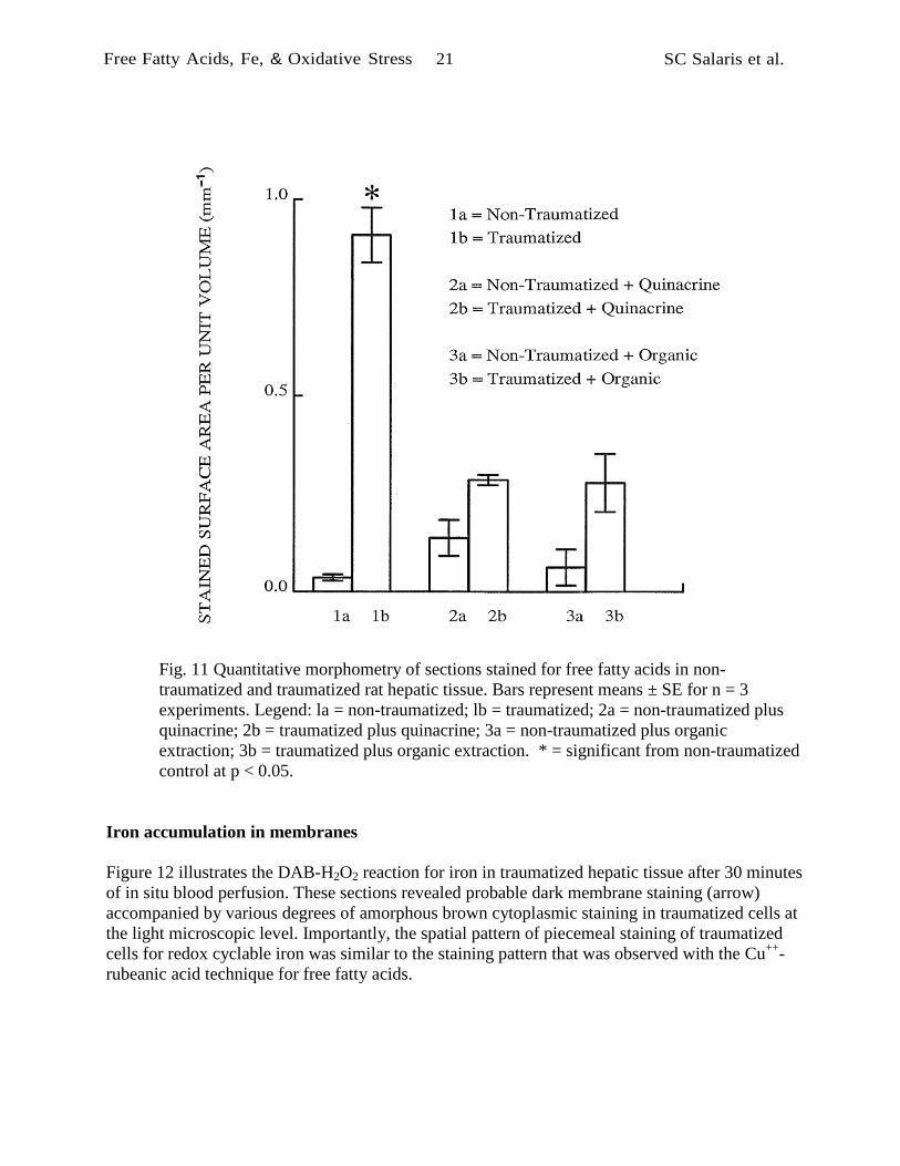
Figure 11 summarizes the results of quantitative morphometry of sections stained for free fatty
acids in non-traumatized and traumatized rat hepatic tissue. The results of stereological
calculations are expressed in square millimeters of positively stained membrane per cubic
millimeter of tissue (mm-1
). In non-traumatized tissues, membrane staining for free fatty acids was
minimal and there was no significant effect of treatments expected to remove free fatty acids or
suppress their formation (F = 1.89, p > 0.05). The area of membranes stained for free fatty acids
was significantly greater in untreated traumatized tissues than in untreated non-traumatized tissues
(t = 11.72, p < 0.05). However, comparable positive staining did not occur in the organic extracted
tissues (t = 1.81, p > 0.05) or the quinacrine treated tissues (t = 2.73, p > 0.05). A group-wise
comparison of the traumatized tissues indicated that both the organic extraction treatment and the
quinacrine treatment significantly reduced membrane staining (F = 37.5, p < 0.01) for free fatty
acids in traumatized tissues.
Free Fatty Acids, Fe, & Oxidative Stress 21 SC Salaris et al.
Fig. 11 Quantitative morphometry of sections stained for free fatty acids in non
traumatized and traumatized rat hepatic tissue. Bars represent means ± SE for n = 3
experiments. Legend: la = non-traumatized; lb = traumatized; 2a = nontraumatized plus
quinacrine; 2b = traumatized plus quinacrine; 3a = non-traumatized plus organic
extraction; 3b = traumatized plus organic extraction. * = significant from non-traumatized
control at p < 0.05.
Iron accumulation in membranes
Figure 12 illustrates the DAB-H2O2 reaction for iron in traumatized hepatic tissue after 30 minutes
of in situ blood perfusion. These sections revealed probable dark membrane staining (arrow)
accompanied by various degrees of amorphous brown cytoplasmic staining in traumatized cells at
the light microscopic level. Importantly, the spatial pattern of piecemeal staining of traumatized
cells for redox cyclable iron was similar to the staining pattern that was observed with the Cu++
-
rubeanic acid technique for free fatty acids.

Free Fatty Acids, Fe, & Oxidative Stress 22 SC Salaris et al.
Fig. 12. DAB-H2O2 reaction for iron in traumatized hepatic tissue after 30 minutes of in
situ blood perfusion. Sections were counterstained with methyl green. These sections
illustrate dark brown membrane staining (arrow) accompanied by various degrees of
cytoplasmic staining in cells in the traumatized region. 400X.
The observed brown cytoplasmic staining of hepatocytes varied in intensity in different areas but
was only associated with the traumatized regions of the tissue, perhaps indicating the presence of
free redox cyclable iron in the cytoplasm of the cell as well as in the intracellular membrane
matrices of the endoplasmic reticulum and mitochondria. Staining due to peroxidase activity in
hepatocytes was unlikely since nontraumatized cells in the same paraffin embedded sections did
not stain with DAB-H2O2 Red blood cells in the specimen consistently showed a positive (internal
control) reaction for iron.
Figures 13 and 14 illustrate the results of negative control experiments. Figure 13 illustrates
control, non-traumatized rat hepatic tissue stained with the DAB-H2O2 reaction and counterstained
with methyl green. Note normal hepatic microstructure with intact liver lobe and central vein.
Figure 14 illustrates freshly traumatized hepatic tissue without the 30 minute blood perfusion time.
Traumatized area is defined by the presence of the indicators described previously. These slides
revealed little membrane staining but a tendency towards an amorphous, brown staining of the
cytoplasmic regions of the cells in the traumatized region (arrow). This cytoplasmic staining was
absent in the non-traumatized tissues and only vaguely present in the traumatized tissues without
blood perfusion. Also note the darkly stained red blood cells (*) which serve as a positive control,
since heme iron does redox cycle [103] to cause DAB polymer formation.

Free Fatty Acids, Fe, & Oxidative Stress 23 SC Salaris et al.
Fig. 13. Negative control DAB-H2O2 reaction for iron in non-traumatized hepatic tissue.
Sections were counterstained with methyl green. These sections illustrate the lack of dark
brown membrane and cytoplasmic staining. Note the darkly stained red blood cells which
served as a positive internal controls for redox cyclable iron. 400X.
Fig. 14. Negative control DAB-H2O2 reaction for iron in traumatized hepatic tissue without
the 30 minutes of in situ blood perfusion. Sections were counterstained with methyl green.
Hepatocytes show little dark brown membrane staining with only a small degree
cytoplasmic staining in cells in the traumatized region. Many positive red cells are present.
400X.

Free Fatty Acids, Fe, & Oxidative Stress 24 SC Salaris et al.
Figure 15 summarizes the results of stereologic analysis of membrane staining for iron by the
DAB-H2O2 reaction. There was a highly significant effect of tissue trauma (F = 73.4, p < 0.01).
Membrane staining was slightly and significantly increased after trauma without blood perfusion
(F = 20.7, p < 0.01), compared to non-traumatized controls, and membrane staining was greatly
increased after trauma followed by 30 min in situ blood perfusion (F = 116.4, p << 0.01). The
density of membrane surface positively stained for low molecular weight chelate iron was on the
same order of magnitude as the density of membrane surface positively stained for free fatty acids.
Fig. 15. Stereological analysis of membrane staining for iron by the DAB-H2O2 reaction.
Bars represent means SE for n = 4 experiments (n = 3 for non-traumatized tissues).
Legend: 1 = non-traumatized tissues; 2 = traumatized tissues plus 0 min blood perfusion; 3
= traumatized tissues plus 30 min blood perfusion. * = significant from nontraumatized
controls at p < 0.05.

Free Fatty Acids, Fe, & Oxidative Stress 25 SC Salaris et al.
Electron microscopy
Because the heavy cytoplasmic component of the DAB-H2O2 reaction product tended to mask
membrane localization at the light microscopic level; we performed electron microscopy. Figure
16 illustrates the ultrastructural appearance of DAB-H2O2 reaction product in traumatized hepatic
tissues. In both non-traumatized and traumatized tissues, peroxisome staining (P) was observed.
This result was expected, since previous work in our laboratory had utilized the DAB-H2O2
reaction to stain for peroxidase activity in peroxisomes. [68] In the application of the DAB-H2O2
reaction employed here, the catalytic activity of low molecular weight chelate iron is visualized as
a pseudoperoxidase. In traumatized tissues, individual cells revealed either of two cytoplasmic
staining patterns: light or heavy. Some cells in the traumatized area showed signs of cytoplasmic
disruption, vacuolization, and severe swelling of the mitochondria.
Fig. 16. Electron micrograph of traumatized hepatic tissue stained for iron using the DAB-
H2O2 reaction. Note individual cells exhibiting evidence of mitochondrial swelling and
cytoplasmic disruption (*). Also note densities in cell membranes (arrows) suggesting that
reaction product is present in traumatized, but not in non-traumatized tissues. Note similar
density of the peroxisome (P) indicating residual peroxidase activity. Bar = 1 micron.
Insert shows red blood cell (RBC) and nearby endoplasmic reticulum (ER) at 40,000X to
illustrate the concentration of the reaction product in and around membrane structures.
Inset bar = 0.5 micron.

Free Fatty Acids, Fe, & Oxidative Stress 26 SC Salaris et al.
Additionally, in these same cells, there was electron dense reaction product associated with
internal and external cell membranes. The piecemeal distribution of reaction product observed by
electron microscopy correlated well with what was observed by light microscopy (see Figure 12).
Intracellularly, the electron microscope revealed reaction product in mitochondrial membranes and
in rough endoplasmic reticulum (RER). The dense reaction product appeared to be "layered" out
on the RER and was readily observed at 40,000X as well as on the plasma membrane of the
nearby red blood cell (insert).
As a control, Figure 17 illustrates traumatized hepatic tissues that did not undergo the DAB-H2O2
reaction for redox cyclable iron. In these tissues, the ultrastructural evidence for trauma, such a
mitochondrial disruption, etc., was observed, but the electron dense reaction product that was
tightly bound to the lipid bilayers of the rough endoplasmic reticulum, mitochondria, and
plasmalemma of injured hepatocytes was not observed.
Fig. 17. Control traumatized hepatic tissues that did not undergo the DAB-H2O2 reaction
for redox cyclable iron. The electron dense reaction product in the lipid bilayers of the
rough endoplasmic reticulum, mitochondria, and plasmalemma was not observed.
Bar = 1 micron.

Free Fatty Acids, Fe, & Oxidative Stress 27 SC Salaris et al.
As an additional control, nontraumatized tissues under electron microscopy did not reveal the
staining patterns mentioned above but did reveal relatively normal liver cell structure (Figure 18).
Fig. 18. Electron micrograph of control, non-traumatized hepatic tissue stained for iron
using the DAB-H2O2 reaction. Note normal tissue structure and membrane staining. The
peroxisomes show increased density expected on the basis of residual peroxidase activity.
Bar = 1 micron
DISCUSSION
The forgoing results demonstrate that products of lipid peroxidation, including conjugated dienes
and MDA-LM can be formed directly by the interaction of free fatty acid-iron chelates with
purified polyunsaturated fatty acids or with tissue slices. The companion results demonstrate that
both free fatty acids and redox cyclable iron can be detected in membranes of traumatized hepatic
tissues known to undergo extensive lipid peroxidation. Taken together, these results provide rather
straightforward evidence for a novel mechanism of tissue injury by free fatty acid-iron complexes.
The original inspiration for this work came from previous studies in our laboratory demonstrating
the importance of trauma as a stimulus for oxidative stress in hepatic tissue [90] and the absence of
detectable increases in hydrogen peroxide [68] or expected lethal bursts of hydroxyl radicals [65]
in models of oxidative stress induced by anoxia and reoxygenation. In turn, we developed the
working hypothesis that oxidative damage following trauma or transient ischemia might be due to

Free Fatty Acids, Fe, & Oxidative Stress 28 SC Salaris et al.
the direct effects of iron rather than the direct effects of reactive oxygen species. To test this
hypothesis, we employed the conjugated diene assay to investigate the ability of pathobiologically
realistic amounts low molecular weight chelate iron [85, 86] to directly oxidize micellar
polyunsaturated fatty acids. Importantly, ferric iron in the absence of oxygen was capable of
directly oxidizing linoleate to form conjugated dienes. Linoleate suspensions treated with ferrous
iron in the absence of oxygen did not form CDs. However, if ferrous iron is added in the presence
of oxygen (about 200 M O2 in air equilibrated water [104] vs. 100 M iron) CD formation
returned. This latter result is explained by the autoxidation of ferrous ions chelated to free fatty
acids
FFA-Fe++
+ O2 FFA-Fe+++
+ O2•,
a phenomenon which is readily observable as a color change (colorless yellow-orange) in 1
mM free fatty acid-iron suspensions.
Upon further reflection, it became evident that the iron would probably be most capable of
initiating lipid oxidation in vivo in a lipid soluble form. Hence, we utilized the complex of ferric
iron and pentanoate. This short chain (5 carbon) free fatty acid salt was chosen because the free
fatty acid itself was soluble in water as well as in a lipid environment and because it formed stable
yellow suspensions with ferric iron that did not adhere to laboratory glassware. Our idea was that
these more hydrophobic short chain free fatty acid-iron complexes could be formed in an aqueous
environment and then migrate into the lipid micelles or lipid bilayers.
In the conjugated diene studies, these free fatty acid-iron complexes could in fact induce formation
of conjugated dienes in micellar linoleate suspensions at pH 7.4. Based on these results, it was
concluded that the initial results with ferric or ferrous iron only were probably due to the
spontaneous formation of lipid soluble free fatty acid-iron salts, which became incorporated into
micellar structures to initiate conjugated diene formation by a non-Fenton mechanism.*
According to this hypothesis, Fenton's reaction may still occur in vivo, but it is important as a
source of ferric iron rather than as a source of HO•.
The next step was to see if these free fatty acid-iron complexes could cause lipid peroxidation in
fresh tissue. For these experiments, we employed our previously published model of short term
organ culture [66-68] and exposed fresh liver tissue to the free fatty acid-iron complexes in the
form of iron-pentanoate. In this model system, the Fe-FFA complex was able to significantly
increase MDA-LM production in liver slices in a time dependent manner quite similar to that
observed when iron was added to air-equilibrated suspensions of polyunsaturated fatty acids. A
slight lag period in MDA-LM production was observed within the first five minutes of
----------------------------- * Regarding non-Fenton mechanisms of lipid peroxidation, Gutteridge [105] argued that in practice, liposomes and
microsomes always some pre-existing lipid hydroperoxides, so that chain initiation can occur without HO• in the
presence of ferrous iron, LOOH + Fe++
LO• + OH- + Fe
+++ , a process that decomposes lipid hydroperoxides into
chain propagating alcoxyl radicals. However, this possibility is excluded as a major factor in the experiments we
report here with purified lipids, since in these experiments ferrous iron added to the lipids anaerobically caused little
CD formation. Only when oxygen was added, did CDs appear, as was the case when ferric iron was added
anaerobically.

Free Fatty Acids, Fe, & Oxidative Stress 29 SC Salaris et al.
incubation, and may be explained either by the time required for the complexes to distribute to the
lipid bilayer of the cell membranes or by local antioxidants such as Vitamin E. The results also
clearly demonstrated that the more lipophilic free fatty acid-iron complex was highly effective in
producing MDA-LM during the incubation period, compared to the water soluble ferricEDTA
chelate. The results with ferric-EDTA were not unexpected since, in contrast to iron pentanoate
complexes, water soluble ferric-ETDA, as previously shown by Borg and Schaich, [106] does not
distribute well to the lipid phase of free fatty acid suspensions.
Finally, in support of the hypothesis that such direct initiation mechanisms may occur in vivo, we
found histochemical evidence of increased free fatty acids in the cell membranes near areas of
trauma in rat liver. Companion histochemical stains for iron revealed a very similar microscopic
distribution of redox cyclable iron in traumatized cells in local concentrations comparable to that
found within normal red blood cells (about 17 mM). In electron micrographs redox cyclable iron
stain was associated with membrane structures.
Accordingly, we suggest that low molecular weight chelate iron and both long chain and short
chain free fatty acids spontaneously form water insoluble complexes that are analogous to soap
scum that is formed from complexes of free fatty acid salts and heavy metals in hard water areas.
Just as do household soap scums, such complexes tend to deposit on surfaces, which in vivo are
comprised of phospholipid membranes. (Indeed, we humorously refer to the mechanism herein
proposed as "the soap scum hypothesis".) To the extent that such complexes enter the lipid
domains of the cell membrane, we suggest that free fatty acid-ferric iron complexes are able to
abstract hydrogen atoms from polyunsaturated fatty acids and directly initiate the lipid
peroxidation cascade:
FFA-Fe+++
+ LH FFA-Fe++
+ L• + H+
L• + O2 LOO•
LOO• + LH LOOH + L• , etc.
We also speculate that the initial reaction of ferric-FFA complexes with the methylene C-H bond
of a polyunsaturated fatty acid may be aided by the 5-membered ring structure of the intermediate
complexes, favoring a concerted reaction that simultaneously protonates the carboxy acid, reduces
the iron, and liberates the free fatty acid radicals. Subsequent redox cycling of the iron, in the
presence of molecular oxygen,
FFA-Fe++
+ O2 FFA-Fe+++
+ O2•
returns the ferrous iron to the ferric state and the complex is able to oxidize another lipid.
Alternatively, complexed ferrous iron may reduce the peroxyl radicals themselves [107, 108]
FFA-Fe++
+ LOO• + H+ FFA-Fe
+++ + LOOH,
to continue the cycle of lipid oxidation. This later reaction may explain previous observations that

Free Fatty Acids, Fe, & Oxidative Stress 30 SC Salaris et al.
mixtures of Fe++
and Fe+++
iron induce greater initial rates of lipid peroxidation than the same total
iron in purely ferrous or ferric forms. [109]
If the forgoing novel mechanism is correct, the question then arises, how do these complexes arise
during naturally occurring instances of oxidative stress? The larger pathophysiologic cascade that
we propose is initiated by non-selective entry of calcium into cells that are stressed by either
ischemia or trauma. [17] It has long been known that rapid influx of calcium occurs in ischemia
due to failure of ATP dependent ion pumps. [110] We further suggest that trauma may physically
disrupt cell membranes making them leaky (at least temporarily) to calcium, therefore allowing
intracellular calcium intoxication.
Elevated levels of cytosolic calcium will result in mitochondrial intoxication, as a result of calcium
uptake, followed by uncoupling of the electron transport chain and subsequent depletion of cellular
energy stores. [111-113] Concurrently, elevated cytosolic calcium may activate membrane
phospholipases [114, 115], which begin liberating free fatty acids in the vicinity of the cell
membrane. In the case of ischemic damage, several reports have demonstrated that free fatty acid
concentrations increase in ischemic dog myocardium. [81, 116] Also, Otani et al showed that the
phospholipase inhibitor, mepacrine, caused significant improvement in high energy phosphates,
coronary blood flow and ventricular function of postischemic pig myocardium. [117]
We found similar results histochemically in traumatized liver tissue, as would be expected as a
result of phospholipase activation. Further, pretreatment with the phospholipase inhibitor,
quinacrine, sharply attenuated histochemical staining for free fatty acids. Theoretically, the
detergent-like action of the free fatty acids may well destabilize the membrane making it more
leaky to ions like calcium or by acting like ionophores and so continuing the cycle of calcium
intoxication. This phenomenon provides an opportunity for positive feedback (Figure 1): calcium
intoxication begets free fatty acid release, which begets more calcium intoxication. In support of
this concept, Philipson and Ward [118] have shown that free fatty acids increased the permeability
of sarcolemmal vesicles to ionized calcium by as much as 150%.
The present studies also suggest that in addition to their direct physical effects on the membrane,
free fatty acids may act chemically as chelators of low molecular weight chelate iron liberated
from stores within the injured hepatocytes. Although the catalytic iron concentration in vivo is in
the low micromolar range, around 5 micromoles/kg except in iron-overload diseases, on a volume
averaged basis, the partitioning of liposoluble iron complexes within membranes, detected in the
present work histochemically, could raise the local iron concentration in the immediate vicinity of
oxidizable fatty acids perhaps 40 fold [119] to 200 micromoles/kg. In traumatized tissues, where
increased iron liberation may occur, the intra-membranal value may be even greater. This value is
clearly sufficient to support lipid peroxidation, as shown both by previous investigations [63] and
by the companion in vitro studies reported here. Iron may be liberated from ferritin by the action
of calcium activated proteases [72] degrading the macromolecular structure of ferritin and thus
liberating low molecular weight chelate iron. [120] The iron may also be released from ferritin by
the increased amounts of superoxide that are generated by calcium intoxicated mitochondria. [73]
Hydrogen peroxide can degrade hemoglobin (which is abundant in traumatized tissue) to release
low molecular weight chelate iron. [63]

Free Fatty Acids, Fe, & Oxidative Stress 31 SC Salaris et al.
Thus, intracellular calcium intoxication and oxidant stress from excess superoxide and hydrogen
peroxide can create the raw materials required for free fatty acid-iron complex formation and
subsequent lipid peroxidation. In turn, oxidants and products of lipid peroxidation themselves may
further reinforce the free original fatty acid-calcium cycle. Au and coworkers, [121] for example,
showed calcium dependent activation of membrane phospholipase A2 activity in isolated brain
capillaries by free radicals generated by xanthine oxidase, hypoxanthine, and iron. EGTA
(ethylene glycol bis(-aminoethyl ether) N, N, N', N' tetraacetic acid) abolished the action of
oxygen radicals on phospholipid breakdown and fatty acid release, suggesting a role for
extracellular calcium ions in phospholipase activation by free radicals. Sevanian and Kim [122]
also demonstrated that phospholipase A2 can be activated in the absence of calcium by
peroxidized fatty acids in the phospholipids. Further, Braughler [17] has reported that lipid
hydroperoxides themselves, in concentrations as low as 1 micromolar increase the permeability of
synaptosome preparations to calcium. Hence, yet another route for positive feedback is provided.
Importantly, in Braughler's system the strong iron chelator, deferoxamine, completely inhibited
both evidence of lipid peroxidation and radiolabelled calcium uptake into synaptosomes. [17]
In conclusion, we present several lines of evidence that iron and free fatty acid complexes form in
the traumatized rat hepatic tissue model and that these complexes are capable of directly initiating
lipid peroxidation without the intermediate formation of hydroxyl radicals. We further propose
that a calcium dependent initiation mechanism of iron liberation and phospholipase activation
furnishes the necessary low molecular weight chelate iron and free fatty acids. Once started, this
cascade leading to lipid oxidation and membrane damage is sustained and amplified by several
potential positive feedback loops embedded in the "soap scum" mechanism of lipid peroxidation
(Figure 1), making it a biochemically explosive process more likely than not to be pathologically
important. An intellectually satisfying virtue of the free fatty acid iron hypothesis proposed herein
is that it unifies the roles of lipid peroxidation and intracellular calcium intoxication in the
pathophysiology of cell death.
Acknowledgements -- Supported by Grant HL-42015 from the National Heart, Lung, and Blood
Institute, U.S. Public Health Service, Bethesda, Maryland.

Free Fatty Acids, Fe, & Oxidative Stress 32 SC Salaris et al.
ABBREVIATIONS
CD--conjugated diene DAB--diaminobenzidine
EDTA--ethylenediaminetetraacetic acid FFA--free fatty acid
H&E--hematoxylin and eosin
MDA-LM--malondialdehyde-like material· PUFA--polyunsaturated fatty acid
SOD--superoxide dismutase
Tris--Tris-hydroxymethylaminomethane
REFERENCES
1. Greenwald, RA and Moy, WW. Effect of oxygen-derived free radicals on hyaluronic acid.
Arthritis Rheum. 23: 455-463; 1980.
2. McCord, JM. Oxygen-derived free radicals in postischemic tissue injury. New England
Journal of Medicine. 312: 159-163; 1985.
3. Babbs, CF. Reperfusion injury of postischemic tissues. Annals of Emergency Medicine 17:
1148-1157; 1988.
4. Freeman, BA and Crapo, JD. Biology of disease--Free radicals and tissue injury.
Laboratory Investigation. 47: 412-426; 1982.
5. Saugstad, OD, Hallman, M, Abraham, JL, Epstein, B, Cochrane, C, and Gluck, L.
Hypoxanthine and oxygen induced lung injury: a possible basic mechanism of tissue
damage?. Pediatric Research. 18: 501-504; 1984.
6. Steinberg, D, Parthasarathy, S, Carew, TE, Khoo, JC, and Witztum, JL. Beyond
cholesterol: modifications of low-density lipoprotein that increase its athrogenicity. New
Engl J Med. 320: 915-924; 1989.
7. Carew, TE, Schwenke, DC, and Steinberg, D. Antiatherogenic effect of probucol unrelated
to its hypocholesterolemic effect: Evidence that antioxidants in vivo can selectively inhibit
low density lipoprotein degradation in atherosclerosis in the Watanabe heritable
hyperlipidimic rabbit. Proc Natl Acad Sci USA. 84: 7725-7729; 1987.
8. Smith, P and Heath, D. Paraquat. CRC Critical Reviews of Toxicology. 4: 411-445; 1976.
9. Ikeda, Y, Anderson, JH, and Long, DM. Oxygen free radicals in the genesis of traumatic
and peritumoral brain edema. Neurosurgery. 24: 679-685; H189.
10. Hall, ED and Braughler, JM. Central nervous system trauma and stroke II. Physiological
and pharmacological evidence for involvement of oxygen radicals and lipid peroxidation .
Free Rad Biol Med. 6: 303-313; 1989.
11. Wolff, SP, Jiang, ZY, and Hunt, N. Protein glycation and oxidative stress in diabetes
mellitus and ageing. Free Rad Biol Med. 10: 339-352; 1991.
12. Slater, TF. Free Radical Mechanisms in Tissue Injury. Pion Limited, London: 1972.
13. Slater, TF. Overview of methods used for detecting lipid peroxidation. Methods in
Enzymology. 105: 283-299; 1984.
14. Bird, RP and Draper, HH. Comparative studies on different methods of malonaldehyde
determination. Methods in Enzymology. 105: 299-305; 1984.
15. Gutteridge, JMC and Toeg, D. Iron-dependent free radical damage to DNA and
deoxyribose. Separation of TBA-reactive intermediates. Int J Biochem. 14: 891-893; 1982.

Free Fatty Acids, Fe, & Oxidative Stress 33 SC Salaris et al.
16. Aust, SD and Svingen, BA. The role of iron in enzymatic lipid peroxidation. 1-28; In Free
Radicals in Biology, Academic Press, New York: 1982.
17. Braughler, JM. Calcium and lipid peroxidation. pp. 99-104; In Oxygen radicals and tissue
injury: proceedings of an Upjohn symposium, Ed. B Halliwell, FASEB, Bethesda: 1988.
18. Hochstein, P and Jain, SK. Association of lipid peroxidation and polymerization of
membrane proteins with erythrocyte aging. Federation Proc. 40: 183-188; 1981.
19. Mead, JF. Free radical mechanisms of lipid damage and consequences for cellular
membranes. pp. 51-68; In Free Radicals in Biology Vol I WA Prior, Editor, Academic
Press, New York: 1976.
20. Gower, JD, Healing, G, Fuller, BJ, Simpkin, S, and Green, CJ. Protection against oxidative
damage in cold-stored rabbit kidneys by desferrioxamine and indomethacin. Cryobiology.
26: 309-317; 1989.
21. Demopolous, HB, Flamm, ES, Pietronigro, DD, and Seligman, L. The free radical
pathology and the microcirculation in the major central nervous system disorders. Acta
Physiol Scand. Suppl. 492: 91-119; 1980.
22. Green, CJ, Healing, G, Lunec, J, Fuller, BJ, and Simpkin, S. Evidence of freeradical-
induced damage in rabbit kidneys after simple hypothermic preservation and
autotransplantation. Transplantation. 41: 161-165; 1986.
23. Meerson, FZ, Kagan, VE, Kozlov, YP, Belkina, LM, and Arkhipenko, YV. The role of
lipid peroxidation in pathogenesis of ischemic damage and the antioxidant protection of the
heart Basic Res Cardiol. 77: 465-485; 1982.
24. Tappel, AL. Lipid peroxidation damage to cell components. Federation Proceedings. 32:
1870-1874; 1973.
25. Benedetti, A, Comporti, M, and Esterbauer, H. Identification of 4-Hydroxynonenal as a
cytotoxic product originating from the peroxidation of liver microsomal lipids. Biochimica
et Biophysica Acta. 620: 281-296; 1980.
26. Curzio, M, Esterbauer, H, DiMauro, C, Cecchini, G, and Dianzani, MU. Chemotactic
activity of the lipid peroxidation product 4-hydroxynonenal and homologous
hydroxyalkenals. Biol Chem Hoppe Seyler. 367: 321-329; 1986.
27. Esterbauer, H, Zollner, H, and Schaur, RJ. Hydroxyalkenals: Cytotoxic products of lipid
peroxidation. ISI Atlas of Science - BioChemistry, Volume 1. 311-317; 1988.
28. Esterbauer, H, Schaur, RJ, and Zollnerj, H. Chemistry and biochemistry of 4-
hydroxynonenal, malondialdehyde and related aldehydes. Free Radical Biology &
Medicine. 11: 81-128; 1991.
29. Hernandez, LA, Grisham, MB, Twohig, B, Arfors, KE, Harlan, JM, and Granger, DN.
Role of neutrophils in ischemia-reperfusion-induced microvascular injury. Am J Physiol.
253: H699-H703; 1987.
30. Zimmerman, BJ, Grisham, MB, and Granger, DN. Mechanisms of oxidantmediated
microvascular injury following reperfusion of the ischemic intestine. pp. 881-886; In
Oxygen Radicals in Biology and Medicine, Ed. C von Sonntag, Plenum Press, New York:
1988.
31. Coughlan, MP. Aldehyde oxidase, xanthine oxidase and xanthine dehydrogenase;
hydroxlases containing molybdenum, iron-sulphur and flavin. pp. 121-183; In
Molybdenum and molybdenum containing enzymes, Ed. MP Coughlan, Pergamon Press,
Oxford: 1980.
32. Walsh, C. Enzymatic Reaction Mechanisms. W.H. Freeman & Co., San Francisco: 1979.

Free Fatty Acids, Fe, & Oxidative Stress 34 SC Salaris et al.
33. Ratych, RE, Chuknyiska, RS, and Bulkley, GB. The primary localization of free radical
generation after anoxia/reoxygenation in isolated endothelial cells. Surgery. 102: 122-131;
1987.
34. Babior, BM, Curnutte, JT, and Okamura, N. The Respiratory Burst Oxidase of the Human
Neutrophil. In Oxygen Radicals and Tissue Injury (Proceedings of a Brook Lodge
Symposium), Ed. B Halliwell, Federation of American Societies of Experimental Biology,
Bethesda, MD: 1988.
35. Cross, AR. Inhibitors of the leukocyte superoxide generating oxidase: mechanisms of
action and methods for their elucidation. Free Rad Biol Med. 8: 71-93; 1990.
36. Halliwell, B and Gutteridge, JMC. Free Radicals in Biology and Medicine. 206-234;
Oxford University Press, 1987.
37. Spinks, JWT and Woods, RJ. An Introduction to Radiation Chemistry, Second Edition.
Wiley-Interscience, New York: 1976.
38. Allen, AO. The Radiation Chemistry of Water and Aqueous Solutions. D. Van Nostrand
Company, Princeton, New Jersey: 1961.
39. Walling, C. Free Radicals in Solution. New York John Wiley & Sons, Inc1957.
40. Benson, SW. The Foundations of Chemical Kinetics. McGraw-Hill, New York: 1960.
41. Emanuel, NM. The kinetic features of the chain mechanism of liquid-phase oxidation
processes. In Problems in Chemical Kinetics, Ed. N Emanuel, MIR Publishers, Moscow:
1981.
42. Aller, LH. Gaseous nebulae and the interstellar medium. pp. 132-184; In Atoms, stars, and
nebulae, Harvard University Press, Cambridge: 1971.
43. Ambrosio, G, Zweier, JL, Jacobus, WE, Weisfeldt, ML, and Flaherty, JT. Improvement of
postischemic myocardial function and metabolism induced by administration of
deferoxamine at the time of reflow: the role of iron in the pathogenesis of reperfusion
injury. Circulation. 75: 906-915; October 1987.
44. Hansson, R, Gustafsson, B, Jonsson, O, Lundstam, S, Pettersson, S, Schersten, T, and
Waldenstrom, J. Effect of Xanthine Oxidase Inhibition on Renal Circulation After
Ischemia. Transplantation Proceedings. XIV: 51-58; 1982.
45. Parks, DA, Bulkley, GB, Granger, DN, Hamilton, SR, and McCord, JM. Ischemic injury in
the cat small intestine, role of superoxide radicals. Gasteroenterology. 82: 9-15; 1982.
46. Akizuki, S, Yoshida, S, Chambers, DE, Eddy, LJ, Parmley, LF, Yellon, DM, and Downey,
JM. Infarct size limitation by the xanthine oxidase inhibitor, allopurinol, in closed-chest
dogs with small infarcts. Cardiovascular Research. 19: 686-692; 1985.
47. DeWall, RA, Vasko, KA, and Stanley, EL. Responses of the ischemic myocardium to
allopurinol. Am Heart J. 82: 262-370; 1971.
48. Jolly, SR, Kane, WJ, Bailie, MB, Abrams, GD, and Lucchesi, BR. Canine myocardial
reperfusion injury. Its reduction by the combined administration of superoxide dismutase
and catalase. Circ Res. 54: 277-285; 1984.
49. Southorn, PA and Powis, G. Free radicals in medicine I. Chemical nature and biological
reactions. Mayo Clin Proc. 63: 381-389; 1988.
50. Ikeda, Y and Long, DM. The molecular basis of brain injury and brain edema: the role of
oxygen free radicals. Neurosurgery. 27: 1-11; 1990.
51. Babbs, CF. Role of iron ions in the genesis of reperfusion injury following successful
cardiopulmonary resuscitation: preliminary data and a biochemical hypothesis. Annals
Emerg Med. 14: 777-783; 1985.

Free Fatty Acids, Fe, & Oxidative Stress 35 SC Salaris et al.
52. Kompala, SD, Babbs, CF, and Blaho, KE. Effect of deferoxamine on late deaths following
cardiopulmonary resuscitation in rats. Annals Emerg Med. 15: 405-407; 1986.
53. Badylak, SF, Babbs, CF, Kougias, C, and Blaho, K. Effect of allopurinol and
dimethylsulfoxide on long-term survival in rats after cardiorespiratory arrest and
resuscitation. American Journal of Emergency Medicine. 4: 313-318; 1986.
54. Sanan, S and Sharma, G. Effect of desferrioxamine mesylate (Desferal) in anesthetized
dogs with clinical haemorrhagic shock. Indian J Med Res. 83: 655-657; 1986.
55. Sanan, S, Sharma, G, Malhotra, R, Sanan, DP, Jain, P, and Vadhera, P. Protection by
desferrioxamine against histopathological changes of the liver in the postoligaemic phase
of clinical haemorrhagic shock in dogs: correlation with improved survival rate and
recovery. Free Rad Res Comms. 6: 29-38; 1989.
56. Parks, DA, Bulkley, GB, and Granger, DN. Role of oxygen free radicals in shock,
ischemia, and organ preservation. Surgery. 94: 428-432; 1983.
57. Crowell, JW, Jones, CE, and Smit, EE. Effect of allopurinol on hemorrhagic shock. Am J
Physiol. 216: 744-748; 1969.
58. Cross, CE. Oxygen Radicals and Human Disease: Davis Conference. Annals of Int Med.
107: 526-545; 1987.
59. Buxton, GV, Greenstock, CL, Helman, WP, and Ross, AB. Critical Review of Rate
Constants for Reactions of Hydrated Electrons, Hydrogen Atoms and Hydroxyl Radicals in
Aqueous Solution. Radiation Chemistry Data Center, Radiation Laboratory University of
Notre Dame, Notre Dame, Indiana, 46556, USA June, 1986.
60. Fukuzawa, K. Site-specific induction of lipid peroxidation by iron in charged micelles and
antioxidant effect of alpha-tocopherol. 25-36; In Clinical and Nutritional Aspects of
Vitamin E, Ed. M Mino, Elsevier Science Publishers, Amsterdam: 1987.
61. Halliwell, B. Oxidants and human disease: some new concepts. FASEB J. 1: 358- 364;
1987.
62. Aust, SD, Morehouse, LA, and Thomas, CE. Hypothesis paper--role of metals in oxygen
radical reactions. J Free Radicals in Biology & Medicine. 1: 3-25; 1985.
63. Halliwell, B. A radical approach to human disease. 139-143; In Oxygen radicals and tissue
injury: proceedings of an Upjohn symposium, Ed. B Halliwell, FASEB, Bethesda: 1988.
64. Rush, JD and Koppenol, WH. Oxidizing intermediates in the reaction of ferrous EDTA
with hydrogen peroxide. J Biol Chem. 261: 6730-6733; 1986.
65. Steiner, MG and Babbs, CF. Hydroxyl radical generation in post-ischemic rat kidney slices
in vivo. Free Rad Biol Med. 9: 67-77; 1990.
66. Salaris, SC and Babbs, CF. A rapid, widely applicable screen for drugs that suppress free
radical formation in ischemia/reperfusion. Journal of Pharmacological Methods. 20: 335-
345; 1988.
67. Salaris, SC and Babbs, CF. The effect of oxygen concentration on the formation of
malondialdehyde-like material in a model of tissue ischemia and reoxygenation. Free Rad
Biol Med. 7: 603-609; 1989.
68. Babbs, CF, Salaris, SC, and Turek, JJ. Cytochemical studies of hydrogen peroxide
generation in postischemic hepatocytes. Am J Physiol 260 (Heart Circ Physiol 29) H123-
H129; 1991.
69. Jaeschke, H, Smith, CV, and Mitchell, JR. Reactive oxygen species during liver ischemia-
reflow injury in isolated perfused rat liver. J Clin Invest. 81: 1240-1246; 1988.
70. Adkison, D, Hollwarth, ME, Benoit, JN, Parks, DA, McCord, JM, and Granger, DN. Role

Free Fatty Acids, Fe, & Oxidative Stress 36 SC Salaris et al.
of free radicals in ischemia-reperfusion injury to the liver. Acta Physiol Scand Suppl. 548:
101-107; 1986.
71. Hitt, ME. Oxygen-derived free radicals: pathophysiology and implications. Compendium
on continuing education for the practicing veterinarian. 10: 939-946; 1988.
72. Nicotera, P, Hartzell, P, and Davis, G. The formation of plasma membrane blebs in
hepatocytes exposed to agents that increase cytosolic Ca++
is mediated by the activation of
a non-lysosomal proteolytic system. FEES Lett. 209: 139-144; 1986.
73. Thomas, CE, Morehouse, LA, and Aust, SD. Ferritin and superoxide-dependent lipid
peroxidation. J Biol Chem. 260: 3275-3280; 1985.
74. Boveris, A and Chance, B. The Mitochondrial Generation of Hydrogen Peroxide (General
Properties and Effect of Hyperbaric Oxygen). Biochem J. 134: 707-716; 1973.
75. Babior, BM. Oxygen-dependent microbial killing by phagocytes. New England Journal of
Medicine. 298: 659-668 and 721-725; 1978.
76. Kim, M and Akera, T. O2 free radicals: cause of ischemia-reperfusion injury to cardiac
Na+- K
+-ATPase. Am J Physiol. 252: 252-257; 1987.
77. Koyama, I, Bulkley, GB, Williams, GM, and Im, MJ. The role of oxygen free radicals in
mediating the reperfusion injury of cold-preserved ischemic kidneys. Transplantation. 40:
590-595; 1985.
78. McEnroe, CS, Pearce, FJ, Ricotta, JJ, and Drucker, WR. Failure of oxygen-free radical
scavengers to improve postischemic liver function. J Trauma 26. 892-896; 1986.
79. Nordstrom, G, Saljo, A, and Hasselgren, PO. Studies on the possible role of oxygen-
derived free radicals for impairment of protein and energy metabolism in liver ischemia.
Circulatory Shock. 26: 115-26; 1988.
80. Otamiri, T, Franzen, L, Lindmark, D, and Tagesson, C. Increased phospholipase A2 and
decreased lysophospholipase activity in the small intestinal mucosa after ischaemia and
revascularization. Gut. 28: 1445-1453; 1987.
81. Vusse, GJ van der, Roemen, THM, Prinzen, FW, Coumans, WA, and Renemen, RS.
Uptake and tissue content of fatty acids in dog myocardium under normoxic and ischemic
conditions. Circ Res. 50: 538-546; 1982.
82. Simpson, RJ and Peters, TJ. Transport of Fe++
across lipid bilayers: possible role of free
fatty acids. Biochim et Biophys Acta. 898: 187-195; 1987.
83. Porter, NE. Chemistry of lipid peroxidation. Methods in Enzymology. 105: 273-282;
1984.
84. Recknagel, RO and Glende, EA. Spectrophotometric detection of lipid conjugated dienes.
Methods in Enzymology. 105: 331-337; 1984.
85. Krause, GS, Joyce, KM, Nayini, NR, Zonia, CL, Garritano, AM, Hoehner, TJ, Evans, AT,
Indrieri, RJ, Huang, RR, Aust, SD, and White, BC. Cardiac arrest and resuscitation: brain
iron delocalization during reperfusion. Ann Emerg Med. 14: 1037-1043; 1985.
86. Holt, S, Gunderson, M, Joyce, K, Nayini, NR, Eyster, GF, Garitano, AM, Zonia, C,
Krause, GS, Aust, SD, and White, BC. Myocardial Tissue Iron Delocalization and
Evidence for Lipid Peroxidation After Two Hours of Ischemia. Annals of Emergency
Medicine. 15: 1155-1159; 1986.
87. O'Haver, TC and Green, GL. Numerical Error Analysis of Derivative Spectrometry for the
Quantitative Analysis of Mixtures. Anal Chem. 48: 312-318; 1976.
88. O'Haver, TC. Potential Clinical Applications of Derivative and Wavelength Modulation
Spectrometry. Clin Chem. 25: 1548-1553; 1979.

Free Fatty Acids, Fe, & Oxidative Stress 37 SC Salaris et al.
89. Chan, HWS and Levett, G. Autoxidation of methyl linoleate. Seperation and analysis of
Isomeric mixtures of methyl linoleate hydroperoxides and methyl hydroxylinoleates.
Lipids. 12: 99-104; 1977.
90. Salaris, SC, Babbs, CF, and Pham, J. Tissue trauma and calcium ions potentiate
ischemia/reoxygenation injury in rat liver slices. Am J Physiol: GI and Liver Physiol.,
submitted Sept, 1991.
91. Buege, JA and Aust, SD. Microsomal Lipid Peroxidation. Methods in Enzymology.51:
302-310; 1978.
92. High, OBB. Lipids. 224; In Theory and Practice of Histological Techniques, Ed. A Steven,
Churchill Livingstone, Edinburgh: 1990.
93. Elleder, M and Lojda, Z. Studies in lipid histochemistry. IX. The specificity of the
Holczinger's reaction for fatty acids. Histochemie. 32: 301-305; 1972.
94. Otamiri, T and Tagesson, C. Role of phospholipase A2 and oxygenated free radicals in
mucosal damage after small intestinal ischemia and reperfusion. Am J Surg. 157: 562-566;
1989.
95. Das, DK, Iyengar, J, Jones, RM, Lu, D, and Maity, S. Protection from nonfreezing cold
injury by quinacrine, a phospholipase inhibitor. Cryobiology. 28: 177-184; 1991.
96. pp. 174-188; In Manual of histologic staining methods of the Armed Forces Institute of
Pathology 3rd Ed., Ed. LG Luna, McGraw-Hill, New York: 1968.
97. Kuo, C and Fridovich, I. A stain for iron containing proteins sensitive to nanogram levels
of iron. Anal Biochem. 170: 183-185; 1988.
98. Weibel, ER. Stereological Methods. Academic Press London1979.
99. Elias, H, Hennig, A, and Schwartz, DE. Stereology: applications to biomedical research.
Physiological Reviews. 51: 158-200; 1971.
100. Cooper, BE. Statistics For Experimentalists. Permagon Press Ltd, Headington Hill Hall,
Oxford UK: 1969.
101. Anderson, VL and McLean, RA. Transformations of Y. p. 23; In Design of Experiments: A
Realistic Approach, Marcel Dekker, Inc., New York: 1974.
102. Gutteridge, JMC. Aspects to consider when detecting and measuring lipid peroxidation.
Free Rad Res Comm. 1: 173-184; 1986.
103. Hebbel, RP and Eaton, JW. Pathobiology of heme interaction with the erythrocyte
membrane. Seminars in Hematology. 26: 136-149; 1989.
104. Washburn, EW, West, CJ, and Hull, C. International critical tables of numerical data,
physics, chemistry, and technology. International Council of Scientific Publishing for
National Research Council, McGraw-Hilll926. National Research Council U.S.
105. Gutteridge, JMC. Lipid peroxidation: some problems and concepts. pp. 9-19; In Oxygen
radicals and tissue injury: proceedings of an Upjohn symposium, Ed. B Halliwell, FASEB,
Bethesda: 1988.
106. Borg, DC and Schaich, KM. Iron and iron-derived radicals. pp. 20-26; In Oxygen radicals
and tissue injury: proceedings of an Upjohn symposium, Ed. B Halliwell, FASEB,
Bethesda: 1988.
107. Reich, 1and Stivala, SS. Autoxidation of Hydrocarbons and Polylegins--Kinetics and
Mechanisms. Marcel Dekker Inc, New York: 1969.
108. Semenov, NN. Some Problems of Chemical Kinetics and Reactivity Volume One.
Pergamon Press, New York NY: 1958.
109. Minotti, G and Aust, SD. The requirement for Iron(III) in the initiation of lipid,

Free Fatty Acids, Fe, & Oxidative Stress 38 SC Salaris et al.
peroxidation by Iron(II) and hydrogen peroxide. J Biol Chem. 262: 1098-1104; 1987.
110. Robbins, SL, Cotran, RS, and Kumar, V. Pathologic Basis of Disease. WB Saunders,
Philadelphia: 1984.
111. Malis, CD and Bonventre, JV. Mechanism of calcium potentiation of oxygen free radical
injury to renal mitochondria. A model for post-ischemic and toxic mitochondrial damage.
J Biol Chem. 261: 14201-14208; 1986.
112. Farber, JL, Chien, KR, and Mittnacht, S Jr.. The pathogenesis of irreversible cell injury in
ischemia. Am J Pathol. 102: 271-281; 1981.
113. Nayler, WG. The role of calcium in the ischemic myocardium. Am J Pathol. 102: 262-270;
1981.
114. Raz, A and Erman, A. Effects of divalent cations on prostaglandin biosynthesis and
phospholipase A2 activation in rabbit kidney medulla slices. pp. 231-238; In Advances in
Prostaglandin and Thromboxane Research Vol 6, Ed. R Paoletti, Raven Press, New York:
1980.
115. Manning, AS. Reperfusion-induced arrhythmias: do free radicals play a critical role?. Free
Rad Biol Med. 4: 305-316; 1988.
116. Prinzen, FW, van der Vusse, GJ, Arts, T, Roemen, THM, Coumans, WA, and Reneman,
RS. Accumulation of nonesterified fatty acids in ischemic canine myocardium. Am J
Physiol. 247: H264-H272; 1984.
117. Otani, H, Engelman, Rm, Breyer, RH, Rousou, JA, Lemeshow, S, and Das, DK.
Mepacrine, a phospholipase inhibitor: a potential tool for modifying myocardial
reperfusion injury. J Thorac Cardiovasc Surg. 92: 247-254; 1986.
118. Philipson, KD and Ward, R. Effects of free fatty acids on Na+ - Ca
++ exchange and Ca
++
permeability of cardiac sarcolemmal vesicles. J Biol Chem. 260: 9666-9671; 1985.
119. Babbs, CF and Steiner, MG. Simulation of Free Radical Reactions in Biology and
Medicine: A New Two-Compartment Kinetic Model of Intracellular Lipid Peroxidation.
Free Rad Biol Med. 8: 471-485; 1990.
120. Aust, SD. Sources of Iron for Lipid Peroxidation in Biological Systems. pp. 27-33; In
Oxygen radicals and tissue injury: proceedings of an Upjohn symposium, Ed. B Halliwell,
FASEB, Bethesda: 1988.
121. Au, AM, Chan, PH, and Fishman, RA. Stimulation of phospholipase A2 activity by
oxygen-derived free radicals in isolated brain capillaries. Journal of Cellular Biochemistry.
27: 449-453; 1985.
122. Sevanian, A and Kim, E. Phospholipase A2 dependent release of fatty acids from
peroxidized membranes. Free Rad Biol Med. 1: 263-271; 1985.
123. Hart, CM, Tolson, JK, and Block, ER. Fatty acid supplementation protects pulmonary
artery endothelial cells from oxidant injury. Am J Respir Cell Mol Biol. 3: 479-489; 1990.
124. Hart, CM, Tolson, JK, and Block, ER. Supplemental fatty acids alter lipid peroxidation and
oxidant injury in endothelial cells. Am J Physiol (Lung Cell. Mol. Physiol.). 260: L481-
488; 1991.
Top Related