


Languages
Pages
Legal


ECG LEADS
Chapter 3

introduction
The heart produces electrical currents
The body acts as a conductor of electricity

introduction
An ECG is a recording of the electrical activity of the heart
Different “views” of the heart can be recorded using different electrodes
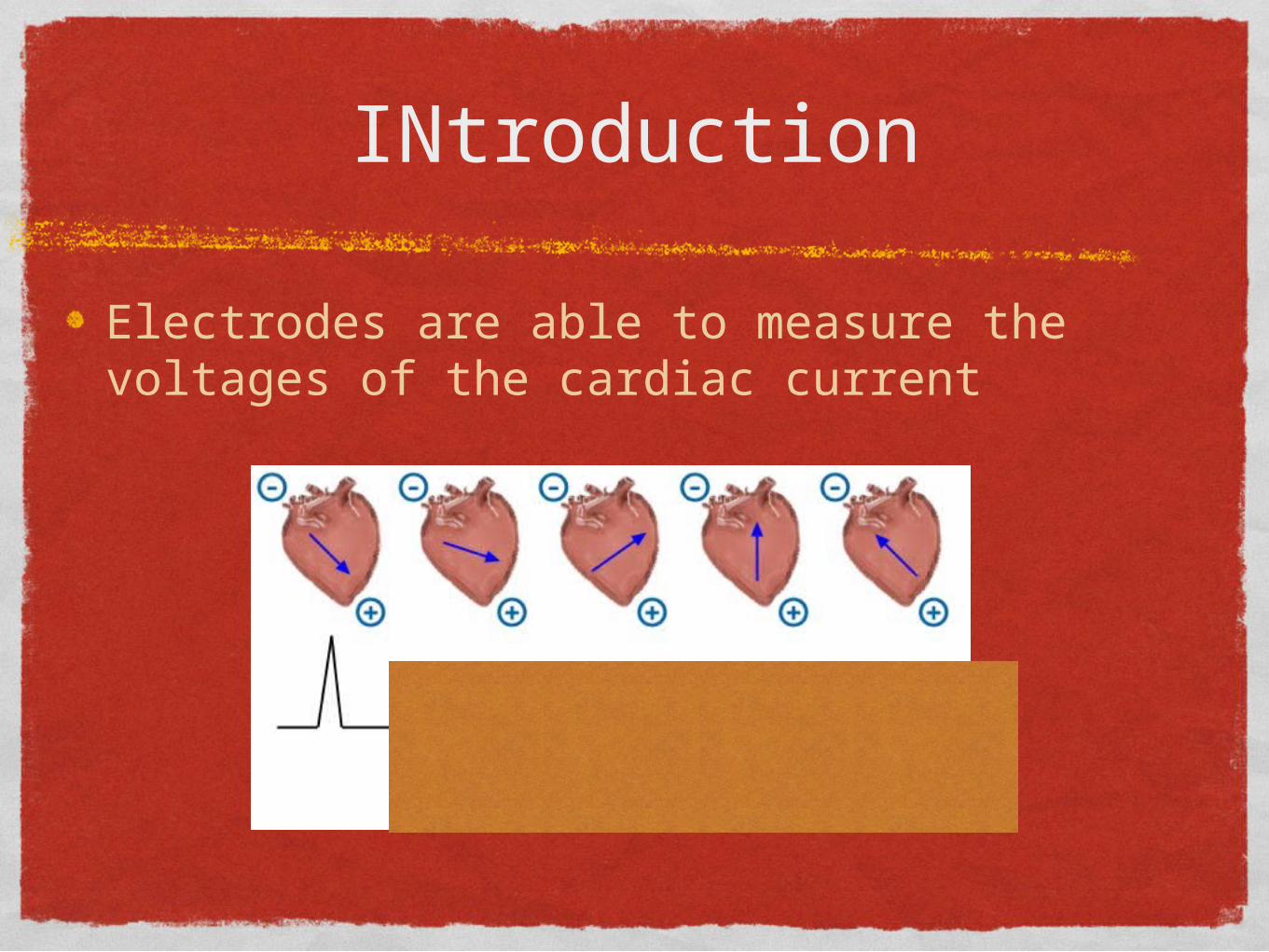
INtroduction
Electrodes are able to measure the voltages of the cardiac current

Electrodes
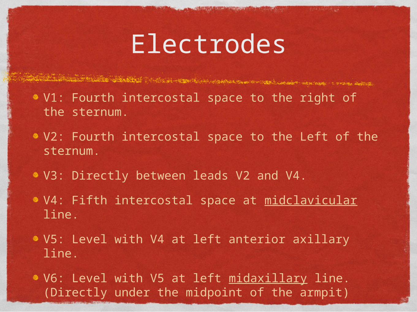
Electrodes
V1: Fourth intercostal space to the right of the sternum.
V2: Fourth intercostal space to the Left of the sternum.
V3: Directly between leads V2 and V4.
V4: Fifth intercostal space at midclavicular line.
V5: Level with V4 at left anterior axillary line.
V6: Level with V5 at left midaxillary line. (Directly under the midpoint of the armpit)

Electrode placement

Electrode and Lead are not the same thing.

Leads
12 possible leads
Six limb (extremity) leads
Bipolar leads: I, II, and III
Unipolar leads: aVR, aVL, aVF
Six chest (precordial) leads

limb leads
Electrodes
Right arm
Left arm
Left leg
Right leg (ground)


lead i
LA = electrical voltages of the heart that are transmitted to the left arm
RA = electrical voltages of the heart that are transmitted to the right arm
The electrocardiograph sustracts RA from LA and the difference appears as lead I.

lead i
Lead one ‘travels’ horizontally.
Its left pole (LA) is postive and its right pole (RA) is negative.
Therefore, lead I = LA minus RA
Shows a positive wave when an impluses moves towards the left arm, negative wave when an impuse moves away from the left arm.
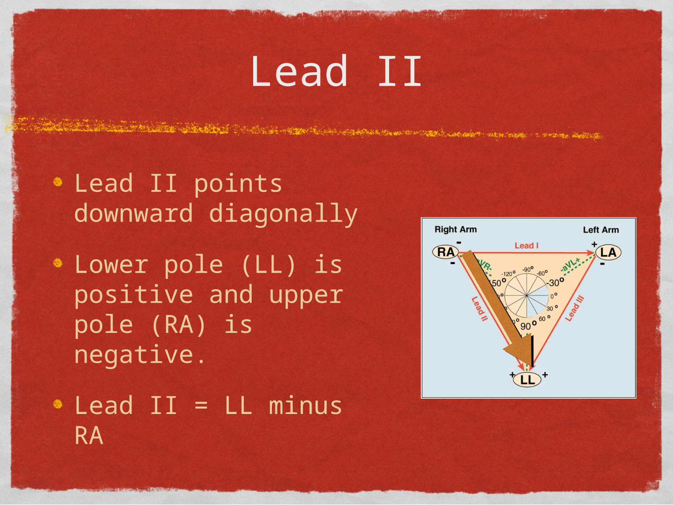
Lead II
Lead II points downward diagonally
Lower pole (LL) is positive and upper pole (RA) is negative.
Lead II = LL minus RA
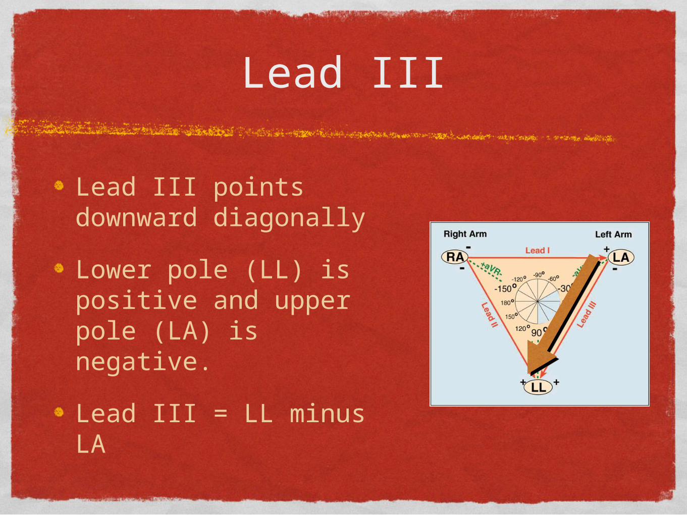
Lead III
Lead III points downward diagonally
Lower pole (LL) is positive and upper pole (LA) is negative.
Lead III = LL minus LA


Bipolar Leads

Unipolar or Augmented limb leads
Record the electrical voltages at one location rather than relative to the voltage at another electrode
limb leads
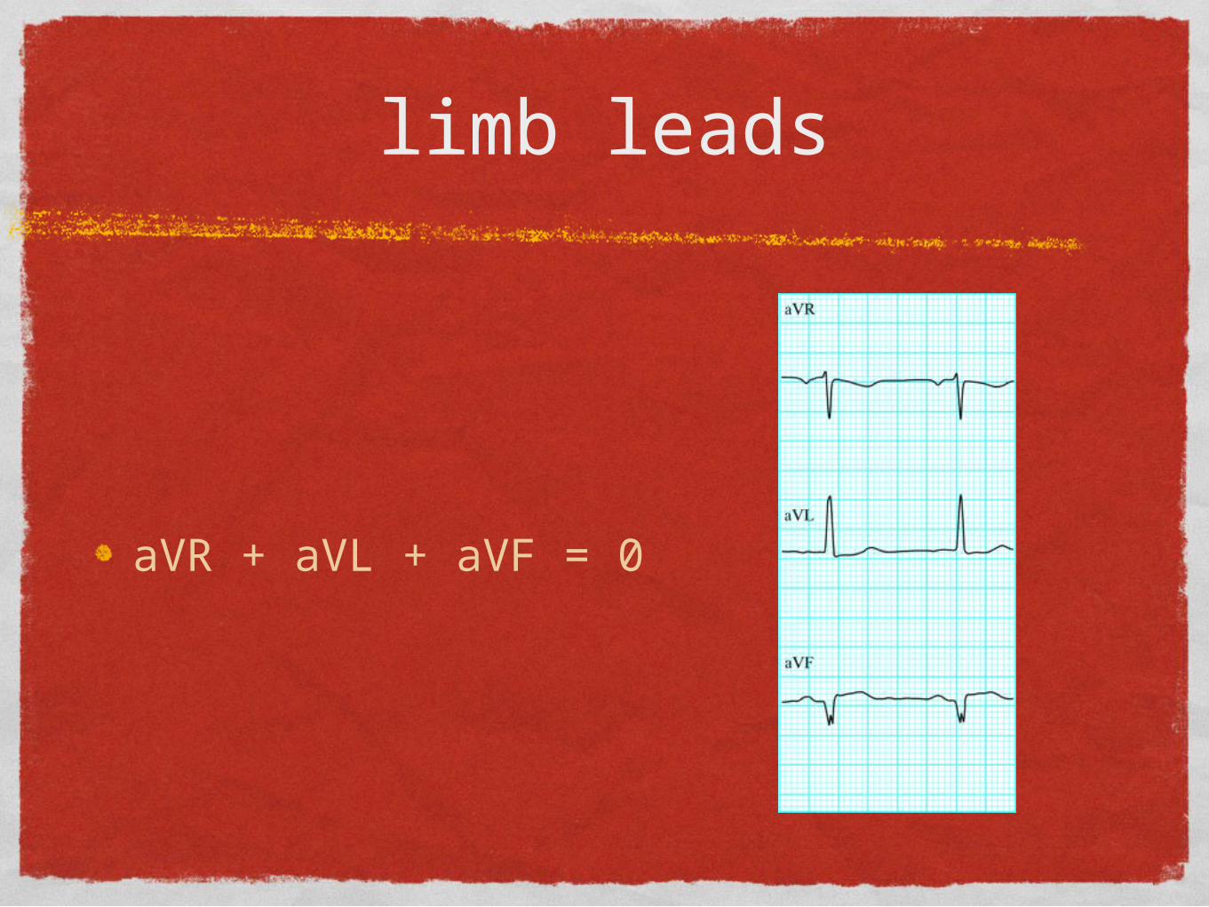
aVR + aVL + aVF = 0

Bipolar leads & Unipolar (or augmented) leads
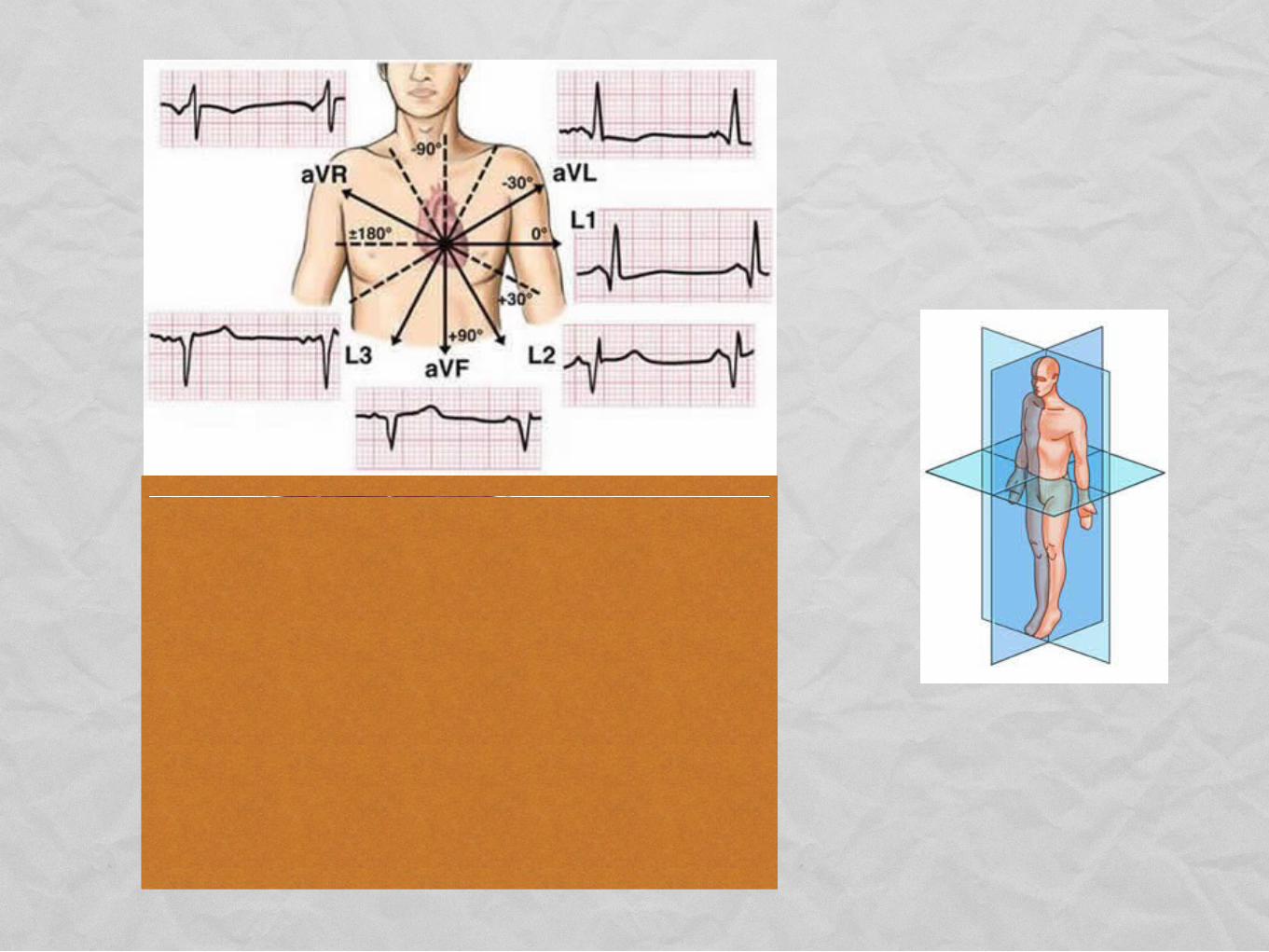
Frontal plane
Limb leads

Limb leads

chest leads
V1 to V6
unipolar
horizontal plane
front to back

Chest Leads

ALL 12 leads
Frontal Plane
Horizontal Plane

Monitor leads
12 leads are not always necessary
Sample of a monitor lead
V1 (positive)
Right shoulder (negative)
Left shoulder (ground)

Holter Monitors
Top Related