


Languages
Pages
Legal


DICOM in Ophthalmology
P. Lloyd Hildebrand, MD
Dean McGee Eye Institute
University of Oklahoma
Oklahoma City, OK USA
Overview
�Ophthalmic imaging needs
�Historical Perspective
�Organizing the Effort
�Priorities
�State of DICOM in Eyecare
�Current Projects

Ophthalmic imaging
�Many devices, many applications
�Primary digital acquisition broadly
used
�Generally no standardized way to
exchange digital images
�Ophthalmic EMR vendors

Special considerations
� Clinic centric
� Image acquisition context
� Angiography
� Stereoscopic views
� Bilateral imaging for comparison
� Clinical trials
� Centralized or distributed services
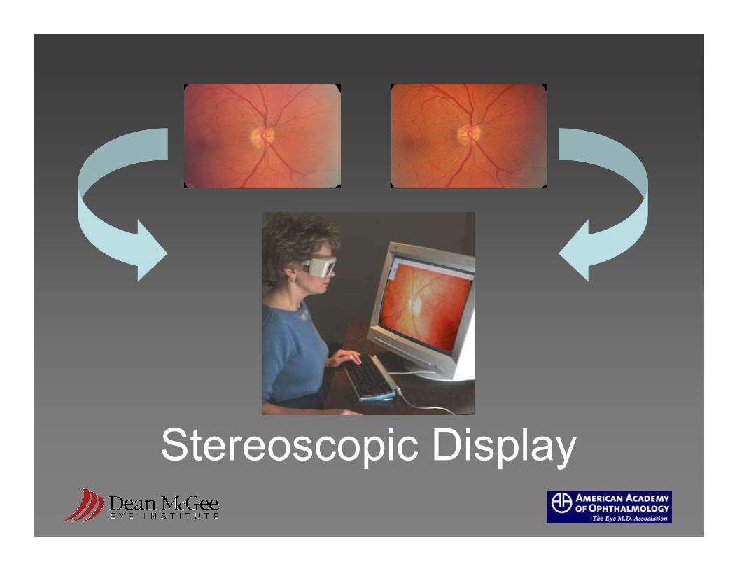
Stereoscopic Display

Ophthalmic imaging examples
Retinal Imaging
color photography
red-free imaging
angiography
optical coherence topography
Retinal imaging examples
QuickTime™ and aTIFF (Uncompressed) decompressorare needed to see this picture.
QuickTime™ and aTIFF (Uncompressed) decompressorare needed to see this picture.
QuickTime™ and aTIFF (Uncompressed) decompressorare needed to see this picture.
QuickTime™ and aTIFF (Uncompressed) decompressorare needed to see this picture.
QuickTime™ and aTIFF (Uncompressed) decompressorare needed to see this picture.

Optical Coherence Tomography

External photography
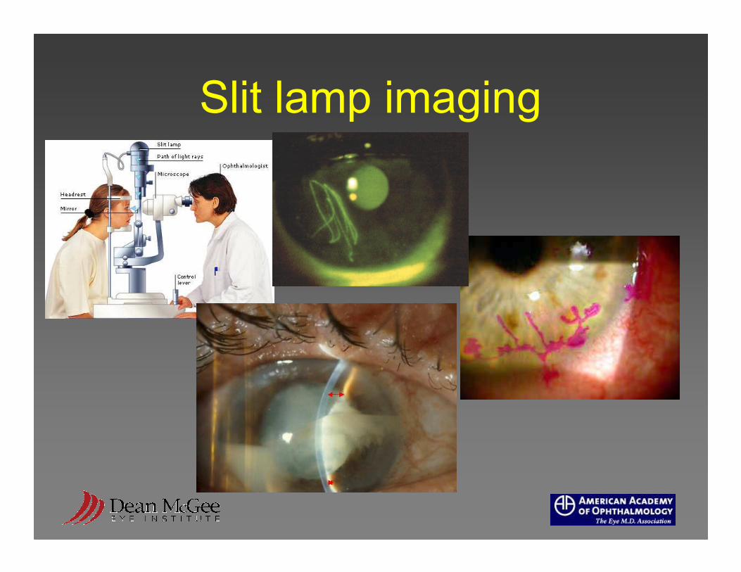
Slit lamp imaging
Specific Activities
�Working Group 9
�SNOMED terminology modeling
�New IOD work proposals
� IHE - multivendor demonstration,
profile

IHE at the AAO Annual Meeting

40’Pete Kuzmak June 28, 2005
Information
PACS Gear
Kowa Sorna/Etiam Canon
Zeiss
Zeiss Nidek
OTI
TopconRADinfo
Systems
OISVist
A
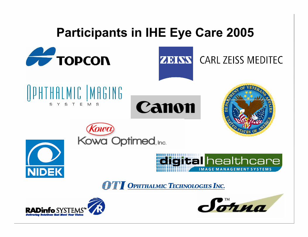
Participants in IHE Eye Care 2005
AAO Strategic Direction
• enhance existing standards
• membership education
• vendor coordination
• advocacy alignment

State of DICOM 2005
� Still in its infancy
� Device vendor interest is high
� Commitment is still needed
� EHR vendor interest is limited
� Support is strong at the AAO
leadership level
State of DICOM 2005
� Secondary capture
� Visible light imaging
� Ophthalmic photography
� Ophthalmic Tomography IOD
�Workflow modifications?
� Extensions workgroups
Top Related